
Autologous chondrocyte implantation for full thickness
Autologous chondrocyte implantation for full thickness
articular cartilage defects of the knee (Review)
Vasiliadis HS, Wasiak J
This is a reprint of a Cochrane review, prepared and maintained by The Cochrane Collaboration and published in The Cochrane Library
2011, Issue 7
http://www.thecochranelibrary.com
Autologous chondrocyte implantation for full thickness articular cartilage defects of the knee (Review)
Copyright © 2011 The Cochrane Collaboration. Published by John Wiley & Sons, Ltd.
TABLE OF CONTENTS
HEADER . . . . . . . . . . . . . . . . . . . . . . . . . . . . . . . . . . . . . . .
ABSTRACT . . . . . . . . . . . . . . . . . . . . . . . . . . . . . . . . . . . . . .
PLAIN LANGUAGE SUMMARY . . . . . . . . . . . . . . . . . . . . . . . . . . . . . .
BACKGROUND . . . . . . . . . . . . . . . . . . . . . . . . . . . . . . . . . . . .
OBJECTIVES . . . . . . . . . . . . . . . . . . . . . . . . . . . . . . . . . . . . .
METHODS . . . . . . . . . . . . . . . . . . . . . . . . . . . . . . . . . . . . . .
RESULTS . . . . . . . . . . . . . . . . . . . . . . . . . . . . . . . . . . . . . . .
Figure 1.
. . . . . . . . . . . . . . . . . . . . . . . . . . . . . . . . . . . . .
Figure 2.
. . . . . . . . . . . . . . . . . . . . . . . . . . . . . . . . . . . . .
Figure 3.
. . . . . . . . . . . . . . . . . . . . . . . . . . . . . . . . . . . . .
DISCUSSION . . . . . . . . . . . . . . . . . . . . . . . . . . . . . . . . . . . . .
AUTHORS’ CONCLUSIONS . . . . . . . . . . . . . . . . . . . . . . . . . . . . . . .
ACKNOWLEDGEMENTS
. . . . . . . . . . . . . . . . . . . . . . . . . . . . . . . .
REFERENCES . . . . . . . . . . . . . . . . . . . . . . . . . . . . . . . . . . . . .
CHARACTERISTICS OF STUDIES . . . . . . . . . . . . . . . . . . . . . . . . . . . . .
DATA AND ANALYSES . . . . . . . . . . . . . . . . . . . . . . . . . . . . . . . . . .
Analysis 1.1. Comparison 1 ACI versus mosaicplasty, Outcome 1 Good or excellent functional results (modified Cincinatti
rating system). . . . . . . . . . . . . . . . . . . . . . . . . . . . . . . . . .
Analysis 1.2. Comparison 1 ACI versus mosaicplasty, Outcome 2 Arthroscopic assessment at one year (ICRS grade 1 or
2). . . . . . . . . . . . . . . . . . . . . . . . . . . . . . . . . . . . . . .
Analysis 1.3. Comparison 1 ACI versus mosaicplasty, Outcome 3 Lysholm scores (0: worst to 100: best).
. . . .
Analysis 1.4. Comparison 1 ACI versus mosaicplasty, Outcome 4 Tegner scores (0: worst to 10: best) at 24 months. .
Analysis 1.5. Comparison 1 ACI versus mosaicplasty, Outcome 5 Meyers scores (higher scores better) at 24 months. .
Analysis 1.6. Comparison 1 ACI versus mosaicplasty, Outcome 6 Excellent outcome (various definitions). . . . .
Analysis 1.7. Comparison 1 ACI versus mosaicplasty, Outcome 7 Satisfactory outcome (various criteria) - exploratory
analysis. . . . . . . . . . . . . . . . . . . . . . . . . . . . . . . . . . . . .
Analysis 2.1. Comparison 2 ACI versus microfracture, Outcome 1 Presence of hyaline cartilage in biopsy. . . . .
Analysis 2.2. Comparison 2 ACI versus microfracture, Outcome 2 Failure and further procedures. . . . . . . .
Analysis 3.1. Comparison 3 MACI versus microfracture, Outcome 1 Lysholm scores (0: worst to 100: best). . . .
Analysis 3.2. Comparison 3 MACI versus microfracture, Outcome 2 Tegner scores (0: worst to 10: best) at 24 months.
Analysis 3.3. Comparison 3 MACI versus microfracture, Outcome 3 ICRS patient score (grade 1 or 2) at 24 months.
Analysis 4.1. Comparison 4 CCI (characterised chrondrocyte implantation) versus microfracture, Outcome 1 Knee function
up to 18 months (KOOS (“overall” minus ’sport’ domain): 0: extreme knee problems, 100: no knee problems).
Analysis 4.2. Comparison 4 CCI (characterised chrondrocyte implantation) versus microfracture, Outcome 2 KOOS
(improvement from baseline at 36 months). . . . . . . . . . . . . . . . . . . . . . . .
Analysis 4.3. Comparison 4 CCI (characterised chrondrocyte implantation) versus microfracture, Outcome 3 KOOS
improvement from baseline at 36 months (adjusted data).
. . . . . . . . . . . . . . . . . .
Analysis 4.4. Comparison 4 CCI (characterised chrondrocyte implantation) versus microfracture, Outcome 4 Treatment
failure requiring re-intervention (up to 36 months). . . . . . . . . . . . . . . . . . . . . .
Analysis 4.5. Comparison 4 CCI (characterised chrondrocyte implantation) versus microfracture, Outcome 5 Adverse
events (at 18 months). . . . . . . . . . . . . . . . . . . . . . . . . . . . . . . .
Analysis 4.6. Comparison 4 CCI (characterised chrondrocyte implantation) versus microfracture, Outcome 6 Adverse
events (at 36 months). . . . . . . . . . . . . . . . . . . . . . . . . . . . . . . .
APPENDICES . . . . . . . . . . . . . . . . . . . . . . . . . . . . . . . . . . . . .
WHAT’S NEW . . . . . . . . . . . . . . . . . . . . . . . . . . . . . . . . . . . . .
HISTORY . . . . . . . . . . . . . . . . . . . . . . . . . . . . . . . . . . . . . . .
CONTRIBUTIONS OF AUTHORS . . . . . . . . . . . . . . . . . . . . . . . . . . . . .
DECLARATIONS OF INTEREST . . . . . . . . . . . . . . . . . . . . . . . . . . . . . .
DIFFERENCES BETWEEN PROTOCOL AND REVIEW . . . . . . . . . . . . . . . . . . . . .
INDEX TERMS
. . . . . . . . . . . . . . . . . . . . . . . . . . . . . . . . . . . .
Autologous chondrocyte implantation for full thickness articular cartilage defects of the knee (Review)
Copyright © 2011 The Cochrane Collaboration. Published by John Wiley & Sons, Ltd.
1
1
2
2
4
4
5
9
10
13
14
18
18
18
25
43
45
45
46
46
47
47
48
48
49
49
50
50
51
51
52
52
53
54
54
58
58
59
59
59
59
i
[Intervention Review]
Autologous chondrocyte implantation for full thickness
articular cartilage defects of the knee
Haris S Vasiliadis1 , Jason Wasiak2
1 Molecular
Cell Biology and Regenerative Medicine, Sahlgrenska University Hospital, Gothenburg University, Gothenburg, Sweden.
Victorian Adult Burns Service and School of Public Health and Preventative Medicine, Monash University, Alfred Hospital, Melbourne,
Australia
2
Contact address: Haris S Vasiliadis, Molecular Cell Biology and Regenerative Medicine, Sahlgrenska University Hospital, Gothenburg,
SE-413 45, Sweden. [email protected]. [email protected].
Editorial group: Cochrane Bone, Joint and Muscle Trauma Group.
Publication status and date: New search for studies and content updated (no change to conclusions), published in Issue 7, 2011.
Review content assessed as up-to-date: 13 April 2011.
Citation: Vasiliadis HS, Wasiak J. Autologous chondrocyte implantation for full thickness articular cartilage defects of the knee.
Cochrane Database of Systematic Reviews 2010, Issue 10. Art. No.: CD003323. DOI: 10.1002/14651858.CD003323.pub3.
Copyright © 2011 The Cochrane Collaboration. Published by John Wiley & Sons, Ltd.
ABSTRACT
Background
Treatments for managing articular cartilage defects of the knee, including drilling and abrasion arthroplasty, are not always effective.
When they are, long-term benefits may not be maintained and osteoarthritis may develop. An alternative is autologous chondrocyte
implantation (ACI), the surgical implantation of healthy cartilage cells into the damaged areas.
Objectives
To determine the efficacy and safety of ACI in people with full thickness articular cartilage defects of the knee.
Search methods
We searched the Cochrane Bone, Joint and Muscle Trauma Group Specialised Register (14 January 2011), the Cochrane Central
Register of Controlled Trials (The Cochrane Library 2010, Issue 4), MEDLINE (1948 to January Week 1 2011), EMBASE (1980 to
Week 1 2011), SPORTDiscus (1985 to 14 January 2011), the WHO International Clinical Trials Registry Platform (26 January 2011),
and Current Controlled Trials (26 January 2011).
Selection criteria
Randomised and quasi-randomised trials comparing ACI with any other type of treatment (including no treatment or placebo) for
symptomatic cartilage defects of the medial or lateral femoral condyle, femoral trochlea or patella.
Data collection and analysis
Review authors selected studies for inclusion independently. We assessed risk of bias based on adequacy of the randomisation and
allocation concealment process, potential for selection bias after allocation and level of masking. We did not pool data due to clinical
and methodological heterogeneity.
Autologous chondrocyte implantation for full thickness articular cartilage defects of the knee (Review)
Copyright © 2011 The Cochrane Collaboration. Published by John Wiley & Sons, Ltd.
1
Main results
Six heterogeneous trials were identified with 442 participants. Methodological flaws of the included trials included incomplete followup and inadequate reporting of outcomes. Three trials compared ACI versus mosaicplasty. One reported statistically significant results
in favour of ACI at one year in the numbers of people with ’good’ or ’excellent’ functional results. Conversely, another trial found
significant improvement for the mosaicplasty group when assessed using one functional scoring system at two years, but no statistically
significant differences based on two other scoring systems. A third trial found no difference between ACI and mosaicplasty, 10 months
on average after the surgery.
There was no statistically significant difference in functional outcomes at two years in a single trial comparing ACI with microfracture
nor in the functional results at 18 months of a single trial comparing characterised chondrocyte implantation versus microfracture.
However, the results at 36 months for this trial seemed to indicate better functional results for characterised chondrocyte implantation
compared with those for microfracture. The sixth trial comparing matrix-guided ACI versus microfracture found significantly better
results for functional outcomes at two year follow-up in the MACI group.
Authors’ conclusions
There is insufficient evidence to draw conclusions on the use of ACI for treating full thickness articular cartilage defects in the knee.
Further good quality randomised controlled trials with long-term functional outcomes are required.
PLAIN LANGUAGE SUMMARY
Autologous chondrocyte implantation for full thickness articular cartilage defects of the knee
A layer of cartilage covering the knee joint surfaces acts to protect the joint and reduce friction. Damage to the cartilage (articular surface)
can decrease mobility of the joint and cause pain on movement. Continuing deterioration of the surface may lead to osteoarthritis.
Treatments for damaged cartilage include relieving symptoms, surgically cleaning up the joint, or surgically re-establishing the cartilage
layer. The latter is done using marrow stimulation techniques (such as microfracture), mosaicplasty (also known as osteochondral
cylinder transplantation), and more recently with implantation of healthy cartilage cells (chondrocytes). In the technique of autologous
chondrocyte implantation (ACI), a small piece of cartilage is retrieved from the knee joint. This piece is brought to a laboratory where
it is digested to free the chondrocyte cells; these cells are subsequently cultured in a culture media in order to expand the numbers of
cells. Then, with a second surgery, the cells are placed into the joint defect in an effort to produce a tissue that substitutes the normal
cartilage.
This review includes six small randomised controlled trials that compared ACI with either mosaicplasty or microfracture. Although
there are some promising results for ACI compared with microfracture from one trial, the evidence from two other trials testing the
same comparison did not confirm these. None of the other three trials testing different comparisons provided conclusive evidence in
favour of ACI, although the longer-term results suggest that the results for some types of ACI may improve over time. The review
identified several ongoing trials that should help to provide evidence to inform on the use of ACI in the future. Meanwhile, there is
insufficient evidence to draw conclusions on the use of ACI.
BACKGROUND
Description of the condition
Cartilage provides coverage for bones in their joint surfaces. Its role
is essential in decreasing the friction between the joining bones
and it also decreases the mechanical load effect on the covered
bone. Loss of cartilage and exposure of the subchondral bone may
produce crepitation and pain during the joint movements, and
repeated joint effusions (Buckwalter 1998).
Cartilage consists of cells (chondrocytes), water and extracellular
matrix of collagen (mainly type II), proteoglycans and noncollagenous proteins. In mature articular cartilage, chondrocytes no
longer divide and receive their nutrition mainly through diffusion
Autologous chondrocyte implantation for full thickness articular cartilage defects of the knee (Review)
Copyright © 2011 The Cochrane Collaboration. Published by John Wiley & Sons, Ltd.
2
from the synovial fluid. This limits their intrinsic capacity for repair and, thus, cartilage lesions are very difficult to heal. If left
untreated lesions are more likely to deteriorate, subsequently exposing the subchondral bone or forming fibrous tissue. The latter,
even when successfully covering underlying bone, does not provide adequate mechanical and functional support and is subject
to wear over time. Thus, no normal hyaline cartilage is formed
and, furthermore, there is usually no improvement of the person’s
symptoms in the long term.
Isolated lesions to cartilage should be differentiated from osteoarthritis, which refers to diffuse damage to the articular surface,
is more common in older people and is generally considered to
be irreversible. Non-osteoarthritis cartilage lesions are most often
found in younger people and are more subject to various treatment
alternatives aiming to cartilage repair or reconstruction (Browne
2000).
Description of the intervention
There are no uniform approaches to managing defects to cartilage. Surgical treatment options, intending primarily to achieve
symptomatic relief with the least amount of invasive intervention
(NICE 2005), are usually divided into marrow-stimulating (reparative) and reconstructive techniques. Marrow-stimulating techniques such as subchondral drilling, abrasion arthroplasty, spongialisation or microfractures allow bone marrow cells derived from
the subchondral bone to migrate into the cartilage lesion area
(Ficat 1979; Johnson 2001; Steadman 2003). The aim of these
techniques is to replenish cartilage through the recruitment of progenitor cells as potential cartilage precursors, allowing the development into chondrogenic cells and, finally, cartilage.
Reconstructive techniques use autografts, allografts or synthetic
material for restoring the lesion area. The use of autogenous periosteal or perichondrial grafts has been proposed in the past but
is not extensively used. Allografts, synthetic polymers or ceramics
are often used, usually in forms of osteochondral cylindrical plugs
to reconstruct or replace the lesion area (Ghazavi 1997). Mosaicplasty (osteochondral cylinder transplantation) uses small cylindrical autografts harvested from less weight-bearing areas of the
femoral condyle articular surface (e.g. intercondylar notch) and
placed in the cartilage defect (Hangody 1998).
Marrow-stimulating techniques have offered an easy-to-perform
treatment option for full thickness cartilage lesions of the knee.
However, according to several studies, repair tissue is mainly fibrotic and lacks the biomechanical and viscoelastic characteristics
of normal hyaline cartilage. Thus, clinical results appear to be inferior, unpredictable and not durable compared to other techniques
(Minas 1998). In addition, reconstructive techniques also have
not managed to provide impressive clinical results. Mosaicplasty,
the most common technique, is considered technically difficult
procedure not easily performed by the average surgeon. The uses
of synthetic grafts have not been extensively studied and only a
few cohort studies have been published.
Autologous chondrocyte implantation (ACI) was introduced in
Sweden in 1987, being the first biological approach to the management of cartilage lesions (Brittberg 1994). ACI of the knee
is a two-stage procedure. The initial stage involves arthroscopy,
where the knee is examined, the lesion is evaluated and small pieces
of healthy cartilage are harvested from a less weight-bearing area
(usually the femoral notch or the medial or lateral rim of trochlea).
Individual chondrocytes are isolated in vitro by collagenase digestion, cultured in media containing patient’s serum, and, following a period of cellular division, chondrocytes are retrieved for reimplantation.
Re-implantation is the second stage of the process. A parapatellar
arthrotomy is undertaken and the defect is debrided to the subchondral bone. Through a second incision, a periosteal patch is
harvested from the proximal medial tibia and sutured to the defect
rim. Fibrin glue or sealant is applied to the peripheral border of the
patch to create a watertight seal. Then, the harvested chondrocytes
are injected beneath the periosteal patch (Brittberg 2008).
Surgical techniques and technologies have undergone substantial
development since the procedure was introduced. For instance, the
above-described operative techniques are considered conventional,
first generation approaches. ’Second generation’ ACI techniques
use manufactured cell carriers such as MACI (Verigen AG, Leverkusen, Germany) aiming to provide and stabilise the cells to the
defect area (Bartlett 2005). Moreover, other materials like collagen
membranes may be used in place of periosteum (type I/III collagen membrane (ChondroGide; Geistlich, Wollhausen, Switzerland or Restore; De Puy, Warzaw, Indiana, USA) (Gooding 2006;
Steinwachs 2007). These materials aim to decrease operation time,
limit surgical trauma and avoid complications attributed to the
use of periosteum (e.g. graft overgrowth).
’Third generation’ ACI uses three-dimensional (3D) matrices such
as hyaluronic acid (Hyalograft-C; Fidia, Italy) as scaffolds (Kon
2009; Marcacci 2005). Chondrocytes are cultured in these scaffolds in a 3D culture before implanting in the lesion area. The
process of implantation (i.e. the second stage of the procedure)
in second and third generation techniques can also be performed
arthroscopically or with a small incision (Brittberg 2008).
A new technique called ’Characterized Chondocyte Implantation’
(CCI) (ChondroCelect, TiGenix NV, Haasrode, Belgium) aims
to improve the results of articular regeneration with chondrocyte
cell therapy through the use of a selected cell population. Characterised chondrocytes are an expanded population of chondrocytes
that expresses a marker profile (a gene score) potentially predictive
of the capacity to form hyaline-like cartilage in vivo in a consistent
and reproducible manner. The surgical technique is performed as
the conventional ACI, however with the use of selected-characterised chondrocytes (Dell’Accio 2003). With the CCI technique,
there is a selection of patients with high potential for success; thus,
there is a possibility that the cells will not be implanted due to
Autologous chondrocyte implantation for full thickness articular cartilage defects of the knee (Review)
Copyright © 2011 The Cochrane Collaboration. Published by John Wiley & Sons, Ltd.
3
a low potential of the patient’s chondrocytes to give satisfactory
clinical results (shown as a low gene score).
We considered any randomised or quasi-randomised (for example,
allocation by hospital record number or date of birth) controlled
trials with the comparisons described in the Types of interventions.
How the intervention might work
Types of participants
A number of studies have suggested the effectiveness of ACI for
cartilage defects of the knee (Peterson 2010; Vasiliadis 2010a;
Vasiliadis 2010c; Zaslav 2009). Cohort studies have also shown a
durability of good and excellent results in terms of clinical evaluation or histological assessment of biopsies, even up to 11 years
after the implantation (Brun 2008; Peterson 2002). Supporters of
this technique highlight that it is the only biological approach to
chondral defects, suggesting the efficiency gained by using chondrocytes for the restoration of cartilage. However, ACI has its limitations. It demands a steeper learning curve, at least compared
with marrow-stimulating techniques. It is also an expensive procedure with a considerable rate of complications (Wood 2006).
We were interested in studies enrolling people between 15 and 55
years of age with symptomatic isolated cartilage defects (surface
area of 1 cm² to 15 cm²) of the medial or lateral femoral condyle,
femoral trochlea or patella. In these studies, the joint should be
free from disease states such as rheumatoid arthritis or osteoarthritis, as determined by radiographic evidence such as joint space
narrowing, osteophyte formation, subchondral bony sclerosis or
cyst formation.
Why it is important to do this review
Cartilage lesions are common in the general population and are
more often anticipated in young and physically active people. Curl
and colleagues report an incidence of chondral lesions in 63% of
the 31,516 performed arthroscopies, with an average of 2.7 lesions
per knee (Curl 1997). In 1000 consecutive arthroscopies examined
by Hjelle and colleagues, chondral or osteochondral lesions of any
type were found in 61% and focal defects were found in 19%
(Hjelle 2002). ACI is a relatively new technique with promising
results. However, the clinical benefits and potential harms remain
unclear.
OBJECTIVES
The objective of this review was to assess the effectiveness and
safety of ACI in people who require repair of clinically significant, symptomatic defects of the medial or lateral femoral condyle,
femoral trochlea and patella caused by acute or repetitive trauma
to the knee joint or osteochondritis dissecans.
Types of interventions
Interventions comparing ACI with placebo, no treatment or another intervention such as mosaicplasty, periosteal grafting and
tibial/femoral osteotomies. We did not include studies comparing
ACI with modified versions of ACI such as porcine-derived type I/
type III collagen as a cover (ACI-C) or matrix-guided autologous
chondrocyte implantation (MACI).
Types of outcome measures
We chose six outcome measures as being most representative of
the clinically important measures of effectiveness. They included
the following:
• knee function scoring systems such as the Lysholm score,
the Tegner score, the Cincinnati Knee Scale (CKS) and the Knee
Society Score (KSS);
• general function or mobility scoring systems such as the
Western Ontario McMaster Osteoarthritis Scale (WOMAC);
• quality of life scoring systems such as Short Form-36 (SF36);
• symptomatology such as pain and swelling;
• hyaline cartilage development as verified by second look
arthroscopy or magnetic resonance imaging (MRI); and
• adverse events.
Search methods for identification of studies
METHODS
Electronic searches
Criteria for considering studies for this review
Types of studies
We searched the Cochrane Bone, Joint and Muscle Trauma Group
Specialised Register (14 January 2011), the Cochrane Central
Register of Controlled Trials (The Cochrane Library 2010, Issue 4), MEDLINE (1948 to January Week 1 2011), EMBASE
(1980 to Week 1 2011), SPORTDiscus (1985 to14 January
2011), the WHO International Clinical Trials Registry Platform
Autologous chondrocyte implantation for full thickness articular cartilage defects of the knee (Review)
Copyright © 2011 The Cochrane Collaboration. Published by John Wiley & Sons, Ltd.
4
(26 January 2011), and Current Controlled Trials (26 January
2011). We applied no language restrictions.
In MEDLINE, the subject specific search strategy was combined
with the Cochrane highly sensitive search strategy for identifying
reports of RCTs (Higgins 2006). The EMBASE subject specific
search strategy was combined with the Scottish Intercollegiate
Guidelines Network (SIGN) RCT filter. The search strategies for
all databases can be found in Appendix 1.
Data synthesis
We planned all analyses to be made on data reported for intention-to-treat results. However, none of the studies used such analyses. We used the fixed-effect model to pool data where there
was no evidence of significant heterogeneity between studies, and
the random-effects model when such heterogeneity was present
(DerSimonian 1986).
Subgroup analysis and investigation of heterogeneity
Data collection and analysis
No specific subgroup analyses were prespecified.
Selection of studies
Records retrieved by the initial search were scanned by review
authors (HV and JW) to exclude obviously irrelevant studies and to
identify trials that met the inclusion criteria. Full-text articles were
retrieved and reviewed independently for the purpose of applying
the inclusion criteria. In all instances, differences of opinion were
resolved by discussion.
Data extraction and management
We extracted data from the studies independently using standardised forms. All differences of opinion between the authors were
resolved by discussion.
Assessment of risk of bias in included studies
We assessed the risk of bias for each study according to the recommendations described in the Cochrane Handbook for Systematic
Reviews of Interventions (Higgins 2006).
The risk of bias tool incorporates assessment of randomisation (sequence generation and allocation concealment), blinding (of participants, treatment providers and outcome assessors), completeness of outcome data, selection of outcomes reported and other
sources of bias. We considered assessors of clinical outcomes and
assessors of histological findings after the biopsy separately in our
assessment of blinding and completeness of outcome data. Discrepancies in ratings were resolved by discussion.
Measures of treatment effect
We expressed dichotomous data as risk ratios (RR) and 95% confidence intervals (CIs). We expressed continuous data as mean differences (MD) and 95% CIs.
Assessment of heterogeneity
We tested statistical heterogeneity using the Chi² test with significance at P < 0.10, and a quantification of the degree of heterogeneity using the I² statistic and further exploration using sensitivity
analyses.
RESULTS
Description of studies
See: Characteristics of included studies; Characteristics of excluded
studies; Characteristics of ongoing studies.
For further details of the included, excluded and ongoing trials, please see the Characteristics of included studies, the
Characteristics of excluded studies and the Characteristics of
ongoing studies.
Results of the search
In our first update in 2006, four trials comparing ACI with any
other type of cartilage repair surgery were included in the systematic review (Basad 2004; Bentley 2003; Horas 2003; Knutsen
2004). In the second update in 2010, we also included two new
trials (Dozin 2005; Saris 2008), and one (Knutsen 2007) which
was a longer-term follow-up of an already included trial (Knutsen
2004). We found another study previously categorised as excluded
(Horas 2000) to be another publication of an already included trial
(Horas 2003). In the current third update in 2011, searches were
performed in January 2011. One trial report, previously in studies
awaiting assessment, Basad 2010 was a full report of an already
included trial (Basad 2004). Given that Basad 2010 reported a
greater number of participants, with a more complete follow-up,
we considered that this should be considered the definitive publication of this trial. A longer term follow-up of Saris 2008 was
also included (Saris 2009). We also found two reports (Van Assche
2009; Van Assche 2010) that reported findings from a subgroup
of trial participants of Saris 2008. Both Saris 2009 and Van Assche
2009 were in ’Studies awaiting assessment’ in the previous version
of the review.
In this update, seven other newly identified publications were excluded; two were abstract reports of the same trial (Park 2008)
and one was a long term follow-up of an already excluded trial
(Gudas 2005). Additionally, two more report of ongoing studies
Autologous chondrocyte implantation for full thickness articular cartilage defects of the knee (Review)
Copyright © 2011 The Cochrane Collaboration. Published by John Wiley & Sons, Ltd.
5
were identified (Fickert; SUMMIT extension study), the second
being an extension study of SUMMIT.
Included studies
Design
Of the six included trials, five were randomised trials and one
(Horas 2003) was quasi-randomised.
Horas 2003 is the same study as Horas 2000 (formerly excluded
as a non-randomised study in our review). Horas 2000 was not
cited in the 2003 report, but the response of the trial authors to a
letter (Smith 2003) commenting on Horas 2003 confirmed that
“the same patient population formed the basis for both the German publication (Horas 2000) and the present article. However,
different individual aspects of the treatment’s results were highlighted, especially in the Discussion sections of the two articles”.
Both these articles report follow-up at 24 months. However, because of differences in the presentation of outcomes between the
two main reports of Horas 2003 and the availability of raw data
for Horas 2003, we chose only to review the 2003 report.
Knutsen 2007 is a five-year follow-up evaluation of Knutsen 2004,
which reported on two-year follow-up after surgery in the 2004
article. As the information on the study design was better described
in Knutsen 2004, we based the risk of bias assessment on this
report.
Saris 2009 presented the 36 month follow-up of Saris 2008. Data
from different follow-up times were extracted from both publications. Van Assche 2009 and Van Assche 2010 studied a subgroup
of Saris 2008 from two centres (one Belgium and one Dutch).
Basad 2010 was a full report of Basad 2004. Basad 2010 was
conducted in the same centre during the same time, and using
the same follow-up time of two years. The authors included more
participants and presented the outcomes in a more complete and
relevant way. Therefore, we considered that Basad 2010 was the
definitive report of this trial.
Sample sizes
The six included studies recruited a total of 442 participants. Based
on reported allocation, 237 had one of the autologous chondrocyte implantation (ACI) techniques and 205 had either microfractures or mosaicplasty. In the former, 94 trial participants had ACI
with the use of periosteum, 46 had ACI with the use of collagen
membrane, 57 had characterised chondrocyte implantation (CCI)
and 40 had matrix-guided ACI (MACI). Bentley 2003 assessed
58 ACI-treated participants, 12 of which had the technique with
periosteum and 46 with collagen membrane as a coverage of the
lesion area. In the control groups, 121 participants had microfractures and 84 had mosaicplasty.
Setting
Three trials were single-centre trials, two of which were based in
Germany (Basad 2010;Horas 2003) and one in the UK (Bentley
2003). Dozin 2005 was a multi-centre study with three surgeons
and involvement from five orthopaedic centres in Italy. Knutsen
2004 was a multi-centre study involving four centres in Norway
and one in the UK. Saris 2008 was a multi-centre study undertaken
in 13 centres in four countries (Belgium, Croatia, Germany and
the Netherlands).
Saris 2008 was sponsored by TiGenix n.x. Eight authors of this
study declared a conflict of interest. One or more of the authors
in Bentley 2003 also declared a conflict of interest (details not
provided).
Participants
The participants of all the studies had isolated cartilage lesions
of the femur or the patella. The average size of the lesions was
homogenous, ranging between 3.8 cm² and 5.1 cm² for three trials
(Bentley 2003; Horas 2003; Knutsen 2004). In Basad 2010, the
acceptable size of the lesion for trial inclusion was between 4 cm²
and 10 cm²; however, Basad 2004 reported a lower limit of 2 cm².
In Dozin 2005 and Saris 2008, the size of treated lesions was much
smaller: Dozin 2005 had an average size of 2.0 cm² and 1.9 cm² for
ACI and mosaicplasty respectively, while the mean size of cartilage
lesions in Saris 2008 was 2.5 cm².
There were important differences in the inclusion criteria of the
included studies (see Characteristics of included studies). For example, Knutsen 2004 and Saris 2008 included only participants
with femoral condyle lesions, while the other trials included lesions at other sites (mainly patella lesions). Cases with osteochondritis dissecans were excluded by Horas 2003 and Saris 2008 but
were included in remaining trials. Osteochondral lesions were also
withdrawn from Basad 2010. Concomitant lesions (anterior cruciate ligament ruptures or meniscal tears) were not excluded in
Saris 2008. Acording to the protocol, Basad 2010 excluded patients with prior or planned meniscectomies (>30% of the meniscus) or knee instability. However, one microfracture patient had
an ACL reconstruction. Five patients (two in the MACI and three
in the microfracture group) had smaller meniscal lesions treated.
Other sources of between-trial variation included differences in
the history of previous surgery and baseline clinical scores: for
instance, the ACI group had a Lysholm score of 24.9 in Horas
2003 and 57.4 in Knutsen 2004.
The average ages of trial participants in the individual trials ranged
between 29 years (Dozin 2005) and 34 years (Basad 2010; Saris
2008). All six trials (Basad 2010; Bentley 2003; Dozin 2005; Horas
2003; Knutsen 2004; Saris 2008) included more male than female
participants (60% or more of all participants were males).
Interventions
Autologous chondrocyte implantation for full thickness articular cartilage defects of the knee (Review)
Copyright © 2011 The Cochrane Collaboration. Published by John Wiley & Sons, Ltd.
6
Four different comparisons were tested by the six included trials. Three trials compared ACI with mosaicplasty (Bentley 2003;
Dozin 2005; Horas 2003). Dozin 2005 and Horas 2003 clarified
that they used autologous periosteum for the coverage of the cartilage lesion and the implanted cells. In the ACI group, Bentley 2003
used either periosteum or collagen membrane. Knutsen 2004 compared ACI (using periosteum) versus microfractures. Basad 2010
compared MACI with microfractures and Saris 2008 compared
characterised chondrocyte implantation (CCI) with microfractures.
Where described, the rehabilitation programmes differed between
the studies (see the Characteristics of included studies). For example, Bentley 2003 used a cast to keep the knee in extension for
the first 10 days, and encouraged full-weight bearing at 24 hours
postoperatively. In contrast, Horas 2003 allowed flexion up to 90
degrees for the first 10 days with partial weight-bearing at two
weeks and full weight-bearing at 12 weeks. Basad 2010 used a
different rehabilitation program for each of the treatment groups.
Outcomes
Several different outcomes were presented by the trials (Lysholm
score, Tegner, modified Cincinnati, visual analogue scale (VAS) for
pain, Mayes, KOOS: Knee Injury Osteoarthritis Outcome Score,
patient rated ICRS and SF-36). Only Horas 2003 provided raw
data of all the outcomes measured (Lysholm, Tegner, Meyers, complications). Knutsen 2004, and the longer term follow-up report
(Knutsen 2007), presented histograms or box plots but not numerical data for functional and pain outcomes (Lysholm, VAS, SF36). Although specified in the methods section, the Tegner score
is not presented in Knutsen 2004 and only the mean baseline and
final scores are presented in Knutsen 2007.
Four trials reported the Lysholm score. However, only Horas 2003
provided exact scores through raw data. Basad 2010 gave the mean
values and standard deviations. Knutsen 2004 presented the results
in a histogram, Dozin 2005 presented them in groups with cut
points at 60 and 90.The Tegner score is presented by Basad 2010,
Horas 2003 and in Knutsen 2007. VAS for pain and the SF-36
results are presented in Knutsen 2004 only. The Meyers score is
presented by Horas 2003. KOOS is given only by Saris 2008.
Four trials also reported on a limited number of second-look
arthroscopies, also providing results from biopsy of the repair tissue. Bentley 2003 and Knutsen 2004 provided the ICRS score
based on the morphology of the repair tissue and also the quality
of the repair tissue after a biopsy retrieval (categorised into hyaline-like, mixed, fibrocartilage or fibrous tissue). Horas 2003 gave
a more case-based narrative description of some biopsies and Saris
2008 provided a mean histology score based on different features
of the histological components of structural repair.
Excluded studies
We excluded 21 studies. Detailed information is given in the
Characteristics of excluded studies.
Ten of the excluded studies were RCTs. Six of these compared
two different ACI techniques. Gooding 2006 and Zeifang 2010
compared ACI-P (use of periosteum) with ACI-C (use of collagen
membrane instead of periosteum). Park 2008 compared ACI-P
with MACI (matrix-guided ACI). Bartlett 2005 and Bickerstaff
compared ACI-C with MACI. (The full manuscript of Bickerstaff
could not be traced.) Schneider 2003 compared conventional ACI
with another ACI technique (CaReS). Visna 2004 addressed a
valid comparison for our review; they compared an ACI-based
treatment (cultivated autologous chondrocytes in a three-dimensional carrier of fibrin glue) with abrasive techniques. However,
20% of the participants had double lesions and 10% had tibia
plateau lesions, which was not consistent with our inclusion criteria. Gudas 2005 did not compare any ACI technique. Two RCTs
(Ebert 2008; Wondrasch 2009) compared different rehabilitation
approaches for patients treated with MACI.
Anderson 2003, Behrens 2006 and Kon 2009 were not RCTs, but
prospective cohort studies.
Nine of the excluded studies could not be traced (Bickerstaff;
Brittberg; Jacobsen; Joergensen; Keating; Trial 1; Trial 2; Trial 3;
Trial 4). Seven of these were identified in a Health Technology
Assessment systematic review (Jobanputra 2001) but remain untraceable (Brittberg; Jacobsen; Joergensen; Trial 1; Trial 2; Trial 3;
Trial 4). At least two of the nine studies were industrially sponsored and at least three more studies were evaluating a product of
interest to industry.
Ongoing studies
Detailed information for the ongoing studies is given in the
Characteristics of ongoing studies.
We found eight ongoing studies evaluating ACI or other ACIbased techniques. Two evaluate conventional ACI (ACTIVE;
Richardson), two evaluate CARTIPATCH (Barnouin; Dubrana),
one evaluates NeoCart (Crawford), one evaluates MACI (
SUMMIT), one evaluates the BioCartT M II (Roth-Ben Arie), and
one the co.Don Chondrosphere (Fickert). There are eight different
comparisons: ACI versus any conventional technique (ACTIVE),
ACI (NeoCart) versus microfracture (Crawford), ACI (MACI)
versus microfracture (SUMMIT), ACI (BioCart TM II) versus
microfracture (Roth-Ben Arie), ACI (CARTIPATCH®) versus
microfracture (Barnouin), ACI (CARTIPATCH®) versus mosaicplasty (Dubrana), co.Don Chondrosphere versus microfracture (Fickert) and ACI and osteotomy versus osteotomy alone
(Richardson). Five of these are multi-centre trials (ACTIVE;
Dubrana; SUMMIT; Roth-Ben Arie; Fickert). Five studies are industrially sponsored (Barnouin; Crawford; SUMMIT; Roth-Ben
Arie; Fickert).
According to the trial registration details, 1459 participants will
be included, 660 of whom are in the ACTIVE study. One of
Autologous chondrocyte implantation for full thickness articular cartilage defects of the knee (Review)
Copyright © 2011 The Cochrane Collaboration. Published by John Wiley & Sons, Ltd.
7
the studies (Richardson) should have been finished in December
2007, but no publication of the results has been traced so far. For
the rest, one trial is planned to finish in 2011, two in 2012, three
(including the five-year follow-up of SUMMIT) in 2015 and two
in 2016.
a subgroup of trial participants of Saris 2008.
We also found two new reports of ongoing studies. One was new
(Fickert) and the other (SUMMIT extension study) was a five-year
follow-up study for patients who have completed the SUMMIT
trial.
Five trials are newly excluded (Ebert 2008; Kon 2009; Park 2008;
Wondrasch 2009; Zeifang 2010).
New studies found for this update
This update included one updated trial (Basad 2010), previously
Basad 2004. Basad 2010 described a larger population with a fuller
account of outcome at two years compared with Basad 2004. We
considered that Basad 2010 is the definitive trial report of this
trial. A longer term follow-up of Saris 2008 was also included
(Saris 2009). We also found two more reports of RCTs ((Van
Assche 2009; Van Assche 2010) that reported on the outcomes of
Risk of bias in included studies
The results of the quality assessment are given in the Characteristics
of included studies and summarised in Figure 1 and Figure 2.
Additionally, a brief descriptive account of the studies is provided
below.
Autologous chondrocyte implantation for full thickness articular cartilage defects of the knee (Review)
Copyright © 2011 The Cochrane Collaboration. Published by John Wiley & Sons, Ltd.
8
Figure 1. Methodological quality summary: review authors’ judgements about each methodological quality
item for each included study.
Autologous chondrocyte implantation for full thickness articular cartilage defects of the knee (Review)
Copyright © 2011 The Cochrane Collaboration. Published by John Wiley & Sons, Ltd.
9
Figure 2. Methodological quality graph: review authors’ judgements about each methodological quality
item presented as percentages across all included studies.
Whenever it was necessary, we contacted the contributing authors
of the included trials by email in order to obtain further clarifications regarding the methodology followed in their trial. Where
the authors’ answers changed our judgement in the ’Risk of bias’
table, we detail this in the Characteristics of included studies.
Note that risk of bias assessment for Knutsen 2004 and Saris 2008
is based on the detailed methodology provided in the 2004 and
2008 papers respectively. The methods given in the follow-up
reports (Knutsen 2007; Saris 2009) are comparable. Basad 2010
has been primarily used for the risk of bias assessment of this trial
but the methods reported in Basad 2004 were also referred to in
our assessment.
Allocation
Appropriate sequence generation to ensure randomisation seemed
likely in all trials except Horas 2003, which was quasi-randomised.
The methods of sequence generation were adequately described in
the reports of three trials (Basad 2010; Dozin 2005; Saris 2008),
but additional clarification provided by the corresponding authors
of Bentley 2003 and Knutsen 2004 demonstrated the use of an
adequate sequence generation method. Allocation concealment
was adequate in two trials (Dozin 2005; Saris 2008), and unclear
in another two (Bentley 2003; Knutsen 2004). Although sealed
envelopes were used in Knutsen 2004, no additional information
was given to specify if the envelopes were opaque. In the earlier
report of Basad 2010, Basad 2004 reported that participants who
did not agree with their allocated therapy were dropped out; thus
the randomisation process was compromised and the trial was
judged at a high risk of selection bias. Basad 2010 did not refer
to this in their later report nor give any information regarding the
allocation concealment. Horas 2003 used alternation, and thus
allocation concealment was not possible in this trial.
Blinding
Two of the studies did not provide enough information to determine the strategies used to blind participants or assessors of clinical
outcomes (Horas 2003; Saris 2008). Knutsen 2004 stated that an
independent observer performed the follow-up clinical examination. Outcome assessors were not blinded in the studies of Basad
2004, Bentley 2003 and Dozin 2005. Knutsen 2004 stated that
an independent observer performed the follow-up clinical examinations, but did not describe blinded assessment. In the five years
follow-up, the evaluation was carried out by the first author, cancelling the claim for assessor’s independency. Hence, the judgement is ’unclear’ regarding risk of bias for this trial. Basad 2010
did not give additional information regarding blinding of the assessors.
We should probably acknowledge here the difficulty of blinding
of the clinical outcomes assessors, that reflects the nature of the
surgical interventions. Furthermore, it should be acknowledged
that the only clinical outcomes assessed in the trials were patientderived scores, thus no clinical assessors were needed.
Three trials (Horas 2003; Knutsen 2004; Saris 2008) reported
blinded assessment of overall histological assessment scores.
Autologous chondrocyte implantation for full thickness articular cartilage defects of the knee (Review)
Copyright © 2011 The Cochrane Collaboration. Published by John Wiley & Sons, Ltd.
10
Incomplete outcome data
In most studies, the number of participants who deviated from the
study protocol was not reported. None of the patients were lost
to the clinical follow-up in Bentley 2003 and Horas 2003. Dozin
2005 provides details on participants lost to follow-up, however
the number is high. The authors reported that 22.7% (5.22) of
the ACI and 31% (7/22) of the mosaicplasty allocated patients did
not proceed to the operation due to spontaneous improvement
after the first surgery and debridement of the defect area. Six of
the patients allocated to the CCI arm of Saris 2008 did not have
the intervention because of a negative ChondroCelect score that
meant that the implant was not viable. Saris 2008 included these
patients when presenting data on complications, but they did not
include them in the results for treatment failure or for assessment
of functional outcome using the KOOS score. Regarding histological assessment carried out in for trial, only Saris 2008 gave adequate information. Bentley 2003 and Horas 2003 only assessed
a subgroup of participants; while 16% (13/80) of patients did not
have biopsies in Knutsen 2004.
Confirmation that no trial participants were lost from follow-up
was received for Knutsen 2004. In the five-year follow-up analyses
of this trial (Knutsen 2007), the patients with failure remained in
the trial, “with their last recorded clinical follow-up scores before
the failure considered to be their final clinical score.”
Bias resulting from incomplete data seemed high in Basad 2010,
when based on an assessment of Basad 2004, where firstly an
unknown number of participants who did not agree with their
allocation were excluded, and secondly a large number of patients
were lost to follow-up. In Basad 2010, there was fewer patients
lost to follow-up compared with Basad 2004. There was, however,
no intention-to-treat analysis and we considered that the risk of
attrition bias remained high.
Selective reporting
Only two of the studies were judged free of selective reporting
(Horas 2003; Knutsen 2004). The outcomes were presented in an
insufficient way by Bentley 2003 did not present the Stanmore
scores. Although the Meyers score was included in the protocol of
Basad 2004, it was not presented in either Basad 2004 or Basad
2010. Lysholm scores were modified and provided at 12 months
only in Dozin 2005, while no International Knee Documentation
Committee data were provided. Saris 2008 presented KOOS score
modified by removing the “sport” domain.
reply to the letter of Smith 2003. Although there are no significant
differences in the outcomes, this is considered a questionable issue
that potentially suggests bias. Basad 2010 was the full report of
Basad 2004, reporting on the same follow-up time (two years).
Although Basad 2004 had a large number of participants lost to
follow-up (only 19 (41%) were available at one year follow-up
and only five (11%) at two years), this is not the case in the later
publication.
Rehabilitation differed considerably between trials. However,
where described in the trial reports, the same programme was provided to both intervention groups of individual trials with the exception of Basad 2010.
Sponsorship - conflict of interest
The trial of Saris 2008 was sponsored by TiGenix n.x. TiGenix is
the company that produces the ChondroCelect, the cell therapy
product which is necessary for the CCI technique. Moreover, eight
authors including the two lead authors of this study declared a
conflict of interest. One or more of the authors in Bentley 2003
also declared a conflict of interest (no details provided).
Patient baseline characteristics
Treatment groups were similar at baseline with respect to defect
size and level of function in four studies (Dozin 2005; Horas 2003;
Knutsen 2004; Saris 2008).
The defect lesion size was reported in all studies. The interim
report of Basad 2010 (Basad 2004) reported differences in the size
of defect at baseline with no further details. However, in Basad
2010 the only difference was the prolonged symptom duration of
MF treated patients, which was 0.3 years longer than in the MACI
group. All trials had clear inclusion and exclusion criteria and
there was some consistency between studies. Within the studies,
participants were generally well matched for location and size of
defect lesion, although Saris 2008 reports that proportionally more
participants in the comparator group had undergone previous knee
surgery.
Effects of interventions
The four comparisons tested by one or more of the six included
trials are presented separately below. The three studies testing the
same comparison (ACI versus mosaicplasty) were sufficiently dissimilar to merit separate descriptions. Only one, primarily exploratory, meta-analysis was performed.
Other potential sources of bias
Horas 2003 gave a graph of the outcomes (figure 1 in the study
report). However, although this study and Horas 2000 are duplicates, the same graph presented in Horas 2000 is slightly different
(crossing of lines of Lysholm and Tegner in Horas 2000 but not in
Horas 2003).The authors failed to explain this difference in their
ACI versus mosaicplasty
Bentley 2003 found no statistically significant differences in functional assessment at one year using either the modified Cincinnati or Stanmore scores (Cincinnati score (“excellent” or “good”
Autologous chondrocyte implantation for full thickness articular cartilage defects of the knee (Review)
Copyright © 2011 The Cochrane Collaboration. Published by John Wiley & Sons, Ltd.
11
results): ACI 51/58 (88%) versus mosaicplasty 29/42 (69%), (reported P = 0.277)). However, our analysis found a statistically
significant result for excellent and good results that favours ACI
over mosaicplasty (risk ratio (RR) 1.27, 95% confidence interval
(CI) 1.02 to 1.59; see Analysis 1.1). A post-hoc subgroup analysis
by defect site was reported by Bentley 2003 to show a statistically
significant difference in function in the ACI group at one year
only for participants with lesions of the medial femoral condyle
(Cincinnati score “excellent” or “good”: ACI 21/24 (88%) versus
mosaicplasty 21/29 (74%), reported P = 0.032). Cincinnati scores
were not statistically significantly different in people with either
lateral femoral condyle or patellar defects.
After one year follow-up, International Cartilage Repair Society (ICRS) grades of 1 (excellent) or 2 (good) assessed using
arthroscopy were given to 30/37 (82%) after ACI and 8/23 (34%)
after mosaicplasty; RR 2.33, 95% CI 1.30 to 4.17 (see Analysis
1.2). In “50%” of participants of the ACI group, tissues were relatively soft on probing compared with the surrounding cartilage.
Seven out of 19 participants who had biopsies after ACI at one
year were found to have hyaline cartilage of normal appearance.
The number of participants having biopsy after mosaicplasty was
not stated and results were only reported for seven participants
with a Cincinnati scale rating of “poor”. In four of these participants, the plugs were in situ but the tissue between them had not
become covered with continuous fibrous tissue; in three, the plugs
had disintegrated; and in one participant, the area of the mosaicplasty had remained reasonably intact but the articular cartilage at
the margins of the defect had broken down to expose subchondral
bone. Bentley 2003 shown an improvement of the quality of the
repair tissue of an ACI patient between a biopsy taken at one and
another at two years. This interesting finding suggests an ongoing
maturation of the repair tissue over time.
Bentley 2003 reported complications but did not mention whether
any further surgery was required. Moreover, the authors did not
split the complications by treatment group. In total, one participant developed calf-vein thrombosis and required anticoagulants
and one developed a superficial infection. Three of the participants
were slow to mobilise and required manipulation under anaesthesia; one of these required arthroscopy and arthrolysis to mobilise
the knee.
Horas 2003 found statistically significant differences in Lysholm
scores at six, 12 and 24 months favouring the mosaicplasty group
(45.75 versus 53.45 at six months; 57.50 versus 68.25 at 12
months and 66.75 versus 72.70 at 24 months; see Analysis 1.3,
results derived from the published raw data for this trial). It is notable that at 24 months postoperatively, 18 of the 20 ACI group
participants and all of the 20 mosaicplasty group participants had
a Lysholm score of 60 or more, which is considered the threshold
for a ’good’ result. The investigators found no significant difference between ACI and mosaicplasty for any time period when
participants were assessed using the Tegner or the Meyers scores.
The Tegner scores for ACI versus mosaicplasty were 1.55 versus
1.55 at three months, 2.95 versus 3.55 at six months, 4.25 versus
5.00 at 12 months and 5.10 versus 5.20 at 24 months (see Analysis
1.4). The Meyers scores for ACI versus mosaicplasty were 8.50
versus 7.85 at three months, 12.05 versus 13.75 at six months,
14.15 versus 15.90 at 12 months and 15.90 versus 16.75 at 24
months (see Analysis 1.5).
The proportions of participants with complications reported in
Horas 2003 were the same in both groups at 24 months (ACI
12/20 (60%) versus mosaicplasty 12/20 (60%)). In both groups,
complications were either surgical (i.e. locking of the joint and
adhesions) or non-surgical (i.e. passing irritation of the infrapatellar branch of the saphenous nerve) and variable in nature. Similar
numbers of participants (8 versus 9) in the two groups had a subsequent surgical procedure, predominantly involving arthroscopy.
However, most operations in the ACI group were to rectify longer
term complications (range of timing of operations 2 to 24 months)
whereas those in the mosaicplasty group generally occurred sooner
after the operation (range 4 days to 22 months) and included treatment for haemarthrosis in two participants.
Dozin 2005 found no significant differences in overall functional
assessment and clinical evaluation using the Lysholm Knee Scoring Scale (LKSS) and the Standard International Knee Documentation Committee Evaluation Form. The LKSS ratings were categorised as complete success (> 90), partial success (60 to 90) or
failure (< 60). Fifteen of the 22 ACI-treated patients (68.2%) and
17 of the 22 mosaicplasty-treated patients (77.3%) had a Lysholm
score of 60 or more. When combined with symptom disappearance to allow for a clearer comparison of the outcome in the two
treatment arms, the percentage of complete success was 68.4%
(13/19) for ACI versus 88.9% (16/18) for mosaicplasty (RR 0.77,
95% CI 0.54 to 1.09; see Analysis 1.6). No adverse events were
reported.
Although outcome measurement differed in the three trials, and
the categorisation of continuous scales into crude categories is
generally unsatisfactory; two analyses featuring all three trials are
presented on an exploratory basis. Analysis 1.6 presenting results
for an ’excellent’ outcome shows the disparity between the results
of the three trials. Analysis 1.7 shows the pooled results for “satisfactory outcome of success”. This is based on the “excellent and
good results” retrieved from the Cincinnati score as presented by
Bentley 2003 and also by the “partial and complete success” as
presented by Dozin 2005 (patients with final Lysholm score of 60
or more with consecutive report of clinical improvement). Partial
and complete success was derived from the raw data provided by
Horas 2003 and was defined as a Lysholm score of 60 or more.
The analysis revealed a non-significant result, with no preference
to one treatment over the other (Figure 3), but also considerable
heterogeneity (I² = 79%).
Autologous chondrocyte implantation for full thickness articular cartilage defects of the knee (Review)
Copyright © 2011 The Cochrane Collaboration. Published by John Wiley & Sons, Ltd.
12
Figure 3. Forest plot of comparison: 1 ACI versus mosaicplasty, outcome: 1.7 Satisfactory outcome (various
criteria) - exploratory analysis.
ACI versus microfracture
Knutsen 2004 found an improvement after both interventions
(ACI and microfracture) at one, two and five years postoperatively.
In both trial reports, the authors presented the majority of the
results graphically, without giving the exact numbers. In Knutsen
2004, the mean values could be extracted from the plots; these
are given below. This was not possible for the five-year follow-up
report (Knutsen 2007).
The authors found that the two intervention groups did not differ
significantly with regards to the Lysholm score and pain score,
assessed using the visual analogue scale (VAS), at all follow-ups.
Mean baseline Lysholm scores were similar in the two groups (ACI:
57.4 versus microfracture: 55.4), and improved in both groups at
one year (ACI: 69.2 versus microfracture: 78) but not at two years
(ACI: 70.8 versus microfracture: 75.4).
The Tegner score also showed no statistically significant difference between the groups in all time points. Based on the physical
component of the Short Form-36 in the first two years, the microfracture group improved significantly more than the ACI group
(reported P = 0.004). No such difference was found at five years
follow-up (reported P = 0.054). The authors reported a significant improvement of SF-36 from baseline to five years for the microfracture group but not for the ACI group. However, the baseline scores were different (reported P = 0.05), which was not addressed in the analysis of the results. SF-36 physical scores changed
from 41.1 (baseline) to 42.6 at one year and 42 at two years for
the ACI group, and from 37.4 (baseline) to 42.9 and 46 at one
and two years respectively in microfracture group (Analysis 5.10).
No difference was detected two years postoperatively in the SF-36
mental health domain. In the microfracture group, patients who
had lesions smaller that 4 cm² were reported to have had significantly better clinical results (according to the Lysholm score, VAS
and SF-36) than those with a bigger defect (P < 0.003). Such an
association was not apparent in the ACI group.
Arthroscopy conducted two years after surgery did not show any
difference between the ACI and microfracture groups using the
ICRS grading system; the findings were graded as “nearly normal”
in both groups. Of the 67 biopsies obtained, the difference in
presence of some hyaline cartilage between the two groups was
not statistically significant (16/32 versus 10/35; RR 1.75, 95% CI
0.93 to 3.28; see Analysis 2.1).
Two years postoperatively, there were two “failures” (5%) in the
ACI group and one (3%) in the microfracture group (see Analysis
2.2). Knutsen 2004 defined a failure as requiring “a re-operation
because of symptoms due to a lack of healing of the primary treated
defect. The need for shaving or trimming a lesion was not defined
as a failure.” These participants received another cartilage treatment and were excluded from further follow-up. Further arthroscopic surgery for trimming and shaving was needed in 10 cases in
the ACI group (25%) and four (10%) in the microfracture group.
In ACI participants, shaving was usually required for symptomatic
tissue hypertrophy. Among microfracture participants, one participant had adhesions needing manipulation and operative release,
and three had minor debridement. No serious complications, such
as deep infections or thromboembolic events, were reported. Five
years after the surgery, the authors report nine failures, including
one total knee replacement, in each treatment group (Knutsen
2007) (see Analysis 2.2).
MACI (matrix-guided ACI) versus microfracture
Basad 2010 found that while participants in both groups had better
Lysholm, Tegner and patient ICRS scores than at baseline, the
improvements were greater for the MACI group and had persisted
at 24 months follow-up. Differences between the two groups in
the mean Lysholm scores were not statistically significant at six
(87 versus 82) or 12 months (92 versus 82) but were significant at
24 months, reflecting a decline in the scores of the microfracture
group (92 versus 69; MD 23.00, 95% CI 9.49 to 36.51; see
Analysis 3.1). Participants in the microfracture group also showed
a much broader scattering of results.
A similar pattern was apparent for level of activities as measured
by Tegner score. The MACI group had statistically significantly
Autologous chondrocyte implantation for full thickness articular cartilage defects of the knee (Review)
Copyright © 2011 The Cochrane Collaboration. Published by John Wiley & Sons, Ltd.
13
better Tegner score results at 24 months (MD 0.65, 95% CI 0.12
to 1.18; see Analysis 3.2). Although more participants of the MACI
group achieved an ICRS subjective score group of either 1 or 2
compared with those in the microfracture group at 24 months (28/
30 versus 6/10, RR 1.56, 95% CI 0.93 to 2.60; see Analysis 3.3),
the reduction in the study population available for this outcome
measure was not explained.
No data were available for complications; however, the authors
stated that there were no treatment-related safety issues during
the study. One participant of the MACI group had persistent
pain and persistent subchondral oedema; the pain was resolved by
retrograde bone grafting.
Characterised chondrocyte implantation (CCI) versus
microfracture (MF)
Saris 2008 did not show a significant difference (either statistical
or clinical) between the two treatments in the ”overall“ KOOS
(Knee Injury Osteoarthritis Outcome Score) scores at six, 12 or
18 months (see Analysis 4.1). Some caution should be exercised in
interpreting these results as the authors elected to exclude one of
the five components of the KOOS score (the ’sports’ domain) due
to inadequate data. Additionally, the data for six participants of
the CCI group who (due to low score of chondrogenic potential)
did not receive allocated treatment and those for two protocol
violations in the microfracture group were not included. In the 36
months follow-up the authors presented the mean improvement
from baseline. Sports domain data of KOOS were also reported at
36 months but still not included in the overall KOOS scores. The
mean improvement from baseline in overall KOOS at 36 months
was greater with CCI compared with microfracture but did not
reach statistical significance (MD 5.42, 95% CI -4.39 to 15.23,
see Analysis 4.2. Note that there are discrepancies in the numbers
available at follow-up for this outcome. Based on a ’mixed linear
model approach’, with time as a categorical variable, the mean
improvement from baseline results become statistically significant
(MD 7.66, 95% CI 0.16 to 15.15; see Analysis 4.3). The better
KOOS results for MACI were reflected in improvements over all
five KOOS domains (see Analysis 4.2). In their 2009 publication,
Saris 2008 found that, based on six monthly assessment intervals,
the KOOS scores from baseline in the MACI group continued to
improve over time, whereas those in the microfracture group did
not, appearing to plateau or decline after 18 months.
Saris 2008 conducted post-hoc subgroup analyses of the overall
KOOS scores at 36 months follow-up based on time since onset
of symptoms, with results presented for two subgroups of participants with symptoms onset before two years and before three years
respectively. However, these results were incompletely reported
with no indication of the numbers of participants in each group.
Saris 2008 reported fewer treatment failures, who had subsequently undergone re-intervention, at 36 months in the CCI
group but the difference between the two groups was not statis-
tically significant (2/51 versus 7/61, RR 0.34, 95% CI 0.07 to
1.57; see Analysis 4.4). Moreover, this does not include the six
CCI participants who did not receive treatment and who thus can
be considered treatment failures also for this group.
At 18 months, the numbers of participants reporting adverse
events, both overall (CCI: 50/57 (88%) versus MF: 50/61 (82%))
and those considered related to the study procedures (CCI: 38/57
(67%) versus MF: 36/61 (61%)) were similar in the two groups.
Seven of the CCI participants (12%) and eight of the microfracture participants (13%) reported serious adverse events, while five
(9%) and eight (13%), respectively, were considered related to the
allocated surgery. However, no criteria were given for the determination of serious adverse events. Two adverse events considered related to the study procedure that required hospitalisation occurred
in the CCI group. These were one case of deep vein thrombosis
occurring 19 days after surgery and one case of severe tendinitis of
the fascia lata occurring approximately 18 months after surgery.
The data for overall and some individual treatment-related adverse events at 18 months are presented in Analysis 4.5; and at 36
months in Analysis 4.6. In both treatment groups, arthralgia (joint
pain) was the most commonly reported adverse event, with no statistically significant difference between the two groups (RR 1.07,
95% CI 0.79 to 1.44 at 18 months and RR 0.99, 95% CI 0.65
to 1.51 at 36 months). More participants in the CCI group experienced joint swelling occurring within 14 days postoperatively
(RR 3.92, 95% CI 1.15 to 13.35: see Analysis 4.5), but difference
between the two groups in joint swelling was not statistically significant at 36 months. Joint crepitations were more common in
the CCI group than the microfracture group; this reached statistical significance at 36 months (RR 4.82, 95% CI 1.09 to 21.35;
see Analysis 4.6).
Van Assche 2009 and Van Assche 2010 assessed the recovery of
physical activity levels after surgery in a subgroup of 67 of the 118
participants of Saris 2008 who were based in Belgium and Dutch
centres. The authors reported a decrease in functional performance
at six months following CCI which resulted in slower recovery
at 9 and 12 months compared with microfracture. However, by
two years follow-up, CCI patients had similar overall functional
outcome compared with microfracture patients. These studies reported no significant difference between the treatment groups in
“overall” sports participation at two years, as assessed by the Modified Baecke Sport Index scores. However, MF-treated patients
showed a significant decrease in Activity Rating Scale (ARS) scores
at one year and two years after surgery. The CCI-treated patients
did not show a significant change in ARS scores.
DISCUSSION
Summary of main results
Autologous chondrocyte implantation for full thickness articular cartilage defects of the knee (Review)
Copyright © 2011 The Cochrane Collaboration. Published by John Wiley & Sons, Ltd.
14
This review included data from six trials, involving a total of 442
participants, comparing ACI with a number of different procedures such as mosaicplasty and microfracture. The heterogeneity
of the trials, especially in the interventions compared and outcomes, precluded pooling, except on an exploratory basis. Hence,
there is very limited evidence available on which to judge the effectiveness of ACI for treating full thickness articular cartilage defects
of the knee.
those from the microfracture group had deteriorated between 12
and 24 months. There was no detailed reporting of adverse effects
for this trial. It is important to mention that Basad 2010 used a
different rehabilitation program appropriate for each of the treatment groups.
ACI versus mosaicplasty
The one trial making this comparison (Saris 2008) found no significant differences between the two treatments in the knee function at 6, 12 or 24 months follow-up; however, there was a greater
improvement in clinical outcomes at 36 months. The authors also
found that improvement continued for the CCI group from 24 to
36 months; while the MF group reached a plateau after 18 months
postoperatively.
Similar numbers of participants of the two groups had treatmentrelated adverse events at 18 and 38 months. While the number of
participants with joint pain were similar in the two groups at both
follow-up times, significantly more in the CCI group reported
postoperative swelling, and there was greater incidence of joint
crepitation after CCI.
The three trials for this comparison reported contradictory findings for functional outcome. While Bentley 2003 found the statistically significant results in favour of ACI for people with ’excellent’ or ’good’ modified Cincinnati scores, the analysis of Lysholm
score data provided for Horas 2003 found in favour of the mosaicplasty group. However, there was no significant difference between groups in the numbers of participants with a good outcome
nor was there in functional outcome measured using the Meyers
or Tegner scores. Dozin 2005 found no statistically significant differences between the two groups for functional outcomes. Pooled
data from measures of ”satisfactory outcome“ demonstrate this
variation in the results of the three trials (Analysis 1.7).
Only Bentley 2003 reported statistically significant results in
favour of ACI based on ICRS grades of ”good“ or ”excellent“ following arthroscopy at one year. However, only 30% of the total
number of randomised participants received arthroscopy and the
lack of blinding also increases the risk of bias in these findings.
Bentley 2003 failed to indicate the treatment group of the five participants with complications, one of which required arthroscopy.
Sixty per cent of participants in both groups had a variety of complications in Horas 2003, whereas no adverse events were reported
in Dozin 2005.
ACI versus microfracture
Knutsen 2004 reported no significant differences between the two
groups in function measured via the Lysholm and Tegner scores or
in pain at follow-up. The difference in favour of the microfracture
group in SF-36 physical domain results at two-year follow-up was
reported as not statistically significant at five years. Similar numbers in both groups were deemed ’failures’ at two and five years.
Arthroscopic examination at two years yielded ”nearly normal“
findings for both groups. Although more ACI group participants
had presence of hyaline cartilage at biopsy, the difference between
the two groups was not statistically significant.
MACI (matrix-guided ACI) versus microfracture
One trial (Basad 2010) compared MACI versus microfracture. At
24 months, the Lysholm and Tegner scores were significantly better in the MACI group compared with the microfracture group.
Notably, the results for the MACI improved over time whereas
Characterised chondrocyte implantation (CCI)
versus microfracture
Overall completeness and applicability of
evidence
The heterogeneity of the available evidence has been referred to
above. In addition, there was a reduction in the available evidence
resulting from loss to follow-up, non-participation in subsequent
invasive procedures, such as biopsies, and post-randomisation exclusions. There is also potential loss of evidence from unreported
trials. As we mentioned in the Excluded studies, nine trials could
not be traced so far. Seven of these were identified in a Health
Technology Assessment systematic review in 2001 (Jobanputra
2001) but still could not be traced. According to their protocols,
most of these studies would be included in this review and thus
provide important evidence to judge the effectiveness of ACI over
other treatments. However, it seems that these trials were either
abandoned due to organising or participating issues or the results
were never published, suggesting a publication bias.
The favourable findings of small, single-centre trials should be
considered provisional and requiring confirmation from other
larger, and preferably multi-centre trials. However, some noteworthy points arise still from the individual trials in terms of applicability. For instance, it should be noted that Horas 2003 did not
use fibrin glue for the watertight seal of the periosteal patch, which
was against the recommendations of the inventors of the technique (Brittberg 1994). However, there is no evidence to attribute
the slightly inferior results of the ACI group to cell leakage; and
studies would be needed to evaluate the need, if any, of the extra
procedure.
Autologous chondrocyte implantation for full thickness articular cartilage defects of the knee (Review)
Copyright © 2011 The Cochrane Collaboration. Published by John Wiley & Sons, Ltd.
15
Based on post-hoc subgroup analysis, the trials provided some preliminary evidence that the location and size of the defect may be
important. Bentley 2003 found more favourable results for ACI
in participants with medial condylar defects. Knutsen 2004 found
that the microfracture technique had poorer results in lesions over
4 cm². This finding was consistent with the results of Gudas 2005
who reduced this size limit to 2 cm². It seems that there is evidence to support the suggestion that small lesions may heal spontaneously or after bone marrow-stimulating procedures (Steadman
2003). The small size of cartilage lesions in Dozin 2005 may explain the high rate of spontaneous improvement and high rate
of symptom relief (and withdrawal from the study) after the first
surgery and debridement of the chondral lesion. There is evidence
to support that a more clear distribution of indications of each of
the cartilage lesion treatments should be performed. The results
given above should be further investigated and confirmed with
additional studies so to potentially conclude in defining specific
indications. However, it is important to note the tentative nature
of these findings so far, which should also be set in the context of
the basic question examined in this review.
Saris 2008 showed a continuous improvement after 18 months, up
to at least 36 months, although the microfracture group reached
a plateau in terms of clinical function after 18 months. In Basad
2010, the Lysholm score similarly declined for the microfracture
group between 12 and 24 months; the MACI group shown a stable
score after 12 months. Those two studies may provide evidence for
a more stable and long standing outcome after ACI compared with
microfracture; the latter may not provide sufficiently high quality
repair tissue that can resists wear and tear over time. The finding is
also compatible with the suggestion of that hyaline cartilage may
mature even 1.5 or 2 years after the surgery (Bentley 2003; Brun
2008; Peterson 2000).
Based on post-hoc and inadequately reported subgroup analyses
involving an unspecified number of participants, Saris 2008 reported significantly better improvement in overall KOOS results
at 36 months follow-up for CCI patients whose onset of symptoms was less than three years before treatment. Notably, similar
findings for the subgroup of patients whose onset of symptoms
was less than two years did not reach statistical significance. The
dangers of subgroup analyses are rife (Sun 2010) and it is essential that the claims in Saris 2009 of a time to treatment effect
are not taken as proven. Another issue that is still questioned and
needs to be clarified is the potential effect of previously performed
subchondral bone surgeries on the outcome after an ACI treatment. Although the outcomes were not systematically reported,
MRI evaluation in Saris 2008 revealed a ”subchondral bone reaction“ in both CCI and microfracture groups. However the microfracture treated patients developed more subchondral bone reaction with more extended elevation of the subchondral bone, 36
months postoperatively. No clinical association was searched by
the authors. This finding is compatible with others showing that
ACI treated patients that had a previous history of microfracture
of the lesion, had a higher incidence of intralesional osteophytes
in the long term (Vasiliadis 2010a). Cohort studies have also suggested that there was a negative clinical effect on patients treated
with ACI, when they had previously treated with microfracture
(Bartlett 2005; Minas 2009). Thus along with the time of surgery,
history of previous surgery may also play an additional role for the
success of the ACI treatment. More studies are needed to confirm
or reject those suggestions.
Most of the included trials failed to present the adverse effects
or failures of the interventions. Where evidence is available, ACI
seems to result in a similar incidence of adverse events and failures
to other methods but the information given is too limited to confirm this.
Bentley 2003 provided some evidence showing that there is an
ongoing maturation of the repair tissue even two years postoperatively after ACI; and Knutsen 2004 found a greater tendency for
hyaline cartilage after ACI at two years. There are studies to show
that the potential of the repair tissue after any cartilage treatment
may change over time due to subsequent maturation (Roberts
2003). Studies have also shown that after microfracture, patients’
knees may deteriorate over time due to failure of the repair tissue
(Kreuz 2006). Thus, there are questions regarding the adequate
timing for biopsies, if strictly necessary, for the evaluation of the
treatment outcome and for the longest follow-up time which is
appropriate for assessing the final outcome of the treatment. The
findings of similar numbers of people in the two treatment groups
with ’treatment failure’, including one in each group having undergone total knee replacement at five years follow-up of Knutsen
2004, show the importance of longer-term follow-up.
Notably, there are limitations of the biopsy as an evaluation tool
for the assessment of the effectiveness of a cartilage treatment therapy. There is no evidence that a biopsy cylinder that is histologically proved to consist of hyaline tissue mechanically behaves as
normal cartilage. Another issue is whether a small cylinder taken
from the repair tissue can predict the consistency, integration and
mechanical behaviour of the entire lesion (Vasiliadis 2010a). It is
also questionable whether it is ethically correct to sacrifice a part of
the repair tissue in order to assess its quality. Even if the retrieved
tissue is relatively small, it may affect the patient’s clinical status
in the future. This concern, and the reluctance of asymptomatic
participants to provide consent for biopsy at one year, formed the
basis for a change in protocol in Basad 2010 to the non-collection
of these samples.
There is a notable heterogeneity among the studies comparing
an ACI technique with other treatments. Among the six studies,
there were four different ACI techniques evaluated (ACI-P, ACIC, MACI, CCI) which were compared with two different interventions (mosaicplasty and microfracture). Besides that, there were
several outcomes assessed and questionnaires were often presented
in different ways (values or groups of values). Therefore there was
a limited opportunity for pooling and to present a meta-analysis
of the outcomes. As a result, the amount of information is not
Autologous chondrocyte implantation for full thickness articular cartilage defects of the knee (Review)
Copyright © 2011 The Cochrane Collaboration. Published by John Wiley & Sons, Ltd.
16
adequate to draw a safe conclusion regarding the effectiveness of
the ACI over other treatments.
This is a request then for more homogenous studies in the future. However, it seems that the launching of new materials in
the market makes the interventions even more heterogeneous. In
the seven ongoing studies, there are seven different comparisons
(see the Characteristics of ongoing studies). There are also three
products to be compared for the first time in a RCT, and all of
them will be compared with microfracture. The only way to produce a valuable conclusion regarding the superiority or not of ACI,
would be the pooling of all ACI techniques or at least of the third
generation ACI techniques. Another interesting finding is that
four of the ongoing studies are industrially sponsored (Barnouin;
Crawford; Roth-Ben Arie; SUMMIT); all four studies compare a
specific product of the sponsoring company with microfracture.
Quality of the evidence
The quality of the individual studies is detailed in the ’Risk of
bias’ tables in the Characteristics of included studies and presented
visually in Figure 1 and Figure 2. Only one trial (Knutsen 2004)
was judged as being at low or unclear risk of bias for the items
assessed. A high risk of selection bias resulting from compromised
randomisation methods was evident in the earlier reports for Basad
2010 due to post-randomisation exclusions; although these were
not mentioned in full report of this trial (Basad 2010), we consider
this is still of concern. Horas 2003, which was quasi-randomised,
was also judged at high risk of selection bias. While assessor blinding, where done, was limited to the examination of biopsy findings, it should be noted that outcome was mainly assessed using
patient-derived scores. Patients could not be blinded due to different incisions of ACI treatment and microfractures (only scars from
arthroscopy apparent) or mosaicplasty (lack of scar for periosteal
retrieval).
An important flaw of the studies is the large proportion of missing
data and also the failure to address incomplete outcome data. In
all but one of the studies (Dozin 2005), details on the outcomes
from patients who deviated from the study protocol or were lost to
clinical follow-up were not reported. That was even more obvious
for the arthroscopic evaluation and biopsies as an outcome (in four
studies). Except in one study (Saris 2008) there was not an organised recruitment of the participants. Only a limited number of the
patients were biopsied and there was no reference to any criteria for
this. Therefore, the validity of finding for this outcome should be
considered doubtful. There was evidence of selective reporting in
most of the studies and also some potential for other bias. At least
in two of the trials, industrial sponsorship was involved (Bentley
2003; Saris 2008); both favoured the ACI intervention.
Given the above, we can conclude that the evidence provided is of
relatively limited validity.
Potential biases in the review process
The comprehensive search undertaken, including for ongoing trials, and the return to trial authors for further details of their studies
should have helped reduce publication and reporting biases. One
of the authors (HV) is undertaking clinical and basic research in
ACI. However, as the review process was carried out independently
by two investigators, it is unlikely that this would introduce bias
in the review.
Agreements and disagreements with other
studies or reviews
There are three recently published systematic reviews addressing
RCTs examining the use of autologous chondrocyte implantation for treating articular cartilage knee defects (Bekkers 2009;
Vasiliadis 2010b; Vavken 2010). Bekkers 2009 includes four RCTs
that compared ACI versus microfracture or mosaicplasty. Bekkers
2009 is partly focused on possible selection criteria for the treatment selection of full thickness cartilage lesions of the knee. However, the limited number of published high quality, level one, relevant studies does not allow for safe conclusions. Vasiliadis 2010b is
a systematic review based on this one, thus following the Cochrane
methodology; it also includes three studies that compared different ACI techniques. Vavken 2010 included also studies comparing ACI versus any other treatment and also comparisons between
different ACI techniques. In Vavken 2010, Horas 2000 and Horas
2003 were assessed as individual studies, thus overestimating the
outcomes of this study (Vasiliadis 2010d). This systematic review
also concluded that there is much inconsistency in methodological
quality and findings among the included studies which precludes
drawing. All the above mentioned systematic reviews agree with
our main findings.
Harris 2010 included 13 studies of Level I or II evidence (i.e.
additionally included prospective cohort studies ), comparing ACI
with microfracture or mosaicplasty with other ACI techniques
(2nd or 3rd generation ACI). The authors also concluded that
additional high quality studies are needed to draw safe conclusions
regarding any superiority of ACI.
Another older systematic review (Ruano-Ravina 2006) included
three RCTs and nine case series comparing ACI to other treatment. The authors concluded that available data afforded no evidence that ACI is more effective than other conventional techniques in treating chondral lesions of the knee. Another systematic
review (Clar 2005), which included four RCTs as well as observational studies, also suggested that ”there is insufficient evidence at
present to say that ACI is cost-effective compared with microfracture or mosaicplasty. Longer term outcomes are required“. The
authors suggested that ”Economic modelling using some assumptions about long-term outcomes that seem reasonable suggests that
ACI would be cost-effective because it is more likely to produce
hyaline cartilage, which is more likely to be durable and to prevent
Autologous chondrocyte implantation for full thickness articular cartilage defects of the knee (Review)
Copyright © 2011 The Cochrane Collaboration. Published by John Wiley & Sons, Ltd.
17
osteoarthritis in the longer term (e.g. 20 years)“. Currently there
is no evidence to confirm this hypothesis.
AUTHORS’ CONCLUSIONS
Implications for practice
There is insufficient evidence from the six trials included in this
review to conclude whether autologous cartilage implantation is
superior to other treatment strategies for treating full thickness
articular cartilage defects in the knee.
generic measure of health-related quality of life. The regular updating of this review is also required as new evidence becomes
available.
ACKNOWLEDGEMENTS
The authors would like to thank Joanne Elliott for her guidance
and contribution to the development of the search strategies, and
feedback on referencing.
Implications for research
The authors would also like to acknowledge the contributing authors of Basad 2010, Bentley 2003, Knutsen 2004 and Saris 2008
for providing additional information, upon our request, on their
trials.
Given the use of ACI and other chondral resurfacing techniques
is becoming increasingly widespread, there is a strong case for further randomised trials of high methodological rigour and longterm follow-up of functional outcomes in order to determine the
effectiveness of ACI for participants with knee defects. Specifically, more information and research is needed to compare chondrocyte techniques with conservative treatment such as intensive
physiotherapy. Further information is needed on the relationship
between clinical, histological and radiological outcomes, and the
most appropriate measure of functional outcomes that relate to a
2010 update: We would like to pay tribute to the contributions
of Christine Clar and Elmer Villanueva, authors of the original
review. They were unable to participate in this update. The authors would also like to thank Peter Herbison, Janet Wale and
Paresh Jobanputra for valuable comments about this version of
the review. In particular, we thank Helen Handoll for very useful
comments that greatly improved earlier versions of this update.
We also acknowledge Lesley Gillespie for her guidance and contribution to the development of the search strategies, and feedback
on referencing.
REFERENCES
References to studies included in this review
Basad 2010 {published data only}
Basad E. personal communication June 12 2005.
Basad E. personal communication November 12 2009.
Basad E, Bachmann G, Sturz H, Steinmeyer J. Treatment
of isolated knee cartilage lesions with MACI versus
microfracture. Poster presented at: American Academy of
Orthopaedic Surgeons 71st Annual Meeting; 2004 Mar 1014; San Francisco, CA.
∗
Basad E, Ishaque B, Bachmann G, Sturz H, Steinmeyer
J. Matrix-induced autologous chondrocyte implantation
versus microfracture in the treatment of cartilage defects of
the knee: a 2-year randomised study. Knee Surgery, Sports
Traumatology, Arthroscopy 2010;18(4):519–27.
Basad E, Sturz H, Steinmeyer J. First results of a comparative
clinical study. The treatment of chondral defects with
MACI. MedReview 2004; 1:25-26. www.medreports.de/
medpdf/medrev1.pdf (accessed 14 June 2005).
Basad E, Stürz H, Steinmeyer J. Treatment of chondral
defects with MACI or microfracture. First results of a
comparative clinical study [Die behandlung chondraler
defekte mit MACI oder microfracture – erste Ergebnisse
einer vergleichenden klinischen Studie]. Orthopädische
Praxis 2004;40:6–10.
Bentley 2003 {published data only}
Bentley G. personal communication December 9 2009.
Bentley G, Biant L, Hunter M, Skinner J, Williams
A, Carrington R, et al.Articular cartilage repair in
the young adult knee: a prospective randomised
controlled trial autologous chondrocyte implantation vs
mosaicplasty [abstract]. 4th International Cartilage Repair
Society Symposium; 2002 Jun 15-18; Toronto (CA).
www.cartilage.org/Files/CONGRESS.pdf (accessed 17
October 2002).
∗
Bentley G, Biant LC, Carrington RW, Akmal M,
Goldberg A, Williams AM, et al.A prospective, randomised
comparison of autologous chondrocyte implantation versus
mosaicplasty for osteochondral defects in the knee. Journal
of Bone & Joint Surgery - British Volume 2003;85(2):223–30.
Bentley G, Biant LC, Hunter M, Nicolau M, Carrington
R, Williams A, et al.Articular cartilage repair in the young
adult knee - a prospective randomised controlled trial
autologous chondrocyte implantation vs mosaicplasty
[abstract]. Journal of Bone & Joint Surgery - British Volume
2003;85 Suppl 2:105.
Dozin 2005 {published data only}
Dozin B, Malpeli M, Cancedda R, Bruzzi P, Calcagno S,
Molfetta L, et al.Comparative evaluation of autologous
Autologous chondrocyte implantation for full thickness articular cartilage defects of the knee (Review)
Copyright © 2011 The Cochrane Collaboration. Published by John Wiley & Sons, Ltd.
18
chondrocyte implantation and mosaicplasty: a
multicentered randomized clinical trial. Clinical Journal of
Sport Medicine 2005;15(4):220–6. [PUBMED: 16003035]
Horas 2003 {published data only}
Horas U, Herr G, Aigner T, Stahl JP, Schnettler R.
Comparitive immunohistochemical and screen-electron
microscopical studies of cartilage regenerates after different
treatment concepts on the knee [abstract]. Hefte zur der
Unfallchirurg 2000;282:108–9.
Horas U, Pelinkovic D, Borner M. Two year results of
a prospective randomised comparative study between
autologous chondrocyte transplantation and osteochondral
cylinder transplantation [abstract [Zweijahresergebnisse
einer prospektiv–randomisierten Vergleichsstudie
zwichen autologer Chondrozyten–transplantation und
osteochondraler Zylindertransplantation]. Hefte zur der
Unfallchirurg 1999;272:689–91.
∗
Horas U, Pelinkovic D, Herr G, Aigner T, Schnettler R.
Autologous chondrocyte implantation and osteochondral
cylinder transplantation in cartilage repair of the knee
joint. A prospective comparative trial. Journal of Bone
& Joint Surgery - American Volume 2003;85(2):185–92.
[PUBMED: 12571292]
Horas U, Schnettler R, Pelinkovic D, Herr G, Aigner
T. Osteochondral transplantation versus autogenous
chondrocyte transplantation. A prospective comparative
clinical study [Knorpelknochentransplantation versus
autogene Chondrocytentransplantation. Eine prospektive
vergleichende Studie]. Chirurg 2000;71(9):1090–7.
Knutsen 2004 {published data only}
Knutsen G. personal communication January 30 2010.
Knutsen G, Drogset JO, Engebretsen L, Grøntvedt
T, Isaksen V, Ludvigsen TC, et al.A randomized trial
comparing autologous chondrocyte implantation with
microfracture. Findings at five years. Journal of Bone &
Joint Surgery - American Volume 2007;89(10):2105–12.
[PUBMED: 17908884]
∗
Knutsen G, Engebretsen L, Ludvigsen TC, Drogset JO,
Grontvedt T, Solheim E, et al.Autologous chondrocyte
implantation compared with microfracture in the knee. A
randomized trial. Journal of Bone & Joint Surgery - American
Volume 2004;86-A(3):455–64.
Saris 2008 {published and unpublished data}
Saris D. personal communication December 4 2009.
Saris D, Vanlauwe J. Prospective multicenter randomized
controlled trial of ChrondroCelect® (in an autologous
chondrocyte transplantation procedure) vs microfracture in
the repair of symptomatic cartilaginous defects of the of
the femoral condyles of the Knee. [http://clinicaltrials.gov/
NCT00414700] (accessed 01/10/2009).
Saris DB, Vanlauwe J, Victor J, Almqvist KF, Verdonk
R, Bellemans J, et al.Treatment of symptomatic cartilage
defects of the knee: characterized chondrocyte implantation
results in better clinical outcome at 36 months in a
randomized trial compared to microfracture. American
Journal of Sports Medicine 2009;37 Suppl 1:10S–19S.
[PUBMED: 19846694]
∗
Saris DB, Vanlauwe J, Victor J, Haspl M, Bohnsack M,
Fortems Y, et al.Characterized chondrocyte implantation
results in better structural repair when treating symptomatic
cartilage defects of the knee in a randomized controlled trial
versus microfracture. American Journal of Sports Medicine
2008;36(2):235–46. [PUBMED: 18202295]
Van Assche D, Staes F, Van Caspel D, Vanlauwe J, Bellemans
J, Saris DB, et al.Autologous chondrocyte implantation
versus microfracture for knee cartilage injury: a prospective
randomized trial, with 2-year follow-up. Knee Surgery,
Sports Traumatology, Arthroscopy 2010;18(4):486–95.
Van Assche D, Van Caspel D, Vanlauwe J, Bellemans
J, Saris DB, Luyten FP, et al.Physical activity levels
after characterized chondrocyte implantation versus
microfracture in the knee and the relationship to objective
functional outcome with 2-year follow-up. American journal
of sports medicine 2009;37 Suppl 1:42S–49S. [PUBMED:
19861694]
References to studies excluded from this review
Anderson 2003 {published data only}
∗
Anderson AF, Hu FH, Mandilas G, Browne JE, Moseley
B, Erggelet C, et al.A controlled study of autologous
chondrocyte implantation versus microfracture for articular
cartilage lesions of the femur [abstract]. American Academy
of Orthopaedic Surgeons 70th Annual Meeting; 2003
Feb 5-9; New Orleans, LA. Available from: http://
www.aaos.org/wordhtml/anmt2003/sciprog/051.htm.
Erggelet C, Browne JE, Fu F, Mandelbaum BR, Micheli
LJ, Moseley JB. The treatment of cartilage defects in
the knee with autologous chondrocyte transplantation
- Results of an international multicenter study [Die
behandlung von nor knorpel defekten des kniegelenkes mit
autologer chondrocyten–transplantation – Ergebnisse einer
internationalen multicenterstudie]. Osteologie 2000;9(1):
38–45. [EMBASE: 2000182716]
Micheli LJ, Browne JE, Erggelet C, Fu F, Mandelbaum B,
Moseley JB, et al.Autologous chondrocyte implantation
of the knee: multicenter experience and minimum 3-year
follow-up. Clinical Journal of Sports Medicine 2001;11(4):
223–8. [MEDLINE: 11753058]
Bartlett 2005 {published data only}
Bartlett W, Krishnan SP, Skinner JA, Carrington RWJ,
Briggs TWR, Bentley G. Collagen-covered versus
matrix-induced autologous chondrocyte implantation
for osteochondral defects of the knee: A comparison of
tourniquet times. European Journal of Orthopaedic Surgery &
Traumatology 2006;16(4):315–7. [EMBASE: 2006586995]
∗
Bartlett W, Skinner JA, Gooding CR, Carrington RW,
Flanagan AM, Briggs TW, et al.Autologous chondrocyte
implantation versus matrix-induced autologous chondrocyte
implantation for osteochondral defects of the knee: a
prospective randomised study. Journal of Bone and Joint
Surgery - British Volume 2005;87(5):640–5. [PUBMED:
15855365]
Autologous chondrocyte implantation for full thickness articular cartilage defects of the knee (Review)
Copyright © 2011 The Cochrane Collaboration. Published by John Wiley & Sons, Ltd.
19
Behrens 2006 {published data only}
Behrens P, Bitter T, Kurz B, Russlies M. Matrix-associated
autologous chondrocyte transplantation/implantation
(MACT/MACI) - 5-year follow-up. Knee 2006;13(3):
194–202. [PUBMED: 16632362]
Bickerstaff {published data only}
Bickerstaff D. Comparison of outcomes of matrix-induced
autologous chondrocyte implantation (MACI®) against
collagen-covered autologous chondrocyte implantation
(ACI) in the surgical treatment of traumatic cartilage
defects of the knee; a multi centre randomised controlled
trial. National Research Register (NRR) Archive. Available
from: https://portal.nihr.ac.uk/Profiles/NRR.aspx?
Publication ID=N0059124772 (accessed 09 July 2009).
Brittberg {published data only}
Brittberg. Identified in Jobanputra P, Parry D, Fry-Smith
A, Burls A. Effectiveness of autologous chondrocyte
transplantation for hyaline cartilage defects in knees: a rapid
and systematic review. Table 3: RCTs currently in progress..
Health Technology Assessment [serial on the Internet]
2001;5(11):11. Available from: http://www.ncchta.org/
fullmono/mon511.pdf.. [MEDLINE: 11368831]
Ebert 2008 {published data only}
Ebert JR, Robertson WB, Lloyd DG, Zheng MH, Wood
DJ, Ackland T. Traditional vs accelerated approaches
to post-operative rehabilitation following matrixinduced autologous chondrocyte implantation (MACI):
comparison of clinical, biomechanical and radiographic
outcomes. Osteoarthritis & Cartilage 2008;16(10):1131–40.
[PUBMED: 18434214]
Gooding 2006 {published data only}
Gooding CR, Bartlett W, Bentley G, Skinner JA, Carrington
R, Flanagan A. A prospective, randomised study comparing
two techniques of autologous chondrocyte implantation
for osteochondral defects in the knee: Periosteum covered
versus type I/III collagen covered. Knee 2006;13(3):
203–10. [PUBMED: 16644224]
Gudas 2005 {published data only}
∗
Gudas R, Kalesinskas RJ, Kimtys V, Stankevicius E,
Toliusis V, Bernotavicius G, et al.A prospective randomized
clinical study of mosaic osteochondral autologous
transplantation versus microfracture for the treatment of
osteochondral defects in the knee joint in young athletes.
Arthroscopy 2005;21(9):1066–75. [PUBMED: 16171631]
Gudas R, Simonaityte R, Cekanauskas E, Tamosiunas R.
A prospective, randomized clinical study of osteochondral
autologous transplantation versus microfracture for the
treatment of osteochondritis dissecans in the knee joint
in children. Journal of Pediatric Orthopedics 2009;29(7):
741–8. [PUBMED: 20104156]
Jacobsen {published data only}
Jacobsen. Identified in Jobanputra P, Parry D, Fry-Smith
A, Burls A. Effectiveness of autologous chondrocyte
transplantation for hyaline cartilage defects in knees: a rapid
and systematic review: Table 3: RCTs currently in progress.
Health Technology Assessment.
Joergensen {published data only}
Joergensen. Identified in Jobanputra P, Parry D, FrySmith A, Burls A. Effectiveness of autologous chondrocyte
transplantation for hyaline cartilage defects in knees: a rapid
and systematic review: Table 3: RCTs currently in progress.
Health Technology Assessment [serial on the Internet]
2001;5(11):11. Available from: http://www.ncchta.org/
fullmono/mon511.pdf.
Keating {published data only}
Keating JF. Randomised controlled trial of tissue engineering
techniques (mosiacplasty and matrix induced autologous
chondrite transplantation) versus arthroscopic washout
control group for osteochondral defects of the knee.
National Research Register (NRR) Archive. Available
from: https://portal.nihr.ac.uk/Profiles/NRR.aspx?
Publication ID=N0519141425 (accessed 09 July 2009).
Kon 2009 {published data only}
Kon E, Gobbi A, Filardo G, Delcogliano M, Zaffagnini S,
Marcacci M. Arthroscopic second-generation autologous
chondrocyte implantation compared with microfracture for
chondral lesions of the knee: prospective non randomized
study at 5 years. American Journal of Sports Medicine 2009;
37(1):33–41. [PUBMED: 19059899]
Park 2008 {published data only}
Jaiswal PK, Park DH, Carrington RWJ, Skinner JA, Briggs
TWR, Flanagan AM, et al.Two to three year follow-up
results of autologous chondrocyte implantation (ACI) versus
matrix-induced autologous chondrocyte implantation
(MACI) for osteochondral defects of the knee. Journal of
Bone and Joint Surgery - British Volume. 2009; Vol. 90
(Suppl 2):393.
∗
Park DH, Krishnan SP, Skinner JA, Carrington RWJ,
Flanagan AM, Briggs TWR, et al.Two to three year followup results of autologous chondrocyte implantation (ACI)
versus matrix-induced autologous chondrocyte implantation
(MACI) for osteochondral defects of the knee. Journal of
Bone and Joint Surgery - British Volume. 2009; Vol. 91
(Suppl 1):84.
Schneider 2003 {published data only}
Schneider U, Andereya S. First results of a prospective
randomized clinical trial on traditional chondrocyte
transplantation vs CaReS-Technology [Erste Ergebnisse
einer prospektiv randomisierten Vergleichsstudie zur
traditionellen ACT und der CaReS–Technologie].
Zeitschrift fur Orthopadie und Ihre Grenzgebiete 2003;141
(5):496–7.
Trial 1 {published data only}
Identified in Jobanputra P, Parry D, Fry-Smith A, Burls A.
Effectiveness of autologous chondrocyte transplantation for
hyaline cartilage defects in knees: a rapid and systematic
review. Table 3 RCTs currently in progress. Health
Technology Assessment 2001; Vol. 5, issue 11:11. Available
from: www.ncchta.org/fullmono/mon511.pdf.
Trial 2 {published data only}
Identified in Jobanputra P, Parry D, Fry-Smith A, Burls A.
Effectiveness of autologous chondrocyte transplantation for
Autologous chondrocyte implantation for full thickness articular cartilage defects of the knee (Review)
Copyright © 2011 The Cochrane Collaboration. Published by John Wiley & Sons, Ltd.
20
hyaline cartilage defects in knees: a rapid and systematic
review. Table 3 RCTs currently in progress. Health
Technology Assessment 2001; Vol. 5, issue 11:11. Available
from: http://www.ncchta.org/fullmono/mon511.pdf.
Trial 3 {published data only}
Identified in Jobanputra P, Parry D, Fry-Smith A, Burls A.
Effectiveness of autologous chondrocyte transplantation for
hyaline cartilage defects in knees: a rapid and systematic
review. Table 3 RCTs currently in progress. Health
Technology Assessment [serial on the Internet] 2001; Vol.
5, issue 11:11. Available from: http://www.ncchta.org/
fullmono/mon511.pdf.
Trial 4 {published data only}
Identified in Jobanputra P, Parry D, Fry-Smith A, Burls A.
Effectiveness of autologous chondrocyte transplantation for
hyaline cartilage defects in knees: a rapid and systematic
review. Table 3 RCTs currently in progress. Health
Technology Assessment 2001; Vol. 5, issue 11:11. Available
from: http://www.ncchta.org/fullmono/mon511.pdf.
Visna 2004 {published data only}
Visna P, Pasa L, Cizmar I, Hart R, Hoch J. Treatment
of deep cartilage defects of the knee using autologous
chondrograft transplantation and by abrasive techniques - a
randomized controlled study. Acta Chirurgica Belgica 2004;
104(6):709–14. [PUBMED: 15663280]
Wondrasch 2009 {published data only}
Wondrasch B, Zak L, Welsch GH, Marlovits S. Effect
of accelerated weightbearing after matrix-associated
autologous chondrocyte implantation on the femoral
condyle on radiographic and clinical outcome after 2 years:
a prospective, randomized controlled pilot study. American
Journal of Sports Medicine 2009;37 Suppl 1:88S–96S.
[PUBMED: 19846693]
Zeifang 2010 {published data only}
Zeifang F, Oberle D, Nierhoff C, Richter W, Moradi
B, Schmitt H. Autologous chondrocyte implantation
using the original periosteum-cover technique versus
matrix-associated autologous chondrocyte implantation:
a randomized clinical trial. American Journal of Sports
Medicine 2010;38(5):924–33. [PUBMED: 19966102]
References to ongoing studies
ACTIVE {unpublished data only}
Ali MS. ACTIVE: Cartilage repair by autologous
chondrocyte transplantation vs existing conventional
treatments. National Research Register (NRR)
Archive. https://portal.nihr.ac.uk/Profiles/NRR.aspx?
Publication ID=N0557164195 (accessed 03 July 2009). [:
N0557164195]
Bowditch M. Active: Autologous chondrocyte
transplantation/implantation versus existing treatments.
National Research Register (NRR) Archive. https://
portal.nihr.ac.uk/Profiles/NRR.aspx?Publication ID=
N0254155976 (accessed 03 July 2009). [: N0254155976]
Chitnis S. Cartilage repair by autologous chondrocyte
transplantation vs existing conventional treatments.
National Research Register (NRR) Archive. https://
portal.nihr.ac.uk/Profiles/NRR.aspx?Publication ID=
N0555151932 (accessed 03 July 2009). [: N0555151932]
Davidson G. ACTIVE Trial - Cartilage repair by
autologous chondrocyte transplantation vs existing
conventional treatments. National Research Register (NRR)
Archive. https://portal.nihr.ac.uk/Profiles/NRR.aspx?
Publication ID=N0210161400 (accessed 03 July 2009). [:
N0210161400]
Geutjens G. Cartilage repair by autologous chondrocyte
transplantation vs existing conventional treatments.
National Research Register (NRR) Archive. https://
portal.nihr.ac.uk/Profiles/NRR.aspx?Publication ID=
N0077196473 (accessed 03 July 2009). [: N0077196473]
Hassan A. Cartilage repair by autologous chondrocyte
transplantation vs existing conventional treatments.
National Research Register (NRR) Archive. https://
portal.nihr.ac.uk/Profiles/NRR.aspx?Publication ID=
N0030177785 (accessed 03 July 2009). [: N0030177785]
House S. ACTIVE: Cartilage repair by autologous
chondrocyte transplantation vs existing conventional
treatments. National Research Register (NRR)
Archive. https://portal.nihr.ac.uk/Profiles/NRR.aspx?
Publication ID=N0283180266 (accessed 03 July 2009). [:
N0283180266]
Hunt N. Cartilage repair by autologous chondrocyte
transplantation vs existing conventional treatments..
National Research Register (NRR) Archive. https://
portal.nihr.ac.uk/Profiles/NRR.aspx?Publication ID=
N0285182557 (accessed 03 July 2009). [: N0285182557]
Learmonth D. Cartilage repair by autologous chrondrocyte
transplantation. National Research Register (NRR)
Archive. https://portal.nihr.ac.uk/Profiles/NRR.aspx?
Publication ID=N0210125036 (accessed 03 July 2009). [:
N0210125036]
Manjure S. Cartilage Repair by Autologous Chondrocyte
Transplantation vs existing conventional treatments.
National Research Register (NRR) Archive. https://
portal.nihr.ac.uk/Profiles/NRR.aspx?Publication ID=
N0594184219 (accessed 03 July 2009). [: N0594184219]
∗
Richardson J. Autologous chondrocyte transplantation/
implantation versus existing treatments. ISRCTN Register
at www.controlled-trials.com/ISRCTN48911177 (accessed
18 June 2009). [: ISRCTN48911177]
Richardson J. Cartilage repair by autologous chondrocyte
transplantation (ACTIVE). National Research Register
(NRR) Archive. https://portal.nihr.ac.uk/Profiles/
NRR.aspx?Publication ID=N0196136104 (accessed 03
July 2009). [: N0196136104]
Richardson J. Cartilage repair by autologous chondrocyte
transplantation vs existing conventional treatments. https:
//portal.nihr.ac.uk/Profiles/NRR.aspx?Publication ID=
N0158153713 (accessed 30 August 2005). [:
N0158153713]
Smith L. Cartilage repair by autologous chondrocyte
transplantation vs existing conventional treatments.
National Research Register (NRR) Archive. https://
Autologous chondrocyte implantation for full thickness articular cartilage defects of the knee (Review)
Copyright © 2011 The Cochrane Collaboration. Published by John Wiley & Sons, Ltd.
21
portal.nihr.ac.uk/Profiles/NRR.aspx?Publication ID=
N0275183400 (accessed 03 July 2009). [: N0275183400]
Spalding T. MRC funded trial: cartilage repair by
autologous chondrocyte transplantation. National Research
Register (NRR) Archive. https://portal.nihr.ac.uk/Profiles/
NRR.aspx?Publication ID=N0295160899 (accessed 03
July 2009):N0295160899. [: N0295160899]
Turner P. Autologous chrondrocyte transplantation/
implantation versus existing treatments (ACTIVE).
National Research Register (NRR) Archive. https://
portal.nihr.ac.uk/Profiles/NRR.aspx?Publication ID=
N0244171341 (accessed 03 July 2009). [: N0244171341]
van Niekerk L. Cartilage repair by autologous chondrocyte
transplantation (ACT) - ACTIVE. National Research
Register (NRR) Archive. https://portal.nihr.ac.uk/Profiles/
NRR.aspx?Publication ID=N0227148951 (accessed 03
July 2009). [: N0227148951]
Barnouin {unpublished data only}
Barnouin L. Comparison of microfracture treatment
and CARTIPATCH® chondrocyte graft treatment
in femoral condyle lesions. Available from:http://
www.clinicaltrials.gov/show/NCT00945399 (accessed 31
May 2010).
Crawford {published and unpublished data}
∗
Confirmatory study of NeoCart in knee cartilage repair.
Available from: http://www.clinicaltrials.gov/ct2/show/
NCT01066702 (accessed 31 May 2010).
Crawford DC, Heveran CM, Cannon WD Jr, Foo LF,
Potter HG. An autologous cartilage tissue implant NeoCart
for treatment of grade III chondral injury to the distal femur:
prospective clinical safety trial at 2 years. American Journal
of Sports Medicine 2009;37(7):1334–43. [MEDLINE:
19448048]
Dubrana {unpublished data only}
Dubrana F. Comparison of autologous chondrocyte
implantation versus mosaicoplasty: a randomized trial
(Cartipatch). Available from: http://www.clinicaltrials.gov/
ct2/show/NCT00560664 (accessed 31 May 2010).
Fickert {unpublished data only}
Fickert S. Efficacy and safety study of co.don chondrosphere
to treat cartilage defects. Available from: http://
clinicaltrials.gov/show/NCT01222559 (accessed 17
February 2011).
femoral condyle. http://www.clinicaltrials.gov/ct2/show/
NCT00729716 (accessed 17 February 2010).
SUMMIT {unpublished data only}
∗
Genzyme. A comparison between the performance
of chondrocytes versus microfracture technique on
knee symptoms (SUMMIT). Available from: http://
www.clinicaltrials.gov/show/NCT00719576 (accessed 17
February 2010).
Genzyme. Extension study for patients who completed
previous study MACI00206 [NCT00719576] of MACI®
implant for the treatment of symptomatic articular cartilage
defects of the femoral condyle. Available from: http:
//clinicaltrials.gov/show/NCT01251588 (accessed 17
February 2011).
Additional references
Bartlett 2005
Bartlett W, Gooding CR, Carrington RW, Skinner
JA, Briggs TW, Bentley G. Autologous chondrocyte
implantation at the knee using a bilayer collagen membrane
with bone graft. A preliminary report. The Journal of
Bone and Joint Surgery - British Volume 2005;87(3):330–2.
[PUBMED: 15773640]
Basad 2004
Basad E, Stürz H, Steinmeyer J. Treatment of chondral
defects with MACI or microfracture. First results of a
comparative clinical study [Die behandlung chondraler
defekte mit MACI oder microfracture – erste Ergebnisse
einer vergleichenden klinischen Studie]. Orthopädische
Praxis 2004;40:6–10.
Bekkers 2009
Bekkers JE, Inklaar M, Saris DB. Treatment selection in
articular cartilage lesions of the knee: a systematic review.
American Journal of Sports Medicine 2009;37 Suppl 1:
148S–55S. [PUBMED: 19934442]
Brittberg 1994
Brittberg M, Lindahl A, Nilsson A, Ohlsson C, Isaksson O,
Peterson L. Treatment of deep cartilage defects in the knee
with autologous chondrocyte transplantation. New England
Journal of Medicine 1994;331(14):889–95.
Brittberg 2008
Brittberg M. Autologous chondrocyte implantation-technique and long-term follow-up. Injury 2008;39 Suppl
1:S40–9. [PUBMED: 18313471]
Richardson {unpublished data only}
Richardson J. Autologous chondrocyte implantation
(ACI) in the treatment of early Osteoarthritis (OA).
www.controlled-trials.com/ISRCTN54356626 (accessed 17
February 2011).
Browne 2000
Browne JE, Branch TP. Surgical alternatives for treatment
of articular cartilage lesions. The Journal of the American
Academy of Orthopaedic Surgeons 2000;8(3):180–9.
[PUBMED: 10874225]
Roth-Ben Arie {published and unpublished data}
Nehrer S, Chiari C, Domayer S, Barkay H, Yayon A. Results
of chondrocyte implantation with a fibrin-hyaluronan
matrix: a preliminary study. Clinical Orthopaedics & Related
Research 2008;466(8):1849–55. [MEDLINE: 18528742]
∗
Roth-Ben Arie Z. Comparison of BioCartII with
microfracture for treatment of cartilage defects of the
Brun 2008
Brun P, Dickinson SC, Zavan B, Cortivo R, Hollander AP,
Abatangelo G. Characteristics of repair tissue in secondlook and third-look biopsies from patients treated with
engineered cartilage: relationship to symptomatology and
time after implantation. Arthritis Research & Therapy 2008;
10(6):R132. [PUBMED: 19014452]
Autologous chondrocyte implantation for full thickness articular cartilage defects of the knee (Review)
Copyright © 2011 The Cochrane Collaboration. Published by John Wiley & Sons, Ltd.
22
Buckwalter 1998
Buckwalter JA. Articular cartilage: injuries and potential for
healing. Journal of Orthopaedic & Sports Physical Therapy
1998;28(4):192–202.
Clar 2005
Clar C, Cummins E, McIntyre L, Thomas S, Lamb J,
Bain L, et al.Clinical and cost-effectiveness of autologous
chondrocyte implantation for cartilage defects in knee
joints: systematic review and economic evaluation.
Health Technology Assessment 2005;9(47):iii-iv, ix-x, 1-82.
[PUBMED: 16336842]
Curl 1997
Curl WW, Krome J, Gordon ES, Rushing J, Smith
BP, Poehling GG. Cartilage injuries: a review of 31,
516 knee arthroscopies. Arthroscopy 1997;13(4):456–60.
[PUBMED: 9276052]
Dell’Accio 2003
Dell’Accio F, De Bari C, Luyten FP. Microenvironment
and phenotypic stability specify tissue formation by human
articular cartilage-derived cells in vivo. Experimental Cell
Research 2003;287(1):16–27. [PUBMED: 12799178]
DerSimonian 1986
DerSimonian R, Laird N. Meta-analysis in clinical trials.
Controlled Clinical Trials 1986;7(3):177–88.
Ficat 1979
Ficat RP, Ficat C, Gedeon P, Toussaint JB. Spongialization: a
new treatment for diseased patellae. Clinical Orthopaedics &
Related Research 1979;(144):74–83. [PUBMED: 535254]
Ghazavi 1997
Ghazavi MT, Pritzker KP, Davis AM, Gross AE. Fresh
osteochondral allografts for post-traumatic osteochondral
defects of the knee. The Journal of Bone & Joint Surgery
- British Volume 1997;79(6):1008–13. [PUBMED:
9393922]
Hangody 1998
Hangody L, Kish G, Karpati Z, Udvarhelyi I, Szigeti I, Bely
M. Mosaicplasty for the treatment of articular cartilage
defects: application in clinical practice. Orthopedics 1998;
21(7):751–6. [PUBMED: 9672912]
Harris 2010
Harris JD, Siston RA, Pan X, Flanigan DC. Autologous
chondrocyte implantation: a systematic review. Journal
of Bone & Joint Surgery - American Volume 2010;92(12):
2220–33. [PUBMED: 20844166]
Higgins 2006
Higgins JPT, Green S, editors. Highly sensitive search
strategies for identifying reports of randomized controlled
trials in MEDLINE. Cochrane Handbook for Systematic
Reviews of Interventions 4.2.6 [updated September 2006];
Appendix 5b. www.cochrane.org/resources/handbook/
hbook.htm (accessed 01 May 2007).
Hjelle 2002
Hjelle K, Solheim E, Strand T, Muri R, Brittberg M.
Articular cartilage defects in 1,000 knee arthroscopies.
Arthroscopy 2002;18(7):730–4. [PUBMED: 12209430]
Horas 2000
Horas U, Schnettler R, Pelinkovic D, Herr G, Aigner
T. Osteochondral transplantation versus autogenous
chondrocyte transplantation. A prospective comparative
clinical study [Knorpelknochentransplantation versus
autogene Chondrocytentransplantation. Eine prospektive
vergleichende Studie]. Chirurg 2000;71(9):1090–7.
Jobanputra 2001
Jobanputra P, Parry D, Fry-Smith A, Burls A. Effectiveness
of autologous chondrocyte transplantation for hyaline
cartilage defects in knees: a rapid and systematic review.
Health Technology Assessment 2001;5(11):1–57. [PUBMED:
11368831]
Johnson 2001
Johnson LL. Arthroscopic abrasion arthroplasty: a review.
Clinical Orthopaedics & Related Research 2001;(391 Suppl):
S306–17. [PUBMED: 11603714]
Knutsen 2007
Knutsen G, Drogset JO, Engebretsen L, Grøntvedt
T, Isaksen V, Ludvigsen TC, et al.A randomized trial
comparing autologous chondrocyte implantation with
microfracture. Findings at five years. Journal of Bone and
Joint Surgery - American Volume 2007;89(10):2105–12.
Kon 2009
Kon E, Gobbi A, Filardo G, Delcogliano M, Zaffagnini S,
Marcacci M. Arthroscopic second-generation autologous
chondrocyte implantation compared with microfracture for
chondral lesions of the knee: prospective nonrandomized
study at 5 years. American Journal of Sports Medicine 2009;
37(1):33–41. [PUBMED: 19059899]
Kreuz 2006
Kreuz PC, Erggelet C, Steinwachs MR, Krause SJ, Lahm
A, Niemeyer P, et al.Is microfracture of chondral defects in
the knee associated with different results in patients aged
40 years or younger?. Arthroscopy - Journal of Arthroscopic
and Related Surgery 2006;22(11):1180–6. [PUBMED:
17084294]
Marcacci 2005
Marcacci M, Berruto M, Brocchetta D, Delcogliano A,
Ghinelli D, Gobbi A, et al.Articular cartilage engineering
with Hyalograft C: 3-year clinical results. Clinical
Orthopaedics & Related Research 2005;(435):96–105.
[PUBMED: 15930926]
Minas 1998
Minas T. Autologous cultured chondrocyte implantation
in the repair of focal chondral lesions of the knee: clinical
indications and operative technique. Journal of Sports
Traumatology & Related Research 1998;20(2):90–102.
Minas 2009
Minas T, Gomoll AH, Rosenberger R, Royce RO, Bryant
T. Increased failure rate of autologous chondrocyte
implantation after previous treatment with marrow
stimulation techniques. American Journal of Sports Medicine
2009;37(5):902–8. [PUBMED: 19261905]
Autologous chondrocyte implantation for full thickness articular cartilage defects of the knee (Review)
Copyright © 2011 The Cochrane Collaboration. Published by John Wiley & Sons, Ltd.
23
NICE 2005
National Institute for Health and Clinical Excellence.
The use of autologous chondrocyte implantation for the
treatment of cartilage defects in knee joints. Technology
Appraisal Guidance 89. http://www.nice.org.uk/page.aspx?
o=259211 (accessed 25 May 2005).
Peterson 2000
Peterson L, Minas T, Brittberg M, Nilsson A, SjogrenJansson E, Lindahl A. Two- to 9-year outcome after
autologous chondrocyte transplantation of the knee.
Clinical Orthopaedics and Related Research 2000;(374):
212–34. [PUBMED: 10818982]
Peterson 2002
Peterson L, Brittberg M, Kiviranta I, Akerlund EL, Lindahl
A. Autologous chondrocyte transplantation. Biomechanics
and long-term durability. American Journal of Sports
Medicine 2002;30(1):2–12. [PUBMED: 11798989]
Peterson 2010
Peterson L, Vasiliadis HS, Brittberg M, Lindahl A.
Autologous chondrocyte implantation: A long-term followup. American Journal of Sports Medicine 2010; Vol. 38,
issue 6:1117–24. [PUBMED: 20181804]
Roberts 2003
Roberts S, McCall IW, Darby AJ, Menage J, Evans H,
Harrison PE, et al.Autologous chondrocyte implantation
for cartilage repair: monitoring its success by magnetic
resonance imaging and histology. Arthritis Research &
Therapy 2003;5(1):R60–73. [PUBMED: 12716454]
Ruano-Ravina 2006
Ruano-Ravina A, Jato Diaz M. Autologous chondrocyte
implantation: a systematic review. Osteoarthritis & Cartilage
2006;14(1):47–51. [PUBMED: 16242355]
Saris 2009
Saris DB, Vanlauwe J, Victor J, Almqvist KF, Verdonk
R, Bellemans J, et al.Treatment of symptomatic cartilage
defects of the knee: characterized chondrocyte implantation
results in better clinical outcome at 36 months in a
randomized trial compared to microfracture. American
Journal of Sports Medicine 2009;37 Suppl 1:10S–19S.
[PUBMED: 19846694]
Schulz 1995
Schulz KF, Chalmers I, Hayes RJ, Altman DG. Empiral
evidence of bias. Dimensions of methodological quality
associated with estimates of treatment effects in controlled
trials. JAMA 1995;273(5):408–12.
Smith 2003
Smith GD, Richardson JB, Brittberg M, Erggelet C,
Verdonk R, Knutsen G, et al.Autologous chondrocyte
implantation and osteochondral cylinder transplantation in
cartilage repair of the knee joint. Journal of Bone & Joint
Surgery - American Volume 2003; Vol. 85–A, issue 12:
2487–8. [PUBMED: 14668530]
Steadman 2003
Steadman JR, Briggs KK, Rodrigo JJ, Kocher MS, Gill TJ,
Rodkey WG. Outcomes of microfracture for traumatic
chondral defects of the knee: average 11-year follow-up.
Arthroscopy 2003;19(5):477–84. [PUBMED: 12724676]
Steinwachs 2007
Steinwachs M, Kreuz PC. Autologous chondrocyte
implantation in chondral defects of the knee with a type I/
III collagen membrane: a prospective study with a 3-year
follow-up. Arthroscopy 2007;23(4):381–7. [PUBMED:
17418330]
SUMMIT extension study
Genzyme. Extension study for patients who completed
previous study MACI00206 [NCT00719576] of MACI®
implant for the treatment of symptomatic articular cartilage
defects of the femoral condyle. Available from: http:
//clinicaltrials.gov/show/NCT01251588 (accessed 17
February 2011).
Sun 2010
Sun X, Briel M, Walter SD, Guyatt GH. Is a subgroup effect
believable? Updating criteria to evaluate the credibility of
subgroup analyses. BMJ 2010;340:c117.
Van Assche 2009
Van Assche D, Van Caspel D, Vanlauwe J, Bellemans
J, Saris DB, Luyten FP, et al.Physical activity levels
after characterized chondrocyte implantation versus
microfracture in the knee and the relationship to objective
functional outcome with 2-year follow-up. American
Journal of Sports Medicine 2009;37 Suppl 1:42S–49S.
[PUBMED: 19861694]
Van Assche 2010
Van Assche D, Staes F, Van Caspel D, Vanlauwe J, Bellemans
J, Saris DB, et al.Autologous chondrocyte implantation
versus microfracture for knee cartilage injury: a prospective
randomized trial, with 2-year follow-up. Knee Surgery,
Sports Traumatology, Arthroscopy 2010;18(4):486–95.
Vasiliadis 2010a
Vasiliadis HS, Danielson B, Ljungberg M, McKeon
B, Lindahl A, Peterson L. Autologous chondrocyte
implantation in cartilage lesions of the knee: longterm evaluation with magnetic resonance imaging and
delayed gadolinium-enhanced magnetic resonance imaging
technique. American Journal of Sports Medicine 2010;38(5):
943–9. [PUBMED: 20185841]
Vasiliadis 2010b
Vasiliadis HS, Wasiak J, Salanti G. Autologous chondrocyte
implantation for the treatment of cartilage lesions of the
knee: a systematic review of randomized studies. Knee
Surgery, Sports Traumatology, Arthroscopy 2010; Vol. Dec;
18, issue 12:1645–55. [PUBMED: 20127071 ]
Vasiliadis 2010c
Vasiliadis HS, Lindahl A, Georgoulis AD, Peterson L.
Malalignment and cartilage lesions in the patellofemoral
joint treated with autologous chondrocyte implantation.
Knee Surgery, Sports Traumatology, Arthroscopy 2011;19(3):
452–7. [PUBMED: 20845030]
Autologous chondrocyte implantation for full thickness articular cartilage defects of the knee (Review)
Copyright © 2011 The Cochrane Collaboration. Published by John Wiley & Sons, Ltd.
24
Vasiliadis 2010d
Vasiliadis HS, Salanti G. Autologous chondrocyte
implantation for the treatment of cartilage lesions:
randomized control trials assessed in a systematic review.
Osteoarthritis & Cartilage 2010; Vol. 18, issue 10:1358-9;
author reply 1360. [PUBMED: 20650324]
Vavken 2010
Vavken P, Samartzis D. Effectiveness of autologous
chondrocyte implantation in cartilage repair of the knee:
a systematic review of controlled trials. Osteoarthritis &
Cartilage 2010;18(6):857–63. [PUBMED: 20346400]
Wood 2006
Wood JJ, Malek MA, Frassica FJ, Polder JA, Mohan
AK, Bloom ET, et al.Autologous cultured chondrocytes:
adverse events reported to the United States Food and
Drug Administration. The Journal of Bone and Joint
Surgery - American Volume 2006;88(3):503–7. [PUBMED:
16510814]
Zaslav 2009
Zaslav K, Cole B, Brewster R, DeBerardino T, Farr J, Fowler
P, et al.A prospective study of autologous chondrocyte
implantation in patients with failed prior treatment for
articular cartilage defect of the knee: results of the Study
of the Treatment of Articular Repair (STAR) clinical trial.
American Journal of Sports Medicine 2009;37(1):42–55.
[PUBMED: 18927254]
References to other published versions of this review
Vasiliadis 2010
Vasiliadis HS, Wasiak J. Autologous chondrocyte
implantation for full thickness articular cartilage defects of
the knee. Cochrane Database of Systematic Reviews 2010,
Issue 10. [DOI: 10.1002/14651858.CD003323.pub3]
Wasiak 2006
Wasiak J, Clar C, Villanueva E. Autologous cartilage
implantation for full thickness articular cartilage defects of
the knee. Cochrane Database of Systematic Reviews 2006,
Issue 3. [DOI: 10.1002/14651858.CD003323.pub2]
∗
Indicates the major publication for the study
Autologous chondrocyte implantation for full thickness articular cartilage defects of the knee (Review)
Copyright © 2011 The Cochrane Collaboration. Published by John Wiley & Sons, Ltd.
25
CHARACTERISTICS OF STUDIES
Characteristics of included studies [ordered by study ID]
Basad 2010
Methods
Randomised controlled trial. Patients were allocated with consecutive numbers in the
order of their study entry and then randomised via computer generated randomisation
list. There was no blinding of participants or outcome assessors. Single centre study
conducted in Germany
Participants
60 participants (40 MACI, 20 microfractures) (mean age 34.2 years; range of defect size
from 4 to 10 cm²) with a post-traumatic, single, symptomatic lesion of the articular
cartilage in the knee
In Basad 2004; 46 participants (mean age 33 years; range of defect size from 2 to 10
cm²) with a post-traumatic, single, symptomatic lesion of the articular cartilage in the
knee suitable for cartilage repair. Nineteen patients participated in 12-month follow-up
(10 MACI, 9 microfracture), whilst only 5 participated at 24 months
Interventions
Matrix-guided ACI (MACI) versus microfracture
A different rehabilitation programme was used for each of the treatment groups
Outcomes
Follow-up: at 3, 6, 12, 18 and 24 months
1. Lysholm-Gillquist score
2. Tegner-Lysholm score
3. ICRS (International Cartilage Repair Society) score (patient and surgeon scores)
Notes
Two different MACIT M membranes, from two different manufacturing sites were used
for the MACI group. Half of the patients received each of the different membranes
Biopsies were not taken although intended in the protocol. The authors explain that
it was due to biopsy site morbidity in the first two cases, as shown on the MRI, and
reluctance to give consent from participants with asymptomatic knees
Risk of bias
Bias
Authors’ judgement
Support for judgement
Random sequence generation (selection Low risk
bias)
A computer generated randomisation list
was used.
Allocation concealment (selection bias)
Patients were allocated with consecutive
numbers in the order of the study entry.
The authors do not clarify the method of
allocation concealment
In Basad 2004, the authors stated that
”after the participants signed the written
informed consent an envelope with the
randomisation number was opened. Patients who didn’t agree with their therapy
High risk
Autologous chondrocyte implantation for full thickness articular cartilage defects of the knee (Review)
Copyright © 2011 The Cochrane Collaboration. Published by John Wiley & Sons, Ltd.
26
Basad 2010
(Continued)
dropped out“
Blinding (performance bias and detection High risk
bias)
Functional and clinical outcomes
No description provided in the Basad
2004 or Basad 2010 papers. However after personal communication the authors
stated that ”the outcome assessors were not
blinded“
Blinding (performance bias and detection Unclear risk
bias)
Biopsies
No description provided in the Basad
2004 or Basad 2010 papers. However after personal communication the authors
stated that ”the outcome assessors were not
blinded“
Incomplete outcome data (attrition bias)
Functional and clinical outcomes
High risk
In Basad 2004, it was reported that participants who did not agree with their allocated
therapy were excluded. 19 participated in
12-month follow-up, whilst only 5 participated at 24 months
Although the authors state in Basad 2010
that they had several missing values, they do
not perform an intention-to-treat analysis
Incomplete outcome data (attrition bias)
Biopsies
Unclear risk
Not performed
Selective reporting (reporting bias)
High risk
Biopsies not taken, although included in
the protocol.
Meyers score was referred to Basad 2004,
although it was not evident in the methods
of Basad 2010.
Other bias
High risk
The study seems to be an extended version
of Basad 2004 with the same follow up time
(2 years). Although Basad 2004 had a large
number of participants lost to follow-up
(only 19 (41%) were available at one year
follow-up and only five (11%) at two years)
, this is not the case in the full report of the
trial in 2010
Additionally, the lower limit of the range
of defect size was 2 cm² in Basad 2004 and
4 cm² in Basad 2010.
Autologous chondrocyte implantation for full thickness articular cartilage defects of the knee (Review)
Copyright © 2011 The Cochrane Collaboration. Published by John Wiley & Sons, Ltd.
27
Bentley 2003
Methods
Randomised controlled trial. Allocation concealment not clear (sealed envelopes were
prepared), randomisation was computer-generated (personal communication). There
was no blinding of participants or outcome assessors (personal communication). Single
centre study conducted in the UK
Participants
100 participants aged between 16 to 49 years (mean age 31.3 years; 57% male; range of
defect size from 1.22 to 12.2 cm², mean 4.66 cm²) with symptomatic lesion of the articular cartilage in the knee suitable for cartilage repair (osteochondral or chondral defect
of more that one centimetre in diameter in a joint that was otherwise biomechanically
normal and free from inflammatory disease). Participants had cartilage defects of varying
aetiologies: trauma 46%, osteochondritis dissecans 19%, chondromalacia patellae 14%,
and other, probably post-traumatic, 21%. Cartilage defects were at various sites (median
femoral condyle 53%, patella 25%, lateral femoral condyle 18%, trochlea 3% and lateral
tibial condyle 1%). Surgery was considered appropriate for participants with persistent
pain and reduction in activities. All but 6 participants had undergone previous surgical
interventions, although all had undergone arthroscopy with the mean number of further
operations at 1.5. No details were given on the types of previous operations
Interventions
ACI (with use of periosteum or collagen membrane) versus mosaicplasty
After surgery, a cast was used to keep the knee in extension for the first 10 days, and
full-weight bearing was encouraged at 24 hours postoperatively. Light jogging could
commence at 6 months but no sports activities were allowed during the first year
Outcomes
Follow-up: at 12 months
1. Clinical improvement rated by the modified Cincinnati rating system and the Stanmore functional rating system: excellent (at least 80), good (55 to 79), fair (30 to 45) or
poor (< 30) based on the Cincinnati Rating System. Improvement defined as excellent
or good results.
2. Arthroscopy used to assess repair according to the International Cartilage Research
Society (ICRS) grading system
Notes
ACI was performed with 2 different methods. Some of the patients were treated with the
use of periosteum for the coverage of the cartilage lesion and the injected suspension of
cells (ACI-P). For the rest of the patients a collagen membrane was used instead of the
autologous periosteal patch (ACI-C). All those patients were summarised into the ACI
arm which was compared with mosaicplasty
Risk of bias
Bias
Authors’ judgement
Random sequence generation (selection Low risk
bias)
Support for judgement
According to the paper, it is unclear due to
lack of description. However, after personal
communication the authors replied that
”the randomisation was done by a computer“
Autologous chondrocyte implantation for full thickness articular cartilage defects of the knee (Review)
Copyright © 2011 The Cochrane Collaboration. Published by John Wiley & Sons, Ltd.
28
Bentley 2003
(Continued)
Allocation concealment (selection bias)
Unclear risk
Sealed envelopes were prepared, but no further information given as whether opaque
or not. (Although the imbalance in numbers between the 2 groups (58 versus 42) is
striking, this may have occurred randomly.
)
Blinding (performance bias and detection High risk
bias)
Functional and clinical outcomes
No information given on patient and outcome assessor blinding from the paper.
However the authors clarified that ”the assessors were not blinded to the operation
performed“
Blinding (performance bias and detection High risk
bias)
Biopsies
No report on blinding in the paper. However the authors clarified that ”the assessors were not blinded to the operation performed“
Incomplete outcome data (attrition bias)
Functional and clinical outcomes
Low risk
The authors did not report any lost to follow-up patients for the clinical assessment
Incomplete outcome data (attrition bias)
Biopsies
High risk
Only 19 biopsies were taken from the ACI
group. The number of participants having
biopsy after mosaicplasty was not stated, results being reported only for 7 participants
with a poor Cincinnati scale rating
Selective reporting (reporting bias)
High risk
Stanmore scores data not provided. Authors report that scores were similar to the
modified Cincinnati Knee Scale score
Other bias
High risk
Study reports the use of ACI-P and ACI-C
as one procedure. No information is given
for how it was decided if the first or the
latter technique was to be used
One or more of the authors declared a conflict of interest
Dozin 2005
Methods
Randomised controlled trial. Method of randomisation: random permuted blocks. Allocation concealment stated (use of central telephone facility). Blinding of participants
investigators and outcome assessors not reported. No intention-to-treat analysis. Multicentre study with 3 surgeons and contribution of 5 orthopaedic centres in Italy
Participants
47 participants (16 to 40 years of age) with a cartilaginous lesion presenting a focal
symptomatic chondral injury of III or IV Outerbridge grade without subchondral bone
injury or loss; traumatic or micro-traumatic injury as diagnosed by arthroscopy and/or
Autologous chondrocyte implantation for full thickness articular cartilage defects of the knee (Review)
Copyright © 2011 The Cochrane Collaboration. Published by John Wiley & Sons, Ltd.
29
Dozin 2005
(Continued)
nuclear magnetic resonance; symptoms characterised by episodes of pain and/or swelling
and no previous surgical treatment (debridement, abrasion, arthroplasty, drilling and/or
microfracture). Overweight participants with associated injury to or loss of subchondral
bone, knee joint instability, associated meniscus damage, injured anterior cruciate ligament etc. were excluded
Of 44 participants, mean age 29 years; males: 61%. Seventy per cent of the lesions were
localised on the femoral condyle (84% medial condyle and 16% lateral condyle) and
30% on the patella. The severity of the lesions was of III grade in 23% of the cases and
of IV grade in the remaining 77%, as evaluated at the arthroscopic examination
Twenty-three participants received allocated surgery
Interventions
ACI with use of periosteum versus mosaicplasty
Outcomes
Follow-up: at 1, 2, 3, 6, 12, 24 and 36 months (scheduled)
1. Lysholm Knee Scoring Scale
2. Standard International Knee Documentation Committee Evaluation Form
Notes
47 were initially registered (ACI 22, MP 25). Two of the mosaicplasty group were
excluded due to malfunction in randomisation and 1 refused mosaicplasty. Twenty-one
more were lost (14 were improved after the debridement, 2 refused due to personal
reasons (pregnancy, change of surgeon), 5 did not show up in the pre-surgery examination
and could not be traced). So finally there were 12 participants allocated ACI and 11
allocated MP who received these interventions
There was poor compliance with follow-up and the reported results were nominally for
12 months (primary endpoint)
Risk of bias
Bias
Authors’ judgement
Support for judgement
Random sequence generation (selection Low risk
bias)
Random lists stratified by orthopaedic surgeon and balanced in permuted blocks of
varying block size in random sequence
Allocation concealment (selection bias)
Central telephone randomisation procedure
Low risk
Blinding (performance bias and detection High risk
bias)
Functional and clinical outcomes
Participants and outcome assessors not
blind to treatment. The clinical evaluations
were performed by their own surgeons
Incomplete outcome data (attrition bias)
Functional and clinical outcomes
High risk
Considerable missing data. Intention-totreat analysis not performed. Missing values at 12 months replaced by last observation
Selective reporting (reporting bias)
High risk
Lysholm scores modified and provided at
12 months only. Data for Standard International Knee Documentation Committee
Autologous chondrocyte implantation for full thickness articular cartilage defects of the knee (Review)
Copyright © 2011 The Cochrane Collaboration. Published by John Wiley & Sons, Ltd.
30
Dozin 2005
(Continued)
evaluation not provided
Other bias
Unclear risk
Insufficient information to assess whether
another important risk of bias exists
Horas 2003
Methods
Quasi-randomised controlled trial. Allocation concealment unlikely, treatment allocated
using alternative consecutive selection, blinding of participants or outcome assessors not
reported
Participants
40 participants were included in the study. Twenty patients (12 women and 8 men)
with a mean age of 31.4 years (range: 18 to 42 years) were treated with ACI. Twenty
patients (5 women and 15 men) with a mean age of 35.4 years (range: 21 to 44 years)
were treated with transplantation of an OCT. The sizes of the cartilage lesions ranged
from 3.2 to 5.6 cm² (mean, 3.75 cm²) in the series as a whole, 3.86 cm² in the group
treated with ACI and 3.63 cm² in the group treated with OCT. Cartilage defects existed
at various sites for those randomly assigned to ACI (median femoral condyle 85%, lateral
femoral condyle 15%) and OCT (median femoral condyle 80%, trochlea 3% and lateral
tibial condyle 1%). Forty per cent of the participants had previous surgery that included
arthroscopy alone (5% of all participants), abrasion (20%), drilling (2.5%), extraction
of osteochondral bodies (5%) and incomplete resection of the medial meniscus (7.5%).
Some participants had more than one type of surgery
Interventions
ACI with use of periosteum versus osteochondral cylinder transplantation (OCT)
After surgery, flexion up to 90 degrees was allowed for the first 10 days with partial
weight-bearing at 2 weeks and full weight-bearing at 12 weeks
Outcomes
Follow-up: at 6, 12 and 24 months
1. Lysholm score
2. Meyers score
3. Tegner score
Histomorphological evaluations of biopsy specimens within 2 years of ACI
Notes
No bio-glue was used for the ACI (water sealing was achieved only with sutures)
Risk of bias
Bias
Authors’ judgement
Support for judgement
Random sequence generation (selection High risk
bias)
Quasi-randomised study. Group assignment using alternating consecutive selection
Allocation concealment (selection bias)
The allocation was based on alternation of
consecutively admitted patients
High risk
Autologous chondrocyte implantation for full thickness articular cartilage defects of the knee (Review)
Copyright © 2011 The Cochrane Collaboration. Published by John Wiley & Sons, Ltd.
31
Horas 2003
(Continued)
Blinding (performance bias and detection Unclear risk
bias)
Functional and clinical outcomes
Information regarding patient and outcome assessor blinding was not reported
Blinding (performance bias and detection Low risk
bias)
Biopsies
The authors mention that the histologist
was blinded with regard to patient allocation
Incomplete outcome data (attrition bias)
Functional and clinical outcomes
Low risk
None of the trial participants was lost to
the follow-up
Incomplete outcome data (attrition bias)
Biopsies
High risk
Only a few participants who had had an
arthroscopy also had a biopsy. No rating
scheme was given and no statistical analysis
performed on the biopsy results (descriptive presentation)
Selective reporting (reporting bias)
Low risk
All the outcomes mentioned in the methods were finally addressed in the results
Other bias
High risk
There is a difference in the outcomes’
graphs in this study and Horas 2000, although those 2 studies are supposed to be
the same. The authors failed to explain this
difference in their reply to the letter of
Smith 2003.
Knutsen 2004
Methods
Randomised controlled trial. Allocation concealment stated (use of sealed envelopes),
block randomisation was used (personal communication). Clinical outcome assessors
were blinded to treatment group. Histological assessment was blinded too. No blinding
of clinical assessors in 5-year follow-up (Knutsen 2007). A multi-centre study performed
in 4 centres in Norway and 1 in the UK
Participants
80 participants (mean age 32.3 years; 60% males) with a history of a single symptomatic
cartilage defect on the femoral condyle in a stable knee. 40 allocated to ACI and 40 to
microfracture (mean, 33.3 and 31.1 years respectively, defect size mean, 5.1 cm² and
4.5 cm², respectively). Each had cartilage defects of varying aetiologies: trauma 65%
or osteochondritis dissecans 28%. Defects were located predominately on the median
femoral condyle (89%) with 11% located on the lateral femoral condyle. Nearly all
participants (94%) had previous surgery including: arthroscopic lavage and debridement
(36%), anterior cruciate ligament reconstruction (19%); meniscal surgery (18%), Pridie
drilling (4%) and operations for osteochondritis dissecans such as drilling or fixation
of a fragment (16%). The patients treated with ACI had undergone an average of 1.6
previous surgical procedures to treat the cartilage defect, and those in the MF group had
undergone an average of 1.4
Autologous chondrocyte implantation for full thickness articular cartilage defects of the knee (Review)
Copyright © 2011 The Cochrane Collaboration. Published by John Wiley & Sons, Ltd.
32
Knutsen 2004
(Continued)
Interventions
ACI with use of periosteum (ACI-P) versus microfracture
After surgery: continuous passive motion and partial weight-bearing with crutches were
started on the first postoperative day. The patients then remained partially weight-bearing
for 8 weeks. Full weight-bearing was introduced between 8 and 12 weeks postoperatively
Outcomes
Follow-up at 1, 2 and 5 years:
1. Lysholm score
2. Tegner score
3. Visual analogue scale (VAS) pain score
4. Short Form-36 (SF-36)
5. ’Failure’ and additional procedures
Arthroscopic evaluation and histomorphological evaluations of biopsy specimens within
2 years of surgery
Notes
Risk of bias
Bias
Authors’ judgement
Support for judgement
Random sequence generation (selection Low risk
bias)
According to the paper, it is unclear due
to lack of description. However, after personal communication the authors replied
that ”block randomisation“ was used
Allocation concealment (selection bias)
Unclear risk
Reported use of sealed envelopes, but no
other description provided regarding if they
were opaque or not
Blinding (performance bias and detection Unclear risk
bias)
Functional and clinical outcomes
Independent outcome assessor performed
follow-up at clinical examination
However, the 5-year follow-up evaluation
(Knutsen 2007) was carried out from the
first author, cancelling the claim for assessor’s independency
Blinding (performance bias and detection Low risk
bias)
Biopsies
Histology assessment and evaluation undertaken by blind assessors
Incomplete outcome data (attrition bias)
Functional and clinical outcomes
No information given regarding any missing data for the clinical evaluation in the
paper. However, after personal communication, the authors confirmed that ”No patients were lost to follow-up“
While clinical data were not collected from
the 9 failures in each group at 5 years, this
loss to follow-up is balanced and may not
Low risk
Autologous chondrocyte implantation for full thickness articular cartilage defects of the knee (Review)
Copyright © 2011 The Cochrane Collaboration. Published by John Wiley & Sons, Ltd.
33
Knutsen 2004
(Continued)
have resulted in bias
Incomplete outcome data (attrition bias)
Biopsies
Unclear risk
Thirteen patients did not have a biopsy at
2 years (8 with ACI, 5 with MF). Missing
data from these may affect the results
Selective reporting (reporting bias)
Low risk
All the outcomes are presented in the results
Other bias
Unclear risk
Insufficient information to assess whether
an important risk of bias exists
Saris 2008
Methods
Randomised controlled trial. Method of randomisation: minimisation method. Allocation concealment and blinding of participants and investigators not reported. Outcome
assessors blinded. Intention-to-treat analysis not performed. A multi-centre study conducted in 13 centres in 4 countries (Belgium, Croatia, Germany and the Netherlands)
Participants
118 participants (18 to 50 years of age) with symptomatic single femoral cartilage lesion
between 1 cm² and 5 cm². Participants with recent osteochondritis dissecans (< 1 year),
microfractures (< 1 year ago), instability, malalignment or extended meniscal resections
(> 50%) were excluded. Mean age 34 years, 64% male. Seven of the patients had also an
anterior cruciate ligament (ACL) lesion, 6 had a meniscal lesion and 2 had both ACL
and meniscal lesion. In the CCI group, 5 patients had previous microfracture and 3
had previous subchondral drilling. In the microfracture group, 1 patient had previous
microfracture and 2 had previous subchondral drilling. In addition, 1 patient in each
group had previous abrasion arthroplasty
Interventions
Characterised chondrocyte implantation (CCI) versus microfracture
After surgery: partial weight-bearing allowed after 2 weeks and full weight-bearing, as
tolerated, at 6 weeks. Low impact training was initiated at 10 months and high-impact
training after 16 months
Outcomes
Follow-up: at 6, 12,18 and 36 months
1. Knee Injury and Osteoarthritis Outcome Scores (KOOS) (symptoms, stiffness, pain,
activities of daily living, functions in sports, quality of life, overall scoring)
2. Adverse events
Biopsy and histology analysis at 12 months
Notes
The CCI technique is performed as the conventional ACI with periosteum (ACI-P),
but with the use of selected-characterised chondrocytes. Characterised chondrocytes are
an expanded population of chondrocytes that expresses a marker profile (a gene score)
potentially predictive of the capacity to form hyaline-like cartilage in vivo
Each biopsy is graded with a ChondroCelect score (CC score). CC score is based on the
quantitative gene expression of a selection of positive and negative markers developed to
predict the cells’ ability to form stable hyaline cartilage in vivo
Additional outcome data presented for 2 centres by Van Assche 2009 and Van Assche
2010.
Autologous chondrocyte implantation for full thickness articular cartilage defects of the knee (Review)
Copyright © 2011 The Cochrane Collaboration. Published by John Wiley & Sons, Ltd.
34
Saris 2008
(Continued)
Risk of bias
Bias
Authors’ judgement
Support for judgement
Random sequence generation (selection Low risk
bias)
Minimisation method was used to achieve
treatment balance with respect to operation
surgeon, location of lesion and presence or
absence of associated lesions
Allocation concealment (selection bias)
Randomisation was done at the time of
surgery, just after the cartilage defect inspection
Low risk
Blinding (performance bias and detection Unclear risk
bias)
Functional and clinical outcomes
No information given regarding the blinding of the assessors of clinical outcomes
Blinding (performance bias and detection Low risk
bias)
Biopsies
The overall histology assessment scores
were determined by 2 blinded histopathologists
Incomplete outcome data (attrition bias)
Functional and clinical outcomes
High risk
Six participants of the CCI group did not
receive allocated treatment because of negative ChondroCelect score and were not included in the outcomes. Two protocol violations in microfracture group
Incomplete outcome data (attrition bias)
Biopsies
Low risk
Missing data balanced across groups
Selective reporting (reporting bias)
High risk
Primary outcome measure was modified by
removing the ’sport’ domain
Other bias
High risk
Possible reporting bias regarding the adverse events. Assessors were not blinded and
they were required to assign a causal relationship to the procedure (CCI or microfracture)
The trial was sponsored by TiGenix n.x.
Eight authors declared a conflict of interest
ACI: autologous cartilage implantation
ACL: anterior cruciate ligament
CCI: characterised chondrocyte implantation
ICRS: the International Cartilage Repair Society’s cartilage injury evaluation package
MACI: matrix-guided autologous cartilage implantation
MF: microfracture
Autologous chondrocyte implantation for full thickness articular cartilage defects of the knee (Review)
Copyright © 2011 The Cochrane Collaboration. Published by John Wiley & Sons, Ltd.
35
OCT: osteochondral cylinder transplantation
Characteristics of excluded studies [ordered by study ID]
Study
Reason for exclusion
Anderson 2003
Not an RCT.
Bartlett 2005
RCT comparing the use of porcine-derived type I/type III collagen as a cover (ACI-C) with MACI using a collagen
bilayer seeded with chondrocytes. Does not meet the inclusion criteria of ACI versus no treatment, placebo or a
form of standard treatment
Behrens 2006
Not an RCT. It is a prospective clinical study of MACI treated patients
Bickerstaff
No full manuscript could be traced. The trial should have finished by 1 December 2006. RCT comparing the
use of MACI with collagen-covered autologous chondrocyte implantation. Does not meet the inclusion criteria
of ACI versus no treatment, placebo or a form of standard treatment
Brittberg
Unable to trace this trial
Trial (located in Göteborg, Sweden) of unknown name. Identified in 2001 in a Health Technology Assessment
systematic review. Sixty participants in total: 30 to undergo subchondral drilling with periosteal flap and 30 to
receive ACI. No further details available
Ebert 2008
RCT comparing traditional versus accelerated approaches to post-operative rehabilitation following MACI. Does
not meet the inclusion criteria of ACI versus any other treatment
Gooding 2006
RCT comparing the use of periosteal covered ACI versus type I/type III collagen covered ACI (ACI-C). Does
not meet the inclusion criteria of ACI versus no treatment, placebo or a form of standard treatment
Gudas 2005
RCT that compares mosaicplasty versus microfracture. Does not compare ACI
Jacobsen
Unable to trace this trial.
Trial (located in Siegsle, Denmark) of unknown name or number identified in 2001 in a Health Technology
Assessment systematic review. Forty participants in total: MACI versus microfracture. No further details available
Joergensen
Unable to trace this trial.
Multi-centre trial (located in Denmark) of unknown name or number identified in 2001 in a Health Technology
Assessment systematic review. Comparison of ACI, surgical debridement and mosaicplasty for lesions less than 2
cm². No further details available
Keating
Unable to trace this trial. The trial should have finished by 30 June 2005
Kon 2009
Not an RCT. Cohort study comparing 2nd generation ACI with microfracture. Not randomised
Park 2008
RCT comparing ACI versus MACI. Does not meet the inclusion criteria of ACI versus no treatment, placebo or
a form of standard treatment (abstract)
Autologous chondrocyte implantation for full thickness articular cartilage defects of the knee (Review)
Copyright © 2011 The Cochrane Collaboration. Published by John Wiley & Sons, Ltd.
36
(Continued)
Schneider 2003
RCT comparing ACI versus CaReS (cartilage regeneration system where chondrocytes are grown directly in a
collagen gel). Does not meet the inclusion criteria of ACI versus no treatment, placebo or a form of standard
treatment
Trial 1
Unable to trace this trial.
Multi-centre trial (located in USA) of unknown name or number identified in 2001 in a Health Technology
Assessment systematic review. Eighty participants in total: 40 to receive ACI (Carticel) and 40 to receive periosteal
graft without chondrocytes. No further details available
Trial 2
Unable to trace this trial.
Trial (located in Austria, Italy and Germany) of unknown name or number identified in 2001 in a Health
Technology Assessment systematic review. Three hundred participants in total: MACI versus other treatments,
including mosaicplasty and microfracture. No further details available
Trial 3
Unable to trace this trial
Trial (located in Malmoe University, Sweden) of unknown name or number identified in 2001 in a Health
Technology Assessment systematic review. Eighty participants in total: 40 to receive ACI (in-house technique)
, 20 to undergo periosteal grafting without chondrocytes and 20 to receive surgical debridement. No further
details available
Trial 4
Unable to trace this trial.
Multi-centre trial (located in USA) of unknown name or number identified in 2001 in a Health Technology
Assessment systematic review. Three hundred participants in total: 150 to receive ACT (Carticel) and 150 to
undergo subchondral drilling/microfracture. No further details available
Visna 2004
Patient characteristics do not meet the inclusion criteria. The study was a RCT comparing an ACI-based treatment
(cultivated autologous chondrocytes in a 3-dimensional carrier-fibrin glue) with abrasive techniques. However,
this trial included a large variety of patients; 20% of the participants presented with double cartilage lesions,
10% with tibia plateau lesions, 20% requiring ACL reconstruction, 46% with partial meniscectomies and 12%
meniscal suturing. No separation of the participants with femoral and patellar lesions was possible
Wondrasch 2009
RCT comparing the outcomes of MACI after accelerated weightbearing group (group A) vs delayed weightbearing
(group B). Does not meet the inclusion criteria of ACI versus any other treatment
Zeifang 2010
RCT comparing the use of periosteal covered ACI versus type I/type III collagen covered ACI (ACI-C). Does
not meet the inclusion criteria of ACI versus no treatment, placebo or a form of standard treatment
ACI: autologous cartilage implantation
ACT: autologous cartilage transplantation
MACI: matrix-associated autologous chondrocyte implantation
RCT: randomised controlled trial
Autologous chondrocyte implantation for full thickness articular cartilage defects of the knee (Review)
Copyright © 2011 The Cochrane Collaboration. Published by John Wiley & Sons, Ltd.
37
Characteristics of ongoing studies [ordered by study ID]
ACTIVE
Trial name or title
Autologous chondrocyte transplantation/implantation versus existing treatments (ACTIVE)
Methods
Multi-centre randomised controlled trial, with 24 centres in UK and 2 in Norway (as detailed in the trial
website on 31 May 2010)
Participants
330 ACI
330 ”conventional“ treatment
Interventions
ACI versus one of the following ”conventional“ treatments (debridement, abrasion, drilling, microfracture,
or mosaicplasty) chosen by the surgeon/patient
Outcomes
Time to cessation of benefit: as defined when 2/3 assessment criteria show no improvement compared with
preoperative assessment levels at least 12 months after surgery:
1. Independently assessed Lysholm Knee score
2. Patient self-assessed Lysholm Knee questionnaire
3. Independent assessor’s judgement based on impact on quality of life, physical examination and functional
observation
4. Cost-effectiveness
Starting date
February 2004 to March 2016
Contact information
Prof James Richardson
Institute of Orthopaedics
The Robert Jones & Agnes Hunt Orthopaedic Hospital
Oswestry
SY10 7AG
UK
Phone: +44 (0)1691 404386
Notes
Several UK centres registered the trial separately in the now archived National Research Register as participating
centres of a multi-centre trial
Barnouin
Trial name or title
Comparison of microfracture treatment and CARTIPATCH® chondrocyte graft treatment in femoral condyle
lesions
Methods
Randomised controlled trial, probably single centre, Belgium
Participants
64 (age 18 to 45 years) with isolated femoral osteochondral lesion (grade 3 or 4 lesion (ICRS) sized 2.5 to 7.
5 cm²; lesion depth under 10 mm), IKDC score below 55, no prior surgical treatment
Interventions
Autologous chondrocyte implantation (CARTIPATCH® procedure) versus microfracture
Outcomes
IKDC (18 months)
Autologous chondrocyte implantation for full thickness articular cartilage defects of the knee (Review)
Copyright © 2011 The Cochrane Collaboration. Published by John Wiley & Sons, Ltd.
38
Barnouin
(Continued)
Starting date
October 2008 to December 2011
Contact information
Contact: Laurence Barnouin +33 (0)4 72 68 69 01; [email protected]
Notes
Sponsored by TBF Genie Tissulaire
Crawford
Trial name or title
A randomised comparison of NeoCart to microfracture for the repair of articular cartilage injuries in the knee
Methods
Randomised controlled trial, 3 centres in USA
Participants
245 participants, aged 18 years to 55 years, with symptomatic articular cartilage lesion of the femur. No prior
surgical intervention other than debridement
Interventions
NeoCart versus microfracture
Outcomes
Follow up: 1 year
KOOS, IKDC, MRI
Starting date
March 2010 to March 2015
Contact information
Histogenics (Theresa G Wingrove, Ph.D. VP Clinical & Regulatory Affairs)
Notes
Sponsored by Histogenics Corporation
Dubrana
Trial name or title
Comparison of autologous chondrocyte implantation versus mosaicplasty: a randomized trial (Cartipatch)
Methods
Multi-centre RCT involving 12 centres in France
Participants
76 (age 18 to 50 years) with isolated femoral osteochondral lesion (grade 3 or 4 lesion (ICRS) sized 2.5 to 7.
5 cm²), IKDC score below 55
Interventions
ACI versus mosaicplasty
Outcomes
Follow-up: 2 years
IKDC
MRI (2 years)
Arthroscopy and biopsy (2 years)
Starting date
April 2007 to July 2012
Contact information
F Dubrana, MD, PhD, +33 298347566; [email protected]
Autologous chondrocyte implantation for full thickness articular cartilage defects of the knee (Review)
Copyright © 2011 The Cochrane Collaboration. Published by John Wiley & Sons, Ltd.
39
Dubrana
(Continued)
Notes
University Hospital, Brest
Institut National de la Sante et de la Recherche Medicale, France
Fickert
Trial name or title
Efficacy and safety study of co.Don Chondrosphere to treat cartilage defects
Methods
Multicentre RCT involving 11 centres in Germany
Participants
150 participants, aged 18 to 50 year, with isolated femoral osteochondral lesion (grade III or IV lesion (ICRS)
sized 1 to < 4 cm2 after debridement to healthy cartilage up to 6 mm in depth)
Interventions
Autologous Chondrocyte Transplantation product co.Don Chondrosphere (ACT3D-CS) versus microfracture
Outcomes
Follow-up: 12, 36, 48, 60 months
Change of overall KOOS
Change of the 5 subscores of the KOOS
MOCART
Bern Score and additional histological assessment scores
Change of ICRS/IKDC
Change of modified Lysholm Score
Days of absence from work (employment) and/or days of inability to follow usual activities
Frequence and type of adverse Events
Starting date
October 2010 to July 2016
Contact information
Stefan Fickert, Ph.D., Universitatsmedizin Mannheim
Notes
Sponsored by co.don® AG
Richardson
Trial name or title
Autologous chondrocyte implantation in the treatment of early osteoarthritis
Methods
Randomised controlled trial, probably single centre, UK
Participants
80 with symptomatic bone of bone cartilage defects
Interventions
ACI and osteotomy versus osteotomy alone
Outcomes
Knee function and quality of life indicators
Starting date
September 2003 to December 2007
Autologous chondrocyte implantation for full thickness articular cartilage defects of the knee (Review)
Copyright © 2011 The Cochrane Collaboration. Published by John Wiley & Sons, Ltd.
40
Richardson
(Continued)
Contact information
Prof James Richardson
Institute of Orthopaedics
The Robert Jones & Agnes Hunt Orthopaedic Hospital
Oswestry
SY10 7AG
UK
Phone: +44 (0)1691 404386
Notes
Study completed but not published so far
Roth-Ben Arie
Trial name or title
Phase II study to investigate the efficacy and safety of BioCartT M II in the treatment of symptomatic cartilage
defects of the femoral condyle in comparison with microfracture
Methods
Multicentre randomised controlled trial with participating centres in USA and Israel
Participants
40 participants, aged 16 to 60 years, with a single contained femoral condyle lesion (medial, lateral or trochlea)
which is symptomatic (moderate to severe pain on VAS) and caused by trauma or osteochondritis dissecans.
Depth of lesion up to 6 mm and size 1.5 to 7.5 cm²
Interventions
BioCart TM II versus microfracture
Outcomes
Follow-up: 12 months with optional follow-up to 5 years
Lysholm joint function score, IKDC, KOOS, ICRS functional status, VAS pain score
Starting date
May 2008 to May 2015
Contact information
Zipi Roth-Ben Arie, PhD , + 972 8 9303021; [email protected]
Notes
Sponsored by ProChon Biotech Ltd
SUMMIT
Trial name or title
A prospective, randomised, open-label, parallel-group, multi-centre study to demonstrate the superiority of
matrix-induced autologous chondrocyte implantation (MACI®) versus arthroscopic microfracture for the
treatment of symptomatic articular cartilage defects of the femoral condyle including the trochlea
Methods
Multicentre, randomised controlled trial with centres in Czech Republic, France, Netherlands, Norway,
Poland, Sweden, UK
Participants
144 participants, age 18 to 55 years, with symptomatic articular cartilage defects in the knee
Interventions
Matrix-Induced Autologous Chondrocyte Implant (MACI® Implant) versus microfracture
Outcomes
Primary outcome measures (1 year): KOOS pain score and function score
Secondary outcome measures: histological score (1 year), MRI assessment (week 52 and 104)
Autologous chondrocyte implantation for full thickness articular cartilage defects of the knee (Review)
Copyright © 2011 The Cochrane Collaboration. Published by John Wiley & Sons, Ltd.
41
SUMMIT
(Continued)
Starting date
July 2008 to March 2012
Contact information
+1 617 252 7832; [email protected]
Notes
Sponsored by Genzyme
Follow-on study
An extension study for this trial is now registered: ”An extension protocol for patients who completed Genzyme-sponsored prospective, randomized, open-label, parallel-group, multicenter study of matrix-induced
autologous chondrocyte implantation (MACI® Implant) for the treatment of symptomatic articular cartilage
defects of the femoral condyle including the trochlea“
Outcomes
Follow-up: 5 years
Change from baseline in overall KOOS and subscales
Treatment failures
Change from baseline in International Knee Documentation Committee (IKDC) Subjective Knee Evaluation
Form
Change from baseline in the 12-Item Short-Form Health Survey (SF-12)
Change from baseline in the European Quality of Life (EuroQOL)
Number of participants reporting treatment-emergent adverse events
Number of participants reporting serious adverse events (SAEs)
Number of participants having subsequent surgical procedures (SSPs)
Starting date
November 2010 to February 2015
Contact information
University Hospital Na Bulovce- Department of Orthopaedic Surgery Postgraduate Medical Institute, Praha
9, Czech Republic, 180 81
ACI: autologous cartilage implantation
IKDC: International Knee Scoring Documentation Committee
ICRS: the International Cartilage Repair Society’s cartilage injury evaluation package
KOOS: Knee Injury Osteoarthritis Outcome Score
MACI: matrix-guided autologous cartilage implantation
MRI: magnetic resonance imaging
VAS: visual analogue score
Autologous chondrocyte implantation for full thickness articular cartilage defects of the knee (Review)
Copyright © 2011 The Cochrane Collaboration. Published by John Wiley & Sons, Ltd.
42
DATA AND ANALYSES
Comparison 1. ACI versus mosaicplasty
Outcome or subgroup title
1 Good or excellent functional
results (modified Cincinatti
rating system)
2 Arthroscopic assessment at one
year (ICRS grade 1 or 2)
3 Lysholm scores (0: worst to 100:
best)
3.1 at 6 months
3.2 at 12 months
3.3 at 24 months
4 Tegner scores (0: worst to 10:
best) at 24 months
5 Meyers scores (higher scores
better) at 24 months
6 Excellent outcome (various
definitions)
6.1 Excellent (Cincinnatti
score > 80; Lysholm score > 90)
6.2 Compete success Lysholm score > 90 and
symptom disappearance
7 Satisfactory outcome (various
criteria) - exploratory analysis
No. of
studies
No. of
participants
Statistical method
Effect size
1
Risk Ratio (M-H, Fixed, 95% CI)
Totals not selected
1
Risk Ratio (M-H, Fixed, 95% CI)
Totals not selected
1
Mean Difference (IV, Fixed, 95% CI)
Totals not selected
1
1
1
1
Mean Difference (IV, Fixed, 95% CI)
Mean Difference (IV, Fixed, 95% CI)
Mean Difference (IV, Fixed, 95% CI)
Mean Difference (IV, Fixed, 95% CI)
0.0 [0.0, 0.0]
0.0 [0.0, 0.0]
0.0 [0.0, 0.0]
Totals not selected
1
Mean Difference (IV, Fixed, 95% CI)
Totals not selected
3
Risk Ratio (M-H, Fixed, 95% CI)
Totals not selected
3
Risk Ratio (M-H, Fixed, 95% CI)
0.0 [0.0, 0.0]
1
Risk Ratio (M-H, Fixed, 95% CI)
0.0 [0.0, 0.0]
Risk Ratio (M-H, Random, 95% CI)
1.02 [0.81, 1.28]
3
177
Comparison 2. ACI versus microfracture
Outcome or subgroup title
1 Presence of hyaline cartilage in
biopsy
2 Failure and further procedures
2.1 ”Failure“ at 2 years
2.2 Further procedures /
arthroscopy
2.3 ”Failure“ at 5 years
No. of
studies
No. of
participants
Statistical method
Effect size
1
Risk Ratio (M-H, Fixed, 95% CI)
Totals not selected
1
1
1
Risk Ratio (M-H, Fixed, 95% CI)
Risk Ratio (M-H, Fixed, 95% CI)
Risk Ratio (M-H, Fixed, 95% CI)
Totals not selected
0.0 [0.0, 0.0]
0.0 [0.0, 0.0]
1
Risk Ratio (M-H, Fixed, 95% CI)
0.0 [0.0, 0.0]
Autologous chondrocyte implantation for full thickness articular cartilage defects of the knee (Review)
Copyright © 2011 The Cochrane Collaboration. Published by John Wiley & Sons, Ltd.
43
Comparison 3. MACI versus microfracture
Outcome or subgroup title
1 Lysholm scores (0: worst to 100:
best)
1.1 at 6 months
1.2 at 12 months
1.3 at 24 months
2 Tegner scores (0: worst to 10:
best) at 24 months
3 ICRS patient score (grade 1 or
2) at 24 months
No. of
studies
No. of
participants
Statistical method
Effect size
1
Mean Difference (IV, Fixed, 95% CI)
Totals not selected
1
1
1
1
Mean Difference (IV, Fixed, 95% CI)
Mean Difference (IV, Fixed, 95% CI)
Mean Difference (IV, Fixed, 95% CI)
Mean Difference (IV, Fixed, 95% CI)
0.0 [0.0, 0.0]
0.0 [0.0, 0.0]
0.0 [0.0, 0.0]
Totals not selected
1
Risk Ratio (M-H, Fixed, 95% CI)
Totals not selected
Comparison 4. CCI (characterised chrondrocyte implantation) versus microfracture
Outcome or subgroup title
1 Knee function up to 18 months
(KOOS (”overall“ minus
’sport’ domain): 0: extreme
knee problems, 100: no knee
problems)
1.1 at 6 months
1.2 at 12 months
1.3 at 18 months
2 KOOS (improvement from
baseline at 36 months)
3 KOOS improvement from
baseline at 36 months (adjusted
data)
3.1 KOOS / overall (no sports
domain)
3.2 KOOS / ADL
3.3 KOOS / pain
3.4 KOOS /
symptoms-stiffness
3.5 KOOS / QoL
3.6 KOOS / sports
4 Treatment failure requiring
re-intervention (up to 36
months)
5 Adverse events (at 18 months)
5.1 Treatment related adverse
events
No. of
studies
No. of
participants
Statistical method
Effect size
1
Mean Difference (IV, Fixed, 95% CI)
Totals not selected
1
1
1
1
Mean Difference (IV, Fixed, 95% CI)
Mean Difference (IV, Fixed, 95% CI)
Mean Difference (IV, Fixed, 95% CI)
Mean Difference (IV, Fixed, 95% CI)
0.0 [0.0, 0.0]
0.0 [0.0, 0.0]
0.0 [0.0, 0.0]
Totals not selected
1
Mean Difference (Fixed, 95% CI)
Totals not selected
1
Mean Difference (Fixed, 95% CI)
0.0 [0.0, 0.0]
1
1
1
Mean Difference (Fixed, 95% CI)
Mean Difference (Fixed, 95% CI)
Mean Difference (Fixed, 95% CI)
0.0 [0.0, 0.0]
0.0 [0.0, 0.0]
0.0 [0.0, 0.0]
1
1
1
Mean Difference (Fixed, 95% CI)
Mean Difference (Fixed, 95% CI)
Risk Ratio (M-H, Fixed, 95% CI)
0.0 [0.0, 0.0]
0.0 [0.0, 0.0]
Totals not selected
1
1
Risk Ratio (M-H, Fixed, 95% CI)
Risk Ratio (M-H, Fixed, 95% CI)
Totals not selected
0.0 [0.0, 0.0]
Autologous chondrocyte implantation for full thickness articular cartilage defects of the knee (Review)
Copyright © 2011 The Cochrane Collaboration. Published by John Wiley & Sons, Ltd.
44
5.2 Treatment related ’serious’
adverse events
5.3 Joint pain
5.4 Joint swelling
5.5 Joint crepitation
6 Adverse events (at 36 months)
6.1 Treatment related adverse
events
6.2 Joint pain
6.3 Joint swelling
6.4 Joint crepitation
1
Risk Ratio (M-H, Fixed, 95% CI)
0.0 [0.0, 0.0]
1
1
1
1
1
Risk Ratio (M-H, Fixed, 95% CI)
Risk Ratio (M-H, Fixed, 95% CI)
Risk Ratio (M-H, Fixed, 95% CI)
Risk Ratio (M-H, Fixed, 95% CI)
Risk Ratio (M-H, Fixed, 95% CI)
0.0 [0.0, 0.0]
0.0 [0.0, 0.0]
0.0 [0.0, 0.0]
Totals not selected
0.0 [0.0, 0.0]
1
1
1
Risk Ratio (M-H, Fixed, 95% CI)
Risk Ratio (M-H, Fixed, 95% CI)
Risk Ratio (M-H, Fixed, 95% CI)
0.0 [0.0, 0.0]
0.0 [0.0, 0.0]
0.0 [0.0, 0.0]
Analysis 1.1. Comparison 1 ACI versus mosaicplasty, Outcome 1 Good or excellent functional results
(modified Cincinatti rating system).
Review:
Autologous chondrocyte implantation for full thickness articular cartilage defects of the knee
Comparison: 1 ACI versus mosaicplasty
Outcome: 1 Good or excellent functional results (modified Cincinatti rating system)
Study or subgroup
Bentley 2003
ACI
Mosaicplasty
n/N
n/N
51/58
29/42
Risk Ratio
Risk Ratio
M-H,Fixed,95% CI
M-H,Fixed,95% CI
1.27 [ 1.02, 1.59 ]
0.5
0.7
1
Favours mosaicplasty
1.5
2
Favours ACI
Analysis 1.2. Comparison 1 ACI versus mosaicplasty, Outcome 2 Arthroscopic assessment at one year
(ICRS grade 1 or 2).
Review:
Autologous chondrocyte implantation for full thickness articular cartilage defects of the knee
Comparison: 1 ACI versus mosaicplasty
Outcome: 2 Arthroscopic assessment at one year (ICRS grade 1 or 2)
Study or subgroup
Bentley 2003
ACI
Mosaicplasty
n/N
n/N
30/37
8/23
Risk Ratio
Risk Ratio
M-H,Fixed,95% CI
M-H,Fixed,95% CI
2.33 [ 1.30, 4.17 ]
0.2
0.5
Favours mosaicplasty
1
2
5
Favours ACI
Autologous chondrocyte implantation for full thickness articular cartilage defects of the knee (Review)
Copyright © 2011 The Cochrane Collaboration. Published by John Wiley & Sons, Ltd.
45
Analysis 1.3. Comparison 1 ACI versus mosaicplasty, Outcome 3 Lysholm scores (0: worst to 100: best).
Review:
Autologous chondrocyte implantation for full thickness articular cartilage defects of the knee
Comparison: 1 ACI versus mosaicplasty
Outcome: 3 Lysholm scores (0: worst to 100: best)
Study or subgroup
ACI
Mean
Difference
Mosaicplasty
Mean
Difference
N
Mean(SD)
N
Mean(SD)
IV,Fixed,95% CI
IV,Fixed,95% CI
20
45.75 (10.13)
20
53.45 (6.43)
-7.70 [ -12.96, -2.44 ]
20
57.5 (7.41)
20
68.25 (7.7)
-10.75 [ -15.43, -6.07 ]
20
66.75 (8.26)
20
72.7 (5.65)
-5.95 [ -10.34, -1.56 ]
1 at 6 months
Horas 2003
2 at 12 months
Horas 2003
3 at 24 months
Horas 2003
-20
-10
0
10
Favours mosaicplasty
20
Favours ACI
Analysis 1.4. Comparison 1 ACI versus mosaicplasty, Outcome 4 Tegner scores (0: worst to 10: best) at 24
months.
Review:
Autologous chondrocyte implantation for full thickness articular cartilage defects of the knee
Comparison: 1 ACI versus mosaicplasty
Outcome: 4 Tegner scores (0: worst to 10: best) at 24 months
Study or subgroup
Horas 2003
ACI
Mean
Difference
Mosaicplasty
N
Mean(SD)
N
Mean(SD)
20
5.1 (1.44)
20
5.2 (1.06)
Mean
Difference
IV,Fixed,95% CI
IV,Fixed,95% CI
-0.10 [ -0.88, 0.68 ]
-1
-0.5
Favours mosaicplasty
Autologous chondrocyte implantation for full thickness articular cartilage defects of the knee (Review)
Copyright © 2011 The Cochrane Collaboration. Published by John Wiley & Sons, Ltd.
0
0.5
1
Favours ACI
46
Analysis 1.5. Comparison 1 ACI versus mosaicplasty, Outcome 5 Meyers scores (higher scores better) at 24
months.
Review:
Autologous chondrocyte implantation for full thickness articular cartilage defects of the knee
Comparison: 1 ACI versus mosaicplasty
Outcome: 5 Meyers scores (higher scores better) at 24 months
Study or subgroup
Horas 2003
ACI
Mean
Difference
Mosaicplasty
N
Mean(SD)
N
Mean(SD)
20
15.9 (2.9)
20
16.75 (1.16)
Mean
Difference
IV,Fixed,95% CI
IV,Fixed,95% CI
-0.85 [ -2.22, 0.52 ]
-1
-0.5
0
Favours mosaicplasty
0.5
1
Favours ACI
Analysis 1.6. Comparison 1 ACI versus mosaicplasty, Outcome 6 Excellent outcome (various definitions).
Review:
Autologous chondrocyte implantation for full thickness articular cartilage defects of the knee
Comparison: 1 ACI versus mosaicplasty
Outcome: 6 Excellent outcome (various definitions)
Study or subgroup
ACI
Mosaicplasty
n/N
n/N
Risk Ratio
Risk Ratio
M-H,Fixed,95% CI
M-H,Fixed,95% CI
1 Excellent (Cincinnatti score > 80; Lysholm score > 90)
Bentley 2003
23/58
9/42
1.85 [ 0.96, 3.58 ]
Dozin 2005
10/22
15/22
0.67 [ 0.39, 1.14 ]
Horas 2003
0/20
0/20
0.0 [ 0.0, 0.0 ]
16/18
0.77 [ 0.54, 1.09 ]
2 Compete success - Lysholm score > 90 and symptom disappearance
Dozin 2005
13/19
0.1 0.2
0.5
Favours mosaicplasty
1
2
5
10
Favours ACI
Autologous chondrocyte implantation for full thickness articular cartilage defects of the knee (Review)
Copyright © 2011 The Cochrane Collaboration. Published by John Wiley & Sons, Ltd.
47
Analysis 1.7. Comparison 1 ACI versus mosaicplasty, Outcome 7 Satisfactory outcome (various criteria) exploratory analysis.
Review:
Autologous chondrocyte implantation for full thickness articular cartilage defects of the knee
Comparison: 1 ACI versus mosaicplasty
Outcome: 7 Satisfactory outcome (various criteria) - exploratory analysis
Study or subgroup
ACI
Mosaicplasty
Risk Ratio
MH,Random,95%
CI
Weight
Risk Ratio
MH,Random,95%
CI
n/N
n/N
Bentley 2003
51/58
29/42
29.9 %
1.27 [ 1.02, 1.59 ]
Dozin 2005
18/19
18/18
36.0 %
0.95 [ 0.82, 1.10 ]
Horas 2003
18/20
20/20
34.1 %
0.90 [ 0.76, 1.07 ]
97
80
100.0 %
1.02 [ 0.81, 1.28 ]
Total (95% CI)
Total events: 87 (ACI), 67 (Mosaicplasty)
Heterogeneity: Tau2 = 0.03; Chi2 = 9.69, df = 2 (P = 0.01); I2 =79%
Test for overall effect: Z = 0.16 (P = 0.87)
Test for subgroup differences: Not applicable
0.5
0.7
Favours mosaicplasty
1
1.5
2
Favours ACI
Analysis 2.1. Comparison 2 ACI versus microfracture, Outcome 1 Presence of hyaline cartilage in biopsy.
Review:
Autologous chondrocyte implantation for full thickness articular cartilage defects of the knee
Comparison: 2 ACI versus microfracture
Outcome: 1 Presence of hyaline cartilage in biopsy
Study or subgroup
Knutsen 2004
ACI
Microfracture
n/N
n/N
16/32
10/35
Risk Ratio
Risk Ratio
M-H,Fixed,95% CI
M-H,Fixed,95% CI
1.75 [ 0.93, 3.28 ]
0.01
0.1
Favours microfracture
1
10
100
Favours ACI
Autologous chondrocyte implantation for full thickness articular cartilage defects of the knee (Review)
Copyright © 2011 The Cochrane Collaboration. Published by John Wiley & Sons, Ltd.
48
Analysis 2.2. Comparison 2 ACI versus microfracture, Outcome 2 Failure and further procedures.
Review:
Autologous chondrocyte implantation for full thickness articular cartilage defects of the knee
Comparison: 2 ACI versus microfracture
Outcome: 2 Failure and further procedures
Study or subgroup
ACI
Microfracture
n/N
n/N
Risk Ratio
Risk Ratio
2/40
1/40
2.00 [ 0.19, 21.18 ]
10/40
4/40
2.50 [ 0.85, 7.31 ]
9/40
9/40
1.00 [ 0.44, 2.26 ]
M-H,Fixed,95% CI
M-H,Fixed,95% CI
1 ”Failure” at 2 years
Knutsen 2004
2 Further procedures / arthroscopy
Knutsen 2004
3 ”Failure” at 5 years
Knutsen 2004
0.01
0.1
1
Favours ACI
10
100
Favours microfracture
Analysis 3.1. Comparison 3 MACI versus microfracture, Outcome 1 Lysholm scores (0: worst to 100: best).
Review:
Autologous chondrocyte implantation for full thickness articular cartilage defects of the knee
Comparison: 3 MACI versus microfracture
Outcome: 1 Lysholm scores (0: worst to 100: best)
Study or subgroup
MACI
Mean
Difference
Microfracture
Mean
Difference
N
Mean(SD)
N
Mean(SD)
IV,Fixed,95% CI
IV,Fixed,95% CI
39
87 (17)
88
82 (18)
5.00 [ -1.53, 11.53 ]
38
92 (11)
17
82 (22)
10.00 [ -1.03, 21.03 ]
33
92 (9)
15
69 (26)
23.00 [ 9.49, 36.51 ]
1 at 6 months
Basad 2010
2 at 12 months
Basad 2010
3 at 24 months
Basad 2010
-50
-25
Favours microfracture
Autologous chondrocyte implantation for full thickness articular cartilage defects of the knee (Review)
Copyright © 2011 The Cochrane Collaboration. Published by John Wiley & Sons, Ltd.
0
25
50
Favours MACI
49
Analysis 3.2. Comparison 3 MACI versus microfracture, Outcome 2 Tegner scores (0: worst to 10: best) at
24 months.
Review:
Autologous chondrocyte implantation for full thickness articular cartilage defects of the knee
Comparison: 3 MACI versus microfracture
Outcome: 2 Tegner scores (0: worst to 10: best) at 24 months
Study or subgroup
Basad 2010
MACI
Mean
Difference
Microfracture
N
Mean(SD)
N
Mean(SD)
37
3.94 (0.91)
17
3.29 (0.92)
Mean
Difference
IV,Fixed,95% CI
IV,Fixed,95% CI
0.65 [ 0.12, 1.18 ]
-2
-1
0
1
Favours microfracture
2
Favours MACI
Analysis 3.3. Comparison 3 MACI versus microfracture, Outcome 3 ICRS patient score (grade 1 or 2) at 24
months.
Review:
Autologous chondrocyte implantation for full thickness articular cartilage defects of the knee
Comparison: 3 MACI versus microfracture
Outcome: 3 ICRS patient score (grade 1 or 2) at 24 months
Study or subgroup
Basad 2010
MACI
Microfracture
n/N
n/N
28/30
6/10
Risk Ratio
Risk Ratio
M-H,Fixed,95% CI
M-H,Fixed,95% CI
1.56 [ 0.93, 2.60 ]
0.5
0.7
Favours microfracture
1
1.5
2
Favours MACI
Autologous chondrocyte implantation for full thickness articular cartilage defects of the knee (Review)
Copyright © 2011 The Cochrane Collaboration. Published by John Wiley & Sons, Ltd.
50
Analysis 4.1. Comparison 4 CCI (characterised chrondrocyte implantation) versus microfracture, Outcome
1 Knee function up to 18 months (KOOS (”overall“ minus ’sport’ domain): 0: extreme knee problems, 100: no
knee problems).
Review:
Autologous chondrocyte implantation for full thickness articular cartilage defects of the knee
Comparison: 4 CCI (characterised chrondrocyte implantation) versus microfracture
Outcome: 1 Knee function up to 18 months (KOOS (”overall” minus ’sport’ domain): 0: extreme knee problems, 100: no knee problems)
Study or subgroup
CCI
Mean
Difference
Microfracture
Mean
Difference
N
Mean(SD)
N
Mean(SD)
IV,Fixed,95% CI
IV,Fixed,95% CI
51
70.56 (12.39)
59
72.63 (15.55)
-2.07 [ -7.30, 3.16 ]
51
73.26 (14.66)
57
73.1 (16.01)
0.16 [ -5.62, 5.94 ]
44
74.73 (14.66)
51
75.04 (14.5)
-0.31 [ -6.19, 5.57 ]
1 at 6 months
Saris 2008
2 at 12 months
Saris 2008
3 at 18 months
Saris 2008
-10
-5
0
Favours microfracture
5
10
Favours CCI
Analysis 4.2. Comparison 4 CCI (characterised chrondrocyte implantation) versus microfracture, Outcome
2 KOOS (improvement from baseline at 36 months).
Review:
Autologous chondrocyte implantation for full thickness articular cartilage defects of the knee
Comparison: 4 CCI (characterised chrondrocyte implantation) versus microfracture
Outcome: 2 KOOS (improvement from baseline at 36 months)
Study or subgroup
Saris 2008
CCI
Mean
Difference
Microfracture
N
Mean(SD)
N
Mean(SD)
39
21.25 (22.48)
43
15.83 (22.82)
Mean
Difference
IV,Fixed,95% CI
IV,Fixed,95% CI
5.42 [ -4.39, 15.23 ]
-20
-10
Favours microfracture
Autologous chondrocyte implantation for full thickness articular cartilage defects of the knee (Review)
Copyright © 2011 The Cochrane Collaboration. Published by John Wiley & Sons, Ltd.
0
10
20
Favours CCI
51
Analysis 4.3. Comparison 4 CCI (characterised chrondrocyte implantation) versus microfracture, Outcome
3 KOOS improvement from baseline at 36 months (adjusted data).
Review:
Autologous chondrocyte implantation for full thickness articular cartilage defects of the knee
Comparison: 4 CCI (characterised chrondrocyte implantation) versus microfracture
Outcome: 3 KOOS improvement from baseline at 36 months (adjusted data)
Study or subgroup
Mean
Difference
Mean Difference (SE)
Mean
Difference
IV,Fixed,95% CI
IV,Fixed,95% CI
1 KOOS / overall (no sports domain)
Saris 2008
7.655 (3.824)
7.66 [ 0.16, 15.15 ]
6.403 (3.409)
6.40 [ -0.28, 13.08 ]
7.332 (3.589)
7.33 [ 0.30, 14.37 ]
5.84 (3.752)
5.84 [ -1.51, 13.19 ]
11.122 (5.221)
11.12 [ 0.89, 21.35 ]
10.818 (6.914)
10.82 [ -2.73, 24.37 ]
2 KOOS / ADL
Saris 2008
3 KOOS / pain
Saris 2008
4 KOOS / symptoms-stiffness
Saris 2008
5 KOOS / QoL
Saris 2008
6 KOOS / sports
Saris 2008
-20
-10
0
Favours microfracture
10
20
Favours CCI
Analysis 4.4. Comparison 4 CCI (characterised chrondrocyte implantation) versus microfracture, Outcome
4 Treatment failure requiring re-intervention (up to 36 months).
Review:
Autologous chondrocyte implantation for full thickness articular cartilage defects of the knee
Comparison: 4 CCI (characterised chrondrocyte implantation) versus microfracture
Outcome: 4 Treatment failure requiring re-intervention (up to 36 months)
Study or subgroup
Saris 2008
CCI
Microfracture
n/N
n/N
2/51
7/61
Risk Ratio
Risk Ratio
M-H,Fixed,95% CI
M-H,Fixed,95% CI
0.34 [ 0.07, 1.57 ]
0.02
0.1
Favours CCI
1
10
50
Favours microfracture
Autologous chondrocyte implantation for full thickness articular cartilage defects of the knee (Review)
Copyright © 2011 The Cochrane Collaboration. Published by John Wiley & Sons, Ltd.
52
Analysis 4.5. Comparison 4 CCI (characterised chrondrocyte implantation) versus microfracture, Outcome
5 Adverse events (at 18 months).
Review:
Autologous chondrocyte implantation for full thickness articular cartilage defects of the knee
Comparison: 4 CCI (characterised chrondrocyte implantation) versus microfracture
Outcome: 5 Adverse events (at 18 months)
Study or subgroup
CCI
Microfracture
n/N
n/N
Risk Ratio
Risk Ratio
38/57
36/61
1.13 [ 0.86, 1.49 ]
5/57
8/61
0.67 [ 0.23, 1.93 ]
35/57
35/61
1.07 [ 0.79, 1.44 ]
11/57
3/61
3.92 [ 1.15, 13.35 ]
7/57
1/61
7.49 [ 0.95, 59.01 ]
M-H,Fixed,95% CI
M-H,Fixed,95% CI
1 Treatment related adverse events
Saris 2008
2 Treatment related ’serious’ adverse events
Saris 2008
3 Joint pain
Saris 2008
4 Joint swelling
Saris 2008
5 Joint crepitation
Saris 2008
0.02
0.1
Favours CCI
1
10
50
Favours microfracture
Autologous chondrocyte implantation for full thickness articular cartilage defects of the knee (Review)
Copyright © 2011 The Cochrane Collaboration. Published by John Wiley & Sons, Ltd.
53
Analysis 4.6. Comparison 4 CCI (characterised chrondrocyte implantation) versus microfracture, Outcome
6 Adverse events (at 36 months).
Review:
Autologous chondrocyte implantation for full thickness articular cartilage defects of the knee
Comparison: 4 CCI (characterised chrondrocyte implantation) versus microfracture
Outcome: 6 Adverse events (at 36 months)
Study or subgroup
CCI
Microfracture
n/N
n/N
Risk Ratio
Risk Ratio
40/57
38/61
1.13 [ 0.87, 1.46 ]
24/57
26/61
0.99 [ 0.65, 1.51 ]
7/57
3/61
2.50 [ 0.68, 9.19 ]
9/57
2/61
4.82 [ 1.09, 21.35 ]
M-H,Fixed,95% CI
M-H,Fixed,95% CI
1 Treatment related adverse events
Saris 2008
2 Joint pain
Saris 2008
3 Joint swelling
Saris 2008
4 Joint crepitation
Saris 2008
0.02
0.1
Favours CCI
1
10
50
Favours microfracture
APPENDICES
Appendix 1. Search strategies
Cochrane Central Register of Controlled Trials (Wiley Online Library)
#1 MeSH descriptor Cartilage, Articular, this term only (150)
#2 MeSH descriptor Cartilage, this term only (56)
#3 MeSH descriptor Chondrocytes, this term only (37)
#4 (cartilage):ti,ab,kw (571)
#5 chondrocyte*:ti,ab,kw (64)
#6 (#1 OR #2 OR #3 OR #4 OR #5) (584)
#7 MeSH descriptor Knee, this term only (389)
#8 MeSH descriptor Knee Joint explode all trees (1487)
#9 MeSH descriptor Knee Injuries, this term only (390)
#10 MeSH descriptor Patella, this term only with qualifier: IN (20)
#11 ((medial or lateral) NEAR condyle*):ti,ab,kw (31)
#12 (trochlea*):ti,ab,kw (11)
#13 (patella*):ti,ab,kw (569)
#14 (knee):ti,ab,kw (6460)
#15 (#7 OR #8 OR #9 OR #10 OR #11 OR #12 OR #13 OR #14) (6682)
Autologous chondrocyte implantation for full thickness articular cartilage defects of the knee (Review)
Copyright © 2011 The Cochrane Collaboration. Published by John Wiley & Sons, Ltd.
54
#16 MeSH descriptor Transplantation, Autologous, this term only (879)
#17 (transplant*):ti,ab,kw (13859)
#18 MeSH descriptor Cell Transplantation, this term only (39)
#19 (implant*):ti,ab,kw (8486)
#20 ((autogen* or autolog*) NEAR (implant* or transplant*)):ti,ab,kw (1850)
#21 (#16 OR #17 OR #18 OR #19 OR #20) (21850)
#22 (#6 AND #15 AND #21) (39)
MEDLINE (OvidSP)
1.
2.
3.
4.
5.
6.
7.
8.
9.
10.
11.
12.
13.
14.
15.
16.
17.
18.
19.
20.
21.
22.
23.
24.
25.
26.
27.
28.
29.
30.
31.
32.
33.
34.
35.
36.
37.
38.
39.
40.
41.
42.
43.
Cartilage, Articular/ (19247)
Cartilage/ (18989)
Chondrocytes/ (8437)
cartilage.tw. (46167)
chondrocyte$.tw. (15792)
or/1-5 (63092)
Knee/ (8880)
exp Knee Joint/ (34816)
Knee Injuries/ (12454)
Patella/in [Injuries] (1712)
((medial or lateral) adj condyle$).tw. (688)
trochlea$.tw. (1851)
patella$.tw. (11090)
knee$.tw. (67892)
or/7-14 (89227)
Transplantation, Autologous/ (38480)
transplant$.tw. (266575)
Cell Transplantation/ (5635)
implant$.tw. (203934)
((autogen$ or autolog$) adj (implant$ or transplant$)).tw. (2670)
tr.fs. (97522)
or/16-21 (537081)
and/6,15,22 (1770)
Randomized controlled trial.pt. (293680)
Controlled clinical trial.pt. (80582)
Randomized Controlled Trials/ (69038)
Random Allocation/ (69139)
Double Blind Method/ (106314)
Single Blind Method/ (14268)
or/24-29 (495792)
Animals/ not Humans/ (3394409)
30 not 31 (459612)
clinical trial.pt. (453459)
exp Clinical Trials as topic/ (232165)
(clinic$ adj25 trial$).tw. (178314)
((singl$ or doubl$ or trebl$ or tripl$) adj25 (blind$ or mask$)).tw. (106849)
Placebos/ (28766)
placebo$.tw. (123750)
random$.tw. (491629)
Research Design/ (60208)
or/33-40 (1046042)
41 not 31 (967346)
42 not 32 (541947)
Autologous chondrocyte implantation for full thickness articular cartilage defects of the knee (Review)
Copyright © 2011 The Cochrane Collaboration. Published by John Wiley & Sons, Ltd.
55
44. or/32,43 (1001559)
45. 23 and 44 (134)
EMBASE (OvidSP)
1.
2.
3.
4.
5.
6.
7.
8.
9.
10.
11.
12.
13.
14.
15.
16.
17.
18.
19.
20.
21.
22.
23.
24.
25.
26.
27.
28.
29.
30.
31.
32.
33.
34.
35.
36.
37.
38.
39.
40.
41.
42.
43.
44.
45.
46.
47.
48.
Articular Cartilage/ (16348)
Cartilage Injury/ or Cartilage/ or Cartilage Degeneration/ (29483)
Cartilage Cell/ (16097)
cartilage.tw. (52558)
chondrocyte$.tw. (18414)
or/1-5 (74810)
Knee/ (29154)
Knee Injury/ (9691)
patella/ (5896)
((medial or lateral) adj condyle$).tw. (806)
trochlea$.tw. (2093)
patella$.tw. (13164)
knee$.tw. (83624)
or/7-13 (102007)
Autotransplantation/ (24200)
transplant$.tw. (317436)
Cell Transplantation/ (12671)
implant$.tw. (239327)
((autogen$ or autolog$) adj (implant$ or transplant$)).tw. (3367)
tr.fs. (1737)
or/15-20 (564728)
and/6,14,21 (1595)
Clinical trial/ (824906)
Randomized controlled trial/ (287318)
Randomization/ (53237)
Single blind procedure/ (13723)
Double blind procedure/ (100818)
Crossover procedure/ (29926)
Placebo/ (172981)
Randomi?ed controlled trial$.tw. (58704)
Rct.tw. (6302)
Random allocation.tw. (1010)
Randomly allocated.tw. (15049)
Allocated randomly.tw. (1689)
(allocated adj2 random).tw. (680)
Single blind$.tw. (10645)
Double blind$.tw. (115284)
((treble or triple) adj blind$).tw. (230)
Placebo$.tw. (153949)
Prospective study/ (161416)
or/23-40 (1111295)
Case study/ (10869)
Case report.tw. (195656)
Abstract report/ or letter/ (765754)
or/42-44 (968630)
41 not 45 (1079070)
limit 46 to human (993397)
22 and 47 (145)
Autologous chondrocyte implantation for full thickness articular cartilage defects of the knee (Review)
Copyright © 2011 The Cochrane Collaboration. Published by John Wiley & Sons, Ltd.
56
SPORTDiscus (Ebsco)
S1 DE ”ARTICULAR cartilage“ (734)
S2 DE ”CARTILAGE“ (931)
S3 DE ”CARTILAGE cells“ (333)
S4 TX cartilage (2916)
S5 TX chondrocyte* (578)
S6 S5 or S4 or S3 or S2 or S1 (2971)
S7 DE ”KNEE“ (11261)
S8 DE ”PATELLA“ (1553)
S9 TX medial N6 condyle* or TX lateral N6 condyle (366)
S10 TX trochlea* (199)
S11 TX patella* (3716)
S12 TX knee* (28176)
S13 S12 or S11 or S10 or S9 or S8 or S7 (29378)
S14 DE ”AUTOTRANSPLANTATION“ (254)
S15 TX transplant* (2575)
S16 TX implant* (8707)
S17 TX autogen* N6 implant* or TX autogen* N6 transplant* or TX autolog* N6 implant* or TX autolog* N6 transplant*
(287)
S18 S17 or S16 or S15 or S14 (11107)
S19 S18 and S13 and S6 (436)
S20 TX ((clinic$ or controlled or comparative or placebo or prospective or randomised or randomized) and (trial or study))
(56886)
S21 TX (random* and (allocat* or allot* or assign* or basis* or divid* or order*)) (8277)
S22 TX ((singl* or doubl* or trebl* or tripl*) and (blind* or mask*)) (4103)
S23 TX (cross?over or (cross over) (710)
S24 TX randomi?ed control* trial* (3737)
S25 TX ((allocat* or allot* or assign* or divid*) and (condition* or experiment* or intervention* or treatment* or therap* or control*
or group*)) (13589)
S26 TX placebo* (5828)
S27 S26 or S25 or S24 or S23 or S22 or S21 or S20 (69428)
S28 S27 and S19 (118)
WHO International Clinical Trials Registry Platform
1.
2.
3.
4.
5.
6.
7.
8.
cartilage AND autologous
cartilage AND autogenous
cartilage AND knee AND transplant*
cartilage AND knee AND implant*
chondrocyte AND autologous
chondrocyte AND autogenous
cartilage AND knee AND transplant*
cartilage AND knee AND implant*
Current Controlled Trials
1.
2.
3.
4.
5.
6.
7.
cartilage AND autologous
cartilage AND autogenous
cartilage AND knee AND transplant%
cartilage AND knee AND implant%
chondrocyte AND autologous
chondrocyte AND autogenous
cartilage AND knee AND transplant%
Autologous chondrocyte implantation for full thickness articular cartilage defects of the knee (Review)
Copyright © 2011 The Cochrane Collaboration. Published by John Wiley & Sons, Ltd.
57
8. cartilage AND knee AND implant%
WHAT’S NEW
Last assessed as up-to-date: 13 April 2011.
Date
Event
Description
13 May 2011
New search has been performed
In this update (Issue 7, 2011), the following changes were made:
1. The search was updated to January 2011.
2. A trial report (Basad 2010) for an already included trial (Basad 2004) was taken
to be the first definitive account of this trial.
3. A longer term follow-up (Saris 2009) of an already included trial (Saris 2008)
was included. Additionally, two reports (Van Assche 2009; Van Assche 2010)
reporting results for a subgroup of trial participants of Saris 2008 were included.
4. New data from the newly included trial reports were added to the ’Data and
Analysis’ section and Results
HISTORY
Protocol first published: Issue 4, 2001
Review first published: Issue 4, 2002
Date
Event
Description
3 September 2010
New search has been performed
In this update, (published Issue 10, 2010), the following changes were made:
1. The title was changed from ’Autologous cartilage
implantation for full thickness articular cartilage defects of the knee’.
2. The search was updated to December 2008. Two
new trials were included (Dozin 2005; Saris 2008);
as well as a long-term follow-up report for an already
included trial (Knutsen 2004).
3. Risk of bias assessment was undertaken and the review was reformatted
3 September 2010
New citation required but conclusions have not There were changes in the authorship.
changed
4 September 2008
Amended
17 May 2006
New citation required but conclusions have not In this substantive update, (published Issue 3, 2006),
changed
the following changes have been made:
1. The search was updated to December 2005.
Converted to new review format.
Autologous chondrocyte implantation for full thickness articular cartilage defects of the knee (Review)
Copyright © 2011 The Cochrane Collaboration. Published by John Wiley & Sons, Ltd.
58
(Continued)
2. Four new studies were included (Basad 2004; Bentley
2003; Horas 2003; Knutsen 2004).
3. Three studies were excluded (Bartlett 2005;
Bickerstaff 2005; Schneider 2003).
4. Data from the four studies comparing ACI versus
any other type of treatment (including no treatment or
placebo) could not be pooled and are described individually.
5. The ’Conclusions’ have been revised.
CONTRIBUTIONS OF AUTHORS
Haris Vasiliadis (HV): background, literature searching, study selection, review development, drafting of written submissions.
Jason Wasiak (JW): conception of revision, background, literature searching, study selection, review development, drafting of written
submissions.
DECLARATIONS OF INTEREST
None known.
DIFFERENCES BETWEEN PROTOCOL AND REVIEW
We clarified in Types of participants that cartilage defects should be ”isolated“.
We addressed the ’Risk of bias’ tool as described by Higgins 2006, rather than the Schulz 1995 criteria, according to the recommendations
of the Cochrane Handbook for Systematic Reviews of Interventions.
INDEX TERMS
Medical Subject Headings (MeSH)
Cartilage, Articular [∗ surgery]; Chondrocytes [∗ transplantation]; Knee Injuries [∗ surgery]; Orthopedic Procedures [methods]; Randomized Controlled Trials as Topic; Transplantation, Autologous
MeSH check words
Humans
Autologous chondrocyte implantation for full thickness articular cartilage defects of the knee (Review)
Copyright © 2011 The Cochrane Collaboration. Published by John Wiley & Sons, Ltd.
59


![[NUBAPATA (“I GOT HER IN THE BUSH”)] ( Tape: 22:17-32:26](http://cdn1.abcdocz.com/store/data/000017604_2-27073880fcf6d1689a8931b7b1e28635-250x500.png)

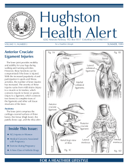
© Copyright 2026