
Dental Research Indian Journal of
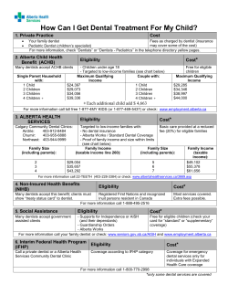
Indian Journal of
Dental Research
ISSN 0970-9290, E-ISSN 1998-3603
Volume: 21 / Issue: 1 / Jan - Mar 2010
CONTENTS
Editorial
Title of scientific papers
B Sivapathasundharam .......................................................................................................................................................................................... 1
Original Research
Osteoblast response to commercially available demineralized bone matrices — An in-vitro study
S Thanga Kumaran, KV Arun, Sabitha Sudarsan, Avaneendra Talwar, N Srinivasan ............................................................................................. 3
Evaluation of circulating immune complexes and serum immunoglobulins in oral cancer patients - A follow up study
Sameena Parveen, Neeraj Taneja, Renuka J Bathi, AC Deka............................................................................................................................... 10
Occlusal morphology of mandibular second molars in Iranian adolescents
Ramin Mosharraf, Behnaz Ebadian, Zeilabi Ali, Akhlaghi Najme, Shamashian Niloofar, Karimi Leila ................................................................. 16
The effect of smoking on gingival crevicular f luid levels of myeloperoxidase
Balwant Rai, Jasdeep Kaur, SC Anand, Kuldeep Laller ........................................................................................................................................ 20
Tensile properties of orthodontic elastomeric ligatures
F Ahrari, T Jalaly, M Zebarjad ............................................................................................................................................................................... 23
Antibacterial effect of bioactive glass in combination with powdered enamel and dentin
AR Prabhakar, Santhosh CH Kumar ..................................................................................................................................................................... 30
Coronal leakage of four intracanal medications after exposure to human saliva in the presence of a
temporary filling material
Rebeca Dibe Veríssimo, Eduardo Diogo Gurgel-Filho,Gustavo De-Deus, Tauby Coutinho-Filho, Francisco José de Souza-Filho ..................... 35
Endodontic sealers: Intratubular penetration and permeability to Enterococcus faecalis
Maria Cecília Tezelli Bortolini, Silvana Soléo Ferreira dos Santos, Sandra Márcia Habitante, Jane Rose Dias Dionísio Rodrigues,
Rodrigo Vance, Antonio Olavo Cardoso Jorge .................................................................................................................................................... 40
Four novel prosthodontic methods for managing upper airway resistance syndrome: An investigative analysis
revealing the efficacy of the new nasopharyngeal aperture guard appliance
R Venkat, N Gopichander, M Vasantakumar ........................................................................................................................................................ 44
Influence of light-curing units and restorative materials on the micro hardness of resin cements
Rosiane Nogueira Kuguimiya, Luciana Bastos Alves, Flávio Roberto Guerra Seabra, Carlos Frederico de Moraes Sarmento,
Alex José Souza Santos, Claudia Tavares Machado............................................................................................................................................ 49
Comparison of the effect of hydrogel and solution forms of sodium ascorbate on orthodontic bracket-enamel
shear bond strength immediately after bleaching: An in vitro study
Soodabeh Kimyai, Siavash Savadi Oskoee, Ali Rafighi, Hadi Valizadeh, Amir Ahmad Ajami, Zahra Norooz Zadeh Helali ................................ 54
Comparison of stress patterns and displacement in conventional cantilever fixed partial denture with resin bonded
cantilever fixed partial denture: A finite element analysis
E Prashanti, S Sajjan, M Kumar ............................................................................................................................................................................ 59
Effects of radio-opacifier addition in dental impression material
Eduardo Gonçalves Mota, Angela Rigo, Maria Ivete Bolzan Rockenbach, Nilza Pereira da Costa ..................................................................... 63
In vitro comparative analysis of resistance to compression of laboratory resin composites and a ceramic system
Alexandre Campos Montenegro, Cíntia Fernandes do Couto, Paulo Roberto Rezende Ventura, Cresus Vinicius Depes Gouvêa,
Aldir Nascimento Machado ................................................................................................................................................................................. 68
Studies on development of controlled delivery of combination drug(s) to periodontal pocket
Gaurav Tiwari, Ruchi Tiwari, Awani K Rai............................................................................................................................................................. 72
Oral health behavior and its determinants in a group of Iranian students
Hossain Neamatollahi, Masoumeh Ebrahimi ...................................................................................................................................................... 84
Copyright © 2010 by Editor-in-chief, IJDR
Prevalence of oral Entamoeba gingivalis and Trichomonas tenax in patients with periodontal disease and healthy
population in Shiraz, southern Iran
J Ghabanchi, M Zibaei, M Daghigh Afkar, AH Sarbazie ....................................................................................................................................... 89
An in vitro study to evaluate the effect of storage time and application of subsequent layers on the variation in
thickness of three commercially available die spacers
Sunil J Jacob, Chethan Hegde, Krishna D Prasad, Manoj Shetty........................................................................................................................ 92
Ex vivo evaluation of coronal and apical microbial leakage of root canal — Filled with gutta-percha or
Resilon/Epiphany root canal filling material
Fábio de Almeida-Gomes, Cláudio Maniglia-Ferreira, Marcelo de Morais Vitoriano, Bruno Carvalho-Sousa,
Nadine Luisa Soares de Lima Guimarães, Roberto Alves dos Santos, Eduardo Diogo Gurgel-Filho,
Márcia Maria de Negreiros Pinto Rocha .............................................................................................................................................................. 98
Effectiveness of transcutaneous electrical nerve stimulation and microcurrent electrical nerve stimulation in
bruxism associated with masticatory muscle pain - A comparative study
Bharat Rajpurohit, Subhash M Khatri, Deepa Metgud, Anjana Bagewadi ........................................................................................................ 104
The influence of torque and manual glide path on the defect or separation rate of NiTi rotary instruments
in root canal therapy
MH Zarrabi, M Javidi, M Vatanpour, H Esmaeili ................................................................................................................................................ 107
Lenticular card: A new method for denture identification
Shreya S Colvenkar ............................................................................................................................................................................................ 112
Review Articles
Hand hygiene among health care workers
Ameet Mani, AM Shubangi, Rajiv Saini ............................................................................................................................................................. 115
Obstructive sleep apnea and its management
C Sunitha, S Aravind Kumar ............................................................................................................................................................................... 119
Case Report
Fixed rehabilitation of patient with aggressive periodontitis using zygoma implants
Gunaseelan Rajan, Mirza Rustum Baig, John Nesan, Jayaram Subramanian................................................................................................... 125
Short Communications
Improving prosthetic prognosis by connective tissue ridge augmentation of alveolar ridge
Niraj Mishra, Balendra P Singh, Jitendra Rao, Pavitra Rastogi .......................................................................................................................... 129
Transient diplopia in dental outpatient clinic: An uncommon iatrogenic event
SM Balaji ............................................................................................................................................................................................................ 132
Numb chin syndrome: A case report and review of the literature
Jeevan Lata, Pramod Kumar .............................................................................................................................................................................. 135
Oral manifestations leading to the diagnosis of familial tuberous sclerosis
Hercílio Martelli Júnior, Leonardo Santos Lima, Paulo Rogério Ferreti Bonan, Ricardo D Coletta ................................................................... 138
An unusual communication between the mylohyoid and lingual nerves in man: Its significance in lingual nerve injury
Bhagath Kumar Potu, Suhani Sumalatha D’Silva, P Thejodhar, Nishita C Jattanna ........................................................................................... 141
Oral abnormalities in the Ellis-van Creveld syndrome
Prashant Babaji................................................................................................................................................................................................... 143
Letters to Editor
Tooth carving
Arathi Rao ........................................................................................................................................................................................................... 146
Has specialization isolated practitioners?
A Gur, JP Majra................................................................................................................................................................................................... 146
To do or not to do? Class II: Reflections of a conscientious dentist
V Susila Anand ................................................................................................................................................................................................... 147
Reviewers, 2009 ....................................................................................................................................................................................... 149
Instructions for Authors ..................................................................................................................................................................... 152
ORIGINAL RESEARCH
www.ijdr.in
Occlusal morphology of mandibular second molars
in Iranian adolescents
Ramin Mosharraf, Behnaz Ebadian, Zeilabi Ali, Akhlaghi Najme, Shamashian Niloofar, Karimi Leila
Department of Prosthodontics,
Isjahan University of Medical
Sciences, Hezar-Jalib, Ave
Isjahan, Iran
ABSTRACT
Received
: 24-06-07
Review completed : 09-01-09
Accepted
: 23-04-09
PubMed ID
: ***
DOI: 10.4103/0970-9290.62802
Context: During human evolution, the morphology of mandibular molar occlusal surface
has changed from pattern “y” to pattern “”. Six types of occlusal patterns were classified
as: 4, 4 y, 5, 5 y, 6 and 6 y.
Aims: To determine the prevalence of these six types of mandibular second molars in Iranian
adolescents.
Settings and Design: This descriptive investigation was undertaken in the high schools of Isfahan
city, Iran. The students were selected by cluster sampling method, and then they were screened
and only those with erupted mandibular second molars bilaterally were selected.
Materials and Methods: A total of 794 cases were randomly selected and the number of cusps
and groove pattern of mandibular second molar were examined intra-orally and by studying
dental casts.
Statistical Analysis Used: Descriptive statistics and chi-square test were used for data analysis.
Results: The most frequent occlusal configuration was the “4” form (76.9%). A total of 683 cases
(86%) were found to have four-cusp form, 104 cases (13.1%) were five-cusp form and 7 cases
(0.9%) were six-cusp form.
Conclusion: The most frequent occlusal configuration was the “4” form; thus, there is a high
evolutionary trend in Iranian mandibular second molars.
Key words: Occlusal morphology, tooth, anatomy, molar
In most dental anatomy textbooks, the permanent mandibular
second molar is often described as having a simple morphological
design, that consists of four cusps, placed on a square occlusal
surface and a cruciform () groove pattern.[1,2] However,
variations in size, cusp number and groove pattern have been
observed in mandibular molars of different populations.[1]
second molar is far more characteristic of Mongoloid and
Negroid populations than Caucasoid.[1,8-11] It is therefore
not uncommon to attempt to differentiate different ethnic
populations by their different morphological features.[12-17]
It is not known whether ethnicity influences dental
morphology. However, it is observed that there are different
degrees of expression and frequency in variation of teeth in
dentitions of different populations.[18] The final tooth form
represents the sum total of its genetic endowment and longterm environmental influences.[1]
In anthropological studies, morphological categories used to
describe these variations in occlusal surfaces of the mandibular
molars are based on a topology developed by Gregory and
Hellman[3] and Hellman:[4] “5 y,” “4 y”, “5” and “4”.
The criterion for determining whether a pattern is a “y” or a
“” is contact of the metaconid with the hypoconid. If contact
occurs, the pattern resembles a “y”; if no contact occurs, the
pattern resembles a “”.[5] The occurrence of the “y” or “”
fissure pattern is independent of the number of cusps.[6] Thus,
groove pattern and cusp number are considered separately
because their evolutionary changes are not well correlated
phenotypically.[7] It is assumed that this trait (fissure pattern) is
polygenic and its expression is determined by combinations of
alleles at two or more loci.[6] Basically, the five-cusp mandibular
Dental anthropology is the study of the origin and the variations
of the human dentition.[13] It is a useful tool to identify
geographic or racial affinities. Dentoanthropologic structures
useful for identification purposes include cusp size, number
and location of cusps, occlusal pattern, root configuration,
number and arrangement of teeth, and individual tooth
measurements.[1,19,20] Few dental anthropological studies have
investigated the associations between these dental features
and crown traits in humans using quantitative methods.[21]
Address for correspondence:
Dr. Ramin Mosharraf,
E-mail: [email protected]
The present descriptive study was undertaken to investigate
the prevalence of six types of mandibular second molars in
Iranian adolescents.
Indian J Dent Res, 21(1), 2010
16
Occlusal morphology of lower second molars
MATERIALS AND METHODS
This descriptive investigation was undertaken in the high
schools of Isfahan city, Iran. The students were selected
by cluster sampling method, and then they were screened
and only those with erupted mandibular second molars
bilaterally were selected. Direct intraoral examination was
done carefully and morphological details of the crown,
namely, the number of cusps and groove patterns of teeth
and gender of the subjects were recorded on prepared forms.
Data were excluded from the investigation in cases in which
the teeth were restored, worn or heavily broken down. After
that, hydrocolloid impressions were taken and dental casts
were immediately poured. Alginate (Alginoplast; Heraeus
Kulzer, Hanau, Germany) was the impression material used
and the casts were made of dental stone type III (Moldano;
Heraeus Kulzer, Hanau, Germany). The study was repeated
on the dental casts and the resultant data were compared with
clinical data, for each subject. If any difference was found,
the examination of that subject was repeated; hence, the final
decision was made on the basis of clinical data. A cusp was
considered as a pronounced elevation on the occlusal surface
of a tooth terminating in a conical, rounded or flat surface.[6]
A total of 794 cases (15-17 years old) were selected and the
number of cusps and groove pattern of mandibular second
molars were examined; 6 types of occlusal fissure pattern
(4 y, 4, 5 y, 5, 6 y and 6) [Figure 1] were
recorded. Descriptive statistics and chi-square test were
used for data analysis.
RESULTS
Of the 794 persons examined, 405 (51%) were males and
Mosharraf, et al.
389 (49%) were females. In this population, we examined
1588 teeth (794 2), and the four-cusp form was the
most frequent occlusal configuration (86%) [Table 1]. The
predominant occlusal pattern was groove form with a “”
shape (87.6%) [Table 2]. The most frequent occlusal surface
configuration was the “4” form (76.9%), and the “6 y”
form was seen only in one case [Table 3]. Most cases (567/794;
71.4%) were bilateral “4” form [Table 4]. The rates of each
type of occlusal patterns between males and females had no
significant differences in “5 y”, “4”, “6 y” and “6”
groups. However, “5” form had significantly higher rate in
males (PLeft 0.001/PRight 0.020). Chi-square analysis also
revealed significantly higher rate in females in the “4 y”
form group (P 0.001).
Using the Pearson correlation test, significant correlations
were observed between left and right occlusal pattern shapes
(P 0.01).
DISCUSSION
Few dental anthropological studies have investigated the
associations between these dental features and crown traits
in humans using quantitative methods.[21]
In this type of studies, some researchers used intraoral
examination,[1,14,17] some studied dental casts,[21-25] and some used
both methods.[26,27] Intraoral examination has the advantages
of accurate recording, proper identification of teeth, and
follow-up of patients when needed. It ensures racial and sexual
identification.[17] A sample of extracted teeth would be less than
ideal.[1,14] For more precise results, we used both methods (intraoral and cast examinations) in the present study.
Hellman classified the mandibular molars based on the
occlusal pattern and the number of cusps.[28] According
to him, the basic pattern is the “y-5” type, with five cusps
and a Y-shaped occlusal configuration. Loh stated that the
distobuccal cusp (hypoconulid) is the most variable and in
the evolutionary advanced type, it disappears and therefore
leads to a four-cusp form.[1] He observed a relatively high
incidence of five-cusp second molars in the Singaporean
population. Observations on the teeth of the Chinese from
mainland China were made by Montelius, who reported a
high incidence (56%) of five-cusp forms in this tooth.[29] In
Table 1: Cusp number of mandibular second molar
No. cusps
4-cusp form
5-cusp form
6-cusp form
Left (%)
683 (86)
104 (13.1)
7 (0.9)
Right (%)
684 (86.1)
102 (12.8)
8 (1)
Total (%)
1367 (86)
206 (13
15 (0.94)
Table 2: Groove patterns of mandibular second molar
Figure 1: Mandibular molar patterns y and fissure patterns are
shown. 1- Protoconid; 2- metaconid; 3- hypoconid; 4- entoconid;
5- hypocoulid; 6- sixth cusp
17
Groove form
“” shaped
“y” shaped
Left (%)
697 (87.8)
97 (12.2)
Right (%)
694 (87.4)
100 (12.6)
Total (%)
1391 (87.6)
197 (12.4)
Indian J Dent Res, 21(1), 2010
Occlusal morphology of lower second molars
Mosharraf, et al.
Table 3: Occlusal morphology of mandibular second molar
Occlusal morphology
4
4
y
5
5
y
6
6
y
Left (%)
612 (77.1)
71 (8.9)
79 (9.9)
25 (3.1)
6 (0.8)
1 (0.1)
Right (%)
609 (76.7)
75 (9.7)
77 (9.7)
25 (3.1)
8 (1.0)
0 (0)
Total (%)
1221 (76.9)
146 (9.2)
156 (9.8)
50 (3.1)
14 (0.9)
1 (0.06)
Table 4: Unilateral or bilateral occlusal morphology of
mandibular second molar
Occlusal
morphology
4
4
y
5
5
y
6
6
y
Bilateral
567
48
57
17
4
0
Total
87
50
42
16
6
1
Unilateral
Left
45
23
22
8
4
1
Right
42
27
20
8
2
0
1997, Guo et al. by observations in new population of china,
stated that the rate of “4” in the second mandibular molars
is the highest, while the rate of “y 5” is the lowest.[30] They
stated that this fact may be due to gradual evolution in the
morphology of mandibular molar occlusal grooves that have
changed from pattern “y” to pattern “”.
In 1985, Hasund and Bang revealed that in Alaskan Eskimo
dentition, the predominant pattern of the lower first molar
was “y 5,” while for the second molar the dominating
patterns were “5” and “4,” and in the lower third molar,
“5” was found in the majority of cases.[31] Devoto and
Perrotto[7] stated that the “” groove pattern appeared on
second molar more than other two molars.
In this study, the most frequent occlusal configuration was
four-cusp form (86%) and the predominant groove pattern
was “” shape (87.6%). The most frequent occlusal surface
configuration was the “4” form (76.9%), and most cases
(71.4%) were bilateral “4” form. This high percentage
of groove pattern with “” shape and low percentage of
primitive “Y” pattern in our study show a high evolutionary
trend persisting in Iranian second molars.
Some of the studies suggested a sex predilection in men
to have the “y 5” or Dryopithecus pattern.[11,12,28] In this
investigation, no significant difference was observed between
males and females in “5 y,” “4,” “6 y” and “6” groups.
However, “5” form had a significantly higher rate in males
and “4 y” form had a significantly higher rate in females. In
study by Guo et al., the rates of each type of groove between
males and females showed no significant difference.[30]
In this study, Pearson correlation test revealed there were
significant correlations between the left and right occlusal
pattern shapes. This would suggest an inherent genetic factor
rather than a casual occurrence.
Indian J Dent Res, 21(1), 2010
The study of dental morphological characteristics and
odontometry is important in anthropological research as it
can provide information on the phylogenetic relationship
between species, as well as variations and diversities within
a population.[20] Furthermore, knowing common variations
in dental anatomy and morphology about each individual
tooth can help in performing some dental treatments such
as restorative, endodontic and orthodontic treatments.[5,6]
Therefore, the results of this anatomical study can be used
in both anthropological researches and clinical aspects of
dental sciences.
REFERENCES
1.
2.
3.
4.
5.
6.
7.
8.
9.
10.
11.
12.
13.
14.
15.
16.
17.
18.
19.
20.
21.
22.
Loh HS. Mongoloid features of the permanent mandibular second
molar in Singaporean Chinese. Aust Dent J 1991;36:442-4.
van beek GC. Dental morphology. Bristol: John Wright and Sons; 1983.
Gregory WK, Hellman M. The crown patterns of Fossils and recent
human molar teeth and their meaning. Nat Hist 1926;26:300-9.
Hellman M. Our third molar teeth: Their eruption, presence and
absence. Dent Cosmos 1936;78:750.
Ash MM, Nelson SJ, editors. Wheeler’s dental anatomy, physiology and
occlusion. St. Louis: Saunders; 2003.
Jordan RE, Abrams L, Kraus BS. Kraus’s dental anatomy and occlusion.
St. Louis: Mosby- year book, Inc; 1992.
Devoto FC, Perrotto BM. Groove pattern and cusp number of
mandibular molars from Tastilian Indians. J Dent Res 1972;51:205.
Goldstein MS. The cusps in the mandibular molar teeth of the Eskimo.
Am J Phys Anthrop 1931;16:215-35.
Brewer-Carias CA, le Blanc S, Neel JV. Genetic structure of a tribal
population, the Yanomama Indians XIII. Dental microdifferentiation.
Am J Phys Anthropol 1976;44:5-14.
Perzigian AJ. The dentition of the Indian Knoll skeletal population:
Odontometrics and cusp number. Am J Phys Anthropol 1976;44:113- 21.
Hasund A, Bang G. Morphologic characteristics of the Alaskan Eskimo
dentition: IV. Cusp number and groove patterns of mandibular molars.
Am J Phys Anthropol 1985;67:65-9.
Moorrees CF. Dentition as a criterion of race with special reference to
the Aleut. J Dent Res 1951;30:815-21.
Dahlberg AA. Dental traits as identification tool. Dent Progress
1963;3:155-60.
Loh HS. Coronal morphology of the mandibular second premolar in
the Singaporean Chinese. Aust Dent J 1993;38:283-6.
Tsai PL, Hsu JW, Lin LM, Liu KM. Logistic analysis of the effects of
shovel trait on Carabelli’s trait in a Mongoloid population. Am J Phys
Anthropol 1996;100:523-30.
Hsu JW, Tsai PL, Hsiao TH, Chang HP, Lin LM, Liu KM, et al. Ethnic dental
analysis of shovel and Carabelli’s traits in a Chinese population. Aust
Dent J 1999;44:40-5.
Mosharraf R, Hajian F. Occlusal morphology of the mandibular first
and second premolars in Iranian adolescents. Inter J Dent Anthropol
2004;5:10-4.
Kieser JA, van der Merwe CA. Classificatory reliability of the Carabelli
trait in man. Arch Oral Biol 1984;29:795-801.
Wood BA, Abbott SA, Graham SH. Analysis of the dental morphology of PlioPleistocene hominids. II. Mandibular molars—study of cusp areas, fissure
pattern and cross sectional shape of the crown. J Anal 1983;137:287-314.
Sharma JC. Dental morphology and odontometry of the Tibetan
immigrants. Am J Phys Anthropol 1983;61:495-505.
Kondo S, Funatsu T, Wakatsuki E, Haung ST, Change SY, Shibasaki Y,
et al. Sexual dimorphism in the tooth crown dimensions of the second
deciduous and first permanent molars of Taiwan Chinese. Okajimas
Folia Anat Jpn 1998;75:239-46.
Huang ST, Miura F, Soma K. A dental anthropological study of Chinese
in Taiwan (2). Teeth size, dental arch dimensions and forms. Gaoxiong
Yi Xue Ke Xue Za Zhi 1991;7:635-43.
18
Occlusal morphology of lower second molars
23.
24.
25.
26.
27.
28.
Huang ST, Miura F, Soma K. A dental anthropological study of Chinese
in Taiwan (3). Teeth size, dental arch dimensions and forms. Gaoxiong
Yi Xue Ke Xue Za Zhi 1992;8:665-78.
Moskona D, Vainder M, Hershkovitz I, Kobyliansky E. Bilateral
asymmetry in dental discrete traits in human isolates: South Sinai
Bedouin tribes. Anthropol Anz 1996;54:289-306.
Macesic M, Kaic Z, Dumancic J, Poje Z, Dumic M. Occlusal molar
surfaces in females with Turner’s syndrome. Coll Antropol
2003;27:761-8.
Liu Kl. Dental condition of two tribes of Taiwan aborigines. Ami and
Atayal. J Dent Res 1977;56:117-27.
Miura F, Soma K, Kuroki T, Fukawa T, Ishida T, Ishijo T, et al. Dental
anthropological study of Mongoloid in China. Kokubyo Gakkai Zasshi
1991;58:566-79.
Hellman M. Racial characters in human dentition. Proc Am Phil Soc
Mosharraf, et al.
29.
30.
31.
1928;67:65-9.
Montelius GA. Observations of teeth of Chinese. J Dent Res
1933;13:501-9
Guo L, Ren L, Sun DL, Shen J. Morphological study on occlusal grove
of mandibular molar of Chinese adults. Shanghai Kou Qiang Yi Xue
1997;6:129-31.
Hasund A, Bang G. Morphologic characteristics of the Alaskan Eskimo
dentition: IV. Cusp number and groove patterns of mandibular molars.
Am J Phys Anthropol 1985;67:65-9.
How to cite this article: Mosharraf R, Ebadian B, Ali Z, Najme A,
Niloofar S, Leila K. Occlusal morphology of mandibular second molars
in Iranian adolescents. Indian J Dent Res 2010;21:16-9.
Source of Support: Nil, Conflict of Interest: None declared.
Author Help: Online submission of the manuscripts
Articles can be submitted online from http://www.journalonweb.com. For online submission, the articles should be prepared in two files (first
page file and article file). Images should be submitted separately.
1)
2)
3)
4)
19
First Page File:
Prepare the title page, covering letter, acknowledgement etc. using a word processor program. All information related to your identity
should be included here. Use text/rtf/doc/pdf files. Do not zip the files.
Article File:
The main text of the article, beginning with the Abstract to References (including tables) should be in this file. Do not include any information
(such as acknowledgement, your names in page headers etc.) in this file. Use text/rtf/doc/pdf files. Do not zip the files. Limit the file size
to 1 MB. Do not incorporate images in the file. If file size is large, graphs can be submitted separately as images, without their being
incorporated in the article file. This will reduce the size of the file.
Images:
Submit good quality color images. Each image should be less than 2048 kb (2 MB) in size. The size of the image can be reduced by
decreasing the actual height and width of the images (keep up to about 6 inches and up to about 1800 x 1200 pixels). JPEG is the most
suitable file format. The image quality should be good enough to judge the scientific value of the image. For the purpose of printing, always
retain a good quality, high resolution image. This high resolution image should be sent to the editorial office at the time of sending a revised
article.
Legends:
Legends for the figures/images should be included at the end of the article file.
Indian J Dent Res, 21(1), 2010


© Copyright 2026