
Ultrathin MoO3 nanocrystalsself-assembled on
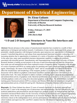
Nano Energy (2015) 12, 510–520
Available online at www.sciencedirect.com
journal homepage: www.elsevier.com/locate/nanoenergy
RAPID COMMUNICATION
Ultrathin MoO3 nanocrystalsself-assembled
on graphene nanosheets via oxygen bonding
as supercapacitor electrodes of high
capacitance and long cycle life
Kai Zhoua, Weijia Zhoua,n, Xiaojun Liua, Yuanhua Sangb,
Shaozheng Jib, Wei Lia, Jia Lua, Ligui Lia, Wenhan Niua,
Hong Liub, Shaowei Chena,c,n
a
New Energy Research Institute, School of Environment and Energy, South China University of Technology,
Guangzhou Higher Education Mega Center, Guangzhou, Guangdong 510006, China
b
State Key Laboratory of Crystal Materials, Center of Bio & Micro/Nano Functional Materials,
Shandong University, 27 Shandanan Road, Jinan, Shandong 250100, China
c
Department of Chemistry and Biochemistry, University of California, 1156 High Street, Santa Cruz,
CA 95064, USA
Received 22 October 2014; received in revised form 16 December 2014; accepted 8 January 2015
Available online 16 January 2015
KEYWORDS
Abstract
MoO3;
3D graphene frameworks;
Oxygen-bonding;
Solid-state symmetric
supercapacitor;
Electrochemical
stability
Ultrathin MoO3 nanocrystals were assembled on 3D graphene oxide frameworks via a hydrothermal
reaction forming a layered structure by oxygen-bonding interactions at the interface. The structure
and morphology of the resulting MoO3-GAs hybrids were characterized by a range of experimental
tools including atomic force microscopy, scanning electron microscopy, transmission electron
microscopy, X-ray photoelectron spectroscopy, infrared and Raman spectroscopy. Because of
abundant exposed active sites of the ultrathin MoO3 and rapid ion diffusion and electron transport
of 3D graphene frameworks, the resulting MoO3-GAs hybrids possess the highest specific
capacitances and excellent cycling stability in both aqueous (527 F g 1 at the current density of
1.0 A g 1, 100% retention after 10,000 cycles) and solid electrolytes (373 F g–1 at 1.0 A g 1, 100%
retention after 5,000 cycles) among leading literature results of similar systems.
& 2015 Elsevier Ltd. All rights reserved.
n
Corresponding authors.
E-mail addresses: [email protected] (W. Zhou), [email protected] (S. Chen).
http://dx.doi.org/10.1016/j.nanoen.2015.01.017
2211-2855/& 2015 Elsevier Ltd. All rights reserved.
Ultrathin MoO3 nanocrystalsself-assembled on graphene nanosheets via oxygen bonding as supercapacitor electrodes
Introduction
Supercapacitors are a promising class of energy storage devices
due to their high power density, ultrafast charging/discharging
rate and excellent cycle performance, which show potential
applications for hybrid electric vehicles, consumer electronics,
backup energy systems, etc [1–4]. In recent years, extensive
research has been devoted to the design and engineering of
new materials and structures to further improve their electrochemical performance, for instance, using pseudocapacitors
which store charges through fast reversible Faradaic reactions
from redox-active materials [5–7]. Among these, layered
transition metal oxides/hydroxides, such as MnO2 [8], Co
(OH)2 [9], V2O5 [10], and Ni(OH)2 [11], have been extensively
explored as supercapacitor electrodes for the enhancement of
specific capacitance, energy and power density.
In particular, α-MoO3 is an n-type semiconductor with an
anisotropic layered structure along the [0 1 0] direction. The
layered structure is formed by stacking bilayer sheets of MoO6
octahedra with van der Waals forces [12,13]. These structures
are presumably advantageous for the insertion/removal of
small ions such as H + and K + . However, its low electronic
conductivity and lack of structural integrity under electrochemical reactions are detrimental to a high-rate and long-term
cycling performance in electrochemical devices [14–16]. Therefore, it remains a challenge to use MoO3 as supercapacitor
electrodes, because the capacitance may diminish significantly
in a prolonged charge–discharge cycling process. For example,
Kim et al. reported that the capacitance of structured MoO3
diminished by 18% after only 1000 cycles of charging and
discharging [17]. Li et al. also reported a significant loss of
25% of the capacitance of α-MoO3 after only 1000 cycles [18].
This is the primary motivation of the present study in which we
employ two effective strategies to overcome this attenuation
problem: (i) fabrication of ultrathin MoO3 nanocrystals on the
surface of mechanically robust graphene nanosheets leading to
a high electrochemically active surface area for faradic reactions at the defects and grain boundaries of the crystalline
lattices, and (ii) formation of strong chemical contacts between
the ultrathin MoO3 nanocrystals and graphene via oxygen
bonding to facilitate interfacial charge transfer and to reduce
the loss of active materials.
Graphene with large specific surface area, high electrical
conductivity, excellent mechanical properties and chemical
stability, has shown great potential for supercapacitor electrode
materials [19–22]. However, because of easy restacking of the
graphene nanosheets due to strong π–π interactions, supercapacitor electrodes based directly on graphene sheets did not
display the expected high rate performance. A variety of threedimensional (3D) graphene materials, for example, graphene
hydrogels [23–26] and graphene papers [27–28], which contain
continuously interconnected pores structures with high surface
area-to-volume ratios have been developed to overcome the
restacking problems of graphene sheets. At the same time, due
to their outstanding electrical properties and mechanical
flexibility, monolithic 3D graphene has been widely explored
as electrode materials for solid-state supercapacitors, which
are considered as promising smart and efficient energy-storage
devices [24,29,30].
In this work, ultrathin MoO3 nanocrystals were epitaxially
grown on the surface of graphene oxide (GO) nanosheets
511
that self-assembled into 3D aerogels (MoO3-GAs). A variety
of experimental techniques including atomic force microscopy (AFM), transmission electron microscopy (TEM), X-ray
photoelectron spectroscopy (XPS), infrared and Raman
spectroscopy were used to characterize the resulting composite structure and interfacial bonding between the MoO3
nanocrystals and graphene nanosheets. Electrochemical
studies showed that MoO3-GAs might be used as effective
supercapacitor electrodes with an excellent specific capacitance of 586 F g 1 at the scan rate of 50 mV s 1 and
527 F g 1 at the current density of 1.0 A g 1 in 1.0 M H2SO4
aqueous electrolyte in a three-electrode configuration. Remarkably, the electrode materials also exhibited excellent
stability in long-term cycling in both aqueous (at a current
density of 10.0 A g 1, 100% retention after 10,000 cycles)
and solid electrolytes (at a current density of 1.0 A g 1,
100% retention after 5000 cycles). To our knowledge, this is
the best supercapacitor performance, in particular, with
regard to specific capacitance and cycling stability, as
compared to those reported in the literature of Mo-based
supercapacitor electrode materials.
Experimental section
Materials
Sodium molybdate (Na2MoO4 2H2O), graphite powers, potassium persulfate (K2S2O8), phosphorus pentoxide (P2O5), potassium permanganate (KMnO4), hydrogen peroxide (H2O2) and
dimethylformamide (DMF) were purchased from Sinopharm
Chemical Reagents Co., Ltd. All reagents were of analytical
grade, and used without further purification. Deionized water
was supplied with a Barnstead Nanopure Water System
(18.3 MΩ cm).
Synthesis of MoO3-GAs
Graphene oxide (GO) was prepared by a modified Hummers
method using natural graphite flakes as the starting materials
[31]. MoO3-graphene aerogels (MoO3-GAs) were synthesized by a
combined hydrothermal and freeze-drying method. In a typical
procedure, 15 mL of a GO (4.0 mg mL 1) solution in DMF
containing 50 mg of sodium molybdate was dispersed evenly
by ultrasonic agitation to obtain an emulsion. The resulting
dispersion was sealed in a Teflon-lined autoclave, and heated at
200 1C for 24 h. After the autoclave naturally cooled to room
temperature, the as-prepared graphene hydrogel was taken out
and cleaned with deionized water several times until the
solution became clear, followed by freeze-drying at 45 1C
for 24 h. For comparison, pristine graphene oxide aerogels
(GAs), and MoO3-GAs at other MoO3 loadings were also prepared
in a similar fashion with GO solution: 10 mg of sodium molybdate
to prepare a lower MoO3 loading of MoO3-GAs (L-MoO3-GAs), and
100 mg to a higher MoO3 loading sample (H-MoO3-GAs).
Structural characterizations
The microstructural and morphological details of the samples
were examined with a NOVA NANOSEM 430 field emission
512
K. Zhou et al.
scanning electron microscope (FESEM) and a JEOL JEM-2100
transmission electron microscope (TEM) at an acceleration voltage of 200 kV. AFM measurements were performed using a
Nanosurf Easyscan2 instrument (Veeco Dimension 3100). The
MoO3-GAs was dispersed in ethanol by ultrasonic agitation to
obtain homogeneously dispersed MoO3-graphene nanosheets,
which were dropped onto the surface of a clean silicon wafer.
Chemical compositions were evaluated by element mapping
(Philips Tecnai F20) and XPS (ESCALAB 250) measurements. XRD
analysis was performed on a Bruker AXS D8 Discover diffractometer with Cu Kα radiation. Raman spectra were recorded on
a RENISHAW inVia instrument with an Ar laser source of 488 nm
in a macroscopic configuration. Infrared spectra were acquired
with a NICOLET 6700 FTIR spectrometer (Thermo Scientific,
USA). The BET surface area was characterized by Micromeritics
ASAP 2010 with nitrogen adsorption at 77 K. Thermogravimetric
analyses (TGA) was carried out under a steady flow of O2,
where the temperature was scanned from 30 to 600 1C at a rate
of 10 1C min 1.
Electrochemical measurements
The monolithic aerogels were cut into small slices with a
thickness of about 1 mm and diameter of about 10 mm, which
were further pressed onto titanium meshes as working electrodes. All electrochemical experiments were carried out with
a CHI 660E Electrochemical Workstation (CH Instruments,
China). In the three-electrode system, a saturated calomel
reference electrode (SCE, Hg/Hg2Cl2 in saturated KCl) and a Pt
counter electrodes were used along with 1.0 M H2SO4 aqueous
electrolyte. In the two-electrode system, two same slices of
the aerogel monoliths that were pressed on titanium meshes
were directly assembled into a symmetric supercapacitor in
1.0 M H2SO4 aqueous electrolyte. In the case of solid-state
symmetric supercapacitors, two slices of aerogel monoliths
pressed on titanium meshes were immersed in the solution of
PVA/H2SO4 gel electrolyte for several times. The resulting
electrolyte-filled electrodes were solidified for 12 h at ambient
conditions. Finally, the two electrodes were assembled into a
solid-state symmetric supercapacitor carefully. The gel electrolyte was fabricated by mixing 3 g H2SO4 and 3 g PVA in
30 mL of deionized water that was subject to heating up to
80 1C for several hours under vigorous stirring until the solution
become transparent.
Specific capacitance C (F/g) for a single electrode was
calculated from the CV and charge-discharge curves by Eqs.
(1) and (2), respectively, where I1 (A) is the response
current, ΔV (V) is the voltage window, ν (V/s) is the scan
rate, I2 (A) is the constant discharge current, Δt (s) is the
discharging time and m1 (g) is the weight of the single
electrode.
R
C ¼ I1 dV
ð1Þ
vm1 ΔV
C¼
I2 Δt
m1 ΔV
ð2Þ
Energy density (E) and power density (P) were calculated
by Eqs. (3) and (4), respectively.
E¼
1
CðΔVÞ2
2
ð3Þ
P¼
E
Δt
ð4Þ
Specific capacitance Cs (F/g) for symmetric supercapacitor was calculated from the CV and charge-discharge curves
by Eqs. (5) and (6), respectively, where I1 (A) is the response
current, ΔV (V) is the voltage window, ν (V/s) is the scan
rate, I2 (A) is the constant discharge current, Δt (s) is the
discharging time and m2 (g) is the total mass of two
electrodes.
R
Cs ¼ 4 I1 dV
ð5Þ
vm2 ΔV
Cs ¼
4I2 Δt
m2 ΔV
ð6Þ
Result and discussion
As manifested in SEM studies (Figure 1a and b), the as-prepared
MoO3-GAs exhibited a 3D interconnected porous network with a
smooth surface, where graphene sheets most likely served as
the structural scaffolds. Note that MoO3-GAs might be exfoliated and dispersed in ethanol by ultrasonic agitation forming a
homogeneous solution of MoO3/graphene nanosheets. AFM
topographic studies showed that the height of the MoO3/
graphene nanosheets was rather uniform at o2.0 nm
(Figure 1c and S1), signifying that MoO3 were likely evenly
distributed on the surface of the graphene nanosheets which
served as a structural unit in the MoO3-GAs hybrids (vide infra).
Note that the resolution of SEM and AFM were not high enough
to identify the exact location of the MoO3 nanocrystals (MoO3
was not detected in XRD measurements either, because of small
size and low loading, Figure S2a). A broad diffraction peak
centered at around 24.51 in Fig. S2a was ascribed to the
graphite (0 0 2), because of reduction of oxygen functional
groups in graphene [24,32]. However, the XRD pattern of HMoO3-GAs in Figure S2b exhibted three diffraction peaks at
2θ=12.81, 25.71 and 39.01, characteristic of the (0 2 0), (0 4 0)
and (0 6 0) diffraction planes of MoO3 (JCPDS no. 05-0508),
respectively. Then, the loading amount of MoO3 was measured
by TGA measurements (Figure S3). It can be seen that the MoO3
loading was quantified at 4.8 wt%, 18.6 wt%, 30.8 wt% of for LMoO3-GAs, MoO3-GAs and H-MoO3-GAs, respectively.
The formation of MoO3-GAs composites was readily manifested in FTIR measurements, as depicted in Figure 1d. It can
be seen that several prominent vibrational bands appear at
1571 cm 1, 1401 cm 1, and 1180 cm 1 with both MoO3-GAs
(black curve) and pristine GAs (red curve). These may be
assigned to the C=C, CC and CO functional moieties of the
graphene nanosheets, respectively. For the MoO3-GAs composites, however, several additional virbational bands emerged
within the range of 500–1000 cm 1. Of these, the vibrational
bands at 945 cm 1 and 896 cm 1 might be assigned to the Moterminal oxygen stretching vibrations, while the peaks at
616 cm 1 and 779 cm 1 were likely due to the stretching
vibrations of oxygen linked with two or three molybdenum
atoms, respectively [16,33,34]. Consistent results were
obtained in Raman measurements, as depicted in the inset to
Figure 1d. One can see that after MoO3 was deposited onto the
Ultrathin MoO3 nanocrystalsself-assembled on graphene nanosheets via oxygen bonding as supercapacitor electrodes
513
Figure 1 (a) and (b) SEM images of MoO3-GAs. (c) Representative AFM topograph of MoO3/graphene nanosheets and the
corresponding line-scan profile. (d) FTIR spectra of the pristine GAs and MoO3-GAs. Inset is the corresponding Raman spectra.
graphene sheets (black curve), the graphene D and G bands
red-shifted somewhat to 1352 and 1589 cm 1 from 1344 and
1584 cm 1 of the pristine graphene areogels (GAs, red curve),
along with an increase of the ratio of the their intensities (ID/IG)
to 1.14 from 1.05. This may be accounted for the enhanced
defect structures of the graphene nanosheets upon the loading
of MoO3 [35].
Further morphological details were resolved in (HR)TEM
analyses, which provided direct evidence of ultrathin MoO3
nanocrystals self-assembled on graphene oxide nanosheets.
Figure 2a and b shows that the MoO3/graphene nanosheets
exhibited a highly wrinkled surface; and MoO3 nanocrystals
can be seen to be rather evenly distributed on the surface of
the graphene nanosheets, as manifested in high-resolution
measurements (Figure 2c) where well-defined lattice fringes
can be resolved and the interplanar spacing of 0.38 nm is
consistent with the (1 1 0) crystalline planes of MoO3 that
were perpendicular to the graphene nanosheets. These observations were in good agreement with results in AFM study
(Figure 1c). The crystalline properties of the composites were
also examined by selected area electron diffraction (Figure S4)
that clearly signified the formation of polycrystalline MoO3
nanocrystals on graphene nanosheets. Note that MoO3 is a
layered semiconductor with an anisotropic layered structure
along the [0 1 0] direction (Figure 2d). The layered structure is
formed by stacking bilayer sheets of MoO6 octahedra by van
der Waals forces [36,37]. The thickness of a single layer of
MoO6 octahedra is about 0.7 nm, and a MoO3/graphene sheet/
MoO3 sandwich structure is about 1.7 nm in thickness. This is
in good agreement with the thickness determined by AFM
measurements of the MoO3/graphene nanosheet composites
(Figure 1c), suggesting the formation of MoO3/graphene/MoO3
sandwiched structures. Furthermore, elemental mapping studies of the MoO3-graphene composites showed a uniform
514
K. Zhou et al.
Figure 2 (a) Low and (b) high resolution TEM images of the MoO3/graphene nanosheets. (c) STEM image of MoO3/graphene
nanosheets. (d) Schematic of MoO3 self-assembled on graphene nanosheets. (e) and (f) Schematic diagrams of MoO3 bonding with
graphene. (g) Element mapping images of MoO3-graphene nanosheets.
distribution of the C, Mo, and O elements (Figure 2g),
consistent with results from SEM (Figure 1b), AFM (Figure 1c)
and HRTEM (Figure 2b and c) measurements.
XPS measurements were then carried out to characterize
the surface elemental compositions and valence states of
the MoO3-GAs. As shown in the survey spectrum in Figure 3a,
carbon, molybdenum and oxygen were the only elements
that could be identified. Deconvolution of the highresolution scan of the C1s electrons exhibited three peaks
at 284.5, 285.5 and 290.2 eV (Figure 3b), corresponding to
the C = C, C O and O =C OH carbons, respectively [38].
This suggests the formation of oxygenated functional moieties in graphene oxide nanosheets. For the Mo3d electrons
(Figure 3c), a doublet can be identified at 235. 6 eV and
232.5 eV, in good agreement with the binding energies of the
Mo (VI) 3d3/2 and 3d5/2 electrons and hence the formation of
MoO3 in the composites [37]. For the O1s electrons
(Figure 3d), two components can be deconvoluted: O1 at
the low binding energy of 530.4 eV which might be assigned to
the lattice oxygen in MoO3, whereas the peak at 532.4 eV (O2)
to C O of graphene oxide [38,39]. In addition, the survey
spectrum of bare GAs was showed in Figure S5a, where carbon
and oxygen were the only elements that could be identified.
Deconvolution of the high-resolution scan of the O1s electrons
exhibited two peaks at 531.1 and 532.4 eV (Figure S5b),
corresponding to the C=O and CO of graphene oxide,
respectively [38]. These comparisons suggest the replacement
of C=O by Mo in MoO3 forming the CO Mo linkage.
Therefore, we concluded that the self-assembly of ultrathin
MoO3 nanocrystals on graphene oxide nanosheets were facilitated via oxygen-bonding interactions (C OMo) at the
interface. Furthermore, based on the integrated peak areas,
the atomic ratio of Mo/C in MoO3-GAswas estimated to be only
2.6 at%, corresponding to a mass loading of MoO3 in MoO3-GAs
at 23.7 wt%. Two additional MoO3-GAs hybrids were prepared
in a similar fashion with the corresponding mass contents of
MoO3 at about 4.9 wt% (L-MoO3-GAs) and 33.8 wt% (H-MoO3GAs), respectively, as shown in Figure S6. The results of MoO3
loading estimated from XPS were consistent with those
obtained by TGA (Figure S3).
In order to further study the assembly mechanism between
MoO3 and graphene nanosheets, a reduced graphene/MoO3 composite (RGO-Mo) was synthesized in the same manner except
that reduced graphene oxide prepared by chemical reduction
with hydrazine hydrate was used instead of graphene oxide
(synthetic details in the Supporting Information). As shown in
Figure 4a and b, molybdenum could not be detected in RGO-Mo
(red curve), whereas Mo3d and Mo3p were very well-defined in
MoO3-GAs (black curve). This indicated that MoO3 nanocrystals
could not be formed on reduced graphene without dangling
oxygen in DMF hydrothermal reaction. Further studies showed
that no MoO3 nanocrystals were produced in the absence of graphene oxide nanosheets in DMF hydrothermal reaction (Figure S7).
Therefore, it is likely that the oxygenated moieties on the
Ultrathin MoO3 nanocrystalsself-assembled on graphene nanosheets via oxygen bonding as supercapacitor electrodes
515
Figure 3 (a) XPS survey spectrum of MoO3-GAs and high resolution scans of (b) C1s, (c) Mo3d and (d) O1s electrons. Black curves are
experimental data and color curves are deconvolution fits.
Figure 4 (a) XPS survey spectra of MoO3-GAs and RGO-Mo. (b) High resolution scans of the Mo3d electrons of MoO3-GAs and RGO-Mo.
graphene oxide nanosheets played an important role in the
assembly of ultrathin MoO3 nanocrystals by Mo OC bonding
linkages at the interface. On the basis of these XPS results
(Figs. 3 and 4), the oxygen-bonding structures between graphene
oxide and MoO3 were depicted in Figure 2e and f. Note that
similar structures have been reported previously, such as Ni
(OH)2/graphene [38] and MoS2/graphene [40], where it was
reported that oxygen on graphene might affect the structure and
morphology of the resulting composites.
Remarkably, the prepared MoO3-GAs composites exhibited a
significant performance as supercapacitor electrodes. Cyclic
voltammetry (CV) and galvanostatic charge/discharge were
employed to examine the electrochemical properties of
MoO3-GAs electrodes in an aqueous electrolyte of 1.0 M H2SO4
in a three-electrode (Figure 5) and two-electrode configuration
(Figure S8). For comparison, pristine GAs and MoO3 were also
tested under the same conditions. Figure 5a shows the CV
curves of the GAs and MoO3-GAs electrodes at the scan rate of
50 mV s 1. It can be seen that the GAs exhibits a typical
rectangular shape (black curve), implying pure electrical
double-layer capacitive behaviors. In contrast, MoO3-GAs displays two pairs of rather well-defined voltammetric peaks (red
516
K. Zhou et al.
Figure 5 (a) CV curves of GAs and MoO3-GAs electrodes at the scan rate of 50 mV s 1. (b) Galvanostatic charge/discharge curves at
a current density of 1 A g 1. (c) CV curves of the MoO3-GAs electrode at different scan rates in 1.0 M H2SO4. (d) Galvanostatic
charge/discharge curves of the MoO3-GAs electrode at different current densities. (e) Specific capacitance of the MoO3-GAs
electrode as a function of scan rate and current density. (f) Cycling stability of the MoO3-GAs electrode at a current density of
10.0 A g 1. Inset is the last 20 charge/discharge profile of the MoO3-GAs at 10.0 A g 1.
curve) with the formal potentials (E10 ) at +0.20 V and +0.30 V,
respectively, which might be ascribed to the redox reactions of
Mo(VI)2Mo(V)2Mo(VI) [18]. Similar results were obtained in
CV measurements of pristine MoO3 (Figure S9). Such voltammetric features have also been observed in Li-ion batteries with
a MoO3 electrode [16,41]. In addition, the peak splitting (ΔEp)
of the two pairs of voltammetric peaks was only 5.1 mV and
4.8 mV indicating facile electron-transfer kinetics, which is
advantageous for pseudo-capacitive contributions. Importantly,
from these CV profiles, the MoO3-GAs composites showed an
impressive specific capacitance of 586 F g 1 at the scan rate
of 50 mV s 1, more than 5-fold larger than that of GAs
(122 F g 1), and markedly higher than that of pristine MoO3
(1.7 F g 1). Furthermore, in the galvanostatic charge/discharge
measurements (Figure 5b), pristine GAs exhibited a rather
symmetrical triangular shape (black curve) whereas marked
deviation can be seen with MoO3-GAs (red curve), as a result of
significant contributions of faradaic capacitance, as described
above. The specific capacitance of MoO3-GAs was estimated to
be 527 F g 1 at the current density of 1.0 A g 1, while that of
GAs was only 91 F g 1. It is worth noting that when normalized
to the mass of redox-active MoO3 alone the specific capacitance
of MoO3-GAs was actually 2223.6 F g 1 at 1.0 A g 1.
As shown in Figure 5c, the voltammetric features of the
MoO3-GAs electrode remained virtually unchanged with the
potential scan rate varied from 1.0 to 200 mV s 1, consistent with fast electron-transfer kinetics. This was further
supported by results from electrochemical impedance
analysis (Figure S10). The equivalent series resistance
(ESR) of MoO3-GAs was only 1.4 Ω, close to that of pure
GAs (1.2 Ω); and the deposition of MoO3 nanocrystals on the
graphene oxide surface did not appear to affect charge
diffusion, as manifested by the similar slope of the lowfrequency linear segment of MoO3-GAs and pure GAs.
Nevertheless, the arc in the high frequency region exhibits
a larger diameter with MoO3-GAs than with GAs alone,
which might be ascribed to the redox reactions of ultrathin
MoO3 nanocrystals deposited on the graphene oxide surface
in MoO3-GAs.
Figure 5d depicted the galvanostatic charge/discharge curves
of the MoO3-GAs electrode in 1.0 M H2SO4 aqueous electrolyte
at different current densities. The specific capacitances were
calculated by the integrated area of the CV curves and
discharging time of the galvanostatic charge/discharge curves,
respectively. The correlation between the specific capacitance
and the scan rate and current density for the MoO3-GAs
electrode was presented in Figure 5e. Notably, the specific
capacitance of MoO3-GAs showed only a slight decrease to
512 F g 1 at a scan rate of 200 mV s 1 from 755 F g 1 at
1.0 mV s 1. Furthermore, with the increase of the current
density up to 10.0 A g 1, the specific capacitance of MoO3-GAs
remains at 328 F g 1. The excellent rate capability of the
MoO3-GAs electrode could be ascribed to the efficient, fast
charge transport of the 3D graphene frameworks.
The electrochemical stability of the MoO3-GAs electrode
was also investigated by galvanostatic charge/discharge
Ultrathin MoO3 nanocrystalsself-assembled on graphene nanosheets via oxygen bonding as supercapacitor electrodes
measurements at a high current density of 10.0 A g 1
(Figure 5f). The specific capacitance increased slightly at the
early stage of cycling (up to 1000 cycles), which may be
ascribed to the repetitive charging/discharging cycles that
improved ion accessibility in MoO3-GAs, leading to increased
accommodation for ions, as reported previously [42]. Remarkably, the specific capacitance remained almost invariant at
about 380 F g 1 even after 10,000 cycles of charging and
discharging. This was in sharp contrast with pristine MoO3
where only 58.7% of the initial capacitance was retained even
at 0.05 A g 1 over 600 cycles (Figure S9d). This result indicated
that the 3D graphene frameworks allowed for large strain
accommodation during the ion insertion/removal process, and
the interactions between MoO3 and graphene were strong
enough to sustain a long cycle life. In order to demonstrate
this deduction, SEM, TEM and Raman analysis of the used MoO3GAs electrode after cycling performance have been accomplished. As shown in Figure S11a-b, it can be clearly observed
that there was almost no change of the morphology. The
nanostructure of used electrode did not present microstructural
agglomeration or pulverization issue. MoO3 nanocrystals still
can be seen to be rather evenly distributed on the surface of
the graphene nanosheets, as manifested in high-resolution
measurements (Figure S11c). Consistent results were obtained
in Raman measurements, as depicted in Figure S11d, two
apparent vibrational bands can beidentified at 1352 and
1589 cm 1, and the ID/IG remained almost invariant after
charging and discharging cycles testing.
The energy and power density of MoO3-GAs electrode
were evaluated by the Ragone plots calculated from galvanostatic charge/discharge curves (Figure S12). The MoO3GAs electrode in 1.0 M H2SO4 aqueous solution delivered a
high energy density of 62 W h kg 1 at a power density of
320 W kg 1, and still remained at 30 W h kg 1 at a power
density of 4000 W kg 1. It should be noted that this
excellent capacitance performance (527 F g 1 at the current density of 1.0 A g 1, which remained unchanged after
10,000 cycles) of MoO3-GAs is the best among literature
results with supercapacitors based on graphene (or carbon
nanotubes) and MoO3 electrodes (Table 1), such as RGO/αMoO3 (291 F g 1 at 2 mV s 1, no result for cycling stability)
[14], MoO3/SWNT (540 F g 1 at 0.1 mV s 1, no result for
cycling stability) [43], MoS2/RGO (265 F g 1 at 10 mV s 1,
97% retention after 1000 cycles) [40], MoO3 hybrids (135 F
g 1 at 1.3 A g 1, 82% retention after 1000 cycles) [17], and
MoO3/MWCNT (103 F g 1 at 25 mV s 1, 80% retention after
1000 cycles) [15].
517
CV measurements of the hybrid materials at different mass
loading of MoO3 were also carried out. As shown in Figure S13a
and b, the L-MoO3-GAs (4.9 wt% by XPS) electrode exhibited a
specific capacitance of 321 F g 1 at the scan rate of 50 mV
s 1, lower than that of MoO3-GAs (23.7 wt%, 586 F g 1). For
H-MoO3-GAs (33.8 wt% by XPS), the initial specific capacitance
was 554 F g 1 at the scan rate of 50 mV s 1, but the cycling
stability was rather poor as the specific capacitance diminished
to 323 F g 1 after only 100 cycles. The same cycling stability
was also demonstrated by charge–discharge cycles testing
(Figure S14). According to the structure and morphology
of H-MoO3-GAs (Figure S13c and d), two kinds of MoO3
(ultrathin nanocrystals and large particles) were on the surface
of graphene in H-MoO3-GAs. The poor cycling stability of
H-MoO3-GAs is possibly due to the large size and mass loss
of the latter large MoO3 particles on the reduced graphene
oxide surface. These results indicate that the mass loading
and existence form of MoO3 on reduced graphene oxide surface is important in the determination of the capacitance
performance.
The remarkable supercapacitor performance of MoO3-GAs
might be accounted for the following factors. Ultrathin MoO3
nanocrystals facilitated fast and reversible redox reactions,
with improved rate capability, and the stacking of monolayers
of MoO6 octahedra on graphene nanosheets that was advantageous to insertion/removal of small ions such as H + and K + .
Usually, a large resistance occurred when the H + and K + ions
were inserted into multilayers and blocks of layered compounds that may cause structural fatigue of the materials. In
the present study, ultrathin MoO3 nanocrystals were assembled
onto graphene nanosheets via strong oxygen-bonding interactions where ion diffusion was facilitated by the highly porous
structures and electron transport effected by the MoOC
interfacial bonding linkages, as manifested by the high electrical conductivity and specific surface area (154.6 m2 g 1,
Figure S15) of 3D graphene frameworks [44,45].
The as-obtained MoO3-GAs can be cut into small pieces with
good mechanical flexibility. These may then be assembled into
a solid-state symmetric supercapacitor using a gel electrolyte
(Figure 6a). The electrochemical performance of the asfabricated devices was shown in Figure 6b–f. The almost
rectangular CV curves and triangular galvanostatic charge–
discharge curves of the device in Figure 6b and c demonstrate
a typical capacitive behavior. As shown in Figure 6d, the MoO3GAs based device delivered a specific capacitance of 373 F g–1
at 1.0 mV s–1 and 181 F g–1 at 100 mV s–1. The excellent performance of MoO3-GAs could be attributed to the unique structural
Table 1 Summary of supercapacitor performance of graphene (or carbon nanotubes) and Mo based supercapacitor
electrodes in a three-electrode configuration.
Electrode materials
Electrolyte
Specific capacitance
Cycling stability
References
MoO3-GAs
RGO/α-MoO3
MoO3/SWNT
MoS2/RGO
MoO3 hybrids
MoO3/MWCNT
α-MoO3 nanorods
1M
1M
1M
1M
1M
1M
1M
755 F g 1
291 F g 1
540 F g 1
265 F g 1
135 F g 1
103 F g 1
214 F g 1
10,000 (100%)
No result
No result
1000 (97%)
1000 (82%)
1000 (80%)
1000 (71.7%)
This work
14
43
40
17
15
18
H2SO4
Na2SO4
LiClO4 propylene carbonate
HClO4
H2SO4
LiClO4 propylene carbonate
Na2SO4
at
at
at
at
at
at
at
1.0 mV s 1
2 mV s 1
0.1 mV s 1
10 mV s 1
1.3 A g 1
25 mV s 1
0.1 A g 1
518
K. Zhou et al.
Figure 6 (a) Photographs of MoO3-GAs monoliths and schematic diagram of a solid-state device with H2SO4-PVA polymer gel as the
electrolyte. (b) CVs of a MoO3-GAs based device at different scan rates. (c) Galvanostatic charge/discharge curves of a MoO3-GAs
based device at different current densities. (d) Specific capacitance of a MoO3-GAs based device in a two-electrode system as a
function of scan rate. (e) Galvanostatic charge/discharge curves at 1.0 A g 1 of a single solid-state supercapacitor (black curves)
and three in series (red curves). Inset is a photograph of a red LED turned on by three devices in series. (f) Cycling stability of the
MoO3-GAs based device at a current density of 1.0 A g 1. Inset shows the last 20 charge/discharge profile at 1.0 A g 1.
features of the graphene oxide aerogels and the ultrathin MoO3
nanocrystals.
The MoO3-GAs based solid-state supercapacitors can be used
to light a commercial red light-emitting diode (LED). Galvanostatic charge/discharge measurements showed that the potential window reached 0.8 V for a single device after being
charged at 1 A g 1 for about 48 s; and when three devices of
an equal mass loading were connected in series, the potential
window reached 2.4 V (Figure 6e), which could easily light up a
red LED after being fully charged, as manifested in the inset to
Figure 6e. Electrochemical stability is another important
property for practical applications of all-solid-state flexible
supercapacitors. The performance durability was further evaluated using galvanostatic charge/discharge at a current density
of 1.0 A g 1. The device capacitance exhibited no obvious
change even after 5000 charge/discharge cycles (Figure 6f).
Conclusions
In summary, we have described an effective strategy to
prepare 3D supercapacitor electrodes based on the selfassembly of ultrathin MoO3 nanocrystals on graphene oxide
nanosheets. The layered structure between ultrathin MoO3
nanocrystals and graphene nanosheets was fabricated via
oxygen-bonding interactions at the interface, which facilitated
charge transport in the perpendicular direction. Moreover, with
abundant exposed active site of ultrathin MoO3 nanocrystals
and 3D porous graphene frameworks, the MoO3-GAs composites
exhibited rapid ion diffusion and electron transport, leading to
a remarkable performance as electrode materials for supercapacitors. To our knowledge, the performance is the best
among those reported in the literature with a high specific
capacitances and excellent cycling stability in both aqueous
(527 F g 1 at the current density of 1.0 A g 1, 100% retention
after 10,000 cycles) and solid electrolytes (373 F g–1 at 1.0 A
g 1, 100% retention after 5000 cycles). Such a remarkable
performance is of significance in the design and fabrication of
high-performance and long-lasting charge storage devices.
Acknowledgements
The authors thank Mr. Longji Shi for assistance in data processing.
This work was supported by the National Recruitment Program of
Global Experts, the PhD Start-up Funds of the Natural Science
Foundation of Guangdong Province (S2013040016465), Zhujiang
New Stars of Science & Technology (2014J2200061), and the
Fundamental Research Funds for Central Universities
(x2hjD2131690), China.
Appendix A.
Supporting information
Supplementary data associated with this article can be found
in the online version at http://dx.doi.org/10.1016/j.nanoen.
2015.01.017.
Ultrathin MoO3 nanocrystalsself-assembled on graphene nanosheets via oxygen bonding as supercapacitor electrodes
References
[1]
[2]
[3]
[4]
[5]
[6]
[7]
[8]
[9]
[10]
[11]
[12]
[13]
[14]
[15]
[16]
[17]
[18]
[19]
[20]
[21]
[22]
[23]
[24]
[25]
[26]
[27]
[28]
[29]
[30]
[31]
[32]
[33]
[34]
[35]
[36]
[37]
[38]
P. Simon, Y. Gogotsi, Nat. Mater. 7 (2008) 845.
J.R. Miller, P. Simon, Science 321 (2008) 651.
L.L. Zhang, X.S. Zhao, Chem. Soc. Rev. 38 (2009) 2520.
H. Jiang, P.S. Lee, C. Li, Energy Environ. Sci 6 (2013) 41.
Y. Wang, Y. Xia, Adv. Mater. 25 (2013) 5336.
W.J. Zhou, K. Zhou, X.J. Liu, R. Hu, H. Liu, S.W. Chen,
J. Mater. Chem. A 2 (2014) 7250.
P. Yang, Y. Ding, Z. Lin, Z. Chen, Y. Li, P. Qiang, M. Ebrahimi,
W. Mai, C.P. Wong, Z.L. Wang, Nano Lett. 14 (2014) 731.
S. Chen, J. Zhu, X. Wu, Q. Han, X. Wang, ACS Nano 4 (2010) 2822.
K.M. Hercule, Q. Wei, A.M. Khan, Y. Zhao, X. Tian, L. Mai, Nano
Lett. 13 (2013) 5685.
J. Zhu, L. Cao, Y. Wu, Y. Gong, Z. Liu, H.E. Hoster, Y. Zhang,
S. Zhang, S. Yang, Q. Yan, P.M. Ajayan, R. Vajtai, Nano Lett. 13
(2013) 5408.
W.J. Zhou, X.J. Liu, Y. Sang, Z. Zhao, K. Zhou, H. Liu,
S.W. Chen, ACS Appl. Mater. Interfaces 6 (2014) 4578.
K. Kalantar-zadeh, J. Tang, M. Wang, K.L. Wang, A. Shailos,
K. Galatsis, R. Kojima, V. Strong, A. Lech, W. Wlodarski,
R.B. Kaner, Nanoscale 2 (2010) 429.
M.B. Sreedhara, H.S.S.R. Matte, A. Govindaraj, C.N.R. Rao,
Chem. Asian J. 8 (2013) 2430.
J. Chang, M. Jin, F. Yao, T.H. Kim, V.T. Le, H. Yue, F. Gunes, B. Li,
A. Ghosh, S. Xie, Y.H. Lee, Adv. Funct. Mater. 23 (2013) 5074.
Q. Mahmood, H.J. Yun, W.S. Kim, H.S. Park, J. Power Sources
235 (2013) 187.
L.Q. Mai, B. Hu, W. Chen, Y.Y. Qi, C.S. Lao, R.S. Yang, Y. Dai,
Z.L. Wang, Adv. Mater. 19 (2007) 3712.
Q. Mahmood, W.S. Kim, H.S. Park, Nanoscale 4 (2012) 7855.
Z. Cui, W. Yuan, C.M. Li, J. Mater. Chem. A 1 (2013) 12926.
Y. Zhu, S. Murali, M.D. Stoller, K.J. Ganesh, W. Cai, P.J. Ferreira,
A. Pirkle, R.M. Wallace, K.A. Cychosz, M. Thommes, D. Su,
E.A. Stach, R.S. Ruoff, Science 332 (2011) 1537.
L.L. Zhang, X. Zhao, M.D. Stoller, Y. Zhu, H. Ji, S. Murali, Y. Wu,
S. Perales, B. Clevenger, R.S. Ruoff, Nano Lett. 12 (2012) 1806.
Y. Yoon, K. Lee, C. Baik, H. Yoo, M. Min, Y. Park, S.M. Lee,
H. Lee, Adv. Mater. 25 (2013) 4437.
T. Kim, G. Jung, S. Yoo, K.S. Suh, R.S. Ruoff, ACS Nano 7 (2013)
6899.
Y. Xu, K. Sheng, C. Li, G. Shi, ACS Nano 4 (2010) 4324.
Y. Xu, Z. Lin, X. Huang, Y. Liu, Y. Huang, X. Duan, ACS Nano 7
(2013) 4042.
H. Gao, F. Xiao, C.B. Ching, H. Duan, ACS Appl. Mater.
Interfaces 4 (2012) 2801.
P. Chen, J. Yang, S. Li, Z. Wang, T.Y. Xiao, Y.H. Qian, S.H. Yu,
Nano. Energy 2 (2013) 249.
H. Gao, F. Xiao, C.B. Ching, H. Duan, ACS Appl. Mater.
Interfaces 4 (2012) 7020.
F. Liu, S. Song, D. Xue, H. Zhang, Adv. Mater. 24 (2012) 1089.
Z.S. Wu, A. Winter, L. Chen, Y. Sun, A. Turchanin, X. Feng,
K. Müllen, Adv. Mater. 24 (2012) 5130.
Y. Xu, Z. Lin, X. Huang, Y. Wang, Y. Huang, X. Duan, Adv. Mater.
25 (2013) 5779.
W.S. Hummers, R.E. Offeman, J. Am. Chem. Soc. 80 (1958) 1339.
H.L. Guo, P. Su, X. Kang, S.K. Ning, J. Mater. Chem. A 1 (2013) 2248.
C.V. Subba Reddy, E.H. Walker Jr, C. Wen, S.-I. Mho, J. Power
Sources 183 (2008) 330.
S. Wang, Q. Gao, Y. Zhang, J. Gao, X. Sun, Y. Tang, Chem. Eur.
J 17 (2011) 1465.
Y. Dong, S. Li, H. Xu, M. Yan, X. Xu, X. Tian, Q. Liu, L. Mai,
Phys. Chem. Chem. Phys. 15 (2013) 17165.
Q. Huang, S. Hu, J. Zhuang, X. Wang, Chem. Eur. J 18 (2012) 15283.
T. Brezesinski, J. Wang, S.H. Tolbert, B. Dunn, Nat. Mater. 9
(2010) 146.
G. Zhou, D.W. Wang, L.C. Yin, N. Li, F. Li, H.M. Cheng, ACS
Nano 6 (2012) 3214.
519
[39] G.R. Li, Z.L. Wang, F.L. Zheng, Y.N. Ou, Y.X. Tong, J. Mater.
Chem. 21 (2011) 4217.
[40] E.G. da SilveiraFirmiano, A.C. Rabelo, C.J. Dalmaschio,
A.N. Pinheiro, E.C. Pereira, W.H. Schreiner, E.R. Leite, Adv.
Energy Mater 4 (2014).
[41] P. Meduri, E. Clark, J.H. Kim, E. Dayalan, G.U. Sumanasekera,
M.K. Sunkara, Nano Lett 12 (2012) 1784.
[42] Z.S. Wu, Y. Sun, Y.Z. Tan, S. Yang, X. Feng, K. Müllen, J. Am.
Chem. Soc. 134 (2012) 19532.
[43] D. Hanlon, C. Backes, T.M. Higgins, M. Hughes, A. O’Neill, P. King,
N. McEvoy, G.S. Duesberg, B. Mendoza Sanchez, H. Pettersson,
V. Nicolosi, J.N. Coleman, Chem. Mater. 26 (2014) 1751.
[44] Z.S. Wu, A. Winter, L. Chen, Y. Sun, A. Turchanin, X. Feng,
K. Müllen, Adv. Mater. 24 (2012) 5130.
[45] Y. Xu, Z. Lin, X. Huang, Y. Wang, Y. Huang, X. Duan, Adv. Mater.
25 (2013) 5779.
Kai Zhou is currently pursuing a Ph.D. under
the supervision of Dr. Weijia Zhou and Prof.
Shaowei Chen in the School of Environment
and Energy at the South China University of
Technology, China. His research is focused
on the designs and synthesis of nanomaterials for energy conversion and storage.
Weijia Zhou completed his Ph.D. with Prof.
Hong Liu and Prof. Jiyang Wang at Shandong
University in 2012. He was doing research in
Prof. Hua Zhang’s group at Nanyang Technological University in 2011. Now, he is working in
New Energy Research Institute, School of Environment and Energy, South China University of
Technology, China. His research interests are
related to the designs and synthesis of low
dimensional nanomaterials for energy conversion and storage, including photo and electro-catalytic water splitting,
and supercapacitor.
Xiaojun Liu is pursuing Ph.D. under the
supervision of Prof. Shaowei Chen at the
School of Chemistry and Chemical Engineering,
South China of Technology University, China.
He acquired his MS from the Shaanxi Normal
University in 2009 working on the development
of novel carbon materials for energy storage
(supercapacitors). Currently, his research is
focused on the design and synthesis of porous
materials and nanostructured materials for
applications in catalysis and energy storage.
Yuanhua Sang obtained his Ph.D. with Prof.
Hong Liu at Shandong University in 2012. He
was working with Dr. Dehong Yu at Bragg
Institute of Australian Nuclear Science and
Technology Organisation from 2011 to 2012.
Now, he works as a lecturer in State Key
Laboratory of Crystal Materials, Shandong University, China. His research interests are structure and property investigation of inorganic
crystal materials with neutron and x-ray diffraction; structural or functional materials by routes of single crystal
growth and ceramic procedure; materials for solar light conversion
including the photocatalytic environmental purification and energy
storage by water splitting.
520
K. Zhou et al.
Shaozheng Ji is currently a master student
of Materials Physics and Chemistry at Shandong University under the supervision of
Prof. Hong Liu. He obtained his B.S. degree
in Materials Physics at Shandong University,
China in 2012. His current research interest
focuses on the synthesis of one-dimensional
metal oxide semiconductors and their application for photodetector and gas sensor.
Wei Li is currently pursuing a master’s
degree under the supervision of Prof. Shaowei Chen in the School of Environment and
Energy at the South China University of
Technology, China. His research is focused
on the synthesis of conductive organic films
for solar cells and gas sensor.
polymer opto-electronic materials, preparation of high performance
organic/polymer solar cells and perovskite solar cells, and nanomaterials
for electrochemical applications.
Wenhan Niu received his Master’s degree from
Wuhan University of Technology in 2012 and he
is pursuing his Ph.D. degree under the supervision of Prof. Shaowei Chen in electrochemistry and novel functional nanomaterials in South
China University of Technology. He is interested
in organometal halide perovskites solar cells,
highly conducting materials for organic electronics and electrochemical applications
Jia Lu is currently pursuing a Master under
the supervision of Dr. Weijia Zhou and Prof.
Shaowei Chen in the school of Environment
and Energy at South China University of
Technology, China. Her research is focused
on the synthesis of nanomaterials as electrocatalyst for oxygen reduction reaction.
Hong Liu is professor in State Key Laboratory
of Crystal Materials, Shandong University, and
adjunct professor in Beijing Institute of Nanoenergy and Nanosystem, CAS. He has published
over 250 peer reviewed paper, such as, Adv.
Mater., J. Am. Chem. Soc., Nano Letters, etc.,
and published more than 25 patents. He was
awarded as Distinguished Young Scholar by
NNSF and Professor of Hundred Talents Program
of CAS. His current research is focused mainly
onnonlinear crystal growth, chemical processing of nanomaterials for
energy related applications including photocatalysis, tissue engineering,
especially the interaction between stem cell and nanostrucuture of
biomaterials.
Ligui Li obtained a B.Sc. degree from Jilin
University in 2004 and received Ph.D. degree
from Changchun Institute of Applied Chemistry,
Chinese Academy of Sciences in 2010. He then
started postdoctoral research in collaboration
with Professor Hai-feng Ji at Drexel University
in 2010 and Professor Ling Zang at the University of Utah in 2011, respectively. In 2013,
he became an associate professor of the College of Environment and Energy at South China
University of Technology. His recent research interests include organic/
Shaowei Chen obtained a B.Sc. degree from
the University of Science and Technology of
China, and then went to Cornell University
receiving his M.Sc. and Ph.D. degrees in 1993
and 1996. Following a postdoctoral appointment in the University of North Carolina at
Chapel Hill, he started his independent career
in Southern Illinois University in 1998. In 2004,
he moved to the University of California at
Santa Cruz and is currently a Professor of
Chemistry. He is also an adjunct professor at South China University of
Technology. His research interest is primarily in the electron transfer
chemistry of nanoparticle materials.


© Copyright 2026