
Antidiabetic effect of Punica Granatum Peel Extract, Spilanthes
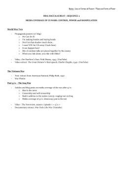
Research Article
Available online at www.ijpras.com
Volume 4, Issue 2 (2015):112-118
ISSN 2277-3657
International Journal of
Pharmaceutical Research &
Allied Sciences
Antidiabetic effect of Punica Granatum Peel Extract, Spilanthes
Paniculata Flower Extract and Selenium in Streptozotocin
Induced Diabetes
Shaikh Asma Afreen, Mohammed Mukhtar Khan, Syed Ayaz Ali*
*Department of pharmacology, Y. B. Chavan College of Pharmacy,
Dr. Rafiq Zakaria Campus, Rauza Bagh, Aurangabad, Maharashtra,
India
*E-mail: [email protected]
Subject: Pharmacology
Abstract
The present study was aimed to evaluate the antidiabetic potential of combination of punica granatum
peel extract, spilanthes paniculata flower extract and selenium in streptozotocin (STZ) induced diabetes
in mice. Group A was used as control and Group B was used as diabetic control (STZ, 40mg/kg for 3
days, i.p.). A placebo was given to both the group orally. Metformin (120mg/kg) was given
intraperitoneally as standard drug to Group C. Ethanolic peel extract of punica granatum (EPP 600mg/kg,
p.o.) was administered to group D, (EPP) in combination with ethyl acetate extract of spilanthes
paniculata flower (EAS) was administered to group E (EPP 300mg/kg, p.o. + EAS 200mg/kg, p.o.),
Sodium selenite was given with combination of herbal extract (EPP 300 mg/kg, p.o.+ EAS 200mg/kg, p.o.
+ Se 0.3mg/kg, i.p.) to group F. STZ was given in multiple low doses of 40mg/kg, i.p. for 3 consecutive
days & hyperglycemia was observed on 7th day. Blood glucose was determined on 0th, 7th, 14th, and 21st
day. Diabetic mice in all group showed significant (P<0.01) increase in serum blood glucose as compared
to control animals. Blood glucose level of animals treated with metformin & herbo-mineral combination
lowers the glucose level significantly (P<0.01) as compared to diabetic control. The herbo-mineral
combination shown significant (P<0.01) increase in the antioxidant enzymes Catalase, SOD, GSH and
reduction in MDA & NO levels as compared to diabetic control. Thus, the present study indicates
decreased blood glucose level & significant improvement in the biomarkers of oxidative stress by using
the combination of sodium selenite with herbal extracts. The results confirm that the herbo-mineral
combination is not only useful in controlling blood glucose level but also helps to prevent oxidative stress
induced damage.
Key words: Punica granatum, Spilanthes paniculata, Selenium Streptozotocin
Introduction
Diabetes mellitus (DM) is a metabolic disorder
characterized by increased blood glucose levels.
This disease is characterized by either lack of
insulin production or deficient activity in the
presence of normal or even elevated levels of
insulin.[1] High amounts of free fatty acids
(FFAs) and glucose badly affects ß cell function
through various pathways such as, defects of
metabolic mechanisms, generation of reactive
oxygen species (ROS), elevation of intracellular
calcium levels and interaction with membrane
potassium channels.[2] Beyond glucose, ROS
formation is also increased by FFAs, through
direct effects on mitochondria. [3] It has been
proposed that over expression and activity of a
mitochondrial inner membrane uncoupling
proteins (UCPs) contribute to an increase in
superoxide
formation
under
diabetic
conditions.[4] Chronic hyperglycemia produces
multiple biochemical impairments and oxidative
stress especially an increased susceptibility to
lipid peroxidation that play role in the progression
of the symptoms of diabetes.[5] Despite advances
in understanding the disorder and management,
the mortality and morbidity of the disease is ever
increasing.[6] Many studies have shown that anti
diabetic herbs with antioxidant property have
proven to be efficient in reducing the risk of
complication of diabetes. [7] Antioxidant like
SOD, CAT, glutathione reductase and some
minerals like Mn, Cu, Zn, selenium comes under
first line defense. [8] Pomegranate is a widely
112
Available online at www.ijpras.com
used plant that has high nutritional value. [9] It
was found that Punica granatum peel contain
tannins, flavonoids, pectin, 3-estrogen compounds
luetolin, quercitin & kemferol. Antioxidant &
antidiabetic activity shown by the peel extract
stimulation of beta-cells, protection of pancreas,
increase the no of beta-cells & subsequent release
of insulin it also reduce the blood sugar through
regeneration of beta-cells.[10] Genus Spilanthes is
a plant of choice for many health related disorder.
The genus Spilanthes (Asteraceae) comprises 30
species and 9 additional intraspecific taxa. [11]
The petroleum ether and ethyl acetate extract of
Spilantes paniculata flowers have shown the
hepatoprotective effect and possible mechanism is
due to the reduction of oxidative stress and
showed protection against increased lipid
peroxidation and maintained the glutathione
contents. [12] Selenium (Se) is an essential
dietary trace element which plays an important
role in a number of biological processes.[8]
Evidence from in vivo & vitro studies suggest that
inorganic selenium can enhance insulin sensitivity
by mediating insulin like action.[13] Specially in
animal models selenite has been shown to
decrease the activity of protein tyrosine phosphate
a negative regulator of insulin signal transmission
& therefore potentially reduced insulin
resistance.[14] Although, Punica granatum and
selenium is well recognized for possessing the
antidiabetic activity but no scientific data have
been published supporting the antidiabetic activity
of Spilanthes paniculata flowers and also the
combination study of these drugs. Hence, the
objective is to evaluate the antidiabetic effect of
combination of Punica granatum peel extract,
Spilanthes paniculata flowers extract and
selenium in streptozotocin induced diabetes in
mice.
Materials and methods
A) Collection of Fruits of Punica granatum: The
fruits were purchased locally and authenticated
from Dept. of Botany, Dr. Babasaheb Ambedkar
Marathwada University Aurangabad. A voucher
specimen no. 0733 has been deposited in the same
department.
B) Collection of flowers of Spilanthes paniculata:
The flowers of Spilanthes paniculata used for the
present studies were collected from Manjlegaon in
a location known as Beed (Maharashtra).
The plant was identified, confirmed and
authenticated by comparing with voucher
specimen available at survey of medicinal plants
& collection unit, Department of Botany, Dr.
Babasaheb Ambedkar Marathwada University,
Aurangabad where voucher specimens have been
deposited and accession no.0730.
Preparation of the extracts
A) Ethanolic extract of Punica granatum: The
powder obtained of Punica granatum peel (1000
g) was defatted using pet ether (60-800C). The
marc was extracted with 95% ethanol using
soxhlet apparatus. The filtrate was air dried and
concentrated under reduced pressure to obtain
37.40g, corresponding to a yield of 24.10% w/w.
B) Ethyl acetate extract of Spilanthes paniculata:
The flowers of Spilanthes paniculata plant were
removed and dried under the shade condition,
powdered with the help of grinder and stored in an
airtight container. The powder of flowers was
weighed (300gm). An extract was prepared by
soxhlet extraction method. The dried powdered
flowers of Spilanthes paniculata were extracted
with ethyl acetate for 36 hrs using soxhlet
extractor. The extracts were concentrated at 60°C
under reduced pressure, to obtain dark brownish
residue the remaining extract was freeze dried.
The yield obtained from the extraction process
was found to be 2.60% w/w.
Animals
Swiss albino mice of either sex weighing between
(22-30 g respectively) were procured from
Wockhardt Ltd, Aurangabad.
They were
maintained at temperature of 25 ± 2°C and
relative humidity of 45 to 55% and under standard
environmental conditions. Animals were housed
under standard laboratory conditions with free
access to food (Amrut rat and mice feed, Sangli,
India.) and water. The mice used in the present
study were maintained in accordance with
guidelines of the CPCSEA, India and the study
was approved by Institutional Ethics committee,
Y.B. Chavan College of pharmacy, Aurangabad.
Induction of Experimental diabetes
Overnight fasted experimental mice were injected
with STZ at a multiple dose of 40 mg/kg
bodyweight for 3 consecutive days.[15]The
solution was injected intraperitoneally (i.p.)
within 5 min after dissolving in citrate buffer pH
4.5[25] The animals were allowed to drink 5%
glucose solution overnight to overcome the drug
induce hypoglycemia. Fasting blood glucose
(FBG) was estimated before the time of induction
of diabetes using commercially available kit
(Accu-Chek Active Test Meter) and on the 7th day
of STZ administration. The mice with moderate
diabetes having glycosuria and hyperglycemia
(blood glucose levels of 250 mg/dl) were included
in the study.
113
Available online at www.ijpras.com
Groups and treatment
(Each group containing 6 animals)
Group A: Normal control (Distilled water,
1ml/100gm p. o).
Group B: Diabetic control (Distilled water, STZ
40 mg/kg for 3 days, i.p).
Group C: Diabetic (STZ) + Metformin
(120mg/kg, i.p).
Group D: Diabetic (STZ) + Ethanolic peel extract
of Punica granatum (600mg/kg, p.o).
Group E: Diabetic (STZ) + Ethanolic peel extract
of Punica granatum (300mg/kg, p.o) + Ethyl
acetate extract of Spilanthes paniculata flowers
(200mg/kg, p.o).
Group F: Diabetic (STZ) + Ethanolic peel extract
of Punica granatum (300mg/kg, p.o)+ Ethyl
acetate extract of Spilanthes paniculata flowers
(200mg/kg, p.o) + Sodium selenite (0.3 mg/kg,
i.p).
Preparation of drug solution
Preparation of STZ solution: Streptozotocin was
dissolved in cold 0.01 M citrate buffer, pH 4.5
and always prepared freshly for immediate use
within 30 min. STZ injections were given
intraperitoneally and the doses were determined
according to the body weight of animals. [16]
Oxidative stress
At the end of study animals were sacrificed and
liver was isolated & used for the estimation of
SOD [17], Catalase [18], MDA [19], GSH [20] &
NO [21] levels.
Statistical analyses
It was carried out by ANNOVA followed by
Dunnett’s t-test at level of significance P<0.05.
All data were shown as the mean ± SEM.
Statistical analysis was performed using Graph
Pad statistical software.
Table 1: Antidiabetic effect of herbo-mineral combination of (EPP + EAS + Sn) on blood glucose levels
in streptozotocin induced diabetes in mice.
GROUPS
Fasting blood glucose level (mg/dl)
Initial week
A. D.W.
First week
Second week
Third week
82.83 ± 3.31
85.50 ± 0.763
86.1 ± 1.06
82.0 ± 2.47
B. STZ + D.W.
86.83 ± 1.302
296 ± 13.297**
401 ± 10.80**
437 ± 18.9**
C. STZ + Metformin
86.16 ± 1.851
268 ± 10.24
191 ±7.34**
103 ± 2.98**
D. STZ + EPP
84.50 ± 4.087
262 ± 14.70
257 ±14.18**
143 ± 5.03**
E. STZ + (EPP + EAS) 85.16 ± 1.046
268 ± 8.41
205 ± 9.5**
128 ± 4.42**
283 ± 15.13
197 ± 5.34**
116 ± 2.88**
F. STZ +
82.33 ± 3.93
(EPP + EAS + Se)
Data is presented as Mean+ SEM (n=6); One way Analysis of Variance (ANOVA) followed by Dunnett’s test
**p<0.01 vs. Diabetic control.
114
Available online at www.ijpras.com
Table 2: Antioxidant effect of herbo-mineral combination of (EPP + EAS + Sn) on liver tissue Catalase,
SOD, MDA, GSH and NO levels in streptozotocin induced diabetes in mice.
Groups
A. D.W.
B. STZ + D.W.
C. STZ + Metformin
D. STZ + EPP
E. STZ + (EPP +
EAS)
F. STZ +
(EPP +EAS + Se)
CATALASE
SOD
nmoles of H2O2
Unit/ml of sample
consumed/min/ mg
protein
MDA
nmole/gm of
tissue
GSH
mmole
/gm of tissue
NO
mmole/gm of
tissue
6.07 ± 0.386
2.00 ± 0.242**
5.34 ± 0.506 **
4.34 ± 0.32 *
6.28 ± 0.78 **
7.00 ± 0.37
3.32 ± 0.395**
7.25 ± 0.407 **
4.79 ± 0.415
5.85 ± 0.28 *
2.46 ± 0.20
4.96 ± 0.33**
1.85 ±0.20**
3.88 ± 0.27*
2.55 ± 0.29**
10.36± 0.420
4.56 ± 0.52**
9.93 ± 0.23**
7.64 ± 0.43**
8.18 ± 0.57 **
8.21 ± 0.33
17.59 ± 1.14**
7.09 ± 0.82**
7.74 ± 0.31**
9.17 ± 0.44**
6.22 ± 1.25 **
8.07 ± 0.51 *
2.76 ±0.18**
9.93 ± 0.30**
7.49 ± 0.73**
Data is presented as Mean+ SEM (n=6); One way Analysis of Variance (ANOVA) followed by Dunnett’s test
*p<0.05, **p<0.01 vs. Diabetic control.
A
B
C
D
E
F
Fig. 1: Photomicrographs of mice liver (H & E stain) Respective treatment showing following changes
in liver section (A) Distilled water showing the normal histology, (B) Streptozotocin showing congested
liver tissue with congested sinusoids and centrilobular necrosis with infiltration of lymphocytes and
neutrophils, (C) metformin showing normal architecture with absence of fatty change and necrosis,
(D) Punica granatum peel extract showing the normal histology, (E) Punica granatum peel extract + S.
paniculata flower extract showing the normal histology and repaired hepatocytes, (F) Punica
granatum peel extract + S. paniculata flower extract + Selenium showing the normal histology with
repairing of hepatocytes. Magnification ×40
115
Available online at www.ijpras.com
Results
Discussion
Diabetic mice in all group showed significant
(p<0.01) increase in serum blood glucose as
compared to control animals. Vehicle treated
diabetic control group showed significant (p<0.01)
increase in serum glucose level as compared to
normal control animals. Blood glucose level of
animals treated with metformin & herbo-mineral
combination lowers the glucose level significantly
(p<0.01) as compared to diabetic control. The
combination of drugs was found to be more
effective in reducing the blood glucose level.
(Table 1).
The present study showed decrease in the
activity of SOD enzymes in diabetic control group
& administration of EPP has not shown any
significant improvement in the activity of enzyme
whereas group D (EPP + EAS) & group E (EPP +
EAS + Se) have shown significant increase in the
activity of this enzyme. The present study showed
decreased in the activity of catalase enzymes in
diabetic control group group C (EPP) showed
protective effect whereas group D (EPP + EAS) &
group E (EPP + EAS + Se) has normalized the
activity of these enzyme. Diabetic control group
showed depletion of GSH levels & the diabetic
mice treated with the herbal extract (EPP) &
(EPP+EAS) showed a marked protective effect.
Whereas the diabetic mice treated with (EPP+EAS
+ Se) normalized the hepatic GSH levels. (Table
2).
Group E (EPP+EAS) have shown marked
protective effect whereas group C (EPP) & group E
(EPP+EAS + Se) normalized elevated total nitrite
level in the liver tissues induced by STZ. A
massive increase in lipid peroxidation was
observed in diabetic control group whereas
treatment with (EPP + EAS) in group E & (EPP +
EAS + Se) in group F decreased the level of MDA,
thus preventing the lipid peroxidation whereas
treatment with (EPP) in group D did not show any
significant decrease in the MDA levels. (Table 2).
In Diabetes mellitus chronic hyperglycemia
produces multiple biochemical impairments and
oxidative
stress
especially
an
increased
susceptibility to lipid peroxidation that play role in
the progression of the symptoms of diabetes. [15]
Multiple low doses of STZ 40 mg/kg, i.p. to mice
for three consecutive days have been found suitable
model to study Type 2 DM. [16] Rise in blood
glucose was observed on the 7th day and the
animals showed symptoms like polyuria & weight
loss which assures that diabetes was successfully
induced.
Oxidative stress induced by hyperglycemia
leads to liver cell damage because liver is subject
to ROS-mediated injury in diabetes. [22] The
histhopathological evaluation of diabetic mice liver
showed degeneration of hepatocytes, centrilobular
necrosis with hydropic changes in surrounding and
diabetic mice treated with the herbal extract (EPP)
& (EPP+EAS) showed mild to moderate
degenerative changes while treatment with
EPP+EAS with sodium selenite showed
regeneration of hepatocytes & normal cord like
pattern.
Many studies have shown that antidiabetic
herbs with natural antioxidant constituents such as
tannins, alkaloids, flavonoids, phenols etc.
enhances free radical scavenging activities & have
proven to be efficient in reducing the risk of
complication of diabetes. Antioxidants like SOD,
CAT, GPx, glutathione reductase and some
minerals like Mn, Cu, and Zn selenium comes
under first line defense. Second line defense
antioxidants include glutathione (GSH), Vit C,
albumin, bilirubin, carotenoids, Vitamin E,
flavonoids, etc. Flavonoids are the phenolic
compounds that inhibit the lipid peroxidation &
lipoxygenase. [8] Tannins contain several
hexahyroxyl diphenoyl groups or galloyl groups
both groups provide protons to stabilize the free
radicals. [23] Ellagic acid reacts with the free
radical & is a potent antioxidant against the lipid
peroxidation in mitochondria & microsomes. [24]
It has been observed that plasma level of βsitosterol is lower among Type 2 diabetes mellitus
with hypoglycemic effects of β-sitosterol being
observed in other studies. [25, 26] β-sitosterol has
been shown to be an AMPK agonist, with the
beneficial effects of β-sitosterol on glucose
metabolism being mediated, in part, through this
mechanism. [27] In animal models selenite has
been shown to decrease the activity of protein
tyrosine phosphatase, a negative regulator of
insulin signal transmission, and can therefore
potentially reduce insulin resistance. [14]
In the present study, the phyto-chemical screening
of the plant extracts was performed and it was
found to contain flavonoids, tannins, phenolic
Histopathological evaluation
Group A: Liver shows normal architecture. The
central vein, portal tract and sinusoids appear
normal (Figure A). Group B: Liver shows
congested liver tissue with congested sinusoids and
centrilobular necrosis with infiltration of
lymphocytes and neutrophils (Figure B). Group C:
Liver shows normal architecture with absence of
fatty change and necrosis (Figure C). Group D:
Liver shows the normal histology (Figure D).
Group E: Liver shows normal hepatic architecture
with regeneration of hepatocytes and absence of
fatty changes and necrosis (Figure E). Group F:
Liver shows normal hepatic architecture with
regeneration of hepatocytes and absence of fatty
change and necrosis (Figure F). (Figure 1).
116
Available online at www.ijpras.com
compounds and steroids and the present data
suggest that the antidiabetic activity and protective
action of herbo-selenium combination against
oxidative stress may be due to its synergistic action
of these constituents thus, synergistic action of the
different chemical constituents appears to be
superior to that of single constituents and the
mechanism underlying such protection may be
mediated by prevention & restoration of
antioxidant defense systems. Restoration of
antioxidant status led to normalization of the
insulin release, decrease in the insulin resistance &
hence maintaining glucose serum levels within the
normal range.
Conclusion
The present study indicates decrease in blood
glucose level & significant improvement in the
biomarkers of oxidative stress by using the
combination of sodium selenite with herbal
extract.Herbal extract showed a marked protective
effect whereas herbo-selenium combination
normalized these levels. Protection may be
mediated by prevention & restoration of
antioxidant defense systems. Restoration of
antioxidant status led to normalization of the
insulin release, decrease in the insulin resistance &
hence maintaining glucose serum levels within the
normal range. This indicates that Punica granatum,
Spilanthes paniculata & sodium selenite are the
three powerful antioxidant which may prove to be
beneficial in diabetes and this supplement could
attenuate diabetic complications.
This finding also indicated beneficial interaction of
selenium at insulin-receptor site resulting in
decrease in insulin resistance & hyperglycemia.
Although the proof of efficient delivery of
selenium at receptor site would need further study
Acknowledgements
We thank Hon’ble Padmashree Mrs. Fatma Rafiq
Zakaria, Chairman, Maulana Azad Educational
Trust and Society for providing the research
facility. We thank Mr. Mohammed Riyaz and Mr.
Bhikan Pathan for assisting in the experimental
work.
“Cite this Article”
SA Afreen, MM Khan, SA Ayaz “Antidiabetic
effect of Punica Granatum Peel Extract,
Spilanthes Paniculata Flower Extract and
Selenium
in
Streptozotocin
Induced
Diabetes”Int. J. of Pharm. Res. & All. Sci.
2015;4(2):112-118
References
1. Lucy, D., Anoja, S., Chu-Su, Y., Alternative
therapies for Type 2 diabetes, Altern. Med. Rev.,
2002, (7), 45-58
2. Prasanth, N., Surampudi, M.D., Kalarickal, J.J.,
et
al.,
Emerging
Concepts
in
the
Pathophysiology of Type 2 Diabetes Mellitus,
Mount Sinai Journal of Medicine., 2009, 76,
216–226
3. Tripathi,
K.D.,
Essentials
of
medical
pharmacology.6th ed. Jaypee Brothers Medical
Publishers (P) Ltd ; New Delhi; 2008.
4. Evans, J.L., Goldfine, I.D., Maddux, B,A., et al.
Oxidative stress and stress activated signaling
pathways: A unifying hypothesis of type 2
diabetes. Endocrine Reviews, 2002, 23,599–622
5. Giugliano, D., Ceriello, A., Paolisso, G.,
Oxidative stress and diabetic vascular
complications, Diabetes Care, 1996, 19, 257-267
6. Dhanbal, S.P., Evaluation of therapeutic activity
and development of quality control profiles for
some antidiabetic herbal drugs, Ind. J. Pharm.
Edu, 2004, 8, 163-165
7. Shokeen, P., Anand, P., Murali, Y.K., et al.,
Antidiabetic activity of 50% ethanolic extract of
Ricinus communis and its purified fractions,
Food. Chem. Toxicol., 2008, 46, 3458-3466
8. Gupta, V.K., Sharma, S.K., Plants as natural
antioxidant. A Review article, Nat. Prod. Rad.,
2006, 5(4), 326-334
9. Poyrazog, E., Knew, W., Atrik, N., Organic
acids and phenolic compounds in pomegranates
(Punica granatum L.) grown in Turkey, J. Food.
Comp. Anal., 2002, 15, 567-575
10. Enas, A.M Khalil., Antidiabetic effect of an
aqueous extract of Pomegranate (Punica
granatum L.) peels in normal and alloxan
diabetic rats, The Egyptian Journal of Hospital
Medicine, 2004, 16, 92-99
11. Jansen, R.K., Acmella oleracea (L.) Syst, Bot,
Monogr., 1985, 8, 110-115
12. Syed. A.A., Shukla, M., Khan, S.W.,
Hepatoprotective
activity
of
Spilanthes
paniculata flower extracts on liver damage
induced by paracetamol in rats, African Journal
of Pharmacy & Pharmacology, 2012, 6(42),
2905-2911
13. Stapleton, S.R., Selenium: An insulin-mimetic.
Cell. Mol. Life. Sci., 2000, 57, 1874-1879
14. Mueller, A.S., Pallauf, J., Compendium of the
antidiabetic effects of supra nutritional selenate
doses. In vivo and invitro investigations with
Type II diabetic db/db mice, J. Nutr. Biochem.
2006, 17, 548-560
15. Mohamed, A.K., Bierhaus, A., Schiekofer, S., et
al., The role of oxidative stress and NF-κB
activation in late diabetic complications,
Biofactors, 1999, 10, 157–167
117
Available online at www.ijpras.com
16. Ventura, S.J., Boone-Villa, V.D., Aguilar, C.N.,
et al., Effect of varying dose and administration
of streptozotocin on blood sugar in male CD1
mice, Proc. West. Pharmacol. Soc., 2011, 54, 5-9
17. Marklund, S.L. Handbook of methods for
oxygen radical research. CRC Press Inc; Boca
Raton; Florida; 1985.
18. Clairborne, A. Handbook of methods for oxygen
radical research. CRC Press; Boca Raton;
Florida; 1985.
19. Ohkawa, H., Ohish, N., Yagi, K., Assay lipid
peroxides in animal tissues by thiobarbituric
acid, Anal. Biochem., 1979, 95, 351-358
20. Ellman, G.L., Tissue sulfhydryl groups, Arch.
Biochem. Biophys., 1959, 82, 70-77
21. Green, L.C., Wagner, D.A., Glogowski, J., et al.,
Analysis of nitrate, nitrite, and [15N] nitrate in
biological fluids, Anal. Biochem., 1982, 126,
131-138
22. Pourkhalili, N., Hosseini, A., Nili-Ahmadabadi,
A., et al., Biochemical and cellular evidence of
the benefit of a combination of cerium oxide
nanoparticles and selenium to diabetic rats,
World. J. Diabetes., 2011, 2(11), 204-210
23. Wang, R.F., Xie, W.B., Zhang, Z., et al.,
Bioactive Compounds from seeds of Punica
granatum, J. Nat. Prod., 2004, 67, 2096-2098
24. Oswa, T., Ide, A., Su, J.D., Inhibiting of Lipid
peroxidation by ellagic acid, J. Agri. & Food.
Chem., 1987, 35, 808-812
25. Sutherland, W.H., Scott, R.S., Lintott, C.J., et al.,
Plasma noncholesterol sterols in patients with
non-insulin dependent diabetes mellitus, Horm.
Metab. Res., 1992, 24, 172–175
26. Ivorra, M.D., D’Ocon, M.P., Paya, M., Villar,
A.,
Anti
hyperglycemic
and
insulinreleasingeffects of beta-sitosterol 3-beta-Dglucoside and its aglycone, beta-sitosterol, Arch.
Int. Pharmacodyn. Ther., 1988, 296, 224–231
27. Hwang, S.L., Kim, H.N., Jung, H.H., et-al.,
Beneficial effects of betasitosterol on glucose
and lipid metabolism in L6 myotube cells are
mediated by AMP activated protein kinase,
Biochem. Biophys. Res. Commun., 2008, 377,
1253–1258
118


© Copyright 2026