
Rare prenylated flavonoids from Tephrosia purpurea
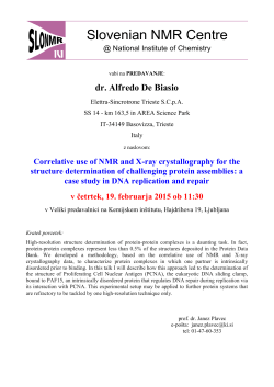
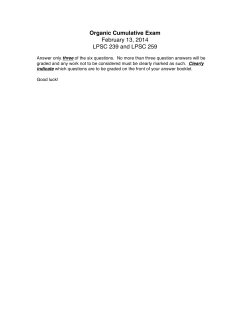
Phytochemistry 70 (2009) 1474–1477
Contents lists available at ScienceDirect
Phytochemistry
journal homepage: www.elsevier.com/locate/phytochem
Rare prenylated flavonoids from Tephrosia purpurea
Mohamed-Elamir F. Hegazy a, Mohamed H. Abd El-Razek b, Fumihiro Nagashima c,
Yoshinori Asakawa c, Paul W. Paré d,*
a
Chemistry of Medicinal Plant Department, National Research Centre, Dokki, Giza 12622, Egypt
Natural Products Chemistry Department, National Research Centre, Dokki, Giza 12622, Egypt
c
Faculty of Pharmaceutical Sciences, Tokushima Bunri University, Yamashiro-Cho, Tokushima 770-8514, Japan
d
Department of Chemistry and Biochemistry, Texas Tech University, Lubbock, Texas 79409-1061, USA
b
a r t i c l e
i n f o
Article history:
Received 18 December 2008
Received in revised form 2 June 2009
Available online 3 September 2009
Keywords:
Tephrosia purpurea
Leguminosae
Prenylated flavonoids
a b s t r a c t
Chemical investigations of aerial parts of Tephrosia purpurea yielded the rare prenylated flavonoids, tephropurpulin A (1) and isoglabratephrin (2), in addition to a previously identified flavonoid, glabratephrin
(3). Structures were established by 1D and 2D NMR spectroscopy, as well as by HR-MS analysis; for compounds 2 and 3, structures were confirmed by X-ray analysis.
Ó 2009 Elsevier Ltd. All rights reserved.
1. Introduction
Tephrosia purpurea (Del.) Pers. (Leguminosae) is a highly
branched, sub-erect herb commonly known in Sanskrit as Sharapunkha. It has been used in Indian traditional medicine for the
treatment of various inflammatory disorders (Santram et al.,
2006; Joshi, 2000). The plant is reported to cure diseases of the kidney, liver, spleen, heart and blood (Despande et al., 2003). The
dried herb is effective as a tonic, laxative, and diuretic. It is also
used in the treatment of bronchitis, bilious febrile attack, boils,
pimples, and bleeding piles. The roots and seeds are reported to
have insecticidal, piscicidal, and vermifugal properties. The roots
are effective in leprous wounds and root juice to skin eruptions.
Ethanolic extracts of aerial plant parts have medicinal properties
including anticancer activity against a human nasopharyngeal epidermoid tumor cell line (KB) (Santram et al., 2006; Zafar et al.,
2004). An aqueous seed extract has significant in vivo hypoglycaemic activity in diabetic rabbits (Rahman et al., 1985). Isolated
flavonoids from this species have also been shown to have antimicrobial activity (Gokhale and Saraf, 2000).
Several reports of T. purpurea have demonstrated the presence
of flavones and flavanones (Gupta et al., 1980; Pelter et al.,
1981); chalcones (Pelter et al., 1981; Sinha et al., 1982; Ventakata
et al., 1984; Chang et al., 2000; Saxena and Choubey, 1997); rotenoids (Ventakata et al., 1984; Chang et al., 2000); and prenylated
flavonoids (Pelter et al., 1981).
* Corresponding author. Tel./fax: +1 (806) 742 3062.
E-mail address: [email protected] (P.W. Paré).
0031-9422/$ - see front matter Ó 2009 Elsevier Ltd. All rights reserved.
doi:10.1016/j.phytochem.2009.08.001
This report focuses on chemical investigations of a dichloromethane extract of aerial parts of T. purpurea resulting in isolation
and structural elucidation of three flavonoid metabolites.
2. Results and discussion
Compound 1 was isolated as a yellowish powder with a specific
25
rotation of ½aD = 2.1 (c 1.92, CHCl3). The structure was established
based on analysis of 1H NMR, 13C NMR, DEPT, 1H–1H COSY, 1H–13C
COSY, HMBC, EIMS and HREIMS data. The EIMS spectrum showed a
molecular ion peak at m/z 410 (20%) in accordance with a molecular formula of C23H22O7. The base peak at m/z 279 (100%) was
attributable to cleavage of the side-chain (2-hydroxy-2-methylpropyl acetate). Exact mass determination of the 410 ion established
the elemental composition C23H22O7, exp. 410.1354, (calcd.
410.1353). The 1H NMR spectrum of 1 (Table 1) had several common signals with tephroapollin F (Abd El-Razek et al., 2007) allowing for the assignment of three singlets at d 1.25 (3H), 1.29 (3H),
and 1.92 (3H) to two gem-Me groups adjacent to an oxygen functionality and an acetate methyl group. The proton spectrum also
established the presence of three singlets each integrating for
one proton at d 6.63, 6.26 and 5.61 and assigned to H-3, H-6 and
H-400 , respectively. 1H NMR signals at d 4.72 (triplet, J = 19.0 Hz)
and 5.11 (dd, J = 10.2, 19.0 Hz), were assigned to H-200 based on
1
H–13C COSY correlations with C-200 at d 73.68. The C-200 resonance
at d 5.11 also exhibited a 1H–1H COSY correlation with the proton
at d 4.25 (dd, J = 10.2, 19.0 Hz) which was assigned to H-30 0 .
The 13C NMR DEPT spectrum showed 23 carbon signals with
two carbonyl carbons at d 182.2 and 169.9; methine and oxyme-
1475
M.-E.F. Hegazy et al. / Phytochemistry 70 (2009) 1474–1477
and C-400 ; H-30 0 with C-7 and C-8; and H-400 with C-8, C-200 and carbonyl acetate. Since only C-7 showed a correlation with H-200 , these
two similar chemical shifts at C-5 and C-7 could be differentiated.
These spectroscopic data were consistent with structure 1 which
has not been previously reported; the compound was assigned
the name tephropurpulin A.
Compound 2 was obtained as colourless crystals with a specific
+
rotation of ½a25
D = 17.15 (c 16.02, CHCl3). An ion peak [M] was observed in the EIMS at m/z 420 (84%), followed by a fragment at m/z
317 [M–OAc–CO2]+ (100%). The high resolution mass spectrum
showed a molecular ion peak [M]+ at m/z 420.1215 (calcd.
420.1209) in accordance with the molecular formula of C24H20O7,
which was supported by 1H, 13C NMR and DEPT experiments. The
IR absorption at 1740, 1685, and 1600 cm1 suggested the presence of acetyl, carbonyl, and phenyl groups, respectively. Examination of the 1H NMR spectroscopic data of 2 indicated the presence
of a flavone structure. Two multiplets at d 7.43 and 7.79 established the presence of B-ring flavone protons at H-20 , H-40 and H60 , as well as H-30 and H-50 . 1H NMR signals at d 1.42 (3H, s) and
1.43 (3H, s), corresponded to a gem-dimethyl group, while resonances at d 6.86 (1H, d, J = 8.5, H-6) and 8.09 (1H, d, J = 8.5), indicated ortho-substitution in the ring A, and a sharp singlet at d
6.69 assigned to H-3. The remaining portion of the molecule contained a dioxaspirononanone functionality with a pair of methylene protons at d 4.88 (1H, d, J = 9.5) and d 4.98 (1H, d, J = 9.5). In
addition, two singlets at d 1.51 (3H) and 5.37 (1H) established
the presence of an acetate group and a methine proton (H-400 ),
respectively. The placement of the dioxaspirononanone moiety at
C-7/8 was determined by HMBC data which exhibited a strong C7 (d 155.8)/C-8 (d 111.7) correlation with H-200 . In addition to
1
H–13C long-range correlation establishing the position of the acetate group, correlations between H-400 (d 5.37) and a carbonyl carbon (OAc, d 168.7) were observed (Table 1).
The relative configuration of compound 2 was deduced from the
NOESY experiment, where the methine proton (H-400 ), one of the
Table 1
1
H (500 MHz) and 13C (125 MHz) NMR spectroscopic data in CDCl3 for compounds 1
and 2 (dH and dC in ppm, J = in Hz).
Position
1
2
dH
dH
2
3
4
4a
5
6
7
8
8a
10
20 , 60
30 , 50
40
20 0
30 0
40 0
50 0
60 0
Me2
OCOCH3
OCOCH3
OH
6.63 s
6.26 s
7.88 m
7.55
4.72
5.11
4.25
5.61
dC
163.4 (s)
106.3 (d)
182.2 (s)
105.4 (s)
163.6 (s)
94.2 (d)
166.9 (s)
103.5 (s)
153.2 (s)
131.7 (s)
126.2 (d)
129.1 (d)
131.7 (d)
73.9 (t)
m
t (19.0)
dd (19.0, 10.2)
dd (10.2, 19.0)
brs
6.69
–
–
8.09
6.86
–
–
–
–
7.43
7.79
7.43
4.88
4.98
–
5.37
–
–
1.42
1.43
1.51
40.1 (s)
76.8 (d)
72.6 (s)
1.25 s
1.29 s
1.92 s
27.1 (q)
27.5 (q)
20.5 (q)
169.9 (s)
163.4 (s)
13.06 s
dC
162.4 (s)
107.1 (d)
176.9 (s)
118.4 (s)
129.9 (d)
109.0 (d)
165.8 (s)
111.7 (s)
153.5 (s)
131.0 (s)
125.9 (d)
128.9 (d)
131.7 (d)
81.3 (t)
s
d (8.5)
d (8.5)
m
m
m
d (9.5)
d (9.5)
58.7 (s)
80.0 (d)
85.2 (s)
174.3 (s)
22.1 (q)
28.4 (q)
19.7 (q)
168.7 (s)
s
s
s
s
thine carbon resonances at d 40.1 and 76.8, respectively; one methylene carbon signal at d 73.9; three methyl carbon signals at d 27.1,
27.5 and 20.5; eight quaternary carbon resonances at d 72.6, 131.7,
163.4, 105.4, 153.2, 163.6, 166.9, 103.5; six aromatic carbon signals
at d 94.2, 126.2, 129.1, 131.7, and one olefinic carbon resonance at
d 106.3 (Table 1). HMBC analysis exhibited correlations of H-3 with
C-2, C-10 and C-4a; H-6 with C-5, C-7, C-8 and C-4a; H-200 with C-7
AcO
4''
AcO
OH
6''
3'
O
4''
3'
6''
2''
2''
O
O
5'
1'
O
O
7
O
1'
7
3
5
OH
3
5
2 O
1 O
AcO
O
4''
3'
6''
2''
O
O
O
1'
7
3
5
3
O
5'
5'
1476
M.-E.F. Hegazy et al. / Phytochemistry 70 (2009) 1474–1477
3. Concluding remarks
A common tetrahydrofurofuran flavone skeleton for pseudosemiglabrin, () -semiglabrin, and semiglabrinol, that has been reported from Tephrosia apollinea and T. purpurea (Waterman and
Khalid, 1980; Pelter et al., 1981; Abou-Douh et al., 2005) supports
the hypothesis that 1 results from enzyme cleavage of the furan
ring. In contrast, the dioxaspirononanone flavonoid structure represent a rare functional type and suggest a novel biogenic route
operative for isoprenoid units to be incorporated in heterocyclic
flavonoid systems. To the best of our knowledge, glabratephrin as
isolated previously from Tephrosia semiglabra (Vleggaar et al.,
1978) is the only example besides 2 of a flavone containing the
dioxaspirononanone flavonoid substitution.
4. Experimental
4.1. General experimental procedures
Fig. 1. Computer-generated ORTEP plot of 2 showing relative configuration.
methylene protons (at d 4.98) and one methyl of the geminal
methyls (at d 1.42) correlated with each other, indicating a co-facial orientation for these protons. X-ray analysis established an axial orientation for the acetoxy group and an equatorial orientation
for the proton at C-400 (Fig. 1).
By comparing spectroscopic data for 2 and 3 (glabratephrin)
(Vleggaar et al., 1978), the compounds were found to be almost
identical. The major proton NMR chemical shift distinction between 2 and 3 was a pair of doublets at d 4.88 and 4.98 (2H,
J = 9.5, AX system) assigned to H-200 for compound 2, while a pair
of doublets at d 4.99 was observed for 3 (AB system). This NMR signal difference was attributed to configurational differences that
were confirmed by X-ray analysis (Fig. 2). Compound 2 was therefore named as the new natural product isoglabratephrin.
Optical rotation was determined using a JASCO DIP-360 digital
Polari meter. IR spectra were recorded with a Perkin–Elmer FT-IR
1725 IR spectrophotometer. 1H and 13C NMR spectra were acquired using a JEOL-LA 500 Lambda (500 and 125 MHz, respectively) spectrometer. Chemical shifts are given on d (ppm) scale.
CI and HR-MS were recorded on a JEOL JMS-DX 303 mass spectrometer. Column chromatography (CC) was carried out on Kieslgel
60 (Merck; 230–400 mesh) and Sephadex LH-20 (Pharmacia Co.,
Tokyo, Japan), whereas TLC was performed on silica gel 60 F254
plated (0.25 mm, Merck Co.).
4.2. Plant material
The aerial parts of T. purpurea (Del.) were collected in spring of
2001, South Sinai, Egypt. A voucher specimen (T-119) has been
deposited in the herbarium: Department of Botany, Faculty of Science, El-Minia University, El-Minia, Egypt.
4.3. Extraction and isolation
Air-dried aerial plant tissue (700 g) was crushed and extracted
with CH2Cl2–MeOH (1:1) (1.2 L) at room temperature. After solvent
removal, the residue (30 g) was subjected to CC on silica gel and
eluted with n-hexanes, CH2Cl2 and MeOH in increasing order of
polarity up to 100% CH2Cl2 and then to 15% MeOH in CH2Cl2 (a total
solvent volume of 3 L).
The n-hexane–CH2Cl2 (1:1) and CH2Cl2 fractions were combined
(11 g) based on TLC similarities, concentrated to remove the solvent and subjected to CC on silica gel except that the eluding solvent was n-hexanes with increasing amounts of EtOAc up to 2:1
n-hexanes:EtOAc. Pooled fractions 12–18 (40 fractions in total)
were purified by Sephadex LH-20 (2 60) eluting with n-hexanes–CH2Cl2–MeOH (7:4:0.25) to afford compounds 2 (6 mg) and
3 (35 mg).
The CH2Cl2–MeOH (85:15) fraction after concentration was further purified by CC on silica gel with n-hexanes and EtOAc as well
as Sephadex LH-20 with n-hexane–CH2Cl2–MeOH (7:4:0.75) to afford compound 1 (8 mg).
4.4. Tephropurpulin A (1)
Fig. 2. Computer-generated ORTEP plot of 3 showing relative configuration.
1
KBr
)
Yellowish powder; ½a25
D = 2.1 (c 1.92, CHCl3); IR (mmax cm
2968 (O–H), 1736 (C@O–OAc), 1680 (C@O), 1596 (C@C); EIMS
[M]+ m/z 410, 279; HREIMS: C23H22O7, exp. 410.1354, (calcd.
410.1353). For 1H and 13C NMR spectroscopic data, see Table 1.
M.-E.F. Hegazy et al. / Phytochemistry 70 (2009) 1474–1477
1477
4.5. Isoglabratephrin (2)
Acknowledgements
Colourless crystals, ½a25
D = +17.2 (c 0.45, CHCl3); mp 234–236 °C,
1
) 2974 (O–H), 1740 (C@O–OAc), 1685 (C@O), 1600
IR (mKBr
max cm
(C@C); EIMS [M]+ m/z 420, 317; HREIMS m/z 420.1205 (calcd.
420.1215, C24H20O7). For 1H and 13C NMR spectroscopic data, see
Table 1.
This research was supported in part by the Robert Welch Foundation (D-1478) and the Frasch Foundation/ACS for Chemical
Research.
References
4.6. X-ray crystallography of compound 2
Single crystal X-ray analysis established the complete structure
and relative configuration of compound (2) and the crystal data are
summarized as follows: C24H20O7, formula wt 420.417, orthorhombic, space group P212121, a = 7.1060 (2) Å, b = 14.6790 (4) Å,
c = 19.9510 (9) Å, V = 2081.07 (12) Å3, Z = 4, Dx = 1.342 Mg m3.
All diagrams and calculations were performed using maXus (Brucker Nonius, Delft & Mac Science, Japan), using graphite monochromated Mo Ka radiation (k = 0.71073 Å). The structures were
refined by full matrix least squares on F2 using Brucker SHELEXL97 (Sheldrick, 1997). The final R and Rw were 0.0448 and 0.1074,
respectively. This crystal structure has been deposited at the Cambridge Crystallographic Data Centre and allocated deposition number CCDC 251441.
4.7. X-ray crystallography of compound 3
Single crystal X-ray analysis established the complete structure
and relative configuration of compound (3) and the crystal data are
summarized as follows: C24H20O7, formula wt 420.42, orthorhombic, space group P212121, a = 14.6740 (4) Å, b = 7.1040 (2) Å,
c = 19.9460 (5) Å, V = 2079.25 (8) Å3, Z = 4, Dx = 1.343 g/cm3. All
diagrams and calculations were performed using maXus (Brucker
Nonius, Delft & Mac Science, Japan), using graphite monochromated Mo Ka radiation (k = 0.71069 Å). The structures were refined by
full matrix least squares on F2 using Brucker SHELEXL-97 (Sheldrick, 1997). The final R and Rw were 0.045 and 0.060, respectively.
Abd El-Razek, M.H., AbouEl-Hamd, H.M., Ahmed, A.A., 2007. Prenylated flavonoids
from Tephrosia apollinea. Heterocycles 71, 2477–2490.
Abou-Douh, A.M., Ito, C., Toscano, R.A., Nariman, N.Y., El-Khrisy, E.E.A., Furukawa, H.,
2005. Prenylated flavonoids from the root of Egyptian Tephrosia apollinea –
crystal structure analysis. Zeitschrift fur Naturforschung B 60, 458–470.
Chang, L.C., Chavez, D., Lynda, L.S., Farnsworth, N.R., Pezzuto, J.M., Kinghorn, A.D.,
2000. Absolute configuration of bioactive flavonoids from Tephrosia purpurea.
Organic Letter 2, 515–518.
Despande, S.S., Shah, G.B., Parmar, N.S., 2003. Antiulcer activity of Tephrosia
purpurea in rats. Indian Journal of Pharmacology 35, 168–172.
Gokhale, A.B., Saraf, M.N., 2000. Tephrosia purpurea: a review of contemporary
literature and medicinal properties. Indian Drugs 37, 553–560.
Gupta, R.K., Krishnamurti, M., Parthasarathi, J., 1980. Purpurin, a flavanone from
Tephrosia purpurea seeds. Phytochemistry 19, 1264.
Joshi, S.G., 2000. Oleaceae. Medicinal Plants. Oxford and IBH Publishing Co. Pvt. Ltd.,
New Delhi, p. 211.
Pelter, A., Ward, R.S., Rao, E.V., Ranga, E., Raju, N., 1981. 8-Substituted flavonoids
and 30 -substituted 7-oxygenated chalcones from Tephrosia purpurea. Journal of
the Chemical Society, Perkin Transactions 1, 2491–2498.
Rahman, H., Kashifudduja, M., Syed, M., Saleemuddin, M., 1985. Hypoglycemic
activity of Tephrosia purpurea seeds. Indian Journal of Medical Research 81, 418.
Santram, L., Singh, P.R., Pal, J.A., Singhai, A.K., 2006. Wound healing potential of
Tephrosia purpurea (Linn.) Pers. in rats. Journal of Ethnopharmacology 108, 204–
210.
Saxena, V.K., Choubey, A., 1997. A neoflavonoid glycoside from Tephrosia purpurea
stem. Fitoterapia 68, 359–360.
Sheldrick, G.M., 1997. SHELXL97. Program for the refinement of crystal structures.
University of Gottingen, Germany.
Sinha, B., Natu, A.A., Nanavati, D.D., 1982. Prenylated flavonoids from Tephrosia
purpurea seeds. Phytochemistry 21, 1468–1470.
Ventakata, R., Raju, E., Ranga, N., 1984. Two flavonoids from Tephrosia purpurea.
Phytochemistry 23, 2339–2342.
Vleggaar, R., Kruger, G.J., Smalberger, T.M., Van den Berg, A.J., 1978. Flavonoids from
Tephrosia XI. The structure of glabratephrin. Tetrahedron 34, 1405–1408.
Waterman, P.G., Khalid, S.A., 1980. The major flavonoids of the seed of T. apollinea.
Phytochemistry 19, 909–915.
Zafar, R., Mujeeb, M., Ahmed, S., 2004. Preliminary phytochemical screening of root
culture of Tephrosia purpurea (Linn.) Pers. Hamdard Medicus XLVIII, 1.


© Copyright 2026