
FUTURE PERSPECTIVES IN SMILE, LASIK AND PRK, AND
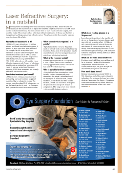
COVER FOCUS
FUTURE PERSPECTIVES IN
SMILE, LASIK AND PRK,
AND PHAKIC IOLs
Where are these technologies headed in the next few years?
BY MARCUS BLUM, MD; MASSIMO BUSIN, MD; AND ALAA EL-DANASOURY, MD
SMILE: The Future is
Minimally Invasive
The lenticule extraction technique is well established after
only 3.5 years.
By Marcus Blum, MD
64 CATARACT & REFRACTIVE SURGERY TODAY EUROPE | APRIL 2015
Figure 1. SMILE is a flapless and bladeless procedure based
on the removal of a disc of tissue, called a lenticule, through a
small incision.
ROUTINE CORRECTION
We now offer SMILE to our patients in Erfurt, Germany, as
a routine refractive correction option. Except in conditions
in which SMILE is not indicated, such as hyperopia, we treat
all suitable patients with SMILE.
The SMILE procedure has become established in the market only 3.5 years after its international commercial introduction. However, there is great potential for further investigations and for broadening the range of indications, and
studies using the SMILE technique to treat hyperopia have
already begun. Still, there is a need for long-term follow-up
studies to further validate the safety and efficacy of this
concept and its assumed potential. I am sure that the future
of laser vision correction will increasingly include minimally
invasive procedures and that we will hear quite a lot about
SMILE during the coming years.
1. Sekundo W, Kunert KS, Blum M. Small incision femtosecond lenticule extraction (SMILE) for the correction of myopia and
myopic astigmatism: Results of a 6 months prospective study. Br J Ophthalmol. 2011;95:335-339.
2. Kamiya K, Igarashi A, Ishii R, Sato N, Nishimoto H, Shimizu K. Early clinical outcomes, including efficacy and endothelial
cell loss, of refractive lenticule extraction using a 500 kHz femtosecond laser to correct myopia. J Cataract Refract Surg.
2012;38:1996-2002.
3. Ivarsen A, Asp S, Hjortdal J. Safety and complications of more than 1500 small-incision lenticule extraction procedures.
Ophthalmology. 2014;121:822-828.
Courtesy of Marcus Blum, MD
For several years, early in its development, the
use of femtosecond laser systems in refractive surgery was restricted to flap creation,
while the actual refractive ablation remained
the domain of the excimer laser. Once flap
creation with femtosecond lasers had demonstrated advantages over mechanical microkeratomes with regard to the quality of the
surgical outcome, it was a logical step to investigate methods
for refractive laser correction using this technology only.
A refractive procedure that required no excimer laser
was brought to market by Carl Zeiss Meditec in September
2011. The procedure was introduced as ReLEx small incision
lenticule extraction (SMILE), and the first series of 91 eyes was
reported by Sekundo.1
SMILE is based on the removal of a disc of tissue—a lenticule—rather than tissue ablation. The refractive lenticule is
created in the intact cornea using the VisuMax femtosecond
laser (Carl Zeiss Meditec). The refractive correction is achieved
by passing a dissector though a small 2- to 3-mm incision,
separating the anterior and posterior lenticular interfaces, and
then removing the lenticule though the incision (Figure 1).
This eliminates the need to create a flap and makes SMILE a
minimally invasive procedure. The cornea above the upper
interface of the lenticule is referred to as the cap.
Practitioners of SMILE have reported that visual outcomes,
predictability, and safety in low, moderate, and high myopes
are comparable with or even better than with femtosecond
LASIK (femto-LASIK).2,3 Additionally, SMILE offers a number
of further potential advantages that have also been reported
(see Potential Advantages of SMILE).
Surface ablation and flap cutting cause loss of the subepithelial nerve plexus. SMILE generates less damage due to the small
size of the pocket incision with a lower level of collateral damage and decellularization. Comparative outcome measurements
have shown fewer dry eye symptoms in SMILE patients in comparison with other corneal refractive procedures.1-3
In SMILE, the Bowman layer is disrupted only along a small
peripheral incision. This minimizes the biomechanical weakening
of the cornea and leaves the anterior stroma more stable than in
LASIK. No case of postoperative kerectasia has been reported after
SMILE, and this procedure is likely to be useful to correct higher
amounts of refractive error than other corneal procedures.4,5
LASIK and PRK: Safety and
Efficacy Continues to Improve
Sophisticated technologies, improved surgical skills, and optimal
patient selection are key in boasting postoperative outcomes.
By Massimo Busin, MD
LASIK has been the dominant refractive surgery procedure in most countries in recent
decades. Its main advantage over PRK consists
in the preservation of the corneal epithelium
over the flap created by a microkeratome or a
femtosecond laser before excimer laser treatment of the exposed stromal surface.
Preservation of the epithelium leads to
increased comfort during the early postoperative period,
faster visual recovery, and reduced wound healing response
in comparison with PRK. Reduced wound healing correlates
with less regression for high corrections and a lower rate of
complications such as significant stromal opacity or haze.
PRK, however, remains an excellent option for correction
of mild to moderate degrees of myopia, particularly in eyes
with thin corneas or recurrent erosions, and for patients
with a predisposition to trauma (eg, those who practice
martial arts or are in the military).
LASIK has been considered a riskier procedure than PRK
because of the possibility of complications occurring during the creation and subsequent manipulation of the flap.
Additionally, if the flap is not properly cut, it may fail to adhere
to the stromal surface and/or present microscopic wrinkles or
striae, leading to poor visual outcomes. Studies suggest that
flap complications occur in 0.3% to 5.7% of LASIK procedures.1
Dry eye syndrome is another drawback experienced by
many post-LASIK patients. Some patients after LASIK experience a marked decrease in tear production that can cause
eye discomfort and blurred vision. In one study, almost half
At 1-year follow-up, better centration of the refractive zone,
significantly less induction of higher-order aberrations, and better
mesopic midterm contrast sensitivity were achieved with ReLEx
than with wavefront-optimized femto-LASIK.6
1. Riau AK, Angunawela RI, Chaurasia SS, Lee WS, Tan DT, Mehta JS. Early corneal wound healing and inflammatory responses
following refractive lenticule extraction (ReLEx). Invest Ophthalmol Vis Sci. 2011;52:6213-6221.
2. Wei S, Wang Y. Comparison of corneal sensitivity between FS-LASIK and femtosecond lenticule extraction (ReLEx flex) or smallincision lenticule extraction (ReLEx smile) for myopic eyes. Graefes Arch Clin Exp Ophthalmol. 2013;251:1645-1654.
3. Vestergaard AH, Gronbech KT, Grauslund J, Ivarsen AR, Hjortdal JO. Subbasal nerve morphology, corneal sensation, and tear film
evaluation after refractive femtosecond laser lenticule extraction. Graefes Arch Clin Exp Ophthalmol. 2013;251:2591-2600.
4. Wu D, Wang Y, Zhang L, Wei S, Tang X. Corneal biomechanical effects: Small-incision lenticule extraction versus femtosecond
laser-assisted laser in situ keratomileusis. J Cataract Refract Surg. 2014;40:954-962.
5. Roy AS, Dupps WJ, Roberts CJ. Comparison of biomechanical effects of small-incision lenticule extraction and laser in situ
keratomileusis: Finite element analysis. J Cataract Refract Surg. 2014;40:971-980.
6. Gertnere J, Solomatin I, Sekundo W. Refractive lenticule extraction (ReLEx flex) and wavefront-optimized femto-LASIK: comparison of contrast sensitivity and high-order aberrations at 1 year. Graefes Arch Clin Exp Ophthalmol. 2013;251:1437-1442.
COVER FOCUS
POTENTIAL ADVANTAGES OF SMILE
of all LASIK patients reported some degree of temporary dry
eye syndrome;1 however, post-LASIK dry eye syndrome is
usually transient and can be effectively treated with lubricating eye drops. It usually disappears when healing of the cornea is completed, which may require up to 6 months.
The safety and effectiveness of LASIK continues to improve,
thanks to increasingly sophisticated technology, improved
surgical skills, and optimal patient selection. In a study of 3,000
consecutive LASIK procedures employing a femtosecond laser
for flap creation, the incidence of flap-related complications was
0.37%, and all complications were managed successfully during
the same surgical session.1 For these reasons and more, I predict
that LASIK will continue to be one of the most dominant keratorefractive surgery techniques employed in the coming years.
COMPARED WITH ALTERNATIVE PROCEDURES
Although alternatives to LASIK exist, including the fairly
recently introduced procedure ReLEx SMILE and implantation
of phakic IOLs, neither will replace LASIK in the near future.
AT A GLANCE
• Practitioners of SMILE have reported that visual
outcomes; predictability; and safety in low, moderate,
and high myopes are comparable with or even better
than with femto-LASIK.
• Although alternatives to LASIK exist, including the
fairly recently introduced procedure ReLEx SMILE and
implantation of phakic IOLs, neither will replace LASIK in
the near future.
• Phakic IOLs offer a lenticular rather than corneal
approach to refractive surgical correction.
APRIL 2015 | CATARACT & REFRACTIVE SURGERY TODAY EUROPE 65
COVER FOCUS
0.37%
The incidence of flap-related complications in
a study of 3,000 consecutive LASIK procedures
employing a femtosecond laser for flap creation.
ReLEx SMILE. This procedure induces fewer higher-order
aberrations (HOAs) and spherical aberration than a femtosecond LASIK procedure,2-4 and it has a less pronounced impact
on the ocular surface and corneal innervation5 and in return a
reduced incidence of dry eye;6-9 but, there are concerns related
to the postoperative risk of keratectasia after SMILE. In my
opinion, these concerns seem unjustified in view of several publications showing that the postoperative tensile strength of the
cornea is considerably higher after SMILE than after either PRK
or LASIK, but, for this and other reasons, the widespread adoption of SMILE is still years away. Considering that the Bowman
layer remains untouched with SMILE, the procedure may allow
the correction of higher levels of myopia in the future.10,11
Phakic IOLs. One year postoperatively, phakic IOL implantation appears to be safer than excimer laser surgical correction
for treatment of moderate to high myopia,12 and it results in
significantly less reduction of BCVA, better contrast sensitivity
than excimer laser correction for moderate to high myopia, and
better acceptance by patients.12 However, analysis of long-term
results of phakic IOL implantation will be needed to rule out an
increase in more severe complications affecting the safety and
efficacy of this procedure, namely early cataract onset, corneal
decompensation, and rhegmatogenous retinal detachment.
CONCLUSION
Results after LASIK surgery are good now, and the trend has
been continued improvement. With the addition of other surgical techniques for the correction of refractive errors surfacing, LASIK volume may go down, but it will not disappear.
66 CATARACT & REFRACTIVE SURGERY TODAY EUROPE | APRIL 2015
The future holds great promise for phakic IOLs.
By Alaa El-Danasoury, MD, FRCS(Ed)
Since the introduction of the excimer laser
to refractive surgery, keratorefractive procedures have become the standard of care for a
wide range of refractive errors. Excimer laser
ablative techniques including PRK and LASIK
have been shown to be safe, effective, and
accurate for the correction of myopia, hyperopia, and astigmatism within certain limits.
As versatile as the excimer laser is, however, not every
refractive error can be corrected on the cornea—particularly
very high myopia. Additionally, although the vast majority of
LASIK and PRK cases are performed without complications or
sequelae, iatrogenic ectasia remains a concern after photoablative procedures, especially in eyes with high refractive errors
or suspicious corneas. In recent years, better understanding of
the effects of flap creation and laser ablation on corneal biomechanics has ensued. At the same time, the effects of HOAs
on quality of vision have been increasingly appreciated, and
investigators have recognized that corneal ablation patterns
can induce increases in an eye’s HOAs.
Phakic IOLs have continued to be commercially available
in a number of styles, offering a lenticular rather than corneal
approach to refractive surgical correction. Phakic IOLs have
passed through many stages of development since Strampelli
and Barraquer first proposed the technology in the 1950s. In
the modern era, there have been three basic phakic IOL designs:
angle-supported and iris-supported lenses in the anterior
chamber and sulcus-supported lenses in the posterior chamber.
Currently no angle-supported phakic IOL model is available. Irissupported phakic IOLs include the rigid Artisan (Ophtec; marketed in the United States as the Verisyse by Abbott Medical
Optics) and the foldable Artiflex (Ophtec; marketed in the
United States as the Veriflex by Abbott Medical Optics).
Figure 2. The KS-Aquaport is designed to restore more natural
aqueous flow and eliminate the need for an iridotomy.
Courtesy of Alaa El-Danasoury, MD, FRCS(Ed)
1. Schallhorn SC, Amesbury EC, Tanzer DJ. Avoidance, recognition, and management of LASIK complications. Am J Ophthalmol.
2006;141(4):733-739. Review.
2. Reinstein DZ, Carp GI, Archer TJ, Gobbe M. Outcomes of small incision lenticule extraction (SMILE) in low myopia. J Refract Surg.
2014;30(12):812-818.
3. Lin F, Xu Y, Yang Y. Comparison of the visual results after SMILE and femtosecond laser-assisted LASIK for myopia. J Refract Surg.
2014;30(4):248-254.
4. Ganesh S, Gupta R. Comparison of visual and refractive outcomes following femtosecond laser-assisted LASIK with SMILE in patients
with myopia or myopic astigmatism. J Refract Surg. 2014;30(9):590-596.
5. Mohamed-Noriega K, Riau AK, Lwin NC, Chaurasia SS, Tan DT, Mehta JS. Early corneal nerve damage and recovery following small
incision lenticule extraction (SMILE) and laser in situ keratomileusis (LASIK). Invest Ophthalmol Vis Sci. 2014;55(3):1823-1834.
6. Denoyer A, Landman E, Trinh L, Faure JF, Auclin F, Baudouin C. Dry eye disease after refractive surgery: comparative outcomes of
small incision lenticule extraction versus LASIK [published online ahead of print November 22, 2014]. Ophthalmology.
7. Gao S, Li S, Liu L, et al. Early changes in ocular surface and tear inflammatory mediators after small-incision lenticule extraction and
femtosecond laser-assisted laser in situ keratomileusis. PLoS One. 2014;9(9):e107370.
8. Xu Y, Yang Y. Dry eye after small incision lenticule extraction and LASIK for myopia. J Refract Surg. 2014;30(3):186-190.
9. Li M, Zhao J, Shen Y, et al. Comparison of dry eye and corneal sensitivity between small incision lenticule extraction and femtosecond
LASIK for myopia. PLoS One. 2013;8(10):e77797.
10. Reinstein DZ, Archer TJ, Randleman JB. Mathematical model to compare the relative tensile strength of the cornea after PRK, LASIK,
and small incision lenticule extraction. J Refract Surg. 2013;29(7):454-460.
11. Wang D, Liu M, Chen Y, et al. Differences in the corneal biomechanical changes after SMILE and LASIK. J Refract Surg.
2014;30(10):702-707.
12. Barsam A, Allan BD. Excimer laser refractive surgery versus phakic intraocular lenses for the correction of moderate to high myopia.
Cochrane Database Syst Rev. 2014 Jun 17.
Phakic IOLs Will Continue to
Complement Cornea-Based
Procedures
Phakic IOLs vs Excimer Laser in High Myopic Patients
One commonly used posterior chamber phakic IOL is the
Visian Implantable Collamer Lens (ICL; STAAR Surgical), also
available in a toric version (Toric ICL; STAAR Surgical). The most
recent iteration of the ICL, the Visian ICL V4c, includes a central
hole that enables more physiologic flow of aqueous through
the pupil, eliminating the need for a peripheral iridectomy.
Candidates for phakic IOL implantation must meet certain anatomic criteria, including an adequately deep anterior
chamber (at least 3 mm), an open angle, a flat or concave iris
configuration, and a healthy endothelium.
There are numerous potential advantages of phakic IOLs
over excimer laser vision correction. Most important may
be the preservation of corneal biomechanics. This eliminates
concerns over iatrogenic ectasia and the induction of HOAs
through laser ablation. In moderate and high corrections, phakic IOLs have minimal effect on quality of vision—a major concern with keratorefractive procedures. Other advantages are
listed in Benefits Associated With Phakic IOL Implantation.
The primary disadvantage of anterior chamber phakic IOLs
is their proximity to the corneal endothelium, making continued monitoring of corneal health a priority. Iris-fixated lenses
are closer to the endothelium than posterior chamber lenses,
and the rigid versions of these lenses require large incisions.
The primary disadvantage of posterior chamber phakic IOLs is
their proximity to the crystalline lens, with the risk of inducing
lenticular opacities. The incidence of induced cataract after
ICL implantation is very low; moreover, the central hole introduced with the latest version of the ICL may help to reduce the
already low incidence of cataract (Figure 2); long-term results
will reveal whether this is the case.
Sizing may be the greatest challenge for certain phakic IOL
styles, the exception being the one-size-fits-all iris-fixated
lenses. Angle-supported lenses must be selected based on
the angle-to-angle distance, which is best measured by anterior chamber OCT. An undersized angle-supported lens can
rotate in the angle and damage the angle or endothelium.
Sulcus-supported lenses are sized based on the sulcus-
BENEFITS ASSOCIATED WITH
PHAKIC IOL IMPLANTATION
• Reversible procedure: Unlike corneal tissue ablation,
phakic IOLs can be removed or exchanged
• High predictability: Refractive outcome is not
modulated by individual corneal tissue response,
especially in high refractive errors
• Preservation: Phakic IOL implantation preserves
the tear film and corneal nerves as well as natural
accommodation (at least in prepresbyopic patients)
COVER FOCUS
WATCH IT NOW
to-sulcus diameter, which is best determined with highfrequency ultrasound. Undersizing of these lenses can leave
a low vault, with increased risk of lens touch. Rotation of an
undersized Toric ICL can lead to a poor refractive result.
THE FUTURE
The future holds great promise for phakic IOLs, as all manufacturers continue to refine their designs. Additionally, development of better diagnostic instruments will assist with lens sizing.
Hopefully an angle-supported lens design will be reintroduced
to the market. A preloaded version of the ICL, introduced last
year, improved the implantation procedure for that lens.
Finally, it must be recognized that phakic IOLs and laser
refractive surgery are complementary modalities, not competitors. Both surgical options are important components
of the comprehensive refractive surgeon’s armamentarium.
While keratorefractive surgery may address patients with low
to moderate refractive errors, phakic IOLs, in my experience,
are a better option for patients who have higher myopia, thin
corneas, or suspicious topographies, or who are at a higher
risk of ectasia, so that safe refractive surgery with a good quality of vision can be offered to all appropriate candidates. n
Marcus Blum, MD
Augenklinik, Helios-Klinikum Erfurt, Erfurt, Germany
[email protected]
n Financial disclosure: None
n
n
Massimo Busin, MD
Head of the Department of Ophthalmology, Villa Serena
Hospital, Forli, Italy
n [email protected]
n Financial disclosure: None
n
Alaa El-Danasoury, MD, FRCS(Ed)
Chief, Cornea and Refractive Surgery Service, Magrabi Eye
Hospitals and Centers, Jeddah, Saudi Arabia
n [email protected]
n Financial disclosure: Consultant (STAAR Surgical)
n
APRIL 2015 | CATARACT & REFRACTIVE SURGERY TODAY EUROPE 67


© Copyright 2026