
Retinal Vascular Fractal Dimension, Childhood IQ, and Cognitive
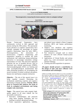
RESEARCH ARTICLE
Retinal Vascular Fractal Dimension,
Childhood IQ, and Cognitive Ability in Old
Age: The Lothian Birth Cohort Study 1936
Adele M. Taylor1, Thomas J. MacGillivray2,3,4*, Ross D. Henderson1, Lasma Ilzina1,
Baljean Dhillon5, John M. Starr6,7, Ian J. Deary1,6*
a11111
1 Department of Psychology, University of Edinburgh, Edinburgh, United Kingdom, 2 Centre for Clinical
Brain Sciences, University of Edinburgh, Edinburgh, United Kingdom, 3 Clinical Research Imaging Centre,
University of Edinburgh, Edinburgh, United Kingdom, 4 Clinical Research Facility, University of Edinburgh,
Edinburgh, United Kingdom, 5 Department of Ophthalmology, Princess Alexandra Eye Pavilion, Edinburgh,
United Kingdom, 6 Centre for Cognitive Ageing and Cognitive Epidemiology, University of Edinburgh,
Edinburgh, United Kingdom, 7 Alzheimer Scotland Dementia Research Centre, University of Edinburgh,
Edinburgh, United Kingdom
* [email protected] (IJD); [email protected] (TJM)
OPEN ACCESS
Citation: Taylor AM, MacGillivray TJ, Henderson RD,
Ilzina L, Dhillon B, Starr JM, et al. (2015) Retinal
Vascular Fractal Dimension, Childhood IQ, and
Cognitive Ability in Old Age: The Lothian Birth Cohort
Study 1936. PLoS ONE 10(3): e0121119.
doi:10.1371/journal.pone.0121119
Academic Editor: Paul Gerard Shiels, University of
Glasgow, UNITED KINGDOM
Received: July 22, 2014
Accepted: February 10, 2015
Published: March 27, 2015
Copyright: © 2015 Taylor et al. This is an open
access article distributed under the terms of the
Creative Commons Attribution License, which permits
unrestricted use, distribution, and reproduction in any
medium, provided the original author and source are
credited.
Data Availability Statement: Due to ethical
restrictions, data are available upon request from the
Lothian Birth Cohort 1936 Study whose authors may
be contacted at [email protected].
Funding: This research and LBC1936 phenotype
collection were supported by Age UK (The
Disconnected Mind project). JMS and IJD are
members of The University of Edinburgh Centre for
Cognitive Ageing and Cognitive Epidemiology (http://
www.ccace.ed.ac.uk) - part of the cross council
Lifelong Health and Wellbeing Initiative - which is
supported by funding from the UK's Biotechnology
Abstract
Purpose
Cerebral microvascular disease is associated with dementia. Differences in the topography of the retinal vascular network may be a marker for cerebrovascular disease. The association between cerebral microvascular state and non-pathological cognitive ageing is
less clear, particularly because studies are rarely able to adjust for pre-morbid cognitive
ability level. We measured retinal vascular fractal dimension (Df) as a potential marker of
cerebral microvascular disease. We examined the extent to which it contributes to differences in non-pathological cognitive ability in old age, after adjusting for childhood
mental ability.
Methods
Participants from the Lothian Birth Cohort 1936 Study (LBC1936) had cognitive ability assessments and retinal photographs taken of both eyes aged around 73 years (n = 648). IQ
scores were available from childhood. Retinal vascular Df was calculated with monofractal
and multifractal analysis, performed on custom-written software. Multiple regression models
were applied to determine associations between retinal vascular Df and general cognitive
ability (g), processing speed, and memory.
Results
Only three out of 24 comparisons (two eyes × four Df parameters × three cognitive measures) were found to be significant. This is little more than would be expected by chance.
No single association was verified by an equivalent association in the contralateral eye.
PLOS ONE | DOI:10.1371/journal.pone.0121119 March 27, 2015
1 / 16
Retinal Vascular Fractal Dimension and Cognitive Ability
and Biological Sciences Research Council, the
Economic and Social Research Council and the
Medical Research Council (MR/K026992/1). The
funders had no role in study design, data collection
and analysis, decision to publish, or preparation of
the manuscript.
Conclusions
The results show little evidence that fractal measures of retinal vascular differences are associated with non-pathological cognitive ageing.
Competing Interests: The authors have declared
that no competing interests exist.
Introduction
Cerebrovascular disease is a major cause of cognitive dysfunction, contributing to cognitive impairment in individuals with and without dementia [1,2]. No single set of cognitive disorders
characterise cerebrovascular disease [3], and due to difficulties in viewing the cerebral small
vessels in vivo, the true burden of cerebral microvascular disease on cognitive function is still
poorly understood [4].
The homology of the retinal and cerebral microvasculatures has led researchers to investigate the potential of the retinal microvasculature as a marker of cerebral microvascular state
[5]. The vascular network is designed for optimal hemodynamic and distributive functions
[6,7] and variation in retinal vascular parameters, such as vessel calibre and branching geometry, may signify a deviation from this. It follows that these retinal changes are a potential marker of cerebrovascular abnormality and disease.
Retinal imaging allows non-invasive, in vivo assessment of the state of the vasculature [8],
and is widely used to study systemic vascular disease [9–11]. Some clinical and populationbased studies show that retinal microvascular abnormalities are associated with recognised
signs of cerebral microvascular pathology, including cerebral microbleeds [12], white matter lesions [13,14], and brain infarcts [15,16]. Importantly, retinal signs are also related to ability in
a range of cognitive domains [17–20], and with increased risk of cognitive impairment [21,22]
and dementia [23]. Ding et al [24] provided a systematic review of this area. The most consistent associations have been found in studies investigating advanced retinal and cognitive abnormalities, such as those using qualitative measures of retinal abnormality, specifically severe
retinopathy, and in those investigating associations with cognitive dysfunction and dementia
rather than differences in ‘normal’ cognitive ability. Associations have been weaker and less
consistent for less severe retinal abnormalities, quantitative retinal measures, and for non-impaired cognitive function. Combined with the inconsistency in sensitivity and robustness of
cognitive measures used across the studies, as yet, there is little hard evidence that retinal vessel
differences are associated with non-impaired cognitive ability in older age.
Recently, researchers have attempted to quantify retinal vascular changes using fractal analysis. This method summarises the global complexity of retinal vascular branching, combining
information about vessel widths, vessel tortuosity, and branching geometry and density, to
give a single dimensionless quantity: the fractal dimension (Df) [25]. Applied to retinal images,
it has potential to be a highly sensitive gauge of microvascular disease. A suboptimal retinal
vascular network increases energy costs and reduces efficiency of metabolic transport; microvascular health is maintained by both adequate vessel diameters and optimal branching architecture [26]. Low Df values, which deviate from the idealised space-filling state of an
“optimized” vascular branching network (Df = 1.7) [27–30] have previously been associated
with stroke [31], diabetic retinopathy [32], coronary heart disease mortality [26], cognitive
dysfunction [22], and dementia [23]. However, it is not known if deviations from optimality
are also related to differences in non-pathologic cognitive ageing. Furthermore, most studies
have relied on monofractal analysis; a method which has generally achieved limited success
[33]. Greater success has been reported by studies using multifractal techniques which
PLOS ONE | DOI:10.1371/journal.pone.0121119 March 27, 2015
2 / 16
Retinal Vascular Fractal Dimension and Cognitive Ability
characterise the retinal vasculature with a hierarchy of exponents rather than a single fractal dimension [34].
An important methodological point is that few studies to date have made adjustments for
differences in prior cognitive ability when investigating the association between retinal vascular
parameters and cognitive ability later in life. This is important because childhood cognitive
ability accounts for a large proportion of variance in cognitive ability in old age [35]. There is
mounting evidence from the field of cognitive epidemiology showing that differences in prior
cognitive ability are associated with health, morbidity and mortality outcomes, including those
related to cerebrovascular disease [36,37]. It is possible that previously found cross-sectional
associations between retinal vascular measures and cognitive ability are confounded by prior
cognitive ability. Shalev et al conducted one of the few studies which reported an association
between non-pathological cognitive ability and retinal vascular measures. They found that
wider retinal venular calibre measured at age 38 years was significantly associated with poorer
cognitive ability measured in childhood, and in adulthood [20]. However, they did not adjust
for childhood cognitive ability in the association between retinal venular calibre and adulthood
cognitive ability, and as a result, the degree to which retinal vascular differences independently
accounted for variance in age 38 cognitive ability was unclear. If retinal vascular differences are
related to normal cognitive function, it is possible that their contribution is small in relation to
that of childhood cognitive ability. Patton and colleagues found childhood IQ to account for almost 20% of the variance in cognitive ability assessed in old age, whilst retinal parameters accounted for 3.7% at the most [19]. The present study is rare in having a valid measure of
childhood cognitive ability, retinal imaging data, and a comprehensive set of cognitive ability
measures from old age.
In this study we investigated the extent to which retinal vascular fractal dimension (as a possible marker of cerebral microvascular disease) contributes to differences in non-pathological
cognitive ability in old age, independently of childhood mental ability.
Methods
Ethical permission for the Lothian Birth Cohort 1936 (LBC1936) study protocol was obtained
from the Multi-Centre Research Ethics Committee for Scotland and from the Lothian Research
Ethics Committee for Scotland. The research was carried out in compliance with the Helsinki
Declaration. Written, informed consent was given by all participants.
LBC1936 Sample
Participants were a sub-sample of the LBC1936: a longitudinal study of cognitive ageing. This
cohort comprises 1091 men and women, most of whom took part in the Scottish Mental Survey
1947 (SMS1947) [35,38]. Initial assessment took place between 2004 and 2007 when participants were about 70 years of age (M = 69.5, ± 0.8). The majority were living in the city of Edinburgh and surrounding Lothian area. A comprehensive description of recruitment and
assessment procedures is available in an open-access protocol paper [39]. Data for the current
study was mainly drawn from a second wave of the study (n = 866) [40]. Briefly, participants
gave sociodemographic and medical history details during structured interviews, underwent
repeat cognitive and physical testing, and completed a series of questionnaires including lifestyle and personality items, at about age 73 (M = 72.5, ± 0.7). Retinal photographs were taken
at this time. Six hundred and sixty three participants had at least one image suitable for fractal
analysis. Individuals with a Mini-Mental State Examination (MMSE) score < 24 were excluded
(n = 6); this cut-off was used to exclude individuals with possible dementia. Participants with
PLOS ONE | DOI:10.1371/journal.pone.0121119 March 27, 2015
3 / 16
Retinal Vascular Fractal Dimension and Cognitive Ability
images of substandard quality for fractal analysis were also excluded (n = 9). A total of 648 remained for statistical analyses (see Fig. 1).
Cognitive Ability
Moray House Test No. 12. The Moray House Test (MHT) No. 12 is a psychometric intelligence test which was validated at age 11 years against the Terman-Merrill revision of the
Binet Scales [41,42]. It encompasses a range of items including reasoning, arithmetic, and
Fig 1. Flow diagram of exclusion factors associated with the study population.
doi:10.1371/journal.pone.0121119.g001
PLOS ONE | DOI:10.1371/journal.pone.0121119 March 27, 2015
4 / 16
Retinal Vascular Fractal Dimension and Cognitive Ability
spatial items, has a time-limit of 45 minutes, and a maximum score of 76. Participants first sat
the MHT on 4th June 1947 as part of the SMS1947, along with almost all schoolchildren in
Scotland aged about 11 years. MHT scores were corrected for age in days at time of testing and
converted to an IQ-type scale for the full LBC1936 sample (mean = 100, SD = 15). This will be
referred to as age 11 IQ. Of the 648 participants included in the analyses, 605 participants had
completed the MHT at age 11.
Cognitive Domains. A general cognitive ability factor (g) was derived from principal components analysis (PCA) of six Wechsler Adult Intelligence Scale-III (WAIS-III) [43] subtests:
Symbol Search, Digit Symbol Coding, Matrix Reasoning, Letter-Number Sequencing, and
Block Design. Likewise, a processing speed factor was derived from scores on a set of speed
measures: Symbol Search and Digit Symbol Coding from WAIS-III; and also measures of Simple and Four-Choice Reaction Time [44], and Inspection Time [45]. The same method was
used to derive a memory factor from two WAIS-III subtests (Letter-Number Sequencing and
Digit Span Backwards) and six Wechsler Memory Scale (WMS-III) subtests [46]: Logical
Memory I Immediate Recall and II Delayed Recall, Spatial Span Forwards and Spatial Span
Backwards, and Verbal Paired Associates I Immediate Recall and II Delayed Recall. The extraction of these factors was described previously [47,48].
Additional Variables
Interview with a trained psychologist elicited self-reported presence or history of hypertension,
cardiovascular disease, stroke, diabetes, and current smoking status (current smoker, ex-smoker,
or non-smoker).
Retinal Image Analysis
Retinal images from both eyes were captured with a nonmydriatic camera at 45o FOV
(CRDGi; Canon USA Inc., Lake Success, NY) and stored with 8 bits per colour plane, at
2048 × 3072 pixels and in TIFF format. Images were down-sampled to 685 x 584 pixels prior to
processing and analysed by an expert retinal image analyst (TM) using custom software built
in MATLAB (The MathWorks, Natwick, MA) [49]. Automatic segmentation of the retinal microvascular network (arterioles and venules) was performed using an algorithm described previously [50] which classifies each pixel of a fundus image as vessel or non-vessel to produce a
segmented map of the retinal vasculature (see Fig. 2). Prior to fractal analysis computationally
segmented images were inspected and obvious artefacts, such as areas of low contrast noise or
“ring” artefacts caused by dust on the camera, were corrected or removed. Fractal analysis may
be more sensitive to changes in vascular patterns when skeletons rather than images containing
vessel width information are considered [51]. Therefore all images were skeletonised by iterative deletion of pixels [52]. Finally, monofractal and multifractal analysis was applied.
In the monofractal approach a single Df (Dbox) was measured using box-counting: the vessel
map or skeletonized version was overlaid with a series of boxes of increasing size l, and the
number of boxes N containing at least one object pixel (representing part of the retinal vascular
tree) was counted. The logarithm of N was plotted against the logarithm of l. Dbox was the
slope of the best fit straight line of the plotted points [53].
In the multifractal approach multiple fractal dimensions were measured at 1000 random
points on the vessel maps and skeletonised images. The generalized sandbox method [34] was
used to investigate the effect of scale on the Df. Multifractal dimensions Df for real f are a more
versatile parameter in describing geometrical properties. Had the retinal vessel tree been a simple monofractal, the Df would have been constant for all scales, f. However, as expected the retinal tree displayed multifractal properties, giving different fractal dimensions as the scale was
PLOS ONE | DOI:10.1371/journal.pone.0121119 March 27, 2015
5 / 16
Retinal Vascular Fractal Dimension and Cognitive Ability
Fig 2. Colour retinal image acquired by fundus photography and retinal blood vessels segmented by computerized procedure.
doi:10.1371/journal.pone.0121119.g002
changed. We computed multifractal dimensions D0, D1, and D2 as these have previously been
reported as likely sensitive markers of small vascular changes [34]. D0, D1 and D2 are commonly referred to as the capacity dimension, the entropy dimension and the correlation dimension,
respectively. As we were unsure of the optimum one to use we investigated all three. Comparing the results enabled us to establish which was the most effective.
Implementation of this algorithm was trained and tested using a set of 20 images, manually
segmented by 2 human observers as well as by the software. Fractal analysis of the manual segmentation performed by the ground truth observer was taken as the reference with which to
compare fractal analysis of a second manual segmentation by a second observer and also the
computational segmentation. A Bland-Altman approach [54] was used to assess the agreement
between Df measurements, with mean differences and coefficients of repeatability (CR) computed. These are summarized in Table 1.
Statistical Analysis
All analyses were conducted using SPSS version 21 (IBM, NY, USA). There were moderate to
strong, but far from perfect, correlations between left and right eye fractal dimensions (Pearson
coefficients between 0.403–0.582); therefore, we conducted analyses on left and right eyes separately. Retinal vascular parameters were analysed as continuous (per 1 SD increase). Cognitive
domain and individual subtest scores were normally distributed; we therefore used multiple
linear regression with cognitive domains and subtest scores as dependent variables, and the
PLOS ONE | DOI:10.1371/journal.pone.0121119 March 27, 2015
6 / 16
Retinal Vascular Fractal Dimension and Cognitive Ability
Table 1. Mean Differences and Coefficient of Repeatability Between Retinal Vascular Fractal Dimension derived by Human Observer and Software Against Ground Truth.
Fractal Dimension
Dbox
D0
D1
D2
Segmentation
Method
Comparison
Mean difference
CR
G
H
-0.02
0.04
G
S
0.01
0.09
G
H
0.02
0.05
G
S
0.01
0.13
G
H
0.01
0.05
G
S
0.02
0.13
G
H
0.00
0.06
G
S
0.01
0.14
*Dbox = monofractal dimension
D0, D1, D2 = multifractal dimension
†
G = ground truth: manual segmentation by first human observer
H = manual segmentation by second human observer
S = automated segmentation by software
doi:10.1371/journal.pone.0121119.t001
retinal microvascular fractal dimensions as independent variables. Three models were fitted to
the data with adjustments for potential confounding factors made in each. Age and sex were
covariates in all three models. Age 11 IQ was included as an additional covariate in the second
model. The third model adjusted for age, sex, age 11 IQ, hypertension, cardiovascular disease,
diabetes, stroke, and current smoking status (non-smoker or ex-smoker vs. current smoker). A
post hoc power analysis using pwr.r.test from the pwr package in R revealed that 648 subjects
would provide 80% power to detect correlation coefficients of small effect size (.11; after
Cohen) [55] with alpha set at. 05, two-tailed.
Results
The mean of the differences between fractal dimensions for two observers and between the first
(ground truth) observer and the computation segmentation were very similar in magnitude,
though the CR was slightly larger for computational segmentation evaluated against first observer (see Table 1). However, the results indicated that the computational segmentation was
within an acceptable level of performance for the identification of vessels (i.e. it was similar to
someone repeating manual segmentation) and so provided suitable input to fractal analysis.
The baseline characteristics for the cohort are summarized in Table 2. Of the 648 participants (326 men, 50.3%) included in the analyses, seven participants did not have a general (g)
cognition score, 32 did not have a processing speed score, 27 did not have a memory score, and
120 did not have the full range of Df measurements. Mean cognitive domain scores and retinal
vascular Df values are summarized in Table 3. Greater Df represents greater branching complexity. Greater g, processing speed, and memory domain scores represent better performance.
The cognitive domain scores reported are standardised variables (mean = 0, SD = 1) for the
whole sample.
Table 4 presents the associations between retinal microvascular Df and ability in the three
cognitive domains. Three of the 24 tests conducted using the basic model were significant at
the conventional p value of <. 05. This is slightly more than one would expect by chance, but
PLOS ONE | DOI:10.1371/journal.pone.0121119 March 27, 2015
7 / 16
Retinal Vascular Fractal Dimension and Cognitive Ability
Table 2. Baseline Characteristics of Participants with Retinal Images of Sufficient and Insufficient Quality for Fractal Analysis.
Characteristics
Image quality
Age, years, mean (SD)
Gradable* (n = 648)
Ungradeable† (n = 9)
72.4 (0.71)
72.4 (0.74)
Male, n (%)
326 (50.3)
6 (66.7)
Age 11 IQ, mean (SD)
102 (14.0)
108 (9.80)
Hypertension, n (%)
313 (48.3)
4 (44.4)
Diabetes, n (%)
61 (9.4)
0 (0)
Current smoker, n (%)
52 (8.0)
2 (22.2)
Cardiovascular disease, mean (%)
177 (27.3)
1 (11.1)
Stroke, n (%)
39 (6.0)
0 (0)
* Participants with at least one retinal image of sufficient quality for fractal analysis
†
Participants with retinal images of insufficient quality for fractal analysis
doi:10.1371/journal.pone.0121119.t002
no association would survive Bonferroni type correction for multiple statistical testing. Full
Bonferroni correction would not be appropriate in this instance given that the retinal measures
are not independent (Pearson coefficients between 0.708–0.982) and neither were the cognitive
measures (Pearson coefficients between 0.438–0.735). In addition to this, no associations were
verified by an equivalent association in the opposite eye, regardless of there being a moderate
correlation between left and right eye measures.
The unadjusted significant associations were as follows. In the basic age- and sex- adjusted
model right eye D1 was significantly associated with g (β = 0.10, P = 0.021) and processing
speed (β = 0.10, P = 0.028). Similarly, right eye D2 was significantly associated with g (β = 0.09,
Table 3. Descriptive Statistics of the Cognitive Domains and Fractal Dimensions of the Retinal Vascular Network.
n
Mean
SD
Range
General (g) Cognitive Ability
641
0.04
0.99
-3.59–3.11
Processing Speed
616
0.08
0.94
-3.18–2.75
Memory
621
0.02
0.98
-3.82–2.21
Dbox
530
1.42
0.02
1.36–1.48
D0
531
1.68
0.02
1.59–1.76
D1
529
1.64
0.03
1.55–1.72
D2
528
1.62
0.03
1.54–1.70
Dbox
537
1.43
0.02
1.35–1.50
D0
538
1.68
0.02
1.60–1.77
D1
539
1.65
0.03
1.57–1.75
D2
538
1.63
0.03
1.55–1.73
Cognitive Domain
Left Eye Fractal Dimension
Right Eye Fractal Dimension
Mean, standard deviation, and range of cognitive domain scores and retinal vascular fractal dimensions.
Dbox = monofractal dimension
D0, D1, D2 = multifractal dimension
N varies due to incomplete range of fractal dimension measurements and/or missing subtest data.
doi:10.1371/journal.pone.0121119.t003
PLOS ONE | DOI:10.1371/journal.pone.0121119 March 27, 2015
8 / 16
Retinal Vascular Fractal Dimension and Cognitive Ability
Table 4. Multiple Regression Models using Retinal Vascular Fractal Dimension to predict Ability in Three Cognitive Domains.
Cognitive Domain, Fractal
Dimension, & Laterality of
Eye
g
Dbox
D0
D1
D2
Speed
Dbox
D0
D1
D2
Memory
Dbox
D0
D1
D2
Model 1
n
β
Model 2
P
n
β
Model 3
P
n
β
P
R
532
0.04
.322
502
0.03
.353
502
0.04
.250
L
523
-0.03
.478
485
-0.03
.432
485
-0.03
.395
R
533
0.08
.063 †
503
0.02
.604
503
0.03
.436
L
524
0.000
.995
486
-0.01
.900
486
-0.01
.825
R
534
0.10
.021*
503
0.03
.404
503
0.04
.324
L
522
0.01
.904
484
0.000
.993
484
-0.01
.749
R
533
0.09
.035*
502
0.04
.259
502
0.04
.244
L
521
0.001
.976
483
-0.01
.812
483
-0.02
.608
R
512
0.04
.385
484
0.03
.447
484
0.04
.315
L
503
-0.08
.094 †
467
-0.06
.195
467
-0.05
.257
R
513
0.07
.094 †
485
0.04
.341
485
0.05
.250
L
504
-0.04
.386
468
-0.02
.625
468
-0.02
.698
R
514
0.10
.028*
485
0.06
.179
485
0.06
.136
L
502
-0.04
.429
466
-0.03
.473
466
-0.03
.452
R
513
0.09
.035*
484
0.07
.115
484
0.07
L
502
-0.03
.583
466
-0.03
.506
466
-0.03
.466
R
513
-0.03
.539
485
-0.02
.673
485
-0.01
.811
L
506
-0.07
.142
470
-0.08
.050*
470
-0.08
.040*
R
514
0.02
.719
486
-0.02
.545
486
-0.02
.611
L
506
-0.05
.232
470
-0.07
.084 †
470
-0.07
.069 †
R
515
0.02
.734
486
-0.03
.421
486
-0.03
.466
L
505
R
514
L
505
-0.06
0.000
-0.06
†
.092 †
.175
469
-0.07
.053
469
-0.08
.038*
.992
485
-0.03
.419
485
-0.03
.439
.188
469
-0.08
.041*
469
-0.08
.029*
Data are presented as standardised beta coefficients reflecting change in cognitive domain score associated with an increase of 1 SD unit in fractal
dimension. Model 1 adjusted for age and sex; Model 2 adjusted for age, sex, and age 11 IQ; Model 3 adjusted for age, sex, age 11 IQ, hypertension,
diabetes, cardiovascular history, stroke, current smoking status.
Dbox = monofractal dimension; D0, D1, D2 = multifractal dimension.
N varies due to incomplete range of fractal dimension measurements and/or missing subtest data.
* Significant (p < 0.05).
Trend (p < 0.10).
†
doi:10.1371/journal.pone.0121119.t004
P = 0.035) and processing speed (β = 0.09, P = 0.035). There was also a statistical trend with
right eye D0 (g: β = 0.08, P = 0.063; processing speed: β = 0.07, P = 0.094). No association was
found between monofractal or multifractal retinal vascular Df and memory in this model. With
the exception of a statistical trend between left eye Dbox and processing speed (β = -0.08,
P = 0.094), no association was found between monofractal dimension Dbox and any
cognitive domain.
IQ at age 11 was not associated with any of the retinal microvascular Df measures at age 73
(Left Dbox: β = 0.007, P = 0.875; Left D0: β = 0.03, P = 0.491; Left D1: β = 0.000, P = 0.996; Left
D2: β = -0.004, P = 0.931; Right Dbox: β = 0.01, P = 0.809; Right D0: β = 0.08, P = 0.063; Right
D1: β = 0.07, P = 0.111; Right D2: β = 0.05, P = 0.236). The addition of age 11 IQ as a covariate
in model 2 weakened the associations between D0, D1, and D2 with g and processing speed, the
β coefficients were reduced by more than half in some cases, and in all cases significance was
PLOS ONE | DOI:10.1371/journal.pone.0121119 March 27, 2015
9 / 16
Retinal Vascular Fractal Dimension and Cognitive Ability
lost. The further addition of all cardiovascular risk factors as covariates in model 3 did not alter
these associations significantly, though there was a statistical trend between right eye D2 and
processing speed (β = 0.07, P = 0.092). Accounting for age 11 IQ in the association between
retinal Df and memory had the converse effect to that seen with the other domain scores. After
adjustment for age 11 IQ, left eye Df analysed using monofractal and multifractal methods significantly predicted memory (Dbox: β = -0.08, P = 0.050; D2: β = -0.08, P = 0.041), and there
was a statistical trend with D0 (β = -0.07, P = 0.084) and D1 (β = -0.07, P = 0.053). Significance
was maintained when all cardiovascular risk factors were accounted for in model 3 (Dbox:
β = -0.08, P = 0.040; D1: B = -0.08, P < 0.038; D2: β = -0.08, P = 0.029), again there was a statistical trend with D0 (β = -0.07, P = 0.069).
The reversed direction of association indicates that increased branching complexity predicted poorer memory performance. However, again these associations were not corroborated
by an equivalent finding when right eye retinal measures were tested and are therefore unlikely
to be meaningful. Taken together, our results show no evidence that differences in retinal vascular fractal dimension contribute to cognitive ability in old age. We suggest that in our sample,
differences in childhood ability, but not retinal vascular parameters, account for much of the
variance in cognitive ability in later life.
S1 Table contains results from the analysis of individual cognitive subtests.
Discussion
Microvascular disease is a major contributor to cognitive dysfunction in individuals with and
without dementia. However, its role in non-pathologic cognitive function is unclear. The present study investigated cross-sectional associations between retinal vascular Df, a putative marker of cerebral microvascular health, and three domains of cognitive ability in a sample of older
adults without cognitive impairment. After adjustment for childhood IQ and traditional cardiovascular risk factors, greater left eye monofractal and multifractal dimensions (Dbox, D1,
and D2) were associated with poorer memory performance, however, neither left nor right eye
fractal dimensions were associated with g or processing speed, and no association was verified
by an equivalent result in the contralateral eye.
The data on retinal microvascular topography and non-pathological cognitive ageing have
been limited, and direct comparison with previous studies is not possible due to various methodological differences in the research to date. The Lothian Birth Cohort 1921 (LBC1921) [19]
study examined the associations between quantitative measures of suboptimal retinal vascular
branching geometry and cognitive ability in 321 men and women aged either 83 or 84. After
adjusting for childhood IQ, cardiovascular risk factors, and socio-demographic variables, deviation from the optimal retinal vascular bifurcation angle accounted for 2.6% of the variance in
logical memory test scores of older adults, while deviation from the optimal retinal vascular
branching coefficient accounted for 3.4% of the variance in a general cognitive ability (g) score,
and 3.7% in verbal fluency test score. Similar results were found in sample of 1007 men and
women from the Dunedin Multidisciplinary Health and Development study, a population
based cohort followed from birth [20]. In this study, wider venular calibre was associated with
lower total and delayed Rey auditory verbal learning scores, lower scores on several tests of executive function, and lower scores on all components of WAIS-IV. Our study results differ
from these data in that no association was found between retinal vascular Df and measures of g
or processing speed, and retinal vascular Df was only weakly associated with memory, though
this was not confirmed by the contralateral eye and therefore unlikely to be meaningful. Some
of this difference may be attributable to the differences in mean age of the cohorts. The
LBC1921 cohort was aged between 83 and 84 years old, an age at which between-person
PLOS ONE | DOI:10.1371/journal.pone.0121119 March 27, 2015
10 / 16
Retinal Vascular Fractal Dimension and Cognitive Ability
variability in cognitive ability scores and retinal vascular state may be increased; however, fractal dimension was not measured in the LBC1921 study, and it is not possible to say if cognitive
ability would have been associated with retinal vascular Df in that sample. The Dunedin study
cohort had a mean age of 38, and their results may be more consistent with the hypothesis that
cognitive impairment is more greatly influenced by cerebral microvascular disease in younger
compared to older adults [18]. Though, it is unlikely that much disease would be present in
people of this age.
Qualitative measures of retinal vascular abnormality have also been associated with cognitive ability in older age. Results from the population-based Atherosclerosis Risk in Communities (ARIC) study showed that in a sample of 8734 persons, individuals with any retinopathy,
microaneurysm, retinal haemorrhage or soft exudates, were 2.6 to 3.39 times more likely to
score below mean on the delayed word recall test than those without [17]. Wong and colleagues
also found an association between presence of retinopathy and low scores on the digit symbol
substitution test, in both the ARIC study and in another sample of 2211 participants from the
population-based Cardiovascular Health Study (CHS) In contrast to quantitative measures designed to identify subtle changes to the vasculature, qualitative measures are indicative of more
severe states and even diseases of the retinal microvasculature, which are often associated with
the breakdown of the blood-brain barrier. This difference in severity of retinal vascular pathology may partly explain why associations between cognitive ability and qualitative measures
have been more consistent than those assessing less severe changes, such as narrowing of the
vessels [18].
The relationship between ‘normal’ cognitive ageing and retinal vascular fractal dimension
has not previously been investigated. The optimal retinal vascular Df is said to be 1.7, and deviations from this have previously been associated with ageing [33], diabetic retinopathy [25]
and death resulting from coronary heart disease (CHD) [26]. Cheung and colleagues recently
found a significant association between reduced retinal vascular Df and cognitive dysfunction.
Individuals with the lowest Df were between 1.03 and 2.82 times more likely to have cognitive
dysfunction [22]. In a separate study the same authors found, that for every 1 SD unit decrease
in Df, the likelihood of having Alzheimer’s Disease rose by 1.54 [23].
This study has several strengths. It makes a novel contribution to research investigating the
relationship between parameters of the retinal microvasculature and cognitive function. To our
knowledge, it is the first to examine the relationship between retinal vascular fractal dimension
and non-pathologic cognitive ability, and the only one to do so using both monofractal and
multifractal methods of analysis. It is important to note that generalized dimension spectrums
for each image analysed, showed that the retinal vessel structures were all geometrical multifractals. Second, we were able to make statistical adjustment for childhood cognitive ability, a
factor known to account for a large proportion of variance in later life cognitive ability [35, 56].
Third, cognitive domain scores were based on a broad range of neuropsychological tests, giving
a robust measure of general (g) cognitive ability, processing speed, and memory. Finally, in
contrast to previous studies [32,33] by analysing left and right eye retinal measures separately
we were able to show that no association was substantiated by the same result using measurements from the contralateral eye (see S1–S3 Figs).
Limitations of the study should also be noted. Problems exist with the automated segmentation algorithm: the edge of the optic disk is sometimes wrongly detected as a vessel, incorrect
detections result from the underlying choroidals, gaps appear in the middle of vessels if the
central light reflex is bright, some vessel paths are broken due to poor image contrast and very
small vessels are sometimes missed. All of these issues lead to a reduction in segmentation accuracy and fractal dimension of the retinal vasculature. Irrespectively, our images have very
good agreement between the segmentations by two observers and so for fractal analysis the
PLOS ONE | DOI:10.1371/journal.pone.0121119 March 27, 2015
11 / 16
Retinal Vascular Fractal Dimension and Cognitive Ability
mean differences and CRs were small (see Table 1). When comparing fractal dimensions for
computerised segmentations and ground truth (first human segmentation), mean differences
were also small and CRs were an acceptable level. These results show that for a reasonable
image quality, repeatability of fractal analysis performed on computerised segmentations is
close to the repeatability found when two observers independently perform manual tracing.
For large epidemiological studies, i.e. greater than fifty images, this would be at acceptable level
in order to detect small changes.
Second, adjustment was not made for myopic refractive errors. Increasing myopia is associated with reduced retinal vascular fractal dimension, though this reduction is likely to be minimal
except in cases of high myopia [57]. Additionally, myopia is less prevalent in older individuals,
and therefore in the LBC participants myopia may not affect results to the same extent as a
younger sample [58]. Differences in retinal vascular fractal dimension have also been found
when comparing images from normal eyes to those from individuals diagnosed with diabetic
retinopathy [25, 32]. Though presence of diabetes was not part of the exclusion criteria for the
present study, no single image judged as ungradeable belonged to a diabetic individual, and adjustment for the presence of diabetes was made in the analyses. Third, the LBC1936 is a self-selecting sample, and as such it is likely to be healthier and more cognitively able than the general
population [56]. It should be noted however, that though retinal vascular Df does not appear to
be associated with cognitive function in this sample at age 73, associations may be found as the
sample ages and vascular pathology progresses. And though an association with cognitive ability
was not found in this study, that is not to say that individual differences in retinal vascular Df
are not indicative of abnormality and disease of small vessels in general. Small vessel disease is a
systemic disorder [2], and though small vessels of the brain may bear the burden of this disease
in some individuals, in others, organs through the body will be most affected [4]. Further research is required to understand the full potential of fractal analysis as an early indicator of disease in the brain and body. Finally, retinal images were taken during only one wave of testing,
restricting us to cross-sectional analyses. Access to retinal images taken at multiple time points
would allow examination of these associations in relation to longitudinal change.
Conclusion
Three out of the 24 comparisons were found to be significant. Although this is more than one
would expect by chance, no association would survive Bonferroni correction for multiple statistical testing. Significant unadjusted associations were weakened and lost significance after covarying for IQ at age 11 and cardiovascular risk factors, and not one association was verified by
an equivalent finding using measurements from the contralateral eye. We cautiously suggest
that our results do not reject the null hypothesis, that differences in retinal vascular Df are not
associated with cognitive ability in old age. Further research is required to elucidate the relationship between parameters of the retinal microvascular network and non-pathological
cognitive function.
Supporting Information
S1 Fig. Change in cognitive domain score associated with 1 SD unit increase in fractal dimension. Dbox = monofractal dimension. D0, D1, D2 = multifractal dimension. β significant
at ± 0.08 (p < 0.05).
(PDF)
S2 Fig. Change in cognitive subtest score associated with 1 SD increase in right eye fractal
dimension. Dbox = monofractal dimension. D0, D1, D2 = multifractal dimension. β
PLOS ONE | DOI:10.1371/journal.pone.0121119 March 27, 2015
12 / 16
Retinal Vascular Fractal Dimension and Cognitive Ability
significant at ± 0.08 (p < 0.05). 1: Symbol search, 2: digit symbol coding, 3: matrix reasoning,
4:letter-number sequencing, 5: digit span backwards, 6: block design, 7: simple reaction time,
8: 4-choice reaction time, 9: inspection time, 10: spatial span forwards, 11: spatial span backwards, 12: verbal paired associates immediate recall, 13: verbal paired associates delayed recall,
14: logical memory immediate recall, 15: logical memory delayed recall.
(PDF)
S3 Fig. Change in cognitive subtest score associated with 1 SD increase in left eye fractal dimension. Dbox = monofractal dimension. D0, D1, D2 = multifractal dimension. β significant
at ± 0.08 (p < 0.05). 1: Symbol search, 2: digit symbol coding, 3: matrix reasoning, 4: letternumber sequencing, 5: digit span backwards, 6: block design, 7: simple reaction time, 8:
4-choice reaction time, 9: inspection time, 10: spatial span forwards, 11: spatial span backwards, 12: verbal paired associates immediate recall, 13: verbal paired associates delayed recall,
14: logical memory immediate recall, 15: logical memory delayed recall.
(PDF)
S1 Table. Multiple Regression Models Using Retinal Vascular Fractal Dimension to predict
Ability in Cognitive Subtests. Note. p < 0.05; † trend p < 0.10. Dbox = monofractal dimension; D0, D1, D2 = multifractal dimension; VPA = verbal paired associates; LM = logical memory. Standardised beta coefficients reflect change in cognitive subtest score associated with an
increase of 1 SD unit in fractal dimension. Model 1 adjusted for age and sex; Model 2 adjusted
for age, sex, and age 11 IQ; Model 3 adjusted for age, sex, age 11 IQ, hypertension, diabetes, cardiovascular history, stroke, current smoking status. N varies due to incomplete range of fractal
dimension measurements and/or missing subtest data.
(PDF)
Acknowledgments
We thank the Scottish Council for Research in Education for allowing access to the SMS1947.
We thank the LBC1936 Study participants who contributed to these studies, the research staff
responsible for data collection, and staff at the Wellcome Trust Clinical Research Facility. JMS
and IJD are members of The University of Edinburgh Centre for Cognitive Ageing and Cognitive Epidemiology, part of the cross council Lifelong Health and Wellbeing Initiative.
Author Contributions
Conceived and designed the experiments: IJD AMT TJM. Performed the experiments: RDH
LI. Analyzed the data: IJD AMT TJM. Contributed reagents/materials/analysis tools: TJM.
Wrote the paper: AMT TJM RDH LI BD JMS IJD. Directed cohort: IJD.
References
1.
O’Brien JT, Erkinjuntti T, Reisberg B, Roman G, Sawada T, Pantoni L, et al. Vascular cognitive impairment. Lancet Neurol. 2003; 2: 89–98. PMID: 12849265
2.
Thompson CS, Hakim AM. Living beyond our physiological means: small vessel disease of the brain
is an expression of a systemic failure in arteriolar function: a unifying hypothesis. Stroke. 2009; 40:
e322–e330. doi: 10.1161/STROKEAHA.108.542266 PMID: 19228835
3.
Knopman DS. Cerebrovascular disease and dementia. Br J Radiol. 2007; 80: S121–S127. doi: 10.
1259/bjr/75681080 PMID: 18445742
4.
Pantoni L. Cerebral small vessel disease: from pathogenesis and clinical characteristics to therapeutic
challenges. Lancet Neurol. 2010; 9: 689–701. doi: 10.1016/S1474-4422(10)70104-6 PMID: 20610345
PLOS ONE | DOI:10.1371/journal.pone.0121119 March 27, 2015
13 / 16
Retinal Vascular Fractal Dimension and Cognitive Ability
5.
Patton N, Aslam T, Macgillivray T, Pattie A, Deary IJ, Dhillon B. Retinal vascular image analysis as a
potential screening tool for cerebrovascular disease: a rationale based on homology between cerebral
and retinal microvasculatures. J Anat. 2005; 206: 319–348. PMID: 15817102
6.
Murray CD. The physiological principle of minimum work: I. The vascular system and the cost of blood
volume. Proc Natl Acad Sci U S A. 1926; 12: 207–214. PMID: 16576980
7.
Zamir M. Fractal dimensions and multifractility in vascular branching. J Theor Biol. 2001; 212: 183–190.
PMID: 11531384
8.
Patton N, Aslam TM, MacGillivray T, Deary IJ, Dhillon B, Eikelboom RH, et al. Retinal image analysis:
concepts, applications and potential. Prog Retin Eye Res. 2006; 25: 99–127. PMID: 16154379
9.
Ikram MK, de Jong FJ, Vingerling JR, Witteman JC, Hofman A, Breteler M, et al. Are retinal arteriolar or
venular diameters associated with markers for cardiovascular disorders? The Rotterdam Study. Invest
Ophthalmol Vis Sci. 2004; 45: 2129–2134. PMID: 15223786
10.
Wong TY, Hubbard LD, Klein R, Marino EK, Kronmal R, Sharrett AR, et al. Retinal microvascular abnormalities and blood pressure in older people: the Cardiovascular Healthy Study. Br J Ophthalmol. 2002;
86: 1007–1013. PMID: 12185128
11.
Klein R, Klein BE, Moss SE, Wong TY, Hubbard L, Cruickshanks KJ, et al. Retinal vascular abnormalities in persons with type 1 diabetes the Wisconsin Epidemiologic Study of Diabetic Retinopathy: XVIII.
Ophthalmology. 2003; 110: 2118–2125. PMID: 14597518
12.
Qiu C, Cotch MF, Sigurdsson S. Cerebral microbleeds, retinopathy, and dementia: the AGES-Reykjavik
Study. Neurology. 2010; 75: 2221–2228. doi: 10.1212/WNL.0b013e3182020349 PMID: 21172845
13.
Longstreth WT, Larsen EK, Klein R, Wong TY, Sharrett AR, Lefkowitz D, et al. Associations between
findings on cranial magnetic resonance imaging and retinal photography in the elderly: the Cardiovascular Health Study. Am J Epidemiol. 2007; 165: 78–84. PMID: 17041135
14.
Kwa VI, van der Sande JJ, Stam J, Tijmes N, Vrooland JL. Retinal arterial changes correlate with cerebral small-vessel disease. Neurology. 2002; 59: 1536–1540. PMID: 12451193
15.
Cooper LS, Wong TY, Klein R, Sharrett AR, Bryan N, Hubbard LD, et al. Retinal microvascular abnormalities and MRI-defined subclinical cerebral infarction: the Atherosclerosis Risk in Communities
Study. Stroke. 2006; 37: 82–86. PMID: 16306463
16.
Cheung N, Mosley T, Islam A. Retinal microvascular abnormalities and subclinical magnetic resonance
imaging brain infarct: a prospective study. Brain. 2010; 133: 1987–1993. doi: 10.1093/brain/awq127
PMID: 20519327
17.
WongT Y, Klein R., Sharrett AR, Nieto J, Boland LL, Couper DJ, et al. Retinal microvascular abnormalities and cognitive impairment in middle-aged persons: The Atherosclerosis Risk in Communities Study.
Stroke. 2002; 33: 1487–1492. PMID: 12052979
18.
Baker ML, Marino Larsen EK, Kuller LH, Klein R, Klein BE, Siscovick DS, et al. Retinal microvascular
signs, cognitive function, and dementia in older persons: the Cardiovascular Health Study. Stroke.
2007; 38: 2041–2047. PMID: 17525385
19.
Patton N, Pattie A, MacGillivray T, Aslam T, Dhillon B, Gow A, et al. The association between retinal
vascular network geometry and cognitive ability in an elderly population. Invest Ophthalmol Vis Sci.
2007; 48: 1995–200. PMID: 17460252
20.
Shalev I, Moffitt TE, Wong TY, Meier MH, Houts RM, Ding J, et al. Retinal vessel caliber and lifelong
neuropsychological functioning: retinal imaging as an investigative tool for cognitive epidemiology. Psychol Sci. 2013; 24: 1198–1207. doi: 10.1177/0956797612470959 PMID: 23678508
21.
Liew G, Mitchell P, Wong TY, Lindley RI, Cheung N, Kaushik S, et al. Retinal microvascular signs and
cognitive impairment. J Am Geriatr Soc. 2009; 57: 1892–1896. doi: 10.1111/j.1532-5415.2009.02459.x
PMID: 19737331
22.
Cheung CY, Ong SY, Ikram MK, Ong YT, Chen CP, Venketasubramanian N, et al. Retinal vascular
fractal dimension is associated with cognitive dysfunction. J Stroke Cerebrovasc Dis. 2012; 23: 43–50.
doi: 10.1016/j.jstrokecerebrovasdis.2012.09.002 PMID: 23099042
23.
Cheung CY, Ong YT, Ikram MK, Ong SY, Li X, Hilal S, et al. Microvascular network alterations in the retina of patients with Alzheimer’s disease. Alzheimers Dement. 2013; 10: 135–142.
24.
Ding J, Patton N, Deary IJ, Strachan MW, Fowkes FG, Mitchell RJ, et al. Retinal microvascular abnormalities and cognitive dysfunction: a systematic review. Br J Ophthalmol. 2008; 92: 1017–1025. doi:
10.1136/bjo.2008.141994 PMID: 18614569
25.
Avakian A, Kalina RE, Sage EH, Rambhia AH, Elliott KE, Chuang EL, et al. Fractal analysis of regionbased vascular change in the normal and non-proliferative diabetic retina. Curr Eye Res. 2002; 24:
274–280. PMID: 12324866
PLOS ONE | DOI:10.1371/journal.pone.0121119 March 27, 2015
14 / 16
Retinal Vascular Fractal Dimension and Cognitive Ability
26.
Liew G, Mitchell P, Rochtchina E, Wong TY, Hsu W, Lee ML, et al. Fractal analysis of retinal microvasculature and coronary heart disease mortality. Eur Heart J. 2011; 32: 422–429. doi: 10.1093/eurheartj/
ehq431 PMID: 21138936
27.
Masters BR. Fractal analysis of the vascular tree in the human retina. Annu Rev Biomed Eng. 2004; 6:
427–452. PMID: 15255776
28.
Daxer A. Characterisation of the neovascularisation process in diabetic retinopathy by means of fractal
geometry: diagnostic implications. Graefes Arch Clin Exp Ophthalmol. 1993; 231: 681–686. PMID:
8299974
29.
Mainster MA. The fractal properties of retinal vessels: embryological and clinical implications. Eye
(Lond). 1990; 4: 235–241.
30.
Landini G, Murray PI, Misson GP. Local connected fractal dimensions and lacunarity analyses of 60°
fluorescein angiograms. Invest Ophthalmol Vis Sci. 1995; 36: 2749–2755. PMID: 7499097
31.
Doubal FN, MacGillivray TJ, Patton N, Dhillon B, Dennis MS, Wardlaw JM Fractal analysis of retinal
vessels suggests that a distinct vasculopathy causes lacunar stroke. Neurology. 2010; 74: 1102–1107.
doi: 10.1212/WNL.0b013e3181d7d8b4 PMID: 20368631
32.
Cheung N, Donaghue KC, Liew G, Rogers SL, Wang JJ, Lim S, et al. Quantitative assessment of early
diabetic retinopathy using fractal analysis. Diabetes Care. 2009; 32: 106–110. doi: 10.2337/dc08-1233
PMID: 18835945
33.
Azemin MZ, Kumar DK, Wong TY, Wang JJ, Mitchell P, Kawasaki R, et al. Age-related rarefaction in
the fractal dimension of retinal vessel. Neurobiol Aging. 2012; 33: 194.e1–194.e4.
34.
Stosić T & Stosić BD. Multifractal analysis of human retinal vessels. IEEE Trans Med Imaging. 2006;
25: 1101–1107. PMID: 16895002
35.
Deary IJ, Whiteman MC, Starr JM, Whalley LJ, Fox HC. The impact of childhood intelligence on later life:
following up the Scottish Mental Surveys of 1932 and 1947. J Pers Soc Psychol. 2004; 86: 130–147.
PMID: 14717632
36.
Gottfredson L, Deary IJ. Intelligence predicts health and longevity, but why? Curr Dir Psychol Sci. 2004;
13: 1–4. PMID: 18414600
37.
Deary IJ, Weiss A, Batty GD. Intelligence and personality as predictors of illness and death: how researchers in differential psychology and chronic disease epidemiology are collaborating to understand
and address health inequalities. Psychol Sci Public Interest. 2010; 11: 53–79.
38.
Deary IJ, Whalley LJ, Starr JM. A lifetime of Intelligence: Follow-up Studies of the Scottish Mental Surveys of 1932 and 1947. Washington, DC: American Psychological Association; 2009.
39.
Deary IJ, Gow AJ, Taylor MD, Corley J, Brett C, Wilson V, et al. The Lothian birth cohort 1936: a study
to examine influences on cognitive ageing from age 11 to age 70 and beyond. BMC Geriatr. 2007; 7:
28. PMID: 18053258
40.
Deary IJ, Gow AJ, Pattie A, Starr JM. Cohort profile: the Lothian Birth Cohorts of 1921 and 1936. Int J
Epidemiol. 2012; 41: 1576–1584. doi: 10.1093/ije/dyr197 PMID: 22253310
41.
Scottish Council for Research in Education (SCRE). The intelligence of Scottish Children: A National
Survey of an Age Group. London, UK: University of London Press; 1933.
42.
Scottish Council for Research in Education (SCRE). The Trend of Scottish Intelligence: A Comparison
of the 1947 and 1932 Surveys of the Intelligence of Eleven-Year-Old Pupils. London, UK: University of
London Press; 1949.
43.
Wechsler D. WAIS-IIIUK Administration and Scoring Manual. London, UK: Psychological Corporation;
1998.
44.
Deary IJ, Der G, Ford G. Reaction times and intelligence differences: a population-based cohort study.
Intelligence. 2001; 29: 389–399.
45.
Deary IJ, Simonotto E, Meyer M, Marshall A, Marshall I, Goddard N, et al. The functional anatomy of inspection time: an event-related fMRI study. Neuroimage. 2004; 22: 1466–1479. PMID: 15275904
46.
Wechsler D. WMS-IIIUK Administration and Scoring Manual. London, UK: Psychological Corporation;
1998.
47.
Luciano M, Gow AJ, Harris SE, Hayward C, Allerhand M, Starr JM, et al. Cognitive ability at age 11 and
70 years, information processing speed, and APOE variation: the Lothian Birth Cohort 1936 study. Psychol Aging. 2009; 24: 129–138. doi: 10.1037/a0014780 PMID: 19290744
48.
Corley J, Gow AJ, Starr JM, Deary IJ. Is body mass index in old age related to cognitive abilities? The
Lothian Birth Cohort 1936 Study. Psychol Aging. 2010; 25: 867–875. doi: 10.1037/a0020301 PMID:
21186916
PLOS ONE | DOI:10.1371/journal.pone.0121119 March 27, 2015
15 / 16
Retinal Vascular Fractal Dimension and Cognitive Ability
49.
MacGillivray TJ, Perez-Rovira A, Trucco E, Chin KS, Giachetti A, Lupascu C, et al. VAMPIRE: Vessel
Assessment and Measurement Platform for Images of the Retina. In: Ng EYK, Tan JH, Acharya UR,
Suri JS, editors. Human Eye Imaging and Modeling. Boca Raton, FL: CRC Press. 2012. pp. 23–38.
50.
Soares JVB, Leandro JJG, Cesar RM, Jelinek HF, Cree MJ. Retinal vessel segmentation using the 2-D
Gabor wavelet and supervised classification. IEEE Trans Med Imaging. 2006; 25: 1214–1222. PMID:
16967806
51.
Parsons-Wingerter P, Elliot KE, Clark JI, Farr AG. Fibroblast growth factor-2 selectively stimulates angiogenesis of small vessels in arterial tree. Arterioscler Thromb Vasc Biol. 2000; 20: 1250–1256. PMID:
10807740
52.
Lam L, Lee S, Suen CY. Thinning-methodologies—a comprehensive survey. IEEE Trans Pattern Anal
Mach Intell.1992; 14: 869–885.
53.
MacGillivray TJ, Patton N, Doubal FN, Graham C, Wardlaw JM. Fractal analysis of the retinal vascular
network in fundus image. Conf Proc IEEE Eng Med Biol Soc. 2007: 6456–6459. PMID: 18003503
54.
Bland JM & Altman DJ. Statistical methods for assessing agreement between two methods of clinical
measurement. Lancet. 1986; 1: 307–310. PMID: 2868172
55.
Cohen J. A power primer. Psychol Bull. 1992; 112: 155–159. PMID: 19565683
56.
Gow AJ, Johnson W, Pattie A, Brett CE, Roberts B, Starr JM, et al. Stability and change in intelligence
from age 11 to ages 70, 79, and 87: the lothian birth cohorts of 1921 and 1936. Psychol Aging. 2011;
26: 232–240. doi: 10.1037/a0021072 PMID: 20973608
57.
Li H, Mitchell P, Liew G, Rochtchina E, Kifley A, Wong TY, et al. Lens opacity and refractive influences
on the measurement of retinal vascular fractal dimension. Acta Ophthalmol. 2010; 88: e234–e240. doi:
10.1111/j.1755-3768.2010.01975.x PMID: 20662797
58.
Wensor M. Prevalence and risk factors of myopia in Victoria, Australia. Arch Ophthalmol. 1999; 117:
658–663. PMID: 10326965
PLOS ONE | DOI:10.1371/journal.pone.0121119 March 27, 2015
16 / 16


© Copyright 2026