
COMPUTATIONAL MECHANICS TOWARDS IMPROVED UNDERSTANDING OF THE BIOMECHANICS OF
Computational Mechanics of the Heart Department of Civil Engineering COMPUTATIONAL MECHANICS TOWARDS IMPROVED UNDERSTANDING OF THE BIOMECHANICS OF MYOCARDIAL INFARCTION Name: Gesant Abed Civil Engineering B.Sc CIV 4044S Dr. S. Skatulla Date : 17/11/2010 The use of finite element analyses and modelling to assist the biomedical and engineering professions to better understand the biomechanics and material properties of the left ventricle, healthy, infarcted and hydrogels assisted cardiac cycles. CIV4044S 2010 University of Cape Town Gesant Abed 1 Computational Mechanics of the Heart Department of Civil Engineering Abstract This thesis uses the Finite element modelling using methods of computational programs to better understand the structural mechanics of the heart. This analysis would consist of modelling the cardiac cycle, the infarcted tissue and the use of hydrogels on these infarcted regions. The results were compared where the movement of the infarcted tissue and the movement after the hydrogel therapy were reviewed to identify its effectiveness on assisting healing of heart muscle tissue. Furthermore research was done on the biology of the heart and the causes of heart diseases its effects. The methods regarding The use of the computer assisted finite element analyses explains the steps that had to be followed which needs to be employed to mimic the muscle contractions (systole) and expansions (diastole), and the interaction of the boundary between infarction region and the health tissue. This thesis explores the potential and limitations of using thermo mechanic loading procedures in finite element modelling especially using it to activate the heart model through the cardiac cycle. CIV4044S 2010 University of Cape Town Gesant Abed 2 Computational Mechanics of the Heart Department of Civil Engineering Contents Abstract ...................................................................................................................................... 2 Contents ..................................................................................................................................... 3 List of Figures .............................................................................................................................. 5 List of Tables ............................................................................................................................... 7 1. Introduction .......................................................................................................................... 8 1.1 Statement of the problem ............................................................................................. 8 1.2 Objectives of the thesis ................................................................................................. 8 2. Role of Bioengineering .......................................................................................................... 9 3. Heart................................................................................................................................... 10 3.1 Healthy Heart .............................................................................................................. 10 3.2 Heart attack/ Myocardial Infarction ............................................................................ 14 3.3 Cardiac cycle ............................................................................................................... 17 3.4 Heart Tissue ................................................................................................................ 22 4. Hydrogels in biomechanics .................................................................................................. 26 5. Engineering Properties ........................................................................................................ 27 6. Finite Element Theory ......................................................................................................... 31 7. Modelling the heart ............................................................................................................ 32 8. Modelling For This Study ..................................................................................................... 36 8.1 ABAQUS Finite Element modelling and Analysis .......................................................... 38 8.2 Modelling Methodology .............................................................................................. 39 8.3 Thermal Stress and Strain............................................................................................ 44 8.4 Fibre Orientations ....................................................................................................... 45 8.5 Boundary Conditions and constraints .......................................................................... 46 8.6 Loading Conditions ...................................................................................................... 48 9. Meshing .............................................................................................................................. 50 CIV4044S 2010 University of Cape Town Gesant Abed 3 Computational Mechanics of the Heart Department of Civil Engineering 10. FE ABAQUS DATA Analysis/ Results ..................................................................................... 51 10.1 Healthy Heart .............................................................................................................. 52 10.2 Heat and temperature ................................................................................................ 56 10.3 Infarcted results .......................................................................................................... 60 10.4 Treated with Hydrogels ............................................................................................... 63 10.5 Different Hydrogel properties ..................................................................................... 65 11. Discussion ........................................................................................................................... 66 12. Conclusions ......................................................................................................................... 68 13. Acknowledgements............................................................................................................. 69 14. Bibliography ........................................................................................................................ 70 CIV4044S 2010 University of Cape Town Gesant Abed 4 Computational Mechanics of the Heart Department of Civil Engineering List of Figures Figure 1 Diagram showing basic structure of the Human heart 11 Figure 2 Indication of the conduction network (Wikipedia) 12 Figure 3 Picture shows the conduction system in relation to the structure of the heart. 13 Figure 4 Diagram example of the extent of tissue that can be affected through ischemia 16 Figure 5 Shows the pathology of the blood through the cardiac cycle. (Wikipedia) 18 Figure 6 The Pressure vs volume change over time (Cardiac Cycle) (DDAH Group) 20 Figure 7 Chart represents all different pressure and volume changes over time 21 Figure 8 Left heart dissection of the heart after boiled, right is left ventricular wall fibre pathways. 23 Figure 9 Results from an analysis done using the pressure on the left and right ventricles. 32 Figure 10. Pressure direction at systole and diastole. 33 Figure 11 Estimation of Mechanical deformations of ellipsoidal heart model 34 Figure 12 Heart model showing fibre orientation and the band shape and fitted volume 35 Figure 13 Eighth of the sphere heart model with blood core 40 Figure 14 Sphere model with a missing quarter slice 40 Figure 15 sphere model in the parts interactions 42 Figure 16 Sphere model missing the Heart wall 42 Figure 17 Illistration of how ABAQUS solves embedded constraints of meshing 47 Figure 18 The cardiac cycle of the heart model in ABAQUS 52 Figure 19 Cross section of heart model of stress 53 Figure 20 Cross section of heart model and where the model is constrained 53 Figure 21 Heart model expanded step 1 54 Figure 22 Heart model contracted step 3 54 Figure 23 Cross section of the heart model contraction 54 Figure 24 Graph of Displacement vs Energy of whole model 55 Figure 25 Heart model and heat at the nodes at expansion stage 56 Figure 26 Heart model contracted and heat at the nodes 56 Figure 27 Graph Pressure vs Temperature nodes of the whole model over cardiac cycle 57 CIV4044S 2010 University of Cape Town Gesant Abed 5 Computational Mechanics of the Heart Department of Civil Engineering Figure 28 Cross section of the model expanded with blood core removed 59 Figure 29 Cross section of the model with blood core removed 59 Figure 30 Cross section and infarcted patch 60 Figure 31 Quatitative stress over the infarcted patch 60 Figure 32 Infarcted patch on the heart model 60 Figure 33 Strain model 61 Figure 34 Displacement of the infarcted region 61 Figure 35 Graph of Pressure vs Time 62 Figure 36 the infarcted patch and heat at nodes 62 Figure 37 Infarcted patch under pressure 63 Figure 38 Infarcted patch under stress 63 Figure 39 Infarcted composite with heart wall part removed 64 Figure 40 the whole model with infarcted patch and improved displacement 64 Figure 41 Infarcted patch and expanded model 65 CIV4044S 2010 University of Cape Town Gesant Abed 6 Computational Mechanics of the Heart Department of Civil Engineering List of Tables Table 1 Material Properties 42 Table 2 Loading Conditions 42 Table 3 Thermal material Properties 42 CIV4044S 2010 University of Cape Town Gesant Abed 7 Computational Mechanics of the Heart 1. Introduction 1.1 Statement of the problem Department of Civil Engineering Heart Disease is the leading cause of death in the Western World and, this area of study is under constant active research. The understanding of the shape and motion is continually being researched and how heart disease greatly affects the over all mechanics and effectiveness, thus will affect the health of individuals. The research of the cardiac mechanics is in constant limelight and attracts the engineering disciplines in order to better understand aspects that contribute to complexities surrounding the many unknowns of this fundamental structure. 1.2 Objectives of the thesis The objectives of this project are, a numerical study of the heart, or the left ventricle thereof, in the presence of an infracted region and the effect of size and mechanical properties of the infracted region. The finite element modelling and analyses help to create a model which uses the commercially available ABAQUS v6.10 software. Parameters of interest i. ii. iii. iv. v. Overall ventricular volume Volume change during a cardiac cycle Deformations Stresses in the infarcted region And the inter action and transition between infarcted and healthy heart tissue CIV4044S 2010 University of Cape Town Gesant Abed 8 Computational Mechanics of the Heart 2. Department of Civil Engineering Role of Bioengineering The link between engineering and medicine has in recent times converged, the professionals on both sides of the fields come together to better understand the structural and mechanical characteristics of varies body parts and organs. This new way of thinking of problems that have simply questions but complex answers, unravel most of the mysteries of the functioning heart. Using engineering ideas, theory and methods we can marry the medical field’s empirical knowledge and experience to advance technologies and novel therapy. This will in turn better the life and quality of life of so many individuals. The subject of this thesis has been explored by many institutions, but there is no actual solution. All the research that has been done uncovers new ways of evaluating the functions of the heart. The theories established may not be correct in its entirety as most unknowns are assumed and thus many analyses are run just focusing on one aspect of the heart at a time, in order to accurately determine its properties. The heart contains many unknowns in terms of the mechanical sciences and as technology, engineering methods and medical sciences improves so does the results of these types of studies. Recent studies, like this one are evaluating the uses of hydrogels in heart therapy, which will be an interest at UCT Cardiovascular Research Unit and Cerecam. In my attempt to better understand the basic function of the human heart, I will explore the loading possibilities on the structure, in three cases on the structural element which is the normal function of a health heart , secondly in the presence of dead tissue and lastly with the introduction of Hydrogels to improve the structural integrity. CIV4044S 2010 University of Cape Town Gesant Abed 9 Computational Mechanics of the Heart 3. Heart 3.1 Healthy Heart 3.1.1 Introduction Department of Civil Engineering The main function of the heart is to pump the blood through the body to every cell, as the blood services as a medium for transporting the waste products to the lungs and the oxygenated blood from the lungs to the rest of the body. The average human heart weighs between 250-350 grams and has an average lifespan of 66 years. (Gray’s H, 1918) The many causes of myocardial tissue failing is attributed to the individual’s Lifestyle and genetic faults effects functioning is eventually disrupted. Thus the rest of the body cannot function to its fullest. The heart is a muscular organ that pumps blood through the vascular system, which is done by repeated, rhythmic contractions. The human heart is composed of cardiac muscle, which is involuntary striated muscle found only within this organ, and connective tissue. It consists of two pumps: the right pump maintaining the pulmonary circulation (blood going to the lungs) and the left pump maintaining the systemic circulation (blood going to the peripheral organs). Each pump consists of two chambers, the atrium and the ventricle. The mechanics in the heart provide cardiac rhythm and transmit action potentials throughout the heart muscle to cause the heart’s rhythmic relaxation (diastole) and contraction (systole). (Greenberg NL, 2001) CIV4044S 2010 University of Cape Town Gesant Abed 10 Computational Mechanics of the Heart 3.1.2 Department of Civil Engineering Anatomy/structure The human heart is about double the size of a clinched fist. It is located anterior to the vertebral column and posterior to the sternum. The apex is the blunt point situated in an inferior (pointing down and left) direction. It is enclosed in a double walled sac called the pericardium. The fibrous pericardium for the superficial part of this sac. This sac protects the heart, anchors it’s surrounding structures and prevents overfilling of the heart with blood. (Gray, H. 1918) The outer layer/wall of the human heart consists of three layers. The outer layer is called the epicardium or visceral pericardium. The middle layer called the myocardium and is composed of muscle which contracts. The inner layer is called the endocardium and is in contract with the blood. This is the blood which the heart is responsible from pumping. This layer also merges with the heart values and the blood vessels of the heart itself. The heart consists of four chambers, the two upper atria and the two lower ventricles. The transportation and the chambers are shown in the figure below and the components involved in the pumping will be explored in the cardiac cycle. The left part of the heart (left ventricle) is stronger as it pumps to the body parts, the walls of the heart is thicker. The outer visceral pericardium contains a serous fluid to reduce friction during heart contractions. (Gray, H. 1918) Figure 1 Diagram showing basic structure of the Human heart CIV4044S 2010 University of Cape Town Gesant Abed 11 Computational Mechanics of the Heart 3.1.3 Department of Civil Engineering Conduction system This system is responsible for the heart muscle contracting and this is the driving force of the loading on the walls. The heart is effectively a syncytium, a network of cardiac muscle cells interconnected by contiguous cytoplasmic bridges. This means the electrical stimulation spreads from one cell to the neighbouring cells. Some of the cardiac tissue is self excitable, contracting without any connection from the nervous system; the rhythm is even in the cell itself. The sinoatrial node sets the rate and timing of all cardiac muscles of the heart. The node generates the electrical signal or impulses. Impulses from the sinotrail node spread rapidly through the walls of the artria , causing the artria and the muscle cells that are electrically coupled to contract in unison. The impulses passes to another region called the atrioventricular node; this is responsible for the delay of impulses before spreading to the walls of the ventricle. This ensures that the artria is empty before the ventricles can contract. (Nucleus 2010)The Purkinje fibres form conducting pathways along the ventricle to the apex. This process takes 0.8 seconds and is a single heartbeat. This function of the heart produces the electrical current that is detected by an electrocardiogram (EKG). (Bailey 2009) Figure 2 Indication of the conduction network (Wikipedia) CIV4044S 2010 University of Cape Town Gesant Abed 12 Computational Mechanics of the Heart Department of Civil Engineering Figure 3 Picture shows the conduction system in relation to the structure of the heart. This illustration also indicates the thickness of the left ventricle compared to the right ventricle. CIV4044S 2010 University of Cape Town Gesant Abed 13 Computational Mechanics of the Heart 3.2 Heart attack/ Myocardial Infarction 3.2.1 Introduction/ Causes Department of Civil Engineering Heart attacks are the leading cause of death for both men and women worldwide. Important risk factors are previous cardiovascular disease, older age, tobacco smoking, high blood levels of certain lipids (triglycerides, low-density lipoprotein) and low levels of high density lipoprotein (HDL), diabetes, high blood pressure, obesity, chronic kidney disease, heart failure, excessive alcohol consumption, the abuse of certain drugs (such as cocaine and methamphetamine), and chronic high stress levels. (R. Beaglehole, et al. (2004)) Many of these risk factors are modifiable; so many heart attacks can be prevented by maintaining a healthier lifestyle. Physical activity, for example, is associated with a lower risk profile. Nonmodifiable risk factors include age, sex, and family history of an early heart attack, which is thought of as reflecting a genetic predisposition. (R. Beaglehole, et al. (2004)) Socioeconomic factors such as a shorter education and lower income, and unmarried cohabitation may also contribute to the risk of MI. To understand epidemiological study results, it's important to note that many factors associated with MI mediate their risk via other factors. For example, the effect of education is partially based on its effect on income and marital status. All highlighted in the World Health Report of 2004. CIV4044S 2010 University of Cape Town Gesant Abed 14 Computational Mechanics of the Heart 3.2.2 Department of Civil Engineering Affects The phrase "heart attack" is sometimes used incorrectly to describe sudden cardiac death, which may or may not be the result of acute myocardial infarction. A heart attack can lead to cardiac arrest, which is the stopping of the heartbeat, and cardiac arrhythmia, an abnormal heartbeat. It is also distinct from heart failure, in which the pumping action of the heart is impaired; severe myocardial infarction may lead to heart failure, but not necessarily. (Mallinson, T 2010) Infarction is a medical condition that relates to the dying of tissue, the arterial supply which has been blocked therefore lowered blood supply results. This restriction of blood supply is known as ischemia, thus the tissue will have lowered oxygen supply. When this happens the patient needs bypass surgery to re establish blood supply. If the procedure is not carry out promptly the individual will die, as the infracted areas spreads to the adjacent myocardium tissue, causing heart failure. There are two classification of Myocardial Infarction (Bailey. R 2009) Transmural: associated with atherosclerosis involving major coronary artery. It can be subclassified into anterior, posterior, or inferior. Transmural infarcts extend through the whole thickness of the heart muscle and are usually a result of complete occlusion of the area's blood supply. Subendocardial: involving a small area in the subendocardial wall of the left ventricle, ventricular septum, or papillary muscles. Subendocardial infarcts are thought to be a result of locally decreased blood supply, possibly from a narrowing of the coronary arteries. The subendocardial area is farthest from the heart's blood supply and is more susceptible to this type of pathology. If impaired blood flow to the heart lasts long enough, it triggers a process called the ischemic cascade; the heart cells in the territory of the occluded coronary artery die (chiefly through necrosis(death of cells caused by no/ little blood supply)) and do not grow back. A collagen scar forms in its place. Recent studies indicate that another form of cell death called apoptosis also plays a role in the process of tissue damage subsequent to myocardial infarction. (Bailey R. 2009) As a result, the patient's heart will be permanently damaged. This Myocardial scarring also puts the patient at risk for potentially life threatening arrhythmias, and may result in the formation of a ventricular aneurysm that can rupture with catastrophic consequences. CIV4044S 2010 University of Cape Town Gesant Abed 15 Computational Mechanics of the Heart Department of Civil Engineering Figure 4 Diagram illustrates an example of the extent of tissue that can be affected through ischemia. (Texas Heart Institute) CIV4044S 2010 University of Cape Town Gesant Abed 16 Computational Mechanics of the Heart 3.3 Cardiac cycle 3.3.1 Introduction Department of Civil Engineering In terms of better understanding the basic function of the heart, the primary movement of the muscles and tissue as well as the strains and stress and deformation of the tissue. Both of which is closely link to the electrical impulses that is traveling throughout the heart. This also contributes and closely links to the volume changes and pressure experienced in regions of the heart at different stages of the cardiac cycle. The cardiac cycle includes the conduction system, the muscle tissue and critical structures. 3.3.2 Normal conditions The cardiac cycle comprises both the first and second stages of diastole and systole phases happen at the same time. This is a simplified and easy representation of the two main phases concerning the cardiac cycle. . Sometimes, the cardiac cycle is divided into two parts, systole, covering isovolumic contraction and ejection, and diastole covering isovolumic relaxation and filling. (This will be explored fully in 4.3) Diastole Phase The atria and ventricles are relaxed. Blood flows into the right and the left atria. The values located between the atria and ventricles are open, thus allowing blood to flow through to the ventricles (Greenberg NL et al 2001) Atrioventricular values areopen The sinoatrial (SA) node, which starts cardiac conduction, contracts causing atrial contraction. Atria empty blood into the ventricules Semilanar values close preventing back flow into the atria Systole Phase The Ventricles contract pumping blood into the arteries. The right ventricle sends blood to the lungs via the pulmonary artery, the left ventricle pumps blood to the atria. The ventricles contract Atrioventricular values close and semilunar value opens Blood flows to either the pulmonary artery or aorta. CIV4044S 2010 University of Cape Town Gesant Abed 17 Computational Mechanics of the Heart Department of Civil Engineering Figure 5 shows the pathology of the blood through the cardiac cycle. (Wikipedia) CIV4044S 2010 University of Cape Town Gesant Abed 18 Computational Mechanics of the Heart 3.3.3 Department of Civil Engineering Volume and pressure changes In a cardiac cycle four phases can be distinguished: the diastolic filling phase, the isovolumic contraction phase, the ejection phase and the isovolumic relaxation phase. (Cardiac Cycle, Regina Bailey) Passive filling; the muscle is relaxed and is filled with blood from the venous system (and the atria). Increase of pressure (small) and volume (large) Isovolumic contraction; the heart muscle contracts while all valves are closed. The cavity pressure increases while the volume stays constant Ejection; the valves open to allow blood to be ejected into the arteries. Pressure increases at first, and then drops. Volume decreases Isovolumic relaxation; the muscle is relaxing while all valves are closed. The volume remains constant while the pressure drops. Typically, the pressure at valve opening is 11 kPa. The first part of the ejection phase is marked by a rapid increase of aortic flow. Aortic and ventricular pressure rise to about 16 kPa. (Modelling cardiac functioning 2008) The Cardiac after load is related ventricle walls and is closely related to the aortic pressure. Afterload is increased when aortic pressure and systemic vascular resistance are increased, by aortic valve stenosis, and by ventricular dilation. When afterload increases, there is an increase in end-systolic volume and a decrease in stroke volume. (Arts. T et al , 2008) CIV4044S 2010 University of Cape Town Gesant Abed 19 Computational Mechanics of the Heart Department of Civil Engineering Figure 6 The Pressure vs volume change over time (Cardiac Cycle) (DDAH Group) CIV4044S 2010 University of Cape Town Gesant Abed 20 Computational Mechanics of the Heart Department of Civil Engineering Figure 7 Chart represents all different pressure and volume changes over time (through one full cardiac cycle (Destiny Q 2007) CIV4044S 2010 University of Cape Town Gesant Abed 21 Computational Mechanics of the Heart 3.4 Heart Tissue 3.4.1 Normal Heart Tissue Department of Civil Engineering The Cardiac muscle is an involuntary striated muscle found in the walls and histologic foundation of the heart, namely the myocardium. Cardiac muscle is adapted to be highly resistant to fatigue. The muscle consists of Cardiac tissues, smooth muscle, endothelial cells. (Notomi Y 2005) Cardiac Muscle found in the hearts chambers right and left atrium and right and left ventricle. These tissues perform the pumping of the heart. Smooth muscle tissues provide stability and flexibility so that the large arteries can contract and expand. Endothelial cells line the chambers and vessels. They stop blood components from moving to the muscle and help prevent clotting. The appearance of the tissue consists of an arrangement of cells, there are three types: Striation: - these are formed by thick and thin protein filaments. These cells may be branched instead of linear and longitudinal. T-Tubules: - These are larger, broader and run along the Z-Dics. T-Tubules play a role in the excitation-contraction coupling (ECC) which was recorded optically by Guixue Bu et al. Intercalated discs: - these are complex adhering structures which connect single cardiac myocytes to an electrochemical syncytium and are mainly responsible for force transmission during muscle contraction. CIV4044S 2010 University of Cape Town Gesant Abed 22 Computational Mechanics of the Heart 3.4.2 Department of Civil Engineering Fibre Orientations The heart consists of four cavities, the left and right atrium, and the left and right ventricle. The atria are separated from the ventricles by the base of the heart. This base consists of three tightly connected fibrous rings, the annuli fibrosi. In these rings, the mitral, tricuspid and aortic valve are mounted. The fourth cardiac valve, the pulmonary valve, lies outside the base. The left and right heart is separated by the intra-atrial and intraventricular septum. The heart is connected to the surrounding tissue at the base by the large arteries and veins that enter and leave its chambers. The pericardium, a fibrous sac that surrounds the heart, is also connected to these vessels. (Bovendeerd, 2008) The geometry of the left ventricle may be considered as a thick-walled truncated ellipsoid [96]. The relatively thin-walled right ventricle is connected to the sub epicardial layers of the left ventricle and covers about half of the surface of the left ventricle. The epicardial free wall of the left ventricle is smooth. This portion of the wall is called the trabecular layer. In addition to these trabeculae, papillary muscles originate from the endocardial wall, which support the mitral valve through fine fibres. Within the cardiac wall, muscle fibres are oriented in a characteristic helical pattern, which is similar across various animal species. The epicardial fibres of the left ventricle run approximately in the axial direction. From the base the epicardial fibres spiral down into the apical vortex. They emerge again from this vortex in the endocardial layers in which they spiral towards the base. Near the basal plane the fibres cross over from the endocardial wall to the epicardial wall. In the mid wall layers the fibres are oriented more circumferentially. The layers, described here, are not anatomically distinct: the orientation of the muscle fibres changes smoothly from epicardium to endocardium. (Bovendeerd, 2008) Figure 8 left heart dissection of the heart after boiled, right is left ventricular wall fibre pathways. CIV4044S 2010 University of Cape Town Gesant Abed 23 Computational Mechanics of the Heart 3.4.3 Department of Civil Engineering Role of calcium in contraction In contrast to skeletal muscle, cardiac muscle requires extracellular calcium ions for contraction to occur. The initiation and propagation of the action potential in ventricular muscle cells is derived from the entry of sodium ions across the sarcolemma in a regenerative process. However, an inward flux of extracellular calcium ions through L-type calcium channels sustains the depolarization of cardiac muscle cells for a longer duration. The reason for the calcium dependence is due to the mechanism of calcium-induced calcium release (CICR) from the sarcoplasmic reticulum that must occur under normal excitation-contraction (EC) coupling to cause contraction. Once the intracellular concentration of calcium increases, calcium ions bind to the protein troponin, which initiates contraction by allowing the contractile proteins, myosin and actin to associate through cross-bridge formation. Cardiac muscle is intermediate between smooth muscle, which has an unorganized sarcoplasmic reticulum and derives its calcium from both the extracellular fluid and intracellular stores. (R D Vaughan-Jones 1999) CIV4044S 2010 University of Cape Town Gesant Abed 24 Computational Mechanics of the Heart 3.4.4 Department of Civil Engineering Myocardial Scar tissue Myocardial scar tissue impaired the ventricular pump function by decreasing the amount of contracting tissue contributing to ejection. We the stretching during the phases of systole and reducing forward stroke volume and by interfering with shortening and thickening of the tissue surrounding the scared tissue. In-vivo studies (Holmes et al. 1994, 1997; Lima et al 1995) shows that scar tissue affects the mechanics of the overall structure and the tissue is increased over time. This leads to irregular, faster and dangerous heart rhythm known as ventricular arrhythmia. The anisotropy of myocardial scar is central to its impact on ventricular function, in studies done with anisotropic porcine myocardial scar (Phil Trans. R. Soc. Lond. A (2001)) resists stretching in the circumferential direction while deforming compatibly with adjacent tissue in the longitudinal and radial directions and thereby preserves systolic function in the infarcted ventricle. CIV4044S 2010 University of Cape Town Gesant Abed 25 Computational Mechanics of the Heart 4. Department of Civil Engineering Hydrogels in biomechanics These hydrogels are polymers with a degree of flexibility very similar to natural tissue. This use of gels in tissue engineering helps to re-establish the layers of myocardial tissue. Which helps the walls and scar tissue to cope with the loading and deformations of the heart. This helps to rehabilitate the heart regions. Many polymers used in tissue engineering is the basis for the growth of new tissue, this can be done in the living tissue and in the lab. The advantage of a hydrogel polymer is that it has characteristics indicative of the tissue it is used in the repair and reinforcing work. The factor that really needs to be considered for the use in such a highly stressed environment is that the Hydrogel needs to convey the mechanical signals that would otherwise exist. Thus not altering the overall mechanical properties and mechanisms of a normal function. (Biomedical Journal 2005 pp. 45) This is achievable if the hydrogel is injected as soon as the infarction occurs, hence ensuring that the laminar layers of myocardial tissue are kept intact. This is one theory that is being experimental and on-going but is in terms of engineering a visual representation of the structural layering. The hydrogels will be explored in the results of this thesis. CIV4044S 2010 University of Cape Town Gesant Abed 26 Computational Mechanics of the Heart 5. Engineering Properties 5.1 Material Properties Department of Civil Engineering Material properties are fundamental for any meaningful results can be obtained and used allows the model to simulate situations and narrow down the endless possible outcomes that would otherwise occur. For the heart model to run properly and extrapolate the desired results from the model, we needed to explore the use of thermo- mechanical coupling which was used to allow the heart wall to react in a similar manner to that of electrical impulses on the muscles. Thus the definitions of these properties are fundamental in the overall outcome of the resulting deformation. The material parameters defined the mechanical and thermal loading establishing the model’s limitations. The two most appropriate mechanical properties were attempted for both hyper elastic and linear elastic materials. Both types of material properties has advantages in terms of analyses whereby the deformation will follow their respective stress and strain graphs on an structural level and with a given deformation which were more realistic. Hyper Elastic in some research papers was used; the use of hyper elastic properties was used to facilitate the large displacements that may be expected from the active myocardial material. It is assumed that the heart muscle tissue would exhibit rubber like properties. Therefore hyper elastic material is closely associated to the kind of properties exhibited by the heart. Neo Hooken equation ( However the hyper elastic material was not chosen for final material model as the displacement were too large caused by specific pressure loadings that were necessary to mimic the true hydrostatic pressures that is experienced in an average heart cardiac cycle. The scale factor of deformation was very small to allow normal deformation to be visualized. Linear elastic The more realistic decision was to use a linear elastic property of the heart, widely used in most simple FE heart models. These properties were used for the heart wall, blood core and the infracted region. The hydrogels elastic properties was also used and varied to get different reactions on the healthy tissue. Hooke’s Law equations The stress and strain equations can be later used in the thermo – stress equations, as all the heat and resulting deformations contribute to the overall CIV4044S 2010 University of Cape Town Gesant Abed 27 Computational Mechanics of the Heart Department of Civil Engineering Density The density of both the heart muscle and the blood is important for the analysis to be carried out.as the loads affects the volumetric expansion and imposed loading. As thermo- mechanical coupling was one of the loading conditions the changes on all parts are mainly volumetrically. The heart wall/ muscle need the most material properties to be defined, as this is the primary part that will incorporate the heat loading to mimic the” wave” of muscle movement throughout the wall of the structure. The properties include mechanical and thermal conditions. In table 1 the values for these properties are indicated. The blood properties is more viscoelastic or viscosity as blood will be able to flow, but for our investigation purpose, we would assume the blood to be at rest in the confines of the heart wall. Therefore not allowing the blood to flow in and out of the heart model, thus we adapted the elastic properties. Allowing the blood core to deform as the heart wall imposes pressure on it and offers resistance to some movement. The infarcted region holds the property of elastic modulus being less elastic and compliant to move with the rest of the healthy tissue. The heat flux of the infarction was agreed to not be compliant with the rest of the muscles this would mean the infarcted region would not contribute to the loading throughout the cardiac cycle, but offers the stiffness and resistance to deforming. Realistically the young’s modulus is four times softer than the normal healthy heart tissue, so in the case of our model we made it three times stiffer, as this analysis was a mechanical static model it gave more accurate deformations this will be explained in the results chapter. Hydrogels value for the elastic modulus was chosen to be close to the heart muscle properties denoting a better movement of the infarct material/ tissue after treatment. The hydrogel was set not to contribute to the loading, no heat conductivity, but does assist in reducing the stress and pressure of the infarcted patch on the surrounding tissue. The hydrogel material property was varied as there are many available hydrogels used for medical purposes. CIV4044S 2010 University of Cape Town Gesant Abed 28 Computational Mechanics of the Heart 5.2 Thermal Mechanical properties 5.2.1 Thermal Conductivity/Heat Transfer Department of Civil Engineering Heat transfer and thermal conductivity is the same principles, thermal conductivity is the material itself conducting heat, where heat transfer can be between two different objects or the surrounding space. Heat transfer is the interaction between different materials. Thermal conductivity is the energy difference through the material over an imposed temperature change. Thermal conductivity and heat transfer are properties that affect one another proportionally, as the thermal conductivity of an object increases so does its ability to transfer heat. During heat transfer, heat from a substance is transferred to a different one. The heat transfer can be provided by conduction (more high-energy molecules transfer a part of the energy to close-by molecules), convection and radiation (electromagnetic waves). Thermal conduction, i.e. the heat flux through a cross section A, is described by Fourier’s law (Beitz, 1987) Same equations applies to the specific heat as wellWith the heat flux, the material constant and the temperature gradient in the heat conducting body in specific linear direction, from one side acting as the heat source and the heat translates through the material/object uniformly. The convectional heat transition between wall and medium depends on the flow of the media (type of the fluid as well as the flow, flow speed and condition of the wall) and is described by wave like motion throughout the part. 5.2.2 Thermal expansion Thermal expansion is the tendency of matter to change in volume in response to a change in temperature. All materials have this tendency thus it is important to be defined to have the resulting compliance. When a material is heated, its particles begin moving and become active thus maintaining a greater average separation. Materials which contract with increasing temperature are rare, and only occur within limited temperature ranges. The degree of expansion divided by the change in temperature is called the material coefficient of thermal expansion and generally varies with temperature. CIV4044S 2010 University of Cape Town Gesant Abed 29 Computational Mechanics of the Heart 5.2.3 Department of Civil Engineering Heat capacity Heat capacity (C) is the measurable physical quantity that characterizes the amount of heat required to change a body’s temperature by a given amount. In the SI units, for heat capacity is expressed as joules per kelvin. Derived quantities include the molar heat capacity, which is the heat capacity per mole of a pure substance. Similarly, specific heat capacity is the heat capacity per unit mass of a body. These quantities are intensive quantities. That is, they are not dependent on amount of material, but directly reflect the type of material, as well as the physical conditions of heating. Temperature reflects the average total kinetic energy of particles in matter. Heat is transfer of thermal energy; it flows from regions of high temperature to regions of low temperature. Thermal energy is stored as kinetic energy and, in solids, also as potential energy in the modes of vibration. These represent degrees of freedom of movement for atoms. These degrees of freedom, and sometimes others, contribute to the heat capacity of a thermodynamic system. As the temperature approaches absolute zero, the specific heat capacity of a system also approaches zero. Quantum theory can be used to quantitatively predict specific heat capacities in simple systems. 5.2.5 Specific heat The specific heat of a substance is defined at the amount of heat that must be absorbed or lost for 1 g of that substance to change its temperature by 1º C. The specific heat of water is 1.00 cal/g ºC. Compared with most other substances, water has an unusually high specific heat. Q = c M (T – T0) Q – Added Heat C- Specific Heat M – Mass (T- T) – Change in temperature The specific heat is the amount of heat per unit mass required to raise the temperature by one degree Celsius. The relationship between heat and temperature change is usually expressed in the form shown below where c is the specific heat. The relationship does not apply if a phase change is encountered, because the heat added or removed during a phase change does not change the temperature. CIV4044S 2010 University of Cape Town Gesant Abed 30 Computational Mechanics of the Heart 6. Department of Civil Engineering Finite Element Theory The basic idea of the finite element method is to break up a continuum into a discrete number of smaller "elements". These elements can be modelled mathematically by a stiffness matrix and are connected by nodes that have degrees of freedom. This is the same way we deal with bending and truss elements. However, beam and truss members have natural locations at which to define nodes. The elements can be expanded upon in order to model fluid flow and temperature, by the use of conversion matrices carry out by the software packages. This enables FEM to solve more complex element behaviour to be modelled. This method is used to solve a modelling problem by dividing the solution domain into discrete regions, these are the finite elements. These elements are connected by nodal points. Depending on the degree of accuracy required, the geometry of the mesh can be altered during analysis. (Basic Finite Element Theory 2009) The basic procedure is to assume a shape function that describes how the nodal displacements are distributed throughout the element based. From the differential equations that is formed an operator matrix that will convert displacements within the element into the desired output variables covering a board range of applications (temperature, pressure impact forces etc) these can be track over time, with the help of finite element analysis packages. Example of common eight nodal elements. Figure 9 Eight node isoperimetric solid element This element has two degrees of freedom in this case are displacement at each node. The number of degrees of freedom becomes finite when each element is defined in terms of nodal values. Therefore the solution can be interpolated using the shape function can the nodal values. Nodal values given for point 1 in terms of coordinates S and T CIV4044S 2010 University of Cape Town Gesant Abed 31 Computational Mechanics of the Heart 7. Department of Civil Engineering Modelling the heart 7.1 Introduction In order to build my model we have to revisit previous research in the field of bioengineering. This process helps to understand what models are appropriate for our study and what the expected results they found were. I have identified four approaches to the modelling of the heart and researched the extent of their studies. Many of the heart modelling, research and collection of measurements has taken many hours to compile, and use a lot of predetermined experimental results, the model are mostly of a certain part, the left ventricle. 7.2 2D model The 2 D model used to model the pericardium was obtained from echocardiograms under control conditions, The FE model of an equatorial plane of the heart was developed by PATRAN. The thickness of the walls was taken at the end of diastole. A 2 D is simple and more convenient in many instances. Although it will not properly represent the main loading on the walls and the visualization of the strains and stresses are not properly seen in the 2 D model. (Carol A. Gibbons Kroeker et al, 2006) Figure 10 Results from an analysis done using the pressure on the left and right ventricles and the stresses on the walls, throughout the cardiac cycle. CIV4044S 2010 University of Cape Town Gesant Abed 32 Computational Mechanics of the Heart Department of Civil Engineering 7.3 3D Model The Fem meshes have been used to model and analysis many of the anatomical structure and organs. The Fem model for the heart was used in many research studies to determine the deformation of the Left ventricle loaded by intraventricular pressure. Using MRI images it is easier to model all the phase and changes in the cardiac cycle. With this tool it makes it easier to get the global and region wall motion, shape and volume analysis. One measured pressure and calculated elastance areadded to the model, near real-time dynamic stressstrain information in obtained. The concern of such a realistic model is that the 3D LV finite element analysis , the input elements need to have time-gaited, reality based structural information, the pressure needs to be continuous and the model needs to have instantaneous tissue elastance For this reason it would not be feasible to carry out such an in depth analysis that is so dependent on the real input elements and individual measurement. (Janko F Verhey et al 2004) Figure 11. Pressure direction at systole and diastole. Rendered LV at systole on the left (a) and (c) and diastole on the right (b) and (d). Shown is the mesh generated with FEM program including all 774 FEM elements rendered with a standard constant shading model in (a) and (b). (c) and (d) show the mesh together with the surface vectors (normals) orthogonally placed on each element (triangle) indicating the pressure directions. CIV4044S 2010 University of Cape Town Gesant Abed 33 Computational Mechanics of the Heart Department of Civil Engineering 7.4 Cylindrical shape The model of the left ventricle using Finite- element mechanical model. The geometrical configuration is cylindrical and . The modelling of active and passive loading based of the contraction in the ventricle, it was given the material properties of Non- linear elastic and linear elastic. It was assumed that the cylinder is fixed at the one end so that the effective stretching pressure load is applied to the opposite end of the cylinder when the pressure load is applied inside the cavity. (Bing Feng et al, 2001) One problem that occurred in this study was that the results are sensitive to the the input values of the radii at the end- diastole. The initial stress of the heart caused by inter action of the tissue itself, was not included in this model. In other studies (Art T. et al, 2004) at a given pressure, wall stress and therefore afterload are increased by an increase in ventricular inside radius (ventricular dilation). Afterload is related to ventricular wall stress (σ), Afterload can be thought of as the "load" that the heart must eject blood against. In simple terms, the afterload is closely related to the aortic pressure. In many shell and cylindrical models this is after loading is easily interpolated. A hypertrophied ventricle (thickened wall) reduces wall stress and afterload. Hypertrophy can be thought of as a mechanism that permits more muscle fibres (actually, sarcomere units) to share in the wall tension that is determined at a give pressure and radius. The thicker the wall, the less tension experienced by each sarcomere unit. Closely related to Law of LaPlace. Figure 12 Estimation of Mechanical deformations of ellipsoidal heart model through end-diastole (left) and end- systole (right). CIV4044S 2010 University of Cape Town Gesant Abed 34 Computational Mechanics of the Heart Department of Civil Engineering 7.5 Elastic Bands The use of bands as shown below is appropriate in the study of only pumping efficiencies and in traces of wave movements of electrical impulses. (Anna Grosberg, Morteza Gharib. 2008) The spiral band tries to properly represent the fibre orientations in order to model torsional loads. The model used Torrent- Guasp , is distinct where there is a strong functional relationship between the heart’s pumping function and its spiral muscle structure as a single band is proposed by A. Grosberg and M. Gharib. The model is not solid and provides great flexibility. This is advantageous in many respects, as is simple and provides a much focused view of the left ventricle. It is a good method of modelling portions of the heart but does not serve the introduction of hydrogels and infracted regions to be introduced and properly modelled. Figure 13 Heart model showing fibre orientation and the band shape and fitted volume around the chambers. CIV4044S 2010 University of Cape Town Gesant Abed 35 Computational Mechanics of the Heart 8. Department of Civil Engineering Modelling For This Study The initial idea of modelling the heart in its entirety would be impractical at this stage of the research. Considering time constraints and the lack of MRI imagery the use of a simplified shape would be more appropriate for academic purposes. It was suggested that we use an egg-shaped shell structure; this shell wall will be relatively thin and uniform. It will represent the live tissue and in a certain random region modelling the dead cardiac tissue as well. The inside of this structure will constitute a gel like structure that fill the inside volume. Thus depicting the resistive nature of blood within the heart structure. This would give a better representation of the contraction and pressure within the structure and the walls. The model will run through the finite element analysis and from the analysis we want to determine the stresses, strains and the deformations of the structure. This would have to be done for three different cases. Or 5 time steps for every different cardiac cycle stage. Namely the ideal/ normal condition, the structure with the presents of an infarcted region and a model with the introduction of hydrogel material. The interested results extrapolated from the analyses would be stress, strain, deformation and contraction over a time period. For the fore mentioned model cases. CIV4044S 2010 University of Cape Town Gesant Abed 36 Computational Mechanics of the Heart Department of Civil Engineering Limitations The electrical coupling cannot be represented on Abaqus as the version does not have the capabilities to explore this type of driving force. The thermo gradient can be used to represent the electrical impulse from one side of the tissue to the other side. This would be allowable in the Abaqus program. Another would be the lack of measurement and real time observation of the heart through varies stages of the cardiac cycle. The time of the modelling and analysis processing is also a factor to consider, as we only have 3 months to interpolate and correct any faults. Therefore the chosen shape and theory will be ideal for such a limited resource study. CIV4044S 2010 University of Cape Town Gesant Abed 37 Computational Mechanics of the Heart 8.1 Department of Civil Engineering ABAQUS Finite Element modelling and Analysis Finite Element modelling provides a simply cost effective way of monitoring and predicting situations that occurs in any sphere of engineering, medicine, aeronautics, and fusing them in order to better understand the operational capacity of certain aspects, that would have been unbeknown unless actual physical experiments were conducted. Thus in this thesis research project it is most suitable as we are working with living tissue and people’s lives would hang in the balance, if experiments were conducted on patients without a definite outcome of such results. In this research project , the FEM package ABAQUS (version 6.10) FE, this package was installed in the Blue Laboratory in the Department of Electrical Engineering., University of Cape Town. FEM package such as ABAQUS is a very useful program as it allows for the material, geometric and boundary conditions to be set and adjusted as information becomes more apparent, as information is exchanged between professional who employ this program. The results of the model, such as stress strain curves, pressure displacement curves etc. has a short turn over time. As is feature helps to address the problems that might occur and alter input parameters. Thus hugely saving on the time factors that would hamper any kind of bioengineering research or study, due to the overwhelming amount of variables the heart and cardiac cycle would expect. CIV4044S 2010 University of Cape Town Gesant Abed 38 Computational Mechanics of the Heart 8.2 Modelling Methodology 8.2.1 Progression to final Shape Department of Civil Engineering As a civil engineering student the bases of the study and analysis had to start at the very basics where the understanding of the heart mechanism and structure had to be examined. Then the understanding of the FEM model had to crease in order to apply these engineering principles to varies models. The understanding of loading and boundary conditions had to be explored on these simple models to gain experience in possible reaction and deformations of the shape. As the development of the final shape progressed by examining the effects on smaller parts and shapes. These parts needed to incorporate the material properties of thermo-mechanical coupling and all its accompanying conditions. After the simple beam member, an eighth of a sphere was explored. Eighth of the sphere was easy to manipulate in terms of mechanical and displacement boundary conditions. The shape had three surfaces that can be restrained in any direction. The eighth of the sphere then had an attached blood core; this core was constructed using a solid deformable part that was revolved around 90 degrees from the main axis. The two parts were joined together in the assembly. (figure13) From the eighth of the sphere model, a ¾ of a Sphere model (figure 14) was made and used to better understand the thermal and displacement coupling that was used on a simply beam tried at an earlier stage. The boundary condition that was needed can be imposed via symmetrical conditions. The part had more similarities to the heart as the opened; missing quarter of a sphere can be thought of as the area where the ventricles and aortas enter the heart. This model exhibited better motion of the blood as the forces imposed on the walls pushed and suction of the inter blood core volume through the cardiac cycle. The problem with this model is that it does not exhibit the desired symmetry conditions and deformations. The final part model that was used for modelling is the full sphere; this is the part that has all the material properties and thermo mechanical loading on the model and will be explained in greater detail in the fore coming chapters. The sphere was then constructed using an arrangement of instances that consists of the origin eighth sphere that was now rotated about the centre datum. Then the fore mentioned resulting shape instance was flipped to create a spherical shape. This was the final shape chosen and will extrapolate the need output information using the FE analyses. The patch shape that was needed has to include the embedded region (infarction) as well as the hydrogel composite material layering. CIV4044S 2010 University of Cape Town Gesant Abed 39 Computational Mechanics of the Heart Department of Civil Engineering Figure 14 Eighth of the sphere heart model with blood core Figure 15 Sphere model with a missing quarter slice CIV4044S 2010 University of Cape Town Gesant Abed 40 Computational Mechanics of the Heart 8.2.2 Department of Civil Engineering Geometry and Model Description The objective of FEM analysis is not to describe reality as accurately as possible, but to find the simplest model resulting in a sufficiently accurate description of reality. The heart model was simplified to a more symmetrical shape and mainly accounts for just the left ventricle. This final shape was developed into a simple sphere shape which was used in this study to better understand the appropriate loadings, material properties and boundary conditions imposed through the cardiac cycle. The spherical model provides a good approximation of the deformation and the expanding and contraction motion of a realistic heart. As the settings for expansion and contraction which are experienced in the cardiac cycle. The model consists of a heart wall that was sketched using a deformable three dimensional shape that was rotated about the datum axis a full revolution. This formed the outer shell of the heart. The top of the sphere was left open to mimic the aorta and the opening where the blood can freely move out. This feature of the model serves a purpose for defining boundary conditions. This spherical geometry was assumed for the mid ventricular section of the left ventricular in the finite element analysis. The wall thickness is similar in proportion to that of the left ventricular which is thicker. The inner core representing the blood volume on the inside was drawn using the similar process. This is a solid deformable sphere which is exposed at the top of the whole part where the heart wall is open. CIV4044S 2010 University of Cape Town Gesant Abed 41 Computational Mechanics of the Heart Department of Civil Engineering The infarcted portion was of cylindrical form and was created as a separate part then was moved in the assembly module this part was then orientated with in the heart wall at the edge. The infarcted region is visible at the edge of the sphere; this region was constrained there, which will be discussed in proceeding chapters. The sphere consists of an outer shell and an inner core, the outer shell has a diameter of 12cm and a thickness of 1cm. The inner core has a diameter of 10 cm and fills the internal section. The whole on top is 2 cm in diameter and Sphere (x- h) 2 +(y-j) 2 + (z-k) 2 = r2 Figure 16 sphere model in the parts interactions CIV4044S 2010 Volume V=4/3 r2 Figure 17 Sphere model missing the Heart wall University of Cape Town Gesant Abed 42 Computational Mechanics of the Heart 8.2.3 Department of Civil Engineering Property Definitions To allow the Finite element analysis to run, the following properties had to be defined, this step needs to be completed to define sections of the model with the appropriate materials and then the thermal loading conditions can be imposed. My research used the following properties. Material Properties Heart Wall Young's Modulus Posson's Ratio 3.00E+05 0.3 Blood Core 2.50E+05 0.3 Infarcted Patch 9.00E+05 0.3 Hydrogels 1 Hydrogels 2 Hydrogels 3 1.00E+06 5.00E+05 7.70E+05 0.4 0.4 0.4 Table.1 indicates the material properties use in the final heart model Loading Conditions STEP 1 Expansion Surface Heat Flux 100 Body Heat Flux 100 STEP 2 Contraction -100 -80 STEP 3 Contraction -100 -20 20 100 1.3 Blood Core 50 50 0.5 Table.2 thermal loading used in the analyses Thermal Material Properties Heart Wall Specfic Heat Conductivity Expansion Coef Table.3 indicating the thermal material properties used in the heart model CIV4044S 2010 University of Cape Town Gesant Abed 43 Computational Mechanics of the Heart 8.3 Department of Civil Engineering Thermal Stress and Strain A change in uniform temperature applied to an unconstrained, three-dimensional elastic element produces an expansion or contraction of the element. Free thermal expansion produces normal strains that are related to the change in temperature by Where 𝛼 is called the coefficient of linear thermal expansion, which is widely tabulated for structural materials, excluding biomaterials. Thermal strain is handled in the same manner as strain due to an applied load. Applying superposition, the thermal strains can be directly added to the stress-strain equations: = Strain = Stress = young’s modulus = Poisson’s ratio 𝛼 = Material Constant = Change in Temp. Note that free thermal expansion of an isotropic material does not induce angular distortion or shear strains. The corresponding stress equations are Common engineering solids usually have thermal expansion coefficients that do not vary significantly over the range of temperatures where they are designed to be used, so where extremely high accuracy is not required, calculations can be based on a constant, average, value of the coefficient of expansion. (Engasp, 2010) CIV4044S 2010 University of Cape Town Gesant Abed 44 Computational Mechanics of the Heart 8.4 Department of Civil Engineering Fibre Orientations The fibre orientation of the muscle tissue affects the active shape deformation of the myocardium. As explained in the literature review, many research papers make use of the fibre orientation. Using different model types to more accurately depict the muscle fibres, as it provides better use of the individual muscle movements on the entire model. In this study the analysis with defined fibre orientation has no effect on the strength of the heart wall model; this is in terms of how the muscle would react. As isotropic properties were used it was the best approximation of the stress and deformation throughout the cardiac cycle. ABAQUS allows for the inclusion of these properties, but the exclusion can be compensated by the appropriate loading and boundary conditions. The infarct patch was set to have also isotropic attributes. With the further investigation into the layout of therapies forming a sandwich of infarct-hydrogel-infarct composite; this will mimic the injection of the hydrogel into the infarcted region. This region will now have support from the stiffness of the hydrogel. The infarcted region is isotropic and has no fibres within the material. The hydrogel itself has no fibre orientation as it will be injected in a gel form and take up a bulbuls shape with isotropic properties. Therefore same stress and strain effect in all directions will be possible. Although the gel well propagate within the infarcted tissue and would be similar to a composite sandwich or layering. CIV4044S 2010 University of Cape Town Gesant Abed 45 Computational Mechanics of the Heart 8.5 Department of Civil Engineering Boundary Conditions and constraints These models are not part of a whole organism, so there is no vasculature or other parts that hold them in place. It is, therefore, necessary for us to fix them in space; otherwise in response to the force produced by excitation, the structure will gain acceleration and fly away. To do that we need to constrain at least three degrees of freedom the degrees of freedom 1 to 6 or the temperature degree of 11, these all needs to be defined. For the boundary constraints the eighth sphere model had symmetry conditions on the inner surface of the opening at the top of the sphere this surface was pinned in all degrees of freedom along the three edge surfaces. (U1 =U2 = U3 = 0) The eighth of the sphere has a tie constraint between the outer wall and the blood centre sphere. Which is a simply form of contact between the interface. The heart wall’s inner surface was defined as the master surface and the outer blood core was defined as the slave surface. This type on constraint helps the user to impose which part would influence the deformation on the other and other stresses and loadings. 8.5.1 Tie Constraints The fore mentioned constraint allows for the inner blood core to change volume and shape with the adjacent heart wall. Constraint is the tie condition between the heart wall instance and the blood core the heart wall inside surface was made the master surface and the outer surface of the inner core the slave. This condition means that the resultant stresses and displacements are imposed on the core, while the blood core provides some resistance, and is allowed to move (flow) like blood because of the heart action. 8.5.2 Embedded Constraint Constraints between embedded region, the infarcted cylindrical part and the heart’s wall are that of an embedded region, this region was placed within the wall of the heart muscle which was set as the host region. The embedded constraint can be used in geometrically linear or nonlinear analysis, and is a reason why the hyper elastic material properties of the host heart wall was not used. The material properties of the infarcted patch are retained and the fibre orientations or composite layering can be altered to include hydrogel material. Thus the interaction between the infarct patch and healthy wall tissue has an effect on each other throughout the cardiac cycle. For the analysis to be successful the embedded region constraint has to consider the differences of the two parts mesh properties. This is done by adjusting the weight factor round off tolerance to 1E-6 and the absolute exterior tolerance and fractional exterior tolerance set at 1, allowing the parts to be merged together in the analyses, consequently not allowing the element parts to create undesired interactions. CIV4044S 2010 University of Cape Town Gesant Abed 46 Computational Mechanics of the Heart 8.5.3 Department of Civil Engineering Defining geometric tolerances A geometric tolerance is used to define how far an embedded node can lie outside the regions of the host elements in the model. By default, embedded nodes must lie within a distance calculated by multiplying the average size of all non-embedded elements in the model by a chosen amount. You can define the geometric tolerance as a fraction of the average size of all non-embedded elements in the model. Alternatively, you can define the geometric tolerance as an absolute distance in the length units chosen for the model. If you specify both exterior tolerances, Abaqus uses the tighter tolerance of the two. The average size of all the non-embedded elements is calculated and multiplied by the fractional exterior, which is then compared to the absolute exterior tolerance to determine the tighter tolerance of the two. The exterior tolerance for embedded elements in host elements is indicated by the shaded region in figure 17. Figure 18 Illustration of how ABAQUS solves embedded constraints of meshing If an embedded node is located inside the specified tolerance zone, the node is constrained to the host elements. The position of this node will be adjusted to move the node precisely onto the host elements. If an embedded node is located outside the specified tolerance zone, an error message will be issued. One of the limitations of the embedded region is that temperature degrees of freedom cannot be constrained, but in the case of this study it did not matter as the embedded patch has no heat generated of conducted through it. CIV4044S 2010 University of Cape Town Gesant Abed 47 Computational Mechanics of the Heart 8.6 Department of Civil Engineering Loading Conditions 8.6.1 Thermo- mechanical loading The coupled temperature –stress analysis was chosen as it was necessary to mimic the effect of electrical coupling, because ABAQUS version used was not able to facilitate the appropriate electrical loading with the proper conditions which allows the heart wall to expansion and the contraction. This thermal loading required the boundary condition to be set; this was set at 20 degrees at the opening on top of the sphere model. The 20 degrees was applied at that open surface and the temperature which heats up the surface travels from this surface throughout the heart wall of the model the steps allowed for the ramping up of the temperature from the 20 degree boundary condition to the applied surface or body load. The opposite would apply to the contraction of the heart model whereby the surface would allow for the heat to dissipate to the surroundings allowing contraction, this happens as the flux moves over the gradient along the outer surface to the boundary condition on the opening portion of the heart. This causes a wave like movement of the displacement and heat at the nodes. The loading conditions was imposed on both the healthy heart model and the heart with an infarct, the models then was loaded with a heat flux that will cause an expansion, contraction to the original state and then to a further contraction to a reduced volume state. Through the cardiac cycle all the possible realistic situations were examined. The following are the heat flux loading situations that can be used to force a reaction of the heart wall that was close to electrical coupling and the expected muscle/tissue response. Body heat flux Body heat flux will be imposed over the heart wall is body heat flux assumes that the heat is generated by the body itself. The loading is started at the midpoint of the heart wall applied over the volume. The loading seems to be propagated over the time scale of 6 increments and the 3 steps. Surface heat flux The surface heat is generated from an outside source acting on the outside surface. It is uniformly applied, which allows for the model to expand and contract give the respective heating and cooling conditions. Of the two possible loading conditions the surface heat flux had a better response and exhibit diastole and systole stages better in the for m of deformations. The sequentially coupled thermal-stress analysis is finished by generating and reading the temperature solution into a stress analysis as a predefined field. One of the major problems is the setting of different loading values in different steps; the problem was that the temperature that would be set at the last step was imposed on all the previous steps. For example the step 1 will be set at 100, the second step being -100, the details of the first step changes but the general reaction that we require does occur. CIV4044S 2010 University of Cape Town Gesant Abed 48 Computational Mechanics of the Heart 8.6.2 Department of Civil Engineering Other Mechanical Properties The pressure from the inside and outside was applied to gain understanding of the Boundary Conditions and deformations, these pressure forces were uniform over the whole lope/ sphere. These loadings were done over three steps; the first was an inner pressure surface load pushing outwards, mimicking expansion, and then second step on the outside surface of the wall pushing inwards, to the original state. The final step was an external surface pressure load allowing the whole model to contract. The pressure loadings are the most appropriate type of loading; this was uniformed over the whole surface throughout the steps. The pressure loading used a general, static method. CIV4044S 2010 University of Cape Town Gesant Abed 49 Computational Mechanics of the Heart 9. Department of Civil Engineering Meshing FEM meshes have been used in many anatomical structures and organs and in this case we can model the deformation and determine the stresses that accompany this type of loading. Meshing is important as it allows for the elements to be defined due to the nature of the loading and the required output values. The Mesh module allows for the generation of meshes on parts and assemblies created within ABAQUS/CAE. As with creating parts and assemblies, the process of assigning mesh attributes to the model such as seeds mesh techniques and element types—are feature based. As a result it can modify the parameter that defines a part or an assembly, and the mesh attributes that you specified within the Mesh module are regenerated automatically. (ABAQUS manual) When performing the heat transfer analysis, first-order hexahedral diffusive heat transfer elements (DC3D8) are used in the heart wall and the rest of the geometry on the blood core. The cylindrical patch is meshed with second-order tetrahedral diffusive heat transfer elements (DC3D10 type of element determines the output). The mesh was independent of the part, as it was found that the blood core, if the mesh on the part was not able to carry out the analysis caused by the temperature loading. Mesh Seeding was different on each part of the model the mesh was not required to match or link up to each other. Seeding allows for the size of the elements to be manipulated before it is automatically computed. CIV4044S 2010 University of Cape Town Gesant Abed 50 Computational Mechanics of the Heart 10. Department of Civil Engineering FE ABAQUS DATA Analysis/ Results The numerical results of this study do not apply to any of the loading conditions and deformations but a more visualization of the effects were able to observed and used for the appropriate situations. Firstly the temperature loading is of an arbitrary nature the temperature was used in order to gain a response from the active material. The field output results that was focused on specifically was that of temperature at the nodes, as this was the driving force of the whole model and caused the other results. The other results which depended on this were the displacement of the whole heart model and the stresses. In all cases they depend and change proportionally with one another. Furthermore, specifics of the reaction which the infarcted region has on the healthy tissue were one of the major objectives. Where the displacements and stresses were explored quantitatively and are visible in colour on the model. As the hydrogel material was introduced the effects was observed too, this would exhibit the differences that increase its elastic properties. CIV4044S Computational Mechanics of the Heart Gesant Abed 51 Computational Mechanics of the Heart 10.1 Department of Civil Engineering Healthy Heart Figure 19 The cardiac cycle of the heart model in ABAQUS This is the first of the objectives of this thesis to obtain a deforming dynamic structure through three deformation modes, rest/passive, expansion and contraction. The previous figures are snap shots of the different stages of the cardiac cycle; it shows the extreme phases of the three steps. It is initially at rest passive state,(top left) and then expands diastolic (filling) (top right), contraction back to the original state( bottom left), and then contraction to systolic (contraction) (bottom right) where blood is forced out. CIV4044S Computational Mechanics of the Heart Gesant Abed 52 Computational Mechanics of the Heart Department of Civil Engineering Figure 20 Cross section of heart model of stress The above figure illustrates the blood core at normal state at early stage of expansion, displaying the tie conditions between the heart wall and the blood core. The dark blue colour of the core indicates that there are no real forces acting on the blood. There is same stress between the blood core and the wall and is visible in yellow within the wall. This is attributed to the tie constraints; this result is not expected realistically but has no effect on the motion and the pressure forces caused by the heat. In the accompanying picture which shows the regions where the model is pinned or constrained. Initially it was pinned at the bottom and the bottom to allow the model to just expand circumferentially, but as the bottom was pinned the loading allowed the wall to expand and pour over the constraint. The reason why it does not work like this is due to the thermal loading Figure 21 cross section of heart model and where the model is constrained and heat fluxes, as the top is defined as a temperature boundary condition as well. Therefore limiting the types of movement one can achieve with a spherical model. So another model was done with the bottom constraint removed so it expands uniformly over the surface area/ volume from top to bottom. This allowed the heart model to displace with all the elements in the bottom hemisphere moving freely. CIV4044S Computational Mechanics of the Heart Gesant Abed 53 Computational Mechanics of the Heart Department of Civil Engineering The most accurate way of visualizing the movement of the elements is by using the displacement which is visible throughout the cardiac cycle, figure 21 shows most of the elements move with relation to the colour change, the blue meaning less of displacement and red denoting the most displacement from the start or rest state of the model. Figure 22 Heart model expanded step 1 Figure 22 illustrates the contraction or systolic phase of the cardiac cycle. And the same colour scale applies to the displacement each element has displaced. Figure 23 heart model contracted step 3 The figure 23 on the right is a cross section of the whole heart model with the blood core intact, the heart is in the contraction phase (systolic), the heart wall is acting on the blood core, and this is interpreted to be like the being forced out of the top hole. If the material properties of the blood were altered to be viscous, the inner space would be emptied by this coupled thermal loading and deformation. Figure 24 Cross section of the heart model contraction CIV4044S Computational Mechanics of the Heart Gesant Abed 54 Computational Mechanics of the Heart Department of Civil Engineering Figure 24 graph shows the energy output of the whole model with relation to the displacement of the whole heart through the cardiac cycle. The energy of this model is the heat applied to the model in terms of heat flux conducted on and along the heart wall. It displays the movement throughout the cardiac cycle, it starts at rest from the 0 position, it increases to the maximum expanded displacement and then continues to the passive state as the energy / heat dissipates to the surroundings. The model further contracts, where blood is expelled from the heart, this is indicated by the up turn of the curve as it reaches zero energy and moves towards zero displacement. Denoting the energy required to displace the heart wall. Figure 25 Graph of Displacement vs Energy (temperature) of whole model CIV4044S Computational Mechanics of the Heart Gesant Abed 55 Computational Mechanics of the Heart 10.2 Department of Civil Engineering Heat and temperature Below is the applied temperature of the loading from surface and body heat flux over the heart wall, the first shows the heat at the nodes through the cardiac cycle. It also displays the expansion of the heart model and the increase of the surface temperature as the heat increases to the lower part when the gradient is increased over the part. As a result the displacement is increased. The second figure shows the decrease of temperature from the bottom of the heart model to the opening causing a negative displacement (contraction). The heat at the nodes decreases over the model and the top of the model as the temperature remains at the set boundary condition. Figure 26 Heart model and heat at the nodes at expansion stage Figure 27 Heart model contracted and heat at the nodes CIV4044S Computational Mechanics of the Heart Gesant Abed 56 Computational Mechanics of the Heart Department of Civil Engineering Figure 27 indicates a graph which displays the relationship between the pressure and the heat at the nodes (nodes are where the elements come to a point(node) and interact with other elements, elements that are formed by meshing in order to calculate the heat through the part) during the cardiac cycle, the plot of the graph starts at the 0 position and as it moves upwards as the heat and temperature at the nodes increases so does the pressure, when the model is fully expanded the temperature decreases causing contract and thus a decrease in pressure, reaching the original size and shape. Finally the model temperature difference is negative causing a contraction, increasing the pressure but inwards on the blood core. Figure 28 Graph Pressure vs Temperature at nodes of the whole model over the cardiac cycle CIV4044S Computational Mechanics of the Heart Gesant Abed 57 Computational Mechanics of the Heart Department of Civil Engineering The figures 29 and 30 below are an indication of the effect heat flux has on the thickness and the displacement, one at full expansion and the other at contraction. The top of the heart model indicated in blue is the region where the temperature boundary condition, this temperature is constrained throughout the cardiac cycle. This means that the movement of displacement originates and ends at the defined boundary condition (the opened top part), the movement is downwards and outwards. The thickness elements are also influenced by the temperature flux, below the displacements changes from the inside of the wall to the outside exhibiting volumetric changes. This is the same as the muscle or heart wall changing volume, and denotes the characteristics of the thicker left ventricular wall. As the internal volume of the heart model increases so does the thickness of the walls and the opposite is true, where the decrease in volume is accompanied by a thinner wall. This feature is because of the imposed temperature loading, and is not a realistic result. The heart under normal circumstances would have an increase in volume and a thinner wall muscle (as they a relaxed), the opposite is true for when it is contracted, it has decreased volume and a thicker muscle wall. Therefore this fact is not favourable for our modelling purposes. This does however open up the possibilities to combine the thermal loading in a separate step to mimic a wall expansion and contraction independent of the cardiac cycle. CIV4044S Computational Mechanics of the Heart Gesant Abed 58 Computational Mechanics of the Heart Department of Civil Engineering Figure 29 Cross section of the model with blood core removed Figure 30 Cross section of the model expanded with blood core removed CIV4044S Computational Mechanics of the Heart Gesant Abed 59 Computational Mechanics of the Heart 10.3 Department of Civil Engineering Infarcted results The pressure forces on the wall of the infarcted region on the healthy tissue, the infarcted region was stiffer and offers greater resistance during the cardiac cycle. This region is embedded and imposes stresses on the area; this increase in stress limits the displacement of the whole region, denoting to realistic behaviour. From the results a small infarcted region resisted the expansion and the contractions with a delay that limited the displacements that is experienced. Below the figure illustrates the blue patch which is the embedded infracted region where the displacement of the part or elements within the part, does not displace from one another as it has stiffer material properties. The effect of this patch is visible on the surrounding tissue, which is visible in the yellow and orange colours denoting the increase in pressure Figure 31 Cross section and infarcted patch Above shows the cross section heart model and the resistance of the infracted patch to move with the expansion. Through this inability to move is seems as if the wall is thinner than the surrounding tissue, Figure 32Quantitative stress over the infarcted patch The picture in figure 32 displays the pressure effects on the infracted material and on the surrounding tissue. The pressure is more to the centre of the patched infracted region. Figure 33 infarcted patch on the heart model CIV4044S Computational Mechanics of the Heart Gesant Abed 60 Computational Mechanics of the Heart Department of Civil Engineering The pressure over the whole surface is of a uniform nature but purely due to the stiffness of the infarcted patch material properties it causes the ease of movement, due to the loading cases is reduced compared to the healthy tissue. In the figures below the change in colour in the deformations is very apparent and affects the quantitative strain, stress and displacement put in the same way. In figure 33 the strain is increased with the displacement in both expansion and contraction. The stresses and pressure is the most apparent when there is expansion in the region and the patch of thinner infracted tissue is acted upon from the surrounding tissue movements. This fact helps to prove that the infracted patch, if left untreated would become bigger due to the stresses that would continue to be imposed on the border between the infracted region and the healthy tissue. Resulting in the tissue being over stressed thus losing their capacity of movement through the cardiac cycle. This is evident in figure 34. Figure 34 Strain model Figure 35 Displacement of the infarcted region CIV4044S Computational Mechanics of the Heart Gesant Abed 61 Computational Mechanics of the Heart Department of Civil Engineering The graph below is depicting the pressure over the patch through the cardiac cycle. Step 1 is the expansion having an increase in pressure, then step 2 having a decrease in the pressure, and finally step 3 with a pressure caused by further contraction. The pressure over time is quite uniform over the time steps and is an indication of the temperature ‘wave’ movement through the heart wall, causing the surrounding muscle tissue to expand and contract uniformly with all the accompanying strain, stress and displacement at the appropriate stages. Figure 36 Graph of Pressure vs Time The figure 36 indicates the temperature at the nodes. It can be observed the temperature at the infracted patch was not allowed to conduct any temperature through the heat flux wave that was moving through the heart wall volume. Therefore creating the desired effects the infracted region would have on the heart. The opening on the top wall a temperature boundary condition and the gradient started at that surface and are allowed to uniformly move through the model. Figure 37 the infarcted patch and heat at nodes CIV4044S Computational Mechanics of the Heart Gesant Abed 62 Computational Mechanics of the Heart 10.4 Department of Civil Engineering Treated with Hydrogels The three objective of this study is to see how the effects of hydrogels included in the infracted patch would improve the displacement, strains and stresses, of the patch itself and the surrounding tissue. Resulting with an ease of movement and reduction of pressure and stress on the surrounding tissue. With the hydrogels stiffness and compressibility being close to the properties of the healthy tissue. This displacement and the stresses on the treated material are very close to that of the healthy material Below the figures 37 and 38 illustrates that the pressure still exists on the patch, and the inclusion of the hydrogel merely takes over the load from the infracted tissue. So there would be no difference in the pressures and stresses imposed on the patch. The benefit of uses a composite of hydrogel in the infracted patch is that the visible displacements is improve due o the stiffness properties are similar to that of the healthy heart tissue. Figure 38 Infarcted patch under pressure Figure 39 Infarcted patch under stress CIV4044S Computational Mechanics of the Heart Gesant Abed 63 Computational Mechanics of the Heart Department of Civil Engineering Figure 40 Infarcted composite with heart wall part removed This figure 39 shows the improved or reduced pressure during the cardiac cycle, namely the expansion stage, distoyle. The heart wall is removed and the patch is visiable, as well as the blood core which is orange showing that there is pressure that is being imposed in is increment from the surrounding heart muscle. Below is the patch exhibiting the same stress as the surrounding tissue and there is no undue stress or restrictive movement of the patch and the healthy heart tissue. The border between the infarcted- hydrogel composite patch is not visible. Figure 41 The whole model with infarcted patch and improved displacement CIV4044S Computational Mechanics of the Heart Gesant Abed 64 Computational Mechanics of the Heart 10.5 Department of Civil Engineering Different Hydrogel properties The hydrogels are man made and can denote the properties which it is given. In the medical field the MeHA Low (7.7 kpa) and MeHA high (43 kpa) was used denoting the elastic moduli (Ifkovits et al, 2010) this in mind the elastic modulus was set at three different properties, but was chosen to be close to MeHA low that were between that of the infarcted tissue and the healthy tissue where if the material properties were the same as or beyond that of the healthy material, it would be ideal for the treatment of the infarction. The displacement time graph and curves relate to one node in the infract hydrogel composite patch. The displacement differences are small due to the close values to one another in table in the material properties. Hydrogels 4.5 4 Displacement 3.5 3 2.5 Hydrogels 2 Hydrogel 2 1.5 Hydrogel 3 1 0.5 0 1 2 3 4 5 6 7 TIme Increments over the 3 steps The figure 41 ABAQUS model shows it at full expansion with the infarct patch visible but with the hydrogel layering, it can be observed that there is no change in the stress and displacement. The movement and displacement is the same as the healthy model, this is favourable in novel therapies. Figure 42 Infarcted patch and expanded model CIV4044S Computational Mechanics of the Heart Gesant Abed 65 Computational Mechanics of the Heart 11. Department of Civil Engineering Discussion One of the major problems of the results was the deformations was needed to be scaled down this was as a result of the thermodynamic and mechanical properties that was base on arbitrary thermal properties that would give a result in the deformations and analyses. The material properties of density and elasticity was that of the heart muscle and infarcted tissue, therefore the use of these two different engineering materials and properties which were chosen to be coupled thermal stress procedure and would not attain meaningful results. One is unable to predict the results from thermal loading and stresses, especially when using these material properties on bio structures. As we are using the thermo loading that ABAQUS has available to compute deformations and then linking it to an electro mechanical loading, which is that would realistically take place within the heart muscle and tissue. There is no way to set the input values of heat loading and temperature to a certain pressure, enabling model setup to be related to an electrical loading. The infarcted material properties have to be changed from being four times softer than the healthy muscle tissue to being much stiffer. This had to be altered to achieve the desired infarction’s inability to move with the rest of the activated muscle. Making the infarcted patch allowed for the part to move more freely at that patch point, therefore it disregarded the realistic behaviour of dead or dying tissue not being able to be activated. The thinning of the infarcted region was achieved in both situations but for different reasons, have a stiffer patch allowed it to remain the same thickness as the body heat flux was applied over the volume, causing the expansion. When the infarction was softer the expansion allowed for the part to displace keeping the same volume because there was no heat loading activating the part, but the resulting pressure squashed the wall, making it seem thinner. Increasing the size of the infarcted region was considered but would provide the similar results, and had to require more model manipulation to partition parts, for the purpose of this study the small patch provide the sufficient results and deformation. The hydrogels material properties was changed or varied between the stiffness of the infarcted state of the infarcted and the material properties of the healthy muscle tissue. As a result the stiffness of the material properties does not influence on the movement of the heart through the cardiac cycle. Layering and fibre orientations of hydrogels did not have a great impact on the results of the displacement and pressure stress values. The layering was useful to have the hydrogels in a sandwich between the infarcted regions. Fibre orientations were set in a certain way the results would be favourable in that direction. Although when further examining the properties of hydrogels, it was more isotropic and incompressible therefore fibre orientations do not apply. The fact that the properties, loadings and boundary conditions can be changed, for future work as the model shape can be refined and the model shape and mesh can be changed to suit the type of results that is required, will benefit this type of study. CIV4044S Computational Mechanics of the Heart Gesant Abed 66 Computational Mechanics of the Heart Department of Civil Engineering In terms of experimental studies done on the rat hearts, the model’s material properties can be input for the different treatment periods over the 21 day periods after the infarction can be modelled and a base of results can be made as it becomes available. This computer modelling will be to carry out the same type of experiments independent of the animal experiments as there will be a library of information available. Limitations of the results The deformations of the heart model during the whole cardiac cycle was scaled down as the variable of temperature from the loading factors and the defined temperature material properties, does not fit with the mechanical material properties. Linking the temperature loading to the electrical loading will and has proved difficult, the pressure in the heart is known in the medical field. The pressure can be used to find the temperature that can cause the same pressure, but ABAQUS is unable to define this pressure to a certain temperature / heat flux. Therefore getting the end temperature to produce a pre-set stress (pressure) is beyond the knowledge of my understanding of ABAQUS. This thermal mechanical property allows for the heart walls to be activated, in the same way as the electrical impulses that exist in all muscular movements. This excitement allows for the wave of muscle tissue contracting in the wall to increase as the temperature gradient is changed. The electrical loading experienced in the muscle tissue moves over an electrical potential these can be defined at two boundary conditions and allows the part to excite causing expansion and contraction along the field. Similar to electrical potential which starts at a cellular level and are caused by the minerals in the tissue e.g. calcium In this model the first problem to overcome would be to complete the whole cardiac cycle, this would include the start at passive state, then step 1 would have the loading to expand the heart, and which continues to step 2 , where the heart model contracts back to the original state . Step 3 would have imposed loading that would contract the model further to a state where it would mimic the change in volume that would force the inner blood, to be pumped out. The use of constraints can change especially for the embedded region, if the part was made separate patch and merging it in the spherical heart wall was considered but would imply the wall boundary to have tie conditions. The opening on top was needed to define the temperature and mechanical boundary conditions, but this also makes the model not that ideal for the spherical model can move through the cardiac cycle. It also would seem that the opening is an infarction in terms of restriction of movement. CIV4044S Computational Mechanics of the Heart Gesant Abed 67 Computational Mechanics of the Heart 12. Department of Civil Engineering Conclusions This was a very simple study and modelling exercise and has highlighted the methods that can be employed and with further exploration and refinement it will be able to stand up to current heart models. The results are useful in understanding the reactions and limitations of all the different materials and loading situations. The package ABAQUS is very useful as it allows for the automation of varies meshing and analysis sections, which saves time and makes the modelling more effective at achieving desired results. Engineering links into the medical field, finite element analysis and bioengineering where they help speed up research and development. This has made me more interested in subject matter; I have seen great potential engineering has outside its mere stone and steel. It is not easy to draw parallels between these two science fields but the one fill the gaps, where the other has shortcomings, in order to improve understanding and achieve these projects objectives. This topic actually helps me to exercise the ability of thinking beyond engineering norms and flexing my aptitude to solve any problem with critical thinking. These skills will be tested and learning the use of Finite elements to model any problem will prove to be valuable now and in future endeavours. This thesis study of computational mechanics of the biomechanics of myocardial infarction and exploring the use of thermal mechanics, have its benefits in engineering as a whole. The fact that we can use basic engineering skills and computing skills to solve this problem concerning the heart by relating it to structures, known to an engineer, will in my opinion far surpass the outcomes I require from this course. Finally what can be achieve at this stage of research and development by using the amalgamation of professional studies and experiences in providing a beneficial study and results from the mechanics of the heart. The fact that engineering principles can be used in biomedical and bio mechanical fields opens up more possibilities for finding cures and solutions to problems by streamlining the experiments and studies that would otherwise take up time and money. By incorporating engineering professionals in the analysis of the heart can highlight issues or facts that are related to their field and the solutions of an unorthodox nature, but the sharing of knowledge and expertise allows for refined improved solutions. In the end all these types of studies benefits society as a whole. Engineering in this day and age where advancement of technological, engineering and medical achievements is reaching the zenith of modern civilization. CIV4044S Computational Mechanics of the Heart Gesant Abed 68 Computational Mechanics of the Heart 13. Department of Civil Engineering Acknowledgements Dr. S. Skatulla (supervisor) and Johnathan Adams Master Student who assisted in consultation about Abaqus and computer modelling Mr.Greg Mitchell who provided guidance throughout the thesis and for the workshops to introduce the FEM package ABAQUS v6.10 Dr.Thomas Franz and Renee Miller for the medical advise on aspects of the heart for modelling and provide the appropriate literature. CIV4044S Computational Mechanics of the Heart Gesant Abed 69 Computational Mechanics of the Heart 14. Department of Civil Engineering Bibliography KRITTIAN, S. JANOSKE, U. OERTEL, H and BO¨ HLKE, T. (2010) Partitioned Fluid–Solid Coupling for Cardiovascular Blood Flow, Left-Ventricular Fluid Mechanics Annals of Biomedical Engineering, Vol. 38, No. 4, April 2010 (_ 2010) pp. 1426–1441 DOI: 10.1007/s10439-009-98957 (Received 31 July 2009; accepted 26 December 2009; published online 8 January 2010) Associate Editor James B. Bassingthwaighte WOODCOCK, J P. (2004) Physical properties of blood and their influence oriblood-flow measurement, Department of Medical Physics, Bristol General Hospital, Guinea Street, Bristol BS1 6SY D.costa K, W.Hoolmes J and D.McCulloch A. (2001), Modelling cardiac mechanical properties in three dimensions, by the Royal society Holmes, J. W., Nunez, J.A. &Covell, J.W. (1997) Functional implications of Myocardial Scar structure. AmJ. Physiol. 272, H2123- H2130 Holmes, J. W., Yamashita, H. , Waldman, L. K. & covell, J. W. (1994) Scar remodelling and transmural deformation after infarction in the pig. Circulation 90, 411-420. M. C. Fishbein, MD, D. Maclean, MB, ChB, PhD, and P. R. Maroko, MD Experimental Myocardial Infarction in the Rat, Qualitative and Quantitative Changes During Pathologic Evolution Jinah Park, Sang-il Park (2000) Dynamic Medical Visualization Strain analysis and visualization: left ventricle of a heart Computers & Graphics 24 (2000) 701}714 J.J. Del Coz D´ıaza P.J. Garc´ıa Nietob, M. Fern´andez Ricoc, J.L. Su´arez Sierraa (2007) Non-linear analysis of the tubular ‘heart’ joint by FEM and experimental validation Journal of Constructional Steel Research 63 1077–1090 www.elsevier.com/locate/jcsr Kyoungju Park, Albert Montillo, Dimitris Metaxas, and Leon Axel. (2005). VOLUMETRIC HEART MODELING AND ANALYSIS. February 2005/Vol. 48, No. 2 COMMUNICATIONS OF THE ACM Guixue Bu, et al. (2009) Uniform action potential repolarization within the sarcolemma of in situ ventricular cardiomyocytes. Biophysical Journal, Vol. 96, No. 6, March 2009, pp. 2532-2546. Janko F Verhey and Nadia S Nathan (2006), Utilizing FEM-Software to quantify pre- and postinterventional cardiac reconstruction data based on modelling data sets from surgical ventricular repair therapy (SVRT) and cardiac resynchronisation therapy (CRT), Published: 31 October 2006 Biomedical Engineering On-Line 2006, 5:58 Janko F Verhey and Nadia S Nathan, (2004), Feasibility of rapid and automated importation of 3D echocardiographic left ventricular (LV) geometry into a finite element (FEM) analysis model, Biomedical Engineering Online. CIV4044S Computational Mechanics of the Heart Gesant Abed 70 Computational Mechanics of the Heart Department of Civil Engineering Zhenhua Hua, Dimitris Metaxasb , Leon Axelc, (2003) In vivo strain and stress estimation of the heart left and right ventricles from MRI images, Z. Hu et al. / Medical Image Analysis 7. www.computerscienceweb.com Kyoungju Park, Albert Montillo, Dimitris Metaxas, and Leon Axel, (2005), VOLUMETRIC HEART MODELING AND ANALYSIS, COMMUNICATIONS OF THE ACM February 2005/Vol. 48, No. 2. Pp. 43-48 Peter H.M. Bovendeerd, (2008) Modeling Cardiac Function - 8W160, lecture notes version 2008, Eindhoven University of University, department of biomedical engineering. Sanjiv kaul, Gary L. Wismer, Thomas J. Brady, Donald L. Johnston, Arthur E. Weyman, Robert D. Okada, Robert E. Dinsmcre. (1985) Measurement of normal Left Heart Dimensions Using Optimally Oriented MR Images, Carol A. Gibbons Kroeker, Samer Adeeb, John V. Tyberg, and Nigel G. Shrive, (2006), A 2D FE model of the heart demonstrates the role of the pericardium in ventricular deformation, the American Physiological Society, http://www.ajpheart.org Anna Grosberg, Morteza Gharib, (2009), Computational models of heart pumping efficiencies based on contraction waves in spiral elastic bands , Journal of Theoretical Biology , pp. 359-370 Bing Feng, Student Member, IEEE, Alexander I. Veress, Member, IEEE, Arkadiusz Sitek, Member, IEEE, Grant T. Gullberg, Senior Member, IEEE, and Dilip Ghosh Roy,(2001) Estimation of Mechanical Properties from Gated SPECT and Cine MRI Data Using a Finite-Element Mechanical Model of the Left Ventricle, IEEE TRANSACTIONS ON NUCLEAR SCIENCE, VOL. 48, NO. 3, JUNE 2001, authorized licensed use limited to: University of Cape Town. Downloaded on June 30, 2010 at 12:34:06 UTC from IEEE Xplore Mark B. Ratcliffe, MD James Hong, MS Ali Salahieh, BS Stuart Ruch, MD, PhD Arthur W. Wallace, MD, PhD, (1998), THE EFFECT OF VENTRICULAR VOLUME REDUCTION SURGERY IN THE DILATED, POORLY CONTRACTILE LEFT VENTRICLE: A SIMPLE FINITE ELEMENT ANALYSIS,SURGERY FOR ADULT CARDIOVASCULAR DISEASE, The Journal of Thoracic and Cardiovascular Surgery Volume 116, Number 4, pp. 566 -577 Robert Beaglehole, et al. (2004) ( PDF). The World Health Report 2004 Changing History. World Health Organization. pp. 120–4. www.who.int/entity/whr/2004/en/report04_en.pdf. Mallinson, T (2010). "Myocardial Infarction". Focus on First Aid (15): 15. http://www.focusonfirstaid.co.uk/Magazine/issue15/index.aspx. Retrieved 2010-07-08 Gray, Henry. (1918). Anatomy of the Human Body, 20th Ed. Greenberg NL, Vandervoort PM, Firstenberg MS, Garcia MJ and Thomas JD. (2001) Estimation of diastolic intraventricular pressure gradients by Doppler M-mode echocardiography. American Journal of Physiology Heart and Circulatory Physiology 280: H2507-2515 Notomi Y, Lysyansky P, Setser RM, Shiota T, Popovic ZB, Martin-Miklovic MG, Weaver JA, Oryszak SJ, Greenberg NL, White RD and Thomas JD. (2005) Measurement of ventricular torsion CIV4044S Computational Mechanics of the Heart Gesant Abed 71 Computational Mechanics of the Heart Department of Civil Engineering by two dimensional ultrasound speckle tracking imaging. Journal of the American College of Cardiology 45: 2034-2041,. Zhenhua H, Dimitris Metaxas, and Leon Axel (2003), Medical Image Analysis Volume 7, Issue 4, December 2003, Pages 435-444, Medical Image Computing and Computer Assisted Intervention Regina Bailey, (2009) About.com, Heart Anatomy Nucleus, John, (2010), Cardiac Conduction System of the Heart, Medical illustration, Human anatomy Drawing. Nucleus Medical Media R D Vaughan-Jones, EXCITATION AND CONTRACTION IN HEART: THE ROLE OF CALCIUM, University Laboratory of Physiology Oxford British Medical Bulletin 42:413-420 (1986) © 1986 The British Council, Volume 42, Number 4 Pp. 413-420 Rhoades RA, Bell DR. (2009) Medical Physiology: Principles for Clinical Medicine. ( 3rd edition edn). Lippincott Williams and Wilkins: Baltimore,. Kundu PK, Cohen IM. (2008) Fluid mechanics. (4th edition edn). Academic Press: San Diego,. Guccione, J. M., G. S. Kassab, et al., Eds. (2010). Computational Cardiovascular Mechanics: Modeling and Applications in Heart Failure. New York, Springer. Glantz, S., G. Misbach, et al. (1978). "The pericardium substantially affects the left ventricular diastolic pressure-volume relationship in the dog." Circulation Research 42(3): 433. Holmes, J. W., T. K. Borg, et al. (2005). "STRUCTURE AND MECHANICS OF HEALING MYOCARDIAL INFARCTS." Annual Review of Biomedical Engineering 7(1): 223-253. Jackson, B., J. Gorman III, et al. (2003). "Border zone geometry increases wall stress after myocardial infarction: contrast echocardiographic assessment." American Journal of PhysiologyHeart and Circulatory Physiology 284(2): H475. Olivetti, G., J. M. Capasso, et al. (1991). "Cellular basis of chronic ventricular remodeling after myocardial infarction in rats." Circulation Research 68(3): 856-869. Wall, S. T., J. C. Walker, et al. (2006). "Theoretical impact of the injection of material into the myocardium - A finite element model simulation." Circulation 114(24): 2627-2635. Courtois, M., S. Kovacs, Jr, et al. (1988). "Transmitral pressure-flow velocity relation. Importance of regional pressure gradients in the left ventricle during diastole." Circulation 78(3): 661-671. How to Calculate Thermal Stress (2010) | eHow.com http://www.ehow.com/how_6640450_calculate-thermal-stress.html#ixzz13Y3cBQgA CIV4044S Computational Mechanics of the Heart Gesant Abed 72


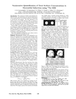


© Copyright 2026