
GS HIV Combo Ag/Ab EIA
GS HIV Combo Ag/Ab EIA Enzyme immunoassay (EIA) for the simultaneous detection of HIV p24 antigen and antibodies to HIV-1 (Groups M and O) and HIV-2 in human serum or plasma. For In Vitro Diagnostic Use GS HIV Combo Ag/Ab EIA 26217 192 Tests 26218 960 Tests GS HIV Combo Ag/Ab EIA CONTENTS 1. INTENDED USE 2. SUMMARY AND EXPLANATION OF THE TEST 3. BIOLOGICAL PRINCIPLES OF THE TEST 4. REAGENTS 5. WARNINGS FOR USERS 6. PRECAUTIONS FOR USERS 7. REAGENT PREPARATION AND STORAGE 8. SPECIMEN COLLECTION, PREPARATION, AND STORAGE 9. GS HIV Combo Ag/Ab EIA PROCEDURE 10. SPECTROPHOTOMETRIC VERIFICATION OF SAMPLE AND REAGENT PIPETTING 11. QUALITY CONTROL – VALIDATION OF RESULTS 12. INTERPRETATION OF RESULTS 13. LIMITATIONS OF THE PROCEDURE 14. PERFORMANCE CHARACTERISTICS 15. REFERENCES 2 GS HIV Combo Ag/Ab EIA 1 - INTENDED USE The GS HIV Combo Ag/Ab EIA is an enzyme immunoassay kit for the simultaneous qualitative detection of Human Immunodeficiency Virus (HIV) p24 antigen and antibodies to HIV Type 1 (HIV-1 groups M and O) and HIV Type 2 (HIV-2) in human serum or plasma. This kit is intended as an aid in the diagnosis of HIV-1 and/or HIV-2 infection, including acute or primary HIV-1 infection. The assay may also be used as an aid in the diagnosis of HIV-1 and/or HIV-2 infection in pediatric subjects (i.e., children as young as 2 years of age). The GS HIV Combo Ag/Ab EIA is intended for manual use and for use with the Bio-Rad EVOLIS™ Automated Microplate System. Results from the GS HIV Combo Ag/Ab EIA cannot be used to distinguish between the presence of HIV-1 p24 antigen, HIV-1 antibody, or HIV-2 antibody in a sample. The GS HIV Combo Ag/Ab EIA is not intended for routine use in screening blood or plasma donors, as the effectiveness of this test for use in the screening of these donors has not been established. However, in urgent situations where traditional licensed blood donor screening tests are unavailable or their use is impractical, this assay can be used as a blood donor screening assay. 2 - SUMMARY AND EXPLANATION OF THE TEST Acquired immunodeficiency syndrome (AIDS) is caused by viruses transmitted by sexual contact, exposure to blood (including sharing contaminated needles and syringes) or certain blood products, or transmitted from an infected mother to her fetus or child during the perinatal period.1 Additionally, transmission of these viruses can occur through tissue transplantation.2 Human Immunodeficiency Virus Type 1 (HIV-1) has been isolated from patients with AIDS and AIDS-related complex (ARC).3-5 HIV-1 was thought to be the sole causative agent of these syndromes until 1986, when a second type of Human Immunodeficiency Virus (HIV-2) was isolated and also reported to cause AIDS.6-7 Since the initial discovery, hundreds of cases of HIV-2 infection have been documented worldwide, including cases of AIDS related to HIV-2.8 In the United States, there have been more than 80 cases of infection with HIV-2 reported, including three potential blood donors.9-15 This second immunodeficiency virus is similar to, but distinct from, HIV-1. Both viruses have similar morphology and lymphotropism,16 and the modes of transmission appear to be identical.8,17 The HIV-1 and HIV-2 genomes exhibit about 60% homology in conserved genes such as gag and pol, and 39-45% homology in the envelope genes.18 Serologic studies have also shown that the core proteins of HIV-1 and HIV-2 display frequent cross-reactivity whereas the envelope proteins are more type-specific.19 Within the two major HIV types, there is significant variation, as well. By analyzing sequences of representative strains, HIV-1 has been divided into four groups: group M (for major), including at least 9 subtypes, 3 sub-subtypes of A, and 2 sub-subtypes of F (A1, A2, A3, B, C, D, F1, F2, G, H, J, and K); group O (for outlier); group N (for non-M, non-O), and group P.20-24 Similarly, the HIV-2 strains have been classified into at least five subtypes (A through E).25 Some HIV-1 variants share ≤50% homology in their envelope genes with the sequences of more common prototype strains. Despite some degree of immunological cross-reactivity between types and subtypes of HIV, reliable detection of the more divergent strains may only be achieved by incorporating specific sequences into the assay design. In one study, detection of HIV-2 positive samples by licensed HIV-1 antibody kits ranged from 60% to 91%, depending on the test used.26 Detection of HIV-1 Group O samples by HIV-1 and HIV-1/HIV-2 assays varied from 0% to 100% in studies with U.S.-licensed and European test kits.27,28 The GS HIV Combo Ag/Ab EIA incorporates highly conserved recombinant and synthetic peptide sequences representing HIV-1 (groups M and O) and HIV-2,29-35 as well as monoclonal antibodies specific for HIV-1 p24 antigen. It was developed to improve sensitivity and specificity of detection of HIV-1 p24 antigen and antibodies to HIV-1 and/or HIV-2 as an aid in the diagnosis of HIV infection. HIV antigens and antibodies appear and are detectable at different stages of seroconversion and of the infection.36,37 The GS HIV Combo Ag/Ab EIA allows for the simultaneous detection of HIV p24 antigen and anti-HIV-1 (M and O groups) and anti-HIV-2 antibodies. This test has been developed to significantly reduce the serological window for detection of HIV. 3 GS HIV Combo Ag/Ab EIA Reactive specimens may contain HIV-1 p24 antigen or antibodies to either HIV-1 or HIV-2. Therefore, additional, more specific or supplemental tests for HIV-1 and HIV-2 such as NAT, Western blot, or immunofluorescence, must be performed to verify the presence of HIV-1 p24 antigen or antibodies to HIV-1 or HIV-2. Recommendations for appropriate use of such additional tests may be issued periodically by the United States Public Health Service. 3 - BIOLOGICAL PRINCIPLES OF THE TEST The GS HIV Combo Ag/Ab EIA is an enzyme immunoassay based on the principle of the sandwich technique for the qualitative detection of HIV-1 p24 antigen and detection of envelope antibodies associated with HIV-1 and/or HIV-2 virus in human serum or plasma. The solid phase is coated with: • Monoclonal antibodies against HIV-1 p24 antigen • HIV antigens: HIV-1 gp160 recombinant protein, a synthetic peptide mimicking a totally artificial (i.e. encoded by no existing virus) HIV-1 group O-specific epitope, and a peptide mimicking the immunodominant epitope of the HIV-2 envelope protein. The conjugates are based upon the use of: • Biotinylated polyclonal antibodies to HIV p24 Ag (Conjugate 1) • Peroxidase-conjugated streptavidin and peroxidase-conjugated HIV-1 antigens (gp41 and gp36 peptides mimicking the immunodominant epitopes of the HIV-1 and HIV-2 envelope glycoproteins, and the same synthetic peptide mimicking a totally artificial HIV-1 group O-specific epitope used for the solid phase) (Conjugate 2) During the assay procedure, Conjugate 1 (biotinylated polyclonal antibody to HIV p24 Ag) is added to the microplate wells, followed by the addition of samples to be assayed, as well as controls and a calibrator. If present, HIV p24 antigen binds to the monoclonal antibody on the solid phase and also binds to the Conjugate 1. HIV-1 and/or HIV-2 antibodies, if present, bind to the antigens immobilized on the solid phase. The addition of Conjugate 1 and sample is validated through a color change from yellow-green to blue. After incubation, excess sample is removed by a wash step. Next, Conjugate 2 is added. Peroxidase-labeled streptavidin reacts with biotinylated Ab-Ag-Ab complexes; peroxidase labeled HIV-1 and HIV-2 antigens bind to the IgG, IgM or IgA antibodies captured on the solid phase. After incubation, unbound Conjugate 2 is removed by washing. Working TMB Solution is added to the plate and allowed to incubate. A blue or blue-green color develops in proportion to the amount of HIV antibody and/or antigen present in the sample. Color development is stopped by the addition of acid, which changes the blue-green color to yellow. The optical absorbances of specimens, controls, and the calibrator are determined spectrophotometrically at a wavelength of 450 nm with a 615 to 630 nm reference. 4 GS HIV Combo Ag/Ab EIA 4 - REAGENTS GS HIV Combo Ag/Ab EIA Product No. 26217 (192 tests), 26218 (960 tests) Component R1 • Microwell Strip Plates (2 or 10 plates) Contents • Microwell plate coated with monoclonal antibodies to HIV p24 (mouse) and HIV-1 and HIV-2 antigens • Tabs are labeled “JJ” • ProClin 300 (trace) Preparation Use as supplied. Return unused strips to the pouch. Do not remove desiccant. R2 • Wash Solution Concentrate (30X) 1 or ** bottles (120 mL) • Sodium Chloride • Tween 20™ Dilute 1:30 with deionized water. Clinical laboratory reagent water (CLRW) is acceptable. C0 • Negative Control 1 or 2 vials (1.5 mL) • Human serum/plasma; negative for antibodies to HIV and HCV; negative for HBsAg and HIV-1 Ag Use as supplied. • 0.005% Gentamicin sulfate • 0.16% ProClin 950 C1 • HIV-1 Ab Positive Control 1 or 2 vials (1.5 mL) • Human HIV-1 antibody in human serum/plasma; negative for HIV-1 Ag, HBsAg and anti-HCV antibodies • 0.005% Gentamicin sulfate • 0.16% ProClin 950 Use as supplied. C2 • HIV-2/O Ab Positive Control 1 or 2 vials (1.5 mL) • Mouse monoclonal antibody to HIV-2 and rabbit antibody to HIV-1 Group O in human serum/plasma; negative for HBsAg and anti-HCV antibodies • 0.005% Gentamicin sulfate • 0.16% ProClin 950 Use as supplied. C3 • HIV Ag Positive Control 1 or 2 vials (1.5 mL) • Purified HIV-1 viral lysate inactivated with heat and a chaotropic agent, in synthetic diluent Use as supplied. • 0.1% ProClin 300 C4 • Cutoff Calibrator 3 or 4 vials (1.7 mL) • Human serum/plasma; negative for antibodies to HIV and HCV; negative for HBsAg and HIV-1 Ag Use as supplied. • 0.005% Gentamicin sulfate • 0.16% ProClin 950 R6 • Conjugate 1 1 or 4 bottles (10 mL) • Biotinylated polyclonal antibodies (sheep) to HIV p24 Ag • 0.5% ProClin 300 Use as supplied. • 0.005% Gentamicin sulfate • Yellow-green dye R7a • Conjugate 2 2 vials (18 mL) or 4 vials (40 mL) • Lyophilized, peroxidase-labeled Streptavidin and peroxidase-labeled HIV-1 and HIV-2 antigens • 0.5% ProClin 300 • 0.005% Gentamicin sulfate Rehydrate in Conjugate 2 Diluent as described. R7b • Conjugate 2 Diluent 1 or 4 bottles (40 mL) • • • • Ready to use as described under Working Conjugate 2 Solution. R8 • Substrate Buffer 1 or 2 bottles (120 mL) • Hydrogen Peroxide • Citric Acid/Sodium Acetate buffer • Dimethylsulfoxide (DMSO) R9 • Chromogen (11X) 1 or 2 bottles (12 mL) • Tetramethylbenzidine (TMB)* R10 • Stopping Solution 1 or ** bottle (120 mL) • 1N H2SO4 (Sulfuric Acid) Plate Sealers • Clear plastic sealers Buffer with protein stabilizers 0.5% ProClin 300 0.005% Gentamicin sulfate Red dye Use as supplied. Dilute with Substrate Buffer as described. Use as supplied. Use as supplied. 38,39 * NOTE: Tetramethylbenzidine is a non-carcinogenic and non-mutagenic chromogen for peroxidase. ** Wash Solution Concentrate and Stopping Solution are supplied separately for the 10 plate (960 test) kit. Refer to catalog number 25261 for the Wash Solution Concentrate and catalog number 25260 for the Stopping Solution. These reagents are included in the 2 plate (192 test) kit. 5 GS HIV Combo Ag/Ab EIA Store kit at 2-8°C. The Stopping Solution component may be stored at 2-30°C. Bring all reagents to room temperature (18-30°C) before use. Return reagents to 2-8°C after use. Return unused strips/plates to pouch and reseal. Do not remove desiccant. Store strip plates at 2-8°C. Reagents that are interchangeable with different kit lots of the GS HIV Combo Ag/Ab EIA are the Chromogen (R9), Substrate Buffer (R8), Wash Solution Concentrate (R2), and Stopping Solution (R10). 5 - WARNINGS FOR USERS For In Vitro Diagnostic Use Warning: FDA has approved this test for use with serum and plasma specimens only. Use of this test kit with specimens other than those specifically approved for use with this test kit may result in inaccurate test results. This test is not intended for use in children younger than 2 years of age. 1. Wear protective clothing, including lab coat, eye/face protection and disposable gloves (synthetic, non-latex gloves are recommended) while handling kit reagents and clinical specimens. Wash hands thoroughly after performing the test. 2. Do not smoke, drink, or eat in areas where specimens or kit reagents are being handled. 3. Do not pipette by mouth. 4. This test kit should be handled only by qualified personnel trained in laboratory procedures and familiar with their potential hazards. Wear appropriate protective clothing, gloves and eye/face protection and handle appropriately with the requisite Good Laboratory Practices. The following is a list of potential chemical hazards contained in some reagents (refer to product description chart in the Reagents section): a. P roClin 300 (0.1% or 0.5%) and ProClin 950 (0.16%) are biocidal preservatives that are irritating to eyes and skin, may be detrimental if enough is ingested, and may cause sensitization by skin contact; prolonged or repeated exposure may cause allergic reaction in certain sensitive individuals. b. 0.005% Gentamicin Sulfate, a biocidal preservative, which is a known reproductive toxin, photosensitizer and sensitizer; prolonged or repeated exposure may cause allergic reaction in certain sensitive individuals. c. The 1 N Sulfuric Acid (H2SO4) Stopping Solution is irritating to skin and severely irritating or corrosive to eyes, depending on the amount and length of exposure; greater exposures can cause eye damage, including permanent impairment of vision. In case of contact with eyes, rinse immediately with plenty of water and seek medical advice. Keep away from strong bases, reducing agents and metals; do not pour water into this component. Waste from this material is considered hazardous acidic waste; however, if permitted by local, regional, and national regulations, it might be neutralized to pH 6-8 for non-hazardous disposal if operators are trained and equipped to do so. 5. The GS HIV Combo Ag/Ab EIA contains human blood components. No known test method can offer complete assurance that infectious agents are absent. Therefore, all human blood derivatives, reagents and human specimens should be handled as if capable of transmitting infectious disease, following recommended Universal Precautions for bloodborne pathogens as defined by OSHA40, the guidelines from the current CDC/NIH Biosafety in Microbiological and Biomedical Laboratories41 and/or local, regional and national regulations.42,43 The following human blood derivatives are found in this kit: a. Human source material used in the preparation of the Negative Control (C0) and Cutoff Calibrator (C4), and as a diluent for the HIV-1 Ab Positive Control (C1) and HIV-2/O Ab Positive Control (C2), has been tested and found nonreactive for Hepatitis B surface antigen (HBsAg), and antibodies to Hepatitis C virus (HCV Ab) and human immunodeficiency virus (HIV-1 and HIV-2). b. Human source material, containing HIV-1 and HIV-2 human antibody used in the preparation of the HIV Ab Positive Controls (C1 and C2) has been heat-treated. It has 6 GS HIV Combo Ag/Ab EIA been tested and found nonreactive for Hepatitis B surface antigen (HBsAg) and antibodies to Hepatitis C virus (HCV Ab). c. The HIV-1 viral lysate used in the HIV Ag Positive Control (C3) has been inactivated using a chaotropic agent and heat. 6. Biological spills: Human source material spills should be treated as potentially infectious. Spills not containing acid should be immediately decontaminated, including the spill area, materials and any contaminated surfaces or equipment, with an appropriate chemical disinfectant that is effective for the potential biohazards relative to the samples involved (commonly a 1:10 dilution of household bleach, 70-80% Ethanol or Isopropanol, an iodophor [such as 0.5% Wescodyne™ Plus, EPA Registration #4959-16-52], or a phenolic, etc.), and wiped dry.44-47 Spills containing acid should be appropriately absorbed (wiped up) or neutralized, the area flushed with water and wiped dry; materials used to absorb the spill may require biohazardous waste disposal. Then the area should be decontaminated with one of the chemical disinfectants. NOTE: DO NOT PLACE SOLUTIONS CONTAINING BLEACH INTO THE AUTOCLAVE. 7. Dispose of all specimens and material used to perform the test as though they contain an infectious agent. Laboratory, chemical or biohazardous wastes must be handled and discarded in accordance with all local, regional and national regulations. 6 - PRECAUTIONS FOR USERS 1. Do not use any kit components beyond their stated expiration dates. 2. The reagents that may be used with different lots of the GS HIV Combo Ag/Ab EIA kit are the Chromogen (R9), Substrate Buffer (R8), Wash Solution Concentrate (R2), and Stopping Solution (R10). Do not mix any other reagents from different lots. Any lot number of the following reagents may be used with this assay provided they have the correct catalog number and are not used beyond their labeled expiration dates: • Chromogen (R9) - Catalog # 26182 • Substrate Buffer (R8) - Catalog # 26181 • Wash Solution Concentrate (R2) - Catalog # 25261 • Stopping Solution (R10) - Catalog # 25260 3. The tabs at the end of the microwell strips are labeled with product code “JJ”. Do not use strips that have other product codes with this kit. 4. Exercise care when opening vials and removing aliquots to avoid microbial contamination of the reagents. 5. Do not allow the microplate to dry between the end of a washing step and addition of reagents. 6. Do not pre-dilute samples in Conjugate 1 (R6) before adding to the microwell plate. 7. The waiting time between dispensing Conjugate 1 and adding the samples may not exceed 30 minutes. 8. Use a clean, disposable container for the Working Conjugate 2 Solution. Exposure of the Conjugate to sodium azide will result in its inactivation. 9. Avoid exposing Chromogen or the Working TMB Solution to strong light during storage or incubation. Do not allow the chromogen solutions to come into contact with an oxidizing agent. 10. The enzyme reaction is very sensitive to metal ions. Do not allow any metal element to come into contact with the various conjugate or substrate solutions. 11. Do not carry out the test in the presence of reactive vapors (acid, alkaline, aldehyde vapors) or dust that could alter the enzyme activity of the conjugates. 12. Use clean, polypropylene containers (do not use polystyrene containers) to prepare and store the Working TMB Solution. If glassware must be used, pre-rinse thoroughly with 1N 7 GS HIV Combo Ag/Ab EIA sulfuric or hydrochloric acid followed by at least three washes of deionized water. Be sure that no acid residue remains on the glassware. If polypropylene containers are to be reused, they should be cleaned in accordance with a cleaning process validated by the testing facility. 13. Bring all reagents to room temperature before use. 14. Clinical samples may contain very high levels of HIV antibody. T herefore, care must be exercised when dispensing samples to avoid cross contamination through aerosols or carryover. For manual pipetting of controls and specimens, use an individual pipette tip for each sample and do not allow other parts of the pipetting device to touch the rim or interior of the specimen container. Consider using new stoppers/caps to seal specimen tubes after use, to avoid errors or contamination of the work area while recapping tubes. Handle the Negative Control and Positive Controls in the same manner as patient specimens. 15. If a specimen or reagent is inadvertently not added to a well, the assay results will read negative. Reagents of this kit have been color-coded to enable confirmation of the addition of specimens/controls and Working Conjugate Solution. 16. Inadequate adherence to package insert instructions may result in erroneous results. 17. Use only adequately calibrated equipment with this assay. 18. Use of dedicated equipment is recommended if equipment performance validations have not precluded the possibility of cross-contamination. 19. The GS HIV Combo Ag/Ab EIA performance is highly dependent upon incubation times and temperatures and effective washing. Temperatures outside of the validated ranges may result in invalid assays. Incubation temperatures should be carefully monitored using calibrated thermometers, or equivalent. 7 - REAGENT PREPARATION AND STORAGE Working Conjugate 2 Solution (R7a + R7b) Note: The lyophilized Conjugate 2 (R7a) may be supplied as a small vial (18 mL) or large vial (40 mL). Check the vial label to determine the size before proceeding. Bring Conjugate Diluent (R7b) to room temperature. Gently tap the vial of the lyophilized Conjugate 2 (R7a) to remove any substance from the rubber cap. Carefully remove the cap and prepare as follows: for the small Lyophilized Conjugate vial (R7a), add 18 mL of Conjugate Diluent (R7b) OR for the large Lyophilized Conjugate vial (R7a), add 40 mL of Conjugate Diluent (R7b) Replace the cap and let stand for 10 minutes, while gently shaking and inverting from time to time to help the dissolution. Working Conjugate 2 Solution is stable for 15 hours at room temperature, and for 4 weeks if stored at 2-8C. Studies have demonstrated no adverse effects from cycling the Working Conjugate Solution between 2-8°C and room temperature (18-30°C) multiple times (e.g., 5 cycles of 3 hours at room temperature followed by 2-8°C). Working Conjugate 2 Solution that is stored frozen (-20°C or colder) may be used until the expiration date of the kit. It may be frozen and thawed 10 times. Note the Concentrate lot number and date and time of preparation of the Working Conjugate Solution. Prepare a user log to record the time and temperature of Working Conjugate Solution storage, to ensure that the recommended limits are not exceeded. Always mix working solution by inverting just prior to use. One vial of prepared Working Conjugate 2 Solution provides enough reagents for testing of approximately 1 plate (18 mL vial) or 2½ plates (40 mL vial). For testing of additional microwell strips, prepare additional Working Conjugate 2 Solution. Ensure that the volume of prepared reagent will be adequate for the entire run. Working TMB Solution (R8 + R9) Bring Chromogen and Substrate Buffer to room temperature. Invert the Chromogen and Substrate Buffer to mix before using. Prepare a 1:11 dilution for each strip to be tested by mixing 8 GS HIV Combo Ag/Ab EIA 100 μL of Chromogen to 1 mL of Substrate Buffer in a clean, polypropylene container (do not use a polystyrene container). Note Chromogen lot number, date and time of preparation, and date and time of expiration (8 hours from preparation) on container. Mix Working Solution gently when combined and again just prior to use. Working TMB Solution should be kept in the dark at room temperature and used within 8 hours. Chromogen should be colorless to slightly yellow. Any other color indicates that the reagent is contaminated. Do not use this reagent. The Working TMB Solution should be colorless. A distinct blue color indicates that the reagent is contaminated. Discard the Working TMB Solution and prepare fresh reagent in a clean container. Prepare only the amount of the reagent to be used within 8 hours, ensuring that the volume of diluted reagent will be adequate for the entire run. Extra Chromogen is provided. Use the following table as a guide: Preparation of Working TMB Solution by Number of Strips Used Number of Strips to be used 1 2 3 Amount of Chromogen (µL) 100 200 300 Amount of Substrate Buffer (mL) 1 2 3 * 1 Complete Plate ** 2 Complete Plates 4 5 6 7 8 9 10 11 12* 24** 400 500 600 700 800 900 1000 1100 1200 2400 4 5 6 7 8 9 10 11 12 24 Working Wash Solution (R2) Prepare working Wash Solution by adding one part Wash Solution Concentrate (30X) to 29 parts of water (e.g., 120 mL of Wash Solution Concentrate to 3480 mL of water). Any lot of Wash Solution Concentrate, provided it is catalog number 25261 and within its labeled shelf life, may be used with this assay. Use deionized or distilled water. Clinical laboratory reagent water is acceptable. The diluted Wash Solution can be stored at room temperature for up to four weeks in a plastic container. Note the lot number, date prepared, and expiration date on the container. The final expiration date of the working Wash Solution should not exceed the expiration date of the Wash Solution Concentrate (30X). Prepare a sufficient quantity of working Wash Solution to complete a full run. 8 - SPECIMEN COLLECTION, PREPARATION, AND STORAGE Serum or plasma specimens may be used in the test. The following tube types and anticoagulants, including those in both glass and plastic tubes, may be used: serum tubes, serum separator tubes (SSTs) with and without activator, potassium EDTA, sodium citrate, sodium and lithium heparin, and plasma separator tubes (PSTs). Samples that are collected into anticoagulant tubes should be filled as labeling indicates to avoid improper dilution. Specimens with observable particulate matter should be clarified by centrifugation prior to testing. Suspended fibrin particles or aggregates may yield falsely positive results. Do not heat-inactivate the samples. Extensive hemolysis may affect test performance. The potential interfering substances at the levels tested below did not produce a change in clinical interpretation. Studies with these substances included HIV negative samples and those spiked with both low and high levels of HIV-1 antigen, HIV-1 antibody, and HIV-2 antibody. In these studies the samples were spiked with the substances at two levels, including the upper levels listed here that were tested in 7 samples each. Hemolyzed: 500 mg/dL of hemoglobin Lipemic: 1000 mg/dL of triglycerides Icteric: 20 mg/dL of bilirubin Proteinemic: 12 g/dL of protein Samples may be stored for no longer than 2 days at room temperature or 7 days at 2-8°C, including the time that samples are in transit. Minimize room temperature storage of samples to the shortest time possible in order to preserve maximum p24 antigen reactivity. For long-term storage, the specimens should be removed from the clot, red blood cells, or separator gel and should be frozen at -20°C or colder. Samples should not be used if they have incurred more than 4 freeze/thaw cycles. Mix samples thoroughly after thawing. 9 GS HIV Combo Ag/Ab EIA Note: If specimens are to be shipped, they should be packed in compliance with Federal Regulations covering the transportation of etiologic agents. Specimens may be shipped at 2-8°C (wet ice) or frozen (dry ice), after removal from the clot, red cells, or separator gel. Do not exceed the storage time limitations described above. This kit is not intended for use on saliva/oral fluid or urine samples. 9 - GS HIV Combo Ag/Ab EIA PROCEDURE Materials Provided See REAGENTS section on page 5. Materials Required But Not Provided 1. Precision pipettes that deliver 20 to 200 μL, 1 mL, 10 mL, 25 mL and 50 mL, as needed (accurate within ±10%), and corresponding pipette tips; multichannel pipettors capable of delivering 25 μL, 75 μL, 80 μL and 100 μL are optional. 2. Appropriately sized graduated cylinders (25 mL, 100 mL, 1000 mL capacity). 3. Dry-heat incubator capable of maintaining 37 ± 2°C. 4. Microwell plate or strip washer qualified for use with this assay. The washer must be capable of dispensing 400 μL per well, cycling 5 times, and soaking for 30-60 seconds between each wash. 5. Microwell strip reader qualified for use with this assay. The spectrophotometer should have the following specifications at wavelengths 450 nm and 615 to 630 nm: Bandwidth: 10 nm HBW (Half Band Width) or equivalent Absorbance Range: 0 to 2 AU (Absorbance Units) Repeatability: ± (0.5% + 0.005) AU Linearity or Accuracy: 1% from 0 to 2.0 AU The instrument should contain a reference filter for reading at 615 to 630 nm. 6. The GS HIV Combo Ag/Ab EIA is approved for use with the Bio-Rad EVOLIS™ Automated Microplate System. 7. Household bleach (5% to 8% sodium hypochlorite) which may be diluted to a minimum concentration of 10% bleach (or 0.5% sodium hypochlorite). Alternative disinfectants include 70-80% ethanol or 0.5% Wescodyne™ Plus. 8. Paper towels or absorbent pads for blotting. 9. Labeled null strips for testing partial plates. 10. Clean polypropylene container for preparation of Working TMB Solution (do not use polystyrene). 11. Deionized or distilled water. Clinical laboratory reagent water is acceptable.48 12. Gloves. 13. Laboratory timer. 14. EIA reagent reservoirs (optional). Preliminary Statements 1. The expected run time for this procedure is approximately 2.5 - 3 hours from initiation of the first incubation step. Each run of this assay must proceed to completion without interruption after it has been started. The allowable time from start of sample pipetting to start of incubation is ≤ 1 hour. 2. One Negative Control, one HIV-1 Ab Positive Control, one HIV-2/O Ab Positive Control, one HIV Ag Positive Control, and three wells of Cutoff Calibrator must be run on each plate. The cutoff for patient samples is determined by the calibrators on each individual plate, and assay validity is determined by the controls on each plate. 10 GS HIV Combo Ag/Ab EIA 3. Do not splash controls, specimens, or reagents between microwells of the plate. 4. Cover plates for each incubation step using plate sealers provided or other appropriate means to minimize evaporation. 5. Avoid exposure of the plates to light during the final incubation step (following the addition of Working TMB Solution). 6. Adhere to the recommended time constraints for the use of the Working TMB Solution (8 hours), Working Conjugate 2 Solution (15 hours at room temperature, 4 weeks at 2-8°C, or until the expiration of the kit at -20°C or colder), and Working Wash Solution (4 weeks). Reagent Preparation Storage Working Conjugate 2 Solution (R7a + R7b) Small (18 mL) Lyophilized Conjugate vial, 15 hours at room temperature OR add 18 mL of Conjugate Diluent OR 4 weeks if stored at 2-8C Large (40 mL) Lyophilized Conjugate vial, OR add 40 mL of Conjugate Diluent Expiration of the kit at -20°C or colder, with up to 10 freeze/thaw cycles Working TMB Solution (R8 + R9) 1:11 dilution (100 μL of Chromogen and 1 mL of Substrate Buffer for each strip to be tested) Lot specific Yes Room temperature (18-30°C) for up to 8 hours Stored in the dark No Working Wash Solution 1:30 dilution (one part Wash Solution (R2) Concentrate [30X] to 29 parts of water) Room temperature for up to 4 weeks (not to exceed the expiration date of the 30X Wash Solution Concentrate) No Stopping Solution (R10) Use as supplied 2-30°C for shelf life of reagent No 7. Avoid the formation of air bubbles in each microwell. 8. Avoid bumping plates containing liquid reagents (especially Working Conjugate 2 Solution) to prevent adherence of liquid to the plate sealer and/or top edges of the microwells. 9. Adequate washing of the microwells with a validated microplate washer is essential to eliminate non-specific binding. 10. Dry residue from the plate blocking process may be visible in the microwells. Assay results will not be affected by this material. Before reading the plates, carefully wipe the bottom of the plates to remove any material that remains on the outside of the wells, and ensure that all strips have been pressed firmly into place. 11. For additional procedural instructions when running this assay with the EVOLIS™ Automated Microplate System, consult the EVOLIS™ Operator’s Manual or the EVOLIS™ Microplate Assay Testing Reference Manual for this assay. EIA Procedure Note: Steps 6 – 16 in this Section, and the steps in Section 10 (Spectrophotometric Verification of Sample and Reagent Pipetting), Section 11 (Quality Control - Validation of Results) and Section 12 (Interpretation of Results) are performed automatically by the EVOLIS™ Automated Microplate System. 1. Perform equipment maintenance and calibration, where necessary, as required by the manufacturer. 2. Bring all of the reagents to room temperature before beginning the assay procedure. 3. Prepare Working Conjugate 2 Solution, Working TMB Solution, and Working Wash Solution if not previously prepared. Mix gently by inversion. Mix again just before use. 4. Remove strips not needed for the assay and replace them with labeled null strips, if necessary. 11 GS HIV Combo Ag/Ab EIA 5. Microwell strips not needed for the assay may be returned to the plate pouch and sealed, and then used at a later time. Strips from different plates can only be mixed to assemble full or partial plates if they are from the same plate lot, and have come from plates that have previously been tested with kit controls and yielded valid runs. When assembling a plate that contains strips from a newly opened, previously untested plate, one of these strips should be placed at the beginning of the plate and tested with the kit controls. 6. If sample identity is not maintained by an automatic procedure, identify the individual wells for each specimen or control on a data sheet. 7. Add 25 μL of Conjugate 1 (R6) to each well. 8. Within 30 minutes, add 75 µL of the controls, calibrator, and the specimens to the appropriate wells. Do not pre-dilute samples in Conjugate 1 (R6) before adding to the microwell plate. One Negative Control, one HIV-1 Ab Positive Control, one HIV-2/O Ab Positive Control, one HIV-1 Ag Positive Control, and three wells of Cutoff Calibrator must be assayed on each plate or partial plate of specimens. NOTE: After addition of samples, controls and calibrator, wells containing Conjugate 1 turn from yellow-green to blue. It is possible to verify the presence of sample in the wells by spectrophotometric reading at 620 nm. (Refer to section 10: Spectrophotometric Verification of Sample and Reagent Pipetting) 9. Cover the microwell plate with a plate sealer or use other means to minimize evaporation. Incubate the plate for 60 ± 5 minutes at 37 ± 2°C. 10. At the end of the incubation period, carefully remove the plate cover, if used, and aspirate the fluid from each well into a biohazard container. Wash the microwell plate or strip a minimum of five times with the Wash Solution (at least 400 μL/well/wash). Soak each well for 30 to 60 seconds between each wash cycle. Aspirate the Wash Solution after each wash. After the last wash, if excess liquid remains, blot the inverted plate on clean, absorbent paper towels. NOTE: Grasp the plate holder firmly at the center of the long sides before inverting to blot. 11. Add 100 μL of Working Conjugate 2 Solution (R7a + R7b) into all wells. The Conjugate must be mixed well before use. NOTE: The Working Conjugate 2 Solution is colored red. It is possible to verify the presence of Conjugate 2 in the wells by spectrophotometric reading at 450/620 nm. Refer to section 10: Spectrophotometric Verification of Sample and Reagent Pipetting. 12. Cover the microwell plate with a plate sealer or use other means to minimize evaporation. Incubate the plate for 30 ± 5 minutes at room temperature (18-30°C). 13. At the end of the incubation period, carefully remove the plate cover, if used, and aspirate the fluid from each well into a biohazard container. Wash the microwell plate or strip a minimum of five times with the Wash Solution (at least 400 μL/well/wash). Soak each well for 30 to 60 seconds between each wash cycle. Aspirate the Wash Solution after each wash. After the last wash, if excess liquid remains, blot the inverted plate on clean, absorbent paper towels. NOTE: Grasp the plate holder firmly at the center of the long sides before inverting to blot. 14. Add 80 µL of the Working TMB Solution to each well containing a specimen, control, or calibrator. Incubate plates in the dark for 30 ± 5 minutes at room temperature (18-30°C). Use of a plate sealer or cover is optional. 15. Carefully remove the plate cover, if used, and add 100 µL of Stopping Solution to each well to terminate the reaction. Use the same sequence and rate of distribution as for the Working TMB Solution addition. Tap the plate gently, or use other means to assure complete mixing. Complete mixing is required for acceptable results. 16. Read absorbance within 30 minutes after adding the Stopping Solution, using the 450 nm filter with 615 nm to 630 nm as the reference. 12 GS HIV Combo Ag/Ab EIA 10 - SPECTROPHOTOMETRIC VERIFICATION OF SAMPLE AND REAGENT PIPETTING (OPTIONAL) Verification of the Conjugate 1 (R6) Pipetting and Sample Pipetting: The presence of this reagent and samples in the well can be verified by spectrophotometric reading at 620 nm. The OD value of each well containing Conjugate 1 and sample must be greater than 0.600 (a lower OD indicates poor dispensing of the Conjugate 1 or of the sample). NOTE: After sample addition, Conjugate 1 (R6) turns from yellow-green to blue in color. Verification of Working Conjugate 2 (R7a + R7b) Pipetting: The presence of Working Conjugate 2 (R7a + R7b) in the well can be verified by spectrophotometric reading at 450 nm with 620 nm as the reference. The OD value of each well must be greater than or equal to 0.100. NOTE: Working Conjugate 2 (R7a + R7b) is red in color. 11 - QUALITY CONTROL – VALIDATION OF RESULTS Calculate the mean absorbance of the Cutoff Calibrator (CCx̄ ) Determine the mean absorbance for the Cutoff Calibrator (CCx̄ ) by dividing the sum of the absorbance values by the number of acceptable Calibrators. The individual absorbance values of the Cutoff Calibrator must be greater than 0.000 AU and less than 0.170 AU. One Cutoff Calibrator absorbance value may be discarded if it is outside this range. The CC x̄ may be calculated from the two remaining absorbance values. The mean of the absorbance of the Cutoff Calibrators (C4) should be less than 0.150. Cutoff Calibrator Sample Number 1 2 3 Absorbance 0.075 Total Absorbance 3 = 0.239 3 = 0.080 (CCx̄ ) 0.083 0.081 0.239 Calculate the cutoff value Determine the cutoff value by adding the CCx̄ to 0.200 as shown in the example below: CCx̄ = 0.080 Cutoff Value = 0.080 + 0.200 = 0.280 Assay validation A run is valid if the following criteria are met: The absorbance of Negative Control (C0) must be greater than 0.000 and less than the assay cutoff (OD 0.000 < C0 < cutoff). The absorbance of HIV-1 Ab Positive Control (C1) must be greater than 0.700 AU (OD C1 > 0.700). The absorbance of HIV-2/O Ab Positive Control (C2) must be greater than 0.700 AU (OD C2 > 0.700). The absorbance of HIV-1 Ag Positive Control (C3) must be greater than 0.700 AU (OD C3 > 0.700). The absorbance of each Cutoff Calibrator (C4) should be greater than 0.000 and less than 0.170 AU (OD 0.000 < C4 < 0.170). One Cutoff Calibrator value may be discarded. If two or more Cutoff Calibrator values are out of limit, the assay must be repeated. The mean of the absorbance of the Cutoff Calibrators (C4) should be less than 0.150. If any of these criteria have not been met, the assay is invalid and must be repeated. 13 GS HIV Combo Ag/Ab EIA 12 - INTERPRETATION OF RESULTS The presence or absence of detectable HIV-1 antigen or antibodies to HIV-1 and/or HIV-2 is determined by comparing the absorbance measured for each sample to the calculated cutoff value. Samples with absorbance values that are <0.000 must be repeated. Those with values greater than the upper linearity limits of the reader should be reported as reactive. Specimens with absorbance values less than the cutoff value are considered nonreactive by the GS HIV Combo Ag/Ab EIA and may be considered negative for HIV-1 (M and O Groups) and HIV-2 antibodies and HIV-1 antigen. Samples with absorbance values equal to or greater than the cutoff value are considered reactive by the GS HIV Combo Ag/Ab EIA. Initially reactive specimens must be retested in duplicate to validate the initial test results. If, after repeat testing, the absorbance values of both duplicate specimens are less than the cutoff value, the original specimen may be considered non-repeatedly reactive and negative for HIV-1 (Groups M and O) and HIV-2 antibodies, as well as HIV p24 antigen. If, after repeat testing, the absorbance value of either of the duplicates is greater than or equal to the cutoff value, the specimen must be considered repeatedly reactive. Reactive specimens must be retested in duplicate; if they are repeatedly reactive, they must be investigated by additional, more specific, or supplemental tests. Refer to CDC guidelines for the current recommended HIV testing algorithm. 13 - LIMITATIONS OF THE PROCEDURE 1. The GS HIV Combo Ag/Ab EIA Procedure and the Interpretation of Results must be followed closely when testing for the presence of HIV-1 antigen or antibodies to HIV-1 and/or HIV-2 in plasma or serum specimens. The user of this kit is advised to read the package insert carefully prior to conducting the test. In particular, the test procedure must be carefully followed for sample and reagent pipetting, plate washing, and time and temperature of the incubation steps. Testing of other body specimens, pooled blood or processed plasma, and products made from such pools is not recommended. 2. The GS HIV Combo Ag/Ab EIA detects circulating antibodies to HIV-1 (Groups M and O) and HIV-2, and it also detects HIV-1 antigen. Thus, it is useful in evaluating patients with signs or symptoms of AIDS, and in establishing prior infection with HIV-1 or HIV-2. Clinical studies continue to clarify and refine the interpretation and medical significance of the presence of 49 antibodies to HIV-1 or HIV-2. Repeatedly reactive specimens must be investigated by additional, more specific, or supplemental tests. Recommendations for appropriate use of such additional tests may be issued periodically by the United States Public Health Service. For individuals who are confirmed positive for HIV antigen or antibodies, appropriate counseling and medical evaluation should be offered. Both confirmation of the test result on a freshly drawn sample and counseling should be considered an important part of testing for HIV antigen and antibody to HIV-1 and HIV-2. 3. AIDS and AIDS-related conditions are clinical syndromes and their diagnosis can only be established clinically. Testing alone cannot be used to diagnose AIDS, even if the recommended investigation of repeatedly reactive specimens suggests a high probability that HIV antigen or antibody to HIV-1 or HIV-2 is present. 4. A negative test result at any point in the investigation of individual subjects does not preclude the possibility of exposure to or infection with HIV-1 and/or HIV-2. 5. Negative results can occur if the quantity of marker present in the sample is too low for the detection limits of the assay, or if the marker which is detected is not present during the stage of disease in which a sample is collected. 6. Failure to add specimen or reagent as instructed in the procedure could result in a falsely negative test. Repeat testing should be considered where there is clinical suspicion of infection or procedural error. 7. The risk of an asymptomatic person with a repeatedly reactive test result developing AIDS or an AIDS-related condition is not known, as the course of HIV infection may vary among 14 GS HIV Combo Ag/Ab EIA 8. 9. 10. 11. 12. 13. individual patients and may be altered by antiretroviral therapy.50,51 However, in a prospective study, AIDS developed in 51% of homosexual men after 10 years of infection.52 Data obtained from testing persons both at increased and at low risk for HIV-1 and/or HIV-2 infection suggest that repeatedly reactive specimens with high reactivity on the GS HIV Combo Ag/Ab EIA may be more likely to demonstrate the presence of antibodies to HIV-1 (Groups M and O) and/or HIV-2 by additional, more specific, or supplemental testing.53 Borderline reactivity is more frequently nonspecific, especially in samples obtained from persons at low risk for infection with HIV-1 or HIV-2; however, the presence of antibodies to HIV-1 and/or HIV-2 in some of these specimens can be demonstrated by additional, more specific, or supplemental testing, or by testing a subsequent sample drawn at a later date (e.g. 3 to 6 months).54 The performance of this assay has not been established for individuals younger than 2 years of age. It is generally recognized that detection of HIV antibody in infants born to seropositive mothers is not adequate to diagnose HIV infection in the infant, since maternal IgG frequently persists for as long as 18 months after birth. Nearly all infants born to HIV-infected mothers passively acquire maternal antibody. Supplemental assays designed specifically for neonatal specimens may be helpful in resolving such cases, including HIV nucleic acid tests or viral culture.55 An absorbance value of less than 0.000 AU may indicate a procedural or instrument error that should be evaluated. That result is invalid and that specimen must be re-run. Factors that can affect the validity of results include failure to add the specimen or reagents to the well, inadequate washing of microplate wells, failure to follow stated incubation times and temperatures, addition of wrong reagents to wells, the presence of metals, or splashing of bleach into wells, improper washing of microwell plates, cross-contamination of nonreactive specimens with HIV antigen or antibody from a high-titered specimen, contamination of the Chromogen or Working TMB Solution by oxidizing agents (sodium hypochlorite, hydrogen peroxide, etc.), and contamination of the Stopping Solution. A person who has antibodies to HIV is presumed to be infected with the virus, except that a person who has participated in an HIV vaccine study may develop antibodies to the vaccine and may or may not be infected with HIV. Clinical correlation is indicated with appropriate counseling, medical evaluation, and possibly additional testing to decide whether a diagnosis of HIV infection is accurate. Heat-inactivated samples may affect the quality of the results, and should not be used. 14 - PERFORMANCE CHARACTERISTICS Testing to determine the performance characteristics of the GS HIV Combo Ag/Ab EIA was performed both manually and with the EVOLIS™ Automated Microplate System. Unless otherwise noted in the tables which follow, the results summarize the manual testing that was performed. The results of the testing with EVOLIS™ are described in text following the data tables, where applicable. Specificity Low Risk Populations Seven thousand (7000) samples (6000 serum and 1000 plasma) from populations at low risk for HIV infection were tested with GS HIV Combo Ag/Ab EIA. Repeatedly reactive (RR) samples were tested by an FDA licensed HIV-1 Western blot and an FDA-approved HIV-1/HIV-2 test that differentiates HIV-1 from HIV-2. Results are presented in Table 1. 15 GS HIV Combo Ag/Ab EIA Table 1 Reactivity in Low Risk Populations Repeatedly Reactive Specimens HIV-1 NonInitially Repeatedly Western Blot HIV-2 Reactive* Reactive Reactive Reactive Positive 1993 7 6 2 0 (99.65%) (0.35%) (0.30%) (0.10%) (0.00%) 2 0 1998 NT NT (99.90%) (0.10%) (0.00%) 996 4 2 0 0 (99.60%) (0.40%) (0.20%) (0.00%) (0.00%) 997 3 2 1 0 (99.70%) (0.30%) (0.20%) (0.10%) (0.00%) 494 4 3 1 0 (99.20%) (0.80%) (0.60%) (0.20%) (0.00%) 502 0 0 NT NT (100%) (0.00%) (0.00%) 6980 20 13 4 0 (99.71%) (0.29%) (0.19%) (0.06%) (0.00%) GS HIV Combo Ag/Ab EIA Number Tested Low Risk Population Health Insurance Applicants 2000 Normal Blood Donors 2000 Low Risk Population - Other 1000 Pregnant Women 1000 Military Recruits - Frozen 498 Military Recruits - Fresh 502 Total 7000 * Based on HIV differentiation test results NT = Not Tested As shown in Table 1, 99.71% (6980/7000) of the low risk populations were initially nonreactive, 0.29% (20/7000) were initially reactive, and 0.19% (13/7000) were repeatedly reactive. Of the 13 repeatedly reactive specimens, 4 specimens were confirmed positive for antibodies to HIV-1 by Western blot and none were reactive for HIV-2 on the HIV-2 differentiation test. Of the 9 repeatedly reactive samples that were not confirmed by HIV-1 Western blot, 8 were also negative by HIV-1 Ag testing (the remaining sample was not available for HIV-1 Ag testing). The specificity of the GS HIV Combo Ag/Ab EIA in the low risk populations in this study was 99.87% (6987/6996) with a 95% confidence interval of 99.76% - 99.93%. The results for the combined samples from low risk populations tested with the GS HIV Combo Ag/Ab EIA compared to results of testing the same samples with a licensed HIV-1/HIV-2 EIA are shown in Table 2. Table 2 Combined Low Risk Populations – Assay Comparison Summary GS HIV Combo Ag/Ab EIA Result HIV-1 Western Blot Licensed HIV-1/HIV-2 Result N a Pos 4 Ind 2 Neg 1 HIV Differentiation Test HIV-1 HIV-2 Reactive Reactive NR 4 0 3 Repeatedly Reactive Repeatedly Reactive 7 Repeatedly Reactive Non-Reactive 6 b 0 1 5 0 0 6 Non-Reactive Repeatedly Reactive 1c 0 0 1 0 0 1 Non-Reactive Non-Reactive 6986 NT NT NT NT NT NT 7000 4 3 7 4 0 10 Total a Of the 3 samples that were nonreactive on the HIV differentiation test, 2 were negative for HIV antigen with an HIV-1 Ag EIA and one was not available for testing. b HIV-1 Ag testing with an HIV-1 Ag EIA was negative for 5 of these samples, and one sample was negative with an HIV-1 RNA assay. c Faint (+/-) gp160 band on the HIV-1 Western Blot. NT= Not Tested Of the 7000 samples from the low risk populations that were tested manually with the GS HIV Combo Ag/Ab EIA, 3002 of the same samples were tested with the GS HIV Combo Ag/Ab EIA on the EVOLIS™ Automated Microplate System. The results by EVOLIS™ testing were equivalent in 16 GS HIV Combo Ag/Ab EIA 99.97% (3001/3002) of the samples from low risk individuals compared to the manual method. One (1) sample was repeatedly reactive when tested on the EVOLIS™ but nonreactive when tested manually. Reactivity in Individuals with Unrelated Medical Conditions The reactivity rate of the GS HIV Combo Ag/Ab EIA was determined in samples from individuals with medical conditions unrelated to HIV infection. Samples that were repeatedly reactive with the GS HIV Combo Ag/Ab EIA were tested with a licensed HIV-1/HIV-2 EIA, an approved HIV-1/HIV-2 differentiation test, an HIV-1 p24 Ag EIA, and a licensed HIV-1 Western blot. Results are shown in Table 3. Table 3 Unrelated Medical Conditions Unrelated Medical Condition Immunological disease Anti-EBV Anti-HCV Anti-HTLV HBsAg + Vaccinia Vaccine Pre-Influenza Vaccine Post-Influenza Vaccine Candida Infection Hemodialysis Hemophilia Multiple Transfusions Multiparous RF + Cord Blood a Total GS HIV Combo Ag/Ab EIA Results N 20 20 20 20 20 20 20 20 20 20 20 20 20 20 20 300 Non-Reactive 20 20 20 20 20 20 20 18 19 20 20 20 20 20 20 297 Repeatedly Reactive 0 0 0 0 0 0 0 b 2 1c 0 0 0 0 0 0 3 a Includes: 6 Scleroderma, 5 Systemic Lupus Erythematosus (SLE), 5 ANA positive, 2 Sjögren’s Syndrome (SS) and 2 Mixed Connective Tissue Disease (MCTD) samples. b Twenty (20) additional post-influenza vaccine samples were tested and all 20 were nonreactive. c This sample was HIV reactive and undifferentiated on the HIV-1/HIV-2 differentiation test. Three (3) specimens from individuals with unrelated medical conditions were repeatedly reactive with the GS HIV Combo Ag/Ab EIA. These 3 specimens were also repeatedly reactive on the licensed HIV-1/HIV-2 EIA. Of the 3 repeatedly reactive samples, 2 were from post-influenza vaccine subjects and 1 was from an individual positive for Candida infection. All 3 specimens were negative for HIV-1 antibodies on an HIV-1 Western blot and an HIV-1 p24 Ag EIA. The two specimens from post-influenza vaccine recipients were nonreactive on the HIV-1/HIV-2 differentiation test and the Candida infection specimen was HIV reactive (undifferentiated) on the same assay. The pre- and post-influenza vaccine specimens were from different individuals. Sensitivity HIV-1 p24 Antigen Analytical Sensitivity The GS HIV Combo Ag/Ab EIA was designed to have an analytical sensitivity of < 50 pg/mL for HIV-1 p24 antigen on a panel derived from the Agence française de sécurité sanitaire des produits de santé (AFSSAPS ) and < 2 IU/mL on the WHO HIV international standard NIBSC 90/636. In an internal study the results demonstrated an antigen sensitivity of 14.78 pg/mL (range of 13.22 15.89 pg/mL) on the AFSSAPS standard and an antigen sensitivity of 0.65 IU/mL (range of 0.40 1.05 IU/mL) on the WHO standard. 17 GS HIV Combo Ag/Ab EIA HIV-1 Antigen Detection in Culture Supernatants Fifty-three (53) HIV-1 culture supernatants were tested on the GS HIV Combo Ag/Ab EIA. These supernatants were from subtypes A (N=10), B (N=10), C (N=9), D (N=4), AE (N=10), F (N=2), G (N=2), H (N=1), J (N=2), N (N=1), and O (N=2). Due to the limited volumes of the HIV-1 culture supernatant samples, all samples were diluted before testing. Of the 53 diluted HIV-1 culture supernatant samples tested with the GS HIV Combo Ag/Ab EIA, 100% (53/53) were reactive. HIV-1 Seroconversion Panels Thirty commercially available seroconversion panels (228 total members) were tested on the GS HIV Combo Ag/Ab EIA and an FDA licensed HIV-1/HIV-2 EIA. A summary of the results is presented in Table 4. Table 4 GS HIV Combo Ag/Ab EIA Reactivity in HIV-1 Seroconversion Panels Panel # # of Panel Members Tested 1 15 2 7 3 8 4 10 5 11 6 9 7 14 8 8 9 6 10 6 11 9 12 7 13 8 14 6 15 6 16 6 17 5 18 6 19 7 20 5 21 5 22 7 23 9 24 9 25 6 26 7 27 6 28 6 29 10 30 4 Total Reactive Bleeds Total Reactive Panels # of Reactive Panel Members GS HIV Licensed Historical Combo HIV1/HIV-2 HIV-1 WB Ag/Ab EIA EIA Results 2 2 3 4 6 2 5 4 2 4 4 2 7 3 4 3 2 4 2 3 2 2 2 2 2 2 0 5 3 2 1 1 2 2 6 1 5 3 2 2 4 1 6 2 2 2 1 1 1 2 0 1 0 0 1 0 0 3 1 1 0 0 0 1 0 0 2 0 0 2 2 1 1 0 0 0 0 0 0 0 0 0 0 0 0 0 0 2 0 0 90 54 11 29 25 7 a Based on bleed date All bleeds in these panels were nonreactive on that test. NA = Not Applicable b 18 Days to First Reactive Result GS HIV Combo Licensed Ag/Ab EIA HIV-1/HIV-2 EIA 33 18 16 28 17 25 36 26 44 7 28 28 7 18 7 13 18 8 17 7 47 23 28 27 14 17 b 5 44 17 35 25 21 35 17 32 36 33 44 27 28 43 11 21 14 15 20 19 21 12 b 28 b b 17 b b 12 51 22 Difference in Days to First Reactive EIA a Result 2 7 5 7 0 7 0 7 0 20 0 15 4 3 7 2 2 11 4 5 NA 5 NA NA 3 NA NA 7 7 5 GS HIV Combo Ag/Ab EIA The GS HIV Combo Ag/Ab EIA detected reactive bleeds in 96.7% (29/30) of the seroconversion panels compared to 83.3% (25/30) detected by an FDA licensed HIV-1/HIV-2 EIA and 23.3% (7/30) detected by the HIV-1 Western blot. Reactivity in 4 of the panels was detected only by the GS HIV Combo Ag/Ab EIA. Of the 25 panels that were reactive by both GS HIV Combo Ag/Ab EIA and the HIV-1/HIV-2 EIA, the first reactive bleed occurred earlier on the GS HIV Combo Ag/Ab EIA in 84.0% (21/25) of the panels. Sixteen percent (16.0%; 4/25) of the panels were detected at the same bleed by both GS HIV Combo Ag/Ab EIA and the HIV-1/HIV-2 EIA. Compared to the FDA licensed third generation HIV-1/HIV-2 EIA, the GS HIV Combo Ag/Ab EIA reduced the time to detection of HIV (i.e. window period), with an overall range of 0 to 20 days for the 30 seroconversion panels tested in this study. Therefore, the GS HIV Combo Ag/Ab EIA demonstrated a greater capability of detecting acute and primary HIV infection than either a third generation HIV-1/HIV-2 EIA or an HIV-1 Western blot. Twenty (20) of the 30 HIV-1 seroconversion panels (N = 153 samples) that were tested manually were also tested using the EVOLIS™ Automated Microplate System. The GS HIV Combo Ag/Ab EIA manual assay results and the EVOLIS™ results were equivalent in reactivity for 98.7% (151/153) of the seroconversion panel specimens tested. Two (2) seroconversion panel members were reactive with the GS HIV Combo Ag/Ab EIA by EVOLIS™ testing one bleed earlier than testing by the manual method. Reactivity in Known HIV-1 Antibody Positive Samples One thousand three hundred (1300) known HIV-1 antibody positive samples (501 sera and 799 plasma) were tested with the GS HIV Combo Ag/Ab EIA and an FDA licensed HIV-1/HIV-2 EIA. These samples included 1000 retrospective HIV-1 antibody positive samples from the U.S., 200 retrospective HIV-1 antibody positive samples (International) from geographic locations outside the U.S, and 100 samples with CDC AIDS Stage Classification. Reactivity with the GS HIV Combo Ag/Ab EIA in the 1300 known antibody positive samples is presented in Table 5. Results are also compared to results of testing the same samples with a licensed HIV-1/HIV-2 EIA. Table 5 Reactivity in Known HIV-1 Antibody Positive Samples Known HIV Antibody Status HIV-1 Antibody Positive (U.S.) N 1000 GS HIV Combo Ag/Ab EIA Repeatedly Reactive 1000 Licensed HIV-1/HIV-2 EIA Repeatedly Reactive 1000 HIV-1 Antibody Positive (International) 200 200 198* AIDS with CDC Stage Classification 100 100 100 Total 1300 1300 1298* * Two (2) samples were initially reactive but had insufficient volume for repeat testing. As shown in Table 5, the sensitivity of the GS HIV Combo Ag/Ab EIA in the HIV-1 known positive population was 100% (1300/1300) with a 95% confidence interval of 99.70% -100%. Of the 1300 samples that were tested manually with the GS HIV Combo Ag/Ab EIA, 349 samples were tested with the GS HIV Combo Ag/Ab EIA on the EVOLIS™ Automated Microplate System. The results by EVOLIS™ testing were equivalent in 100% (349/349) of the known HIV-1 positive samples when compared to the manual method. Reactivity in Known HIV-2 Antibody Positive Samples Two hundred (200) known HIV-2 antibody positive samples obtained from individuals from different geographic locations were tested with the GS HIV Combo Ag/Ab EIA and an FDA licensed HIV-1/HIV-2 EIA. Results are presented in Table 6. 19 GS HIV Combo Ag/Ab EIA Table 6 Reactivity in Known HIV- 2 Antibody Positive Samples Known HIV Antibody Status N GS HIV Combo Ag/Ab EIA Repeatedly Reactive Licensed HIV-1/HIV-2 EIA Repeatedly Reactive HIV-2 Antibody Positive 200 200 200 As shown in Table 6, the sensitivity of the GS HIV Combo Ag/Ab EIA with HIV-2 antibody positive samples was 100% (200/200) with a 95% confidence interval of 98.11% - 100%. Of the 200 known HIV-2 antibody positive samples that were tested manually with the GS HIV Combo Ag/Ab EIA, 100 were tested with the GS HIV Combo Ag/Ab EIA on the EVOLIS™ Automated Microplate System. The results by EVOLIS™ testing were equivalent in 100% (100/100) of the known HIV-2 positive samples when compared to the manual method. HIV-1 Group O Antibody Positive Samples Sixty-three (63) HIV-1 Group O samples were tested with the GS HIV Combo Ag/Ab EIA. Results are summarized in Table 7. Table 7 HIV-1 Group O Positive Samples HIV-1 Group O Country of Origin N Cameroon 61 GS HIV Combo Ag/Ab EIA Repeatedly Reactive 61 United States 1 1 France 1 1 63 63 (100%) Total Of the 63 HIV-1 Group O samples tested with the GS HIV Combo Ag/Ab EIA, 100% (63/63) were repeatedly reactive. HIV-1 Group M Subtype Infectivity Panel A commercially available 15 member HIV-1 Group M Subtype Infectivity Panel [subtypes A, A1, B, C, D, F1, F2, G, CRF01, CRF02, CRF05, CRF06, CRF09, CRF11, and CRF13] was tested with the GS HIV Combo Ag/Ab EIA. This subtype panel was repeatedly reactive in 100% (15/15) of the members on the GS HIV Combo Ag/Ab EIA. Low Titer Panel A commercially available HIV-1 Low Titer Panel consists of 15 frozen plasma members with HIV-1 antibody responses near the sensitivity limit of anti-HIV screening tests. One anti-HIV-1 negative specimen is included in the panel as a negative control. Criteria for inclusion in the panel were signal-to-cutoff ratio (S/CO) within the dynamic range of the assay on at least one FDA licensed anti-HIV screening test and one of the following: a minimally positive, indeterminate, or negative Western blot, plus detectable HIV-1 RNA. Reactivity of the GS HIV Combo Ag/Ab EIA with the HIV-1 Low Titer Panel (N = 15) was compared to an FDA licensed HIV-1/HIV-2 EIA and a licensed HIV-1 Western blot. The GS HIV Combo Ag/Ab EIA and the HIV-1/HIV-2 EIA were equivalent in 100% (14/14) of the reactive HIV-1 Low Titer Panel samples. Only 50% (7/14) of the reactive HIV-1 Low Titer panel members were positive on the HIV-1 Western blot. Ten (10) members of the Low Titer Panel were also tested using the EVOLIS™ Automated Microplate System. The GS HIV Combo Ag/Ab EIA tested with the EVOLIS™ was equivalent in 20 GS HIV Combo Ag/Ab EIA reactivity in 100% (10/10) of the samples in the HIV-1 Low Titer Panel compared to the manual method. HIV-1 Incidence/Prevalence Panel A commercially available HIV-1 Incidence/Prevalence Panel consists of 15 members (plasma) from different donors. Seven members are characterized as incident and eight as prevalent, based on consensus results from nine tests using five different methods. Specimens are undiluted aliquots from plasma units collected between 1996 and 1998, from HIV positive deferred donors in the United States whose dates of infection and seroconversion are unknown. The HIV-1 Incidence/Prevalence Panel (N = 15) was tested on the GS HIV Combo Ag/Ab EIA and on a licensed HIV-1/HIV-2 EIA, and compared to the HIV-1 Western blot historical data provided in the Certificate of Analysis (C of A). The GS HIV Combo Ag/Ab EIA, the licensed HIV-1/HIV-2 EIA, and the HIV-1 Western blot were equivalent in 100% (15/15) of the HIV-1 Incidence/Prevalence Panel samples. The HIV-1 Incidence/Prevalence Panel (N = 15) members were also tested using the EVOLIS™ Automated Microplate System. The GS HIV Combo Ag/Ab EIA tested with the EVOLIS™ was equivalent in reactivity in 100% (15/15) of the samples in the HIV-1 Incidence/Prevalence Panel when compared to the manual method. AIDS with CDC Stage Classification A total of 100 CDC AIDS stage classification samples were tested with the GS HIV Combo Ag/Ab EIA and a licensed HIV-1/HIV-2 EIA. Results are summarized in Table 8. Table 8 AIDS with CDC Stage Classification Repeatedly Reactive GS HIV Combo Ag/Ab EIA Licensed HIV-1/HIV-2 EIA 11 (100%) 11 (100%) CDC Stage CDC AIDS Stage A3 N 11 CDC AIDS Stage B3 9 9 (100%) 9 (100%) CDC AIDS Stage C1 11 11 (100%) 11 (100%) CDC AIDS Stage C2 20 20 (100%) 20 (100%) CDC AIDS Stage C3 49 49 (100%) 49 (100%) Total 100 100 (100%) 100 (100%) As shown in Table 8, the sensitivity of the GS HIV Combo Ag/Ab EIA in AIDS patients with CDC Stage Classification was 100% (100/100) with a 95% confidence interval of 96.29% - 100%. Of the 100 samples with known CDC AIDS stage classifications that were tested manually with the GS HIV Combo Ag/Ab EIA, 50 were tested with the GS HIV Combo Ag/Ab EIA on the EVOLIS™ Automated Microplate System. The results by EVOLIS™ testing were equivalent in 100% (50/50) of the samples from known AIDS CDC stage classification when compared to the manual method. Individuals with Acute HIV Infection and Follow-Up Sample Testing Twenty-one (21) frozen serially drawn serum samples from 9 individuals with acute HIV infection were tested in duplicate with the GS HIV Combo Ag/Ab EIA. The mean signal-to-cutoff (S/CO) ratio is presented in Table 9. The GS HIV Combo Ag/Ab EIA results are compared to historical data. The historical data provided include HIV-1 RNA copies, and results from a licensed HIV-1/HIV-2 EIA, a licensed HIV-1 EIA, and a licensed HIV-1 Western blot. 21 GS HIV Combo Ag/Ab EIA Table 9 Reactivity with Acute HIV Infection N = 9 Patients, 21 Samples Acute HIV Patient 1 Days HIV-1 RNA st from 1 copies/ mL bleed 0 >500,000 56 NA 2 0 16 42 183,850 10,479 NA 3 0 141 >500,000 NA 4 0 19 >500,000 NA 5 0 21 64 >500,000 NA NA 6 0 25 32 795,520 NA NA 7 0 34 72,000 NA 8 0 15 29 460,790 NA NA 9 0 20,420 Total GS HIV Combo Ag/Ab EIA S/CO Result 15.09 RR 15.09 RR 2.97 RR 15.09 RR 15.09 RR 15.09 RR 15.09 RR 15.09 RR 15.09 RR 14.90 RR 15.09 RR 15.09 RR 12.49 RR 15.09 RR 15.09 RR 2.07 RR 15.09 RR 7.19 RR 15.09 RR 15.09 RR 0.31 NR 20/21 (95.24%) NA = Not Available RR = Repeatedly Reactive Pos = Positive Ind = Indeterminate * These values are the mean of three results. Historical Results HIV-1/HIV-2 EIA HIV-1 EIA S/CO Result S/CO Result 0.38 NR 0.12 NR 10.6 IR 6.52 IR 0.14 NR 0.45 NR 10.4 IR 6.42 IR 10.3 IR 6.08 IR 8.88* RR* NA NA 10.50 IR 6.42 IR 0.02 NR 0.05 NR 10.20 IR 2.98 IR 10.99* RR* 1.29* RR* 10.90 IR 3.70 IR 10.60 IR 8.59 IR 0.16 NR 0.05 NR 10.80 IR 5.42 IR 10.40 IR 5.95 IR 0.08 NR 0.28 NR 9.87 IR 6.98 IR 1.80* RR* 0.15* NR* 10.50 IR 5.01 IR 10.90 IR 7.57 IR 0.09 NR 0.09 NR 15/21 13/20 (71.43%) (65.00%) IR = Initially reactive NR = Nonreactive HIV-1 Western blot Neg Pos Neg Pos Pos Neg Pos Neg Pos Neg Ind Pos Neg Pos Pos Neg Pos Neg Pos Pos Neg 11/21 (52.38%) Neg = Negative As shown in Table 9, the GS HIV Combo Ag/Ab EIA was repeatedly reactive with 95.24% (20/21) of the acute HIV-1 infection specimens tested. One sample that appeared to be a very early infection (20,420 RNA copies) was nonreactive with GS HIV Combo Ag/Ab EIA. In comparison for these 21 acute samples, the licensed HIV-1/HIV-2 EIA was reactive with 71.43% (15/21), the licensed HIV-1 EIA was reactive with 65.00% (13/20) and the licensed HIV-1 Western blot was reactive with 52.38% (11/21). Table 10 is a summary of the GS HIV Combo Ag/Ab EIA reactivity with the nine acute HIV-1 infected patients, studied in Table 9, in comparison to three licensed assays (HIV-1/HIV-2 EIA, HIV-1 EIA, and HIV-1 Western blot). Table 10 Acute Infection - Assay Comparison Summary # of Specimens for which GS HIV Combo Ag/Ab EIA is: Comparator Assay vs. Licensed HIV-1/HIV-2 EIA vs. Licensed HIV-1 EIA vs. Licensed HIV-1 Western blot More Sensitive 5/9 (55.56%) 6/9 (66.67%) 8/9 (88.89%) Equivalent 4/9 (44.44%) 3/9 (33.33%) 1/9 (11.11%) 22 Less Sensitive 0/9 (0.00%) 0/9 (0.00%) 0/9 (0.00%) GS HIV Combo Ag/Ab EIA Overall, the GS HIV Combo Ag/Ab EIA was more sensitive in reactivity with the acute HIV infection specimens than all three licensed assays used in the comparison. The GS HIV Combo Ag/Ab EIA was more sensitive than the HIV-1/HIV-2 EIA in 55.56% (5/9) of patients, more sensitive than the HIV-1 EIA in 66.67% (6/9) of the patients and more sensitive than the HIV-1 Western blot in 88.89% (8/9) of the patients. HIV-1 NAT-yield Samples Thirty (30) HIV-1 NAT-yield plasma samples from a U.S. blood bank (mainly clade B)56-59 were evaluated with the GS HIV Combo Ag/Ab EIA and a licensed HIV-1/HIV-2 EIA. In this testing, 33.33% (10/30) of the HIV-1 NAT-yield plasma samples were reactive with the GS HIV Combo Ag/Ab EIA by the manual method and 36.67% (11/30) were reactive using the EVOLIS™ Automated Microplate System. One sample near the cutoff (mean S/CO 0.97) by the manual method was reactive (mean S/CO 1.14) using EVOLIS™. In comparison, 10.00% (3/30) were reactive with the licensed HIV-1/HIV-2 EIA, a 3rd generation HIV antibody test. All samples that were reactive on the licensed HIV-1/HIV-2 EIA were also reactive on the GS HIV Combo Ag/Ab EIA. An additional 71 HIV-1 NAT-yield plasma samples from blood banks outside the U.S. were tested with the GS HIV Combo Ag/Ab EIA and a licensed HIV-1/HIV-2 EIA60. These HIV-1 NAT-yield plasma samples included subtypes A1 and C, and they were from South Africa, Germany, Poland and France. In this study, 45.07% (32/71) of the HIV-1 NAT-yield plasma samples were reactive with the GS HIV Combo Ag/Ab EIA in comparison to 8.45% (6/71) that were reactive with the licensed HIV-1/HIV-2 EIA. All samples that were reactive on the licensed HIV-1/HIV-2 EIA were also reactive on the GS HIV Combo Ag/Ab EIA. p-NAAT (Pooled Nucleic Acid Amplification Test) Screening Samples The GS HIV Combo Ag/Ab EIA was evaluated in the testing of 50 retrospective p-NAAT screening samples (using a research NAT)61. Forty-four (44) were known positive for HIV and 6 were known negative. In this testing, 86.36% (38/44) HIV-1 p-NAAT positive screening samples were reactive with the GS HIV Combo Ag/Ab EIA in comparison to 59.09% (26/44) that were reactive with a 3rd generation HIV-1/HIV-2 antibody test. All samples that were reactive on the licensed HIV-1/HIV-2 EIA were also reactive on the GS HIV Combo Ag/Ab EIA. Individuals at High Risk for HIV Infection - STD Clinic A total of 1000 samples obtained from individuals at high risk for HIV infection were tested with the GS HIV Combo Ag/Ab EIA and a licensed HIV-1/HIV-2 EIA. The plasma samples were from individuals belonging to groups recognized to be at a higher risk for HIV infection due to the following factors: lifestyle, behavior, or known exposure event. All samples repeatedly reactive on either test were tested by a licensed HIV-1 Western blot and an FDA approved HIV-1/HIV-2 test that differentiates HIV-1 from HIV-2. Results are presented in Table 11. Table 11 Reactivity in Individuals at High Risk for HIV Infection Licensed HIV-1/HIV-2 EIA GS HIV Combo Ag/Ab EIA Repeat Reactive Nonreactive Total Repeat Reactive 41* 0 41 Nonreactive 0 959 959 Total 41 959 1000 * 40/41 samples that were repeat reactive with both the GS HIV Combo Ag/Ab EIA and the licensed HIV-1/HIV-2 EIA were HIV-1 Western Blot positive; one sample was HIV-1 Western blot negative and also negative on an approved HIV-1/HIV-2 differentiation test and HIV-1 RNA negative. As shown in Table 11, 41/1000 (4.10%) individuals at high risk for infection were repeatedly reactive with the GS HIV Combo Ag/Ab EIA. Of the 41 repeatedly reactive samples, 40 samples were positive by HIV-1 Western blot and by the HIV-1/HIV-2 differentiation test. All samples that 23 GS HIV Combo Ag/Ab EIA were repeat reactive by the licensed HIV-1/HIV-2 EIA and HIV-1 Western blot positive were repeat reactive on the GS HIV Combo Ag/Ab EIA. The GS HIV Combo Ag/Ab EIA was repeatedly reactive in 100% (40/40) of the HIV positive samples in this high risk population with a 95% confidence interval of 91.22% - 100%. Of the 1000 samples from high risk individuals that were tested manually with the GS HIV Combo Ag/Ab EIA, 504 samples (485 nonreactive and 19 repeat reactive) were also tested with the GS HIV Combo Ag/Ab EIA on the EVOLIS™ Automated Microplate System. The results by EVOLIS™ testing were equivalent in 100% (504/504) of the samples from high risk individuals as compared to the manual method. Individuals from an HIV-2 Endemic Region A total of 500 samples from an HIV-2 endemic region (Sierra Leone and Guinea Bissau) were tested with the GS HIV Combo Ag/Ab EIA and a licensed HIV-1/HIV-2 EIA. Repeatedly reactive samples were tested by a licensed HIV-1 Western blot and an HIV-1/HIV-2 differentiation test kit. Results are presented in Tables 12 and 13. Table 12 Reactivity in Individuals from HIV-2 Endemic Region Repeatedly Reactive Specimens GS Licensed HIV Differentiation Test HIV-1 Western Blot HIV Combo HIV-1/HIV-2 Ag/Ab EIA EIA Reactive Repeat Repeat High Risk Not Reactive Reactive Population N Pos Ind Neg NR HIV-1 HIV-2 Differentiated HIV-2 b c a d e 24 8 150 144 118 102 18 5 25 500 Endemic (30.00%) (28.80%) (23.60%) (4.80%) (1.60%) (20.40%) (3.60%) (1.00%) (5.00%) Region a Of these 118 samples positive for HIV-1 on an FDA-licensed HIV-1 Western blot, 102 were reactive for antibodies to HIV-1, 10 were reactive for antibodies to HIV-2, 5 were undifferentiated, and 1 was nonreactive with an FDA-approved HIV differentiation test. b Of these 24 samples Indeterminate for HIV-1 on the FDA-licensed HIV-1 Western blot, 8 were reactive for antibodies to HIV-2 and 16 were nonreactive with the FDA-approved HIV differentiation test. c All 8 HIV-1 Western blot negative samples were nonreactive with the FDA-approved HIV differentiation test. d Many of the 18 samples that were HIV-2 reactive by the FDA-approved HIV differentiation test exhibited atypical patterns on the FDA-licensed HIV-1 Western blot. e All 5 undifferentiated samples were positive on the FDA-licensed HIV-1 Western blot. Table 13 Reactivity in Individuals from HIV-2 Endemic Region - Assay Comparison Summary Licensed HIV-1/HIV-2 EIA GS HIV Combo Ag/Ab EIA Repeat Reactive Nonreactive a Total b Repeat Reactive 139 11 150 Nonreactive 5c 345 350 Total 144 356 500 a In HIV-1 Western blot testing, 0/11 samples were positive, 8/11 were indeterminate, and 3/11 were negative. On the approved HIV differentiation test, all were nonreactive. b In HIV-1 Western blot testing, 118/150 samples were positive, 24/150 were indeterminate, and 8/150 were negative. On the approved HIV differentiation test, 102 were HIV-1 reactive, 18 were HIV-2 reactive, and 5 were undifferentiated. c In HIV-1 Western blot testing, 0/5 samples were positive, 1/5 were indeterminate, and 4/5 were negative. On the approved HIV differentiation test, all were nonreactive. As shown in Tables 12 and 13, 150/500 (30.00%) samples in this HIV-2 endemic population were repeatedly reactive with the GS HIV Combo Ag/Ab EIA. Of the 150 GS HIV Combo Ag/Ab EIA repeatedly reactive samples, 126 samples were also positive by the licensed HIV-1 Western blot or the approved HIV differentiation test (102 were HIV-1 positive by both HIV-1 Western blot and the HIV differentiation test, 6 samples were HIV-1 reactive by the HIV-1 Western blot only, and 18 samples were HIV-2 reactive with the HIV differentiation test. 24 GS HIV Combo Ag/Ab EIA Of the 500 samples from an HIV-2 endemic region tested manually with the GS HIV Combo Ag/Ab EIA, 100 samples were tested with the GS HIV Combo Ag/Ab EIA on the EVOLIS™ Automated Microplate System. The results by EVOLIS™ testing were equivalent in 99.00% (99/100) of the HIV-2 endemic region samples compared to the manual method. One (1) sample was repeatedly reactive when tested on the EVOLIS™ but nonreactive (near the cutoff) when tested manually. This sample was HIV-1 Western blot indeterminate, negative with the HIV differentiation test, and HIV-1 Ag nonreactive. Pediatric Populations (2 - 21 years) Specificity in Low and High Risk Pediatric Populations The reactivity rate of the GS HIV Combo Ag/Ab EIA was determined using 100 plasma samples from healthy pediatric subjects (age 2 - 21 years), 87 samples from pregnant females (age 15 - 21 years), 125 blood donors (age 16 - 21 years), 34 samples from a low risk population (age 3 – 21 years) and 57 samples from a high risk U.S. population that were collected at an STD clinic (age 18 - 21 years), for a total of 403 samples from a pediatric population at low and high risk for HIV infection. Samples were tested with the GS HIV Combo Ag/Ab EIA and a licensed HIV-1/HIV-2 EIA. Results are presented in Table 14. Table 14 Low and High Risk Pediatric Population Age Range Gender In Years Female 2-5 Male 6 - 10 11 - 15 16 - 21 N 8 8 GS HIV Combo Ag/Ab EIA NonRepeatedly Reactive Reactive 8 0 8 0 FDA Licensed Confirmed HIV-1/HIV-2 EIA NonRepeatedly Positive Samples Reactive Reactive 8 0 0 8 0 0 Female 7 7 0 7 0 0 Male 11 11 0 11 0 0 Female 24 24 0 24 0 0 Male 21 21 0 21 0 0 Female 210 209 1 210 0 0 Male 114 111 399 (99.01%) 3 4 (0.99%) 111 400 (99.26%) 3 3 (0.74%) 3 3 (0.74%) 403 The specificity of the GS HIV Combo Ag/Ab EIA in the low and high risk pediatric population was 99.75% (399/400) with a 95% confidence interval of 98.60% - 99.96%. Reactivity of the GS HIV Combo Ag/Ab EIA in Known Positive Pediatric Subjects The reactivity rate of the GS HIV Combo Ag/Ab EIA was determined for known positive specimens from pediatric subjects (ranging in age from 2 – 21 years). A total of 41 (32 serum and 9 plasma) frozen retrospective HIV-1 antibody positive pediatric samples were tested with the GS HIV Combo Ag/Ab EIA and a licensed HIV-1/HIV-2 EIA. Repeatedly reactive samples on either of these assays were also tested with a licensed HIV-1 Western blot. Results are presented in Table 15. 25 GS HIV Combo Ag/Ab EIA Table 15 HIV-1 Antibody Positive Pediatric Samples Age Gender N GS HIV Combo Ag/Ab EIA Range Non-Reactive Repeatedly In Years Reactive 2-5 Male 2 0 2 6 - 10 11 - 15 16 - 21 Licensed HIV-1/HIV-2 EIA Non-Reactive Repeatedly Reactive 0 2 HIV-1 Western blot Positive 2 Female 3 0 3 0 3 3 Male 6 0 6 0 6 6 Female 7 0 7 0 7 7 Male 3 0 3 0 3 3 Female 7 0 7 0 7 7 Male 13 Total 41 0 13 0 13 13 0 (0.00%) 41 (100.00%) 0 (0.00%) 41 (100.00%) 41 (100.00%) As can be seen in Table 15, 100% (41/41) of the known HIV-1 antibody positive pediatric samples were repeatedly reactive with the GS HIV Combo Ag/Ab EIA. All of the 41 repeatedly reactive samples were positive for antibodies to HIV-1 by Western blot. Reproducibility and Precision Testing A panel of 19 specimens was used for determining the reproducibility and precision of the GS HIV Combo Ag/Ab EIA. The 19-member reproducibility panel included 8 serum members (6 positive and 2 negative), 6 plasma (EDTA) members (4 positive and 2 negative) and the 5 GS HIV Combo Ag/Ab EIA kit controls/calibrator. The composition of the panel was as follows: # 1 2 3 4 5 6 7 8 9 10 Panel Member Composition HIV-1 Ab Positive Control HIV2/O Ab Positive Control HIV Ag Positive Control Cutoff Calibrator Negative Control HIV-1 Positive (Serum) HIV-1 Low Positive (Serum) HIV-1 High Negative (Serum) HIV-1 Low Positive (EDTA) HIV-1 Group O Low Positive (Serum) # 11 12 13 14 15 16 17 18 19 Panel Member Composition HIV-1 Group O Low Positive (EDTA) HIV-2 Positive (Serum) HIV-2 Low Positive (Serum) HIV-2 Low Positive (EDTA) HIV-2 High Negative (EDTA) HIV-1 Ag Low Positive (Serum) HIV-1 Ag Low Positive (EDTA) Negative (Serum) Negative (EDTA) Reproducibility Reproducibility of the GS HIV Combo Ag/Ab EIA was determined for both manual microplate and EVOLIS™ Automated Microplate System processing. Three (3) lots of the GS HIV Combo Ag/Ab EIA were used in the evaluation, and testing was performed at three sites. Each panel member was tested in triplicate (x3) on 1 run per day for 5 days on 1 lot of the GS HIV Combo Ag/Ab EIA at each site (15 replicates per member per lot/site). The data were analyzed for within-run and between-day reproducibility according to the principles described in the Clinical Laboratory Standards Institute (CLSI) EP15-A2. The standard deviation (SD) and percent coefficient of variation (% CV) were calculated. Results were analyzed for both manual (Table 16) and EVOLIS™ testing (Table 17) separately. 26 GS HIV Combo Ag/Ab EIA Table 16 Reproducibility Results - Manual Testing By Signal to Cutoff Ratio (S/CO), N = 15 at each site Grand Panel Mean Member (S/CO) 1 7.55 2 4.99 3 7.60 4 0.19 5 0.19 6 2.63 7 1.10 8 0.72 9 1.08 10 0.84 11 0.80 12 1.75 13 1.04 14 0.86 15 0.57 16 1.16 17 1.24 18 0.23 19 0.18 1 Within Run SD %CV 0.426 6.4 0.117 1.9 0.330 4.0 0.013 5.9 0.010 4.7 0.084 3.7 0.029 2.8 0.015 2.3 0.047 4.7 0.033 4.0 0.029 3.5 0.164 7.2 0.069 6.2 0.084 7.9 0.034 5.1 0.041 3.4 0.062 4.8 0.011 4.2 0.016 6.9 Site 1 Between Day SD %CV 0.186 2.8 0.574 9.5 0.228 2.8 0.026 11.9 0.026 12.2 0.077 3.4 0.049 4.8 0.025 3.8 0.029 2.8 0.065 7.9 0.048 5.8 0.159 7.0 0.082 7.4 0.131 12.4 0.052 7.8 0.087 7.3 0.076 5.8 0.057 21.6 0.031 13.7 Overall SD %CV 0.465 7.0 0.586 9.6 0.401 4.8 0.029 13.3 0.027 13.1 0.114 5.1 0.057 5.6 0.029 4.4 0.055 5.5 0.072 8.9 0.056 6.8 0.228 10.0 0.107 9.6 0.155 14.7 0.062 9.4 0.096 8.1 0.098 7.5 0.059 22.0 0.034 15.3 Within Run SD %CV 0.477 5.9 0.199 4.4 0.352 4.2 0.007 3.6 0.012 6.3 0.303 10.4 0.046 3.8 0.033 4.1 0.186 15.8 0.059 8.1 0.051 6.4 0.064 5.2 0.056 6.7 0.092 13.4 0.016 3.7 0.048 3.5 0.106 7.1 0.017 6.6 0.016 8.7 Site 2 Between Day SD %CV 0.982 12.2 0.727 16.1 0.533 6.3 0.017 8.8 0.026 13.4 0.525 17.9 0.238 19.5 0.150 18.9 0.142 12.0 0.089 12.2 0.142 17.9 0.105 8.6 0.111 13.4 0.037 5.4 0.061 14.1 0.053 3.9 0.092 6.1 0.007 2.8 0.017 9.3 Overall SD %CV 1.092 13.6 0.754 16.7 0.639 7.6 0.018 9.5 0.028 14.8 0.606 20.7 0.242 19.8 0.153 19.3 0.234 19.8 0.107 14.6 0.151 19.0 0.123 10.1 0.124 15.0 0.099 14.4 0.063 14.6 0.071 5.2 0.141 9.4 0.018 7.2 0.023 12.7 Within Run SD %CV 0.738 9.2 0.180 4.1 0.551 9.0 0.008 4.5 0.008 4.5 0.153 5.6 0.041 3.8 0.017 2.4 0.084 8.1 0.028 2.9 0.030 3.9 0.075 4.2 0.047 4.0 0.046 5.4 0.029 4.6 0.043 4.6 0.049 5.3 0.009 5.2 0.009 7.2 Site 3 Between Day SD %CV 0.415 5.2 0.273 6.2 0.605 9.9 0.015 8.8 0.005 3.1 0.176 6.5 0.099 9.4 0.041 5.9 0.064 6.1 0.089 9.2 0.048 6.3 0.039 2.2 0.081 6.8 0.042 4.9 0.024 3.8 0.046 5.0 0.029 3.2 0.021 12.3 0.013 10.3 Overall SD %CV 0.847 10.6 0.327 7.4 0.819 13.4 0.017 9.9 0.010 5.5 0.234 8.6 0.107 10.1 0.044 6.4 0.105 10.1 0.093 9.6 0.057 7.4 0.084 4.8 0.093 7.9 0.062 7.3 0.037 6.0 0.063 6.8 0.057 6.2 0.023 13.4 0.016 12.6 Total Overall SD %CV 1.126 14.9 1.072 21.5 1.425 18.7 0.032 16.5 0.027 13.8 0.489 18.6 0.178 16.1 0.109 15.2 0.169 15.7 0.146 17.5 0.096 12.0 0.546 31.2 0.210 20.2 0.213 24.6 0.131 23.0 0.228 19.7 0.312 25.1 0.062 27.1 0.053 29.7 Site 3 Between Day SD %CV 0.0001 0.0 0.349 7.5 0.102 1.3 0.013 6.2 0.022 10.3 0.061 1.9 0.051 4.0 0.037 4.5 0.048 3.9 0.066 6.3 0.060 7.6 0.103 5.3 0.123 9.3 0.055 5.8 0.051 7.3 0.066 6.1 0.0001 0.0 0.034 13.4 0.021 10.1 Overall SD %CV 0.510 5.5 0.376 8.0 0.197 2.6 0.017 8.0 0.026 12.2 0.205 6.5 0.061 4.8 0.041 5.0 0.102 8.1 0.099 9.4 0.063 7.9 0.143 7.4 0.130 9.8 0.069 7.4 0.060 8.6 0.072 6.6 0.067 5.9 0.037 14.5 0.032 15.2 Total Overall SD %CV 0.665 7.3 1.311 23.1 0.730 8.7 0.036 18.2 0.023 11.3 0.366 11.6 0.133 10.3 0.077 9.4 0.143 10.8 0.179 15.3 0.195 19.7 0.770 31.3 0.245 17.5 0.292 24.7 0.129 16.6 0.139 11.7 0.186 14.2 0.042 18.9 0.031 16.3 Negative variances were rounded to zero, per statistical convention. Table 17 Reproducibility Results – EVOLIS™ Testing By Signal to Cutoff Ratio (S/CO), N = 15 at each site Site 1 Grand Panel Mean Within Run Between Day Overall Member (S/CO) SD %CV SD %CV SD %CV 1 9.16 0.585 6.7 0.137 1.6 0.601 6.9 2 5.67 0.222 3.2 0.507 7.2 0.554 7.9 3 8.39 0.209 2.3 0.177 2.0 0.274 3.0 4 0.20 0.006 3.9 0.009 5.7 0.011 6.8 5 0.20 0.012 6.2 0.004 2.2 0.012 6.6 6 3.16 0.103 3.6 0.167 5.9 0.196 6.9 7 1.29 0.014 1.2 0.076 6.4 0.077 6.5 8 0.82 0.019 2.5 0.055 7.2 0.058 7.6 9 1.32 0.087 6.9 0.062 4.9 0.106 8.5 10 1.17 0.071 5.6 0.069 5.5 0.099 7.8 11 0.99 0.048 4.3 0.028 2.5 0.055 5.0 12 2.46 0.378 11.6 0.118 3.6 0.396 12.1 13 1.40 0.147 9.3 0.190 12.0 0.240 15.2 14 1.18 0.189 13.2 0.0001 0.0 0.189 13.2 15 0.78 0.085 10.6 0.117 14.6 0.144 18.1 16 1.19 0.049 4.2 0.049 4.2 0.069 5.9 17 1.31 0.044 3.4 0.027 2.1 0.051 4.0 18 0.22 0.011 5.7 0.007 3.6 0.012 6.7 19 0.19 0.006 3.6 0.013 8.0 0.015 8.8 1 Within Run SD %CV 0.382 4.0 0.509 9.6 0.129 1.5 0.008 3.6 0.007 3.5 0.123 3.5 0.043 3.0 0.019 2.1 0.073 5.1 0.130 11.0 0.064 6.0 0.135 6.2 0.049 3.7 0.159 13.5 0.081 9.6 0.046 3.5 0.034 2.3 0.012 5.0 0.008 4.2 Site 2 Between Day SD %CV 0.145 1.5 0.451 8.5 0.223 2.6 0.010 4.7 0.011 5.5 0.175 5.0 0.033 2.3 0.022 2.5 0.060 4.2 0.194 16.4 0.144 13.6 0.333 15.4 0.229 17.6 0.130 11.1 0.102 12.1 0.052 3.9 0.077 5.2 0.014 6.0 0.008 4.2 Negative variances were rounded to zero, per statistical convention. 27 Overall SD %CV 0.408 4.3 0.680 12.8 0.257 3.0 0.013 6.0 0.013 6.5 0.214 6.2 0.054 3.8 0.029 3.3 0.094 6.5 0.233 19.7 0.158 14.9 0.360 16.6 0.234 18.0 0.206 17.5 0.130 15.4 0.070 5.3 0.084 5.7 0.018 7.8 0.011 5.9 Within Run SD %CV 0.510 5.5 0.138 3.0 0.169 2.2 0.011 5.0 0.014 6.5 0.195 6.2 0.034 2.7 0.018 2.2 0.090 7.2 0.074 7.0 0.017 2.2 0.100 5.2 0.042 3.1 0.042 4.5 0.032 4.6 0.027 2.5 0.067 5.9 0.014 5.5 0.024 11.4 GS HIV Combo Ag/Ab EIA Precision A precision study was performed with the GS HIV Combo Ag/Ab EIA on the EVOLIS™ Automated Microplate System using a panel of 19 samples that were tested in duplicate, twice a day, for 20 days. The results are summarized in Table 18. Table 18 Precision Results - EVOLIS Testing By Signal to Cutoff Ratio (S/CO), N = 80 Panel Member 1 2 3 4 5 6 7 8 9 10 11 12 13 14 15 16 17 18 19 1 2 Mean S/CO 9.80 7.46 8.25 0.18 0.20 3.36 1.48 1.01 1.65 1.42 1.32 2.80 1.66 1.34 0.80 1.30 1.45 0.25 0.20 Within Run SD %CV 0.385 3.9 0.566 7.6 0.203 2.5 0.010 5.2 0.016 7.9 0.137 4.1 0.037 2.5 0.069 6.8 0.094 5.7 0.158 11.2 0.096 7.2 0.389 13.9 0.234 14.1 0.164 12.3 0.076 9.4 0.050 3.9 0.058 4.0 0.021 8.3 0.024 12.1 Between Run SD %CV 0.277 2.8 0.123 1.6 0.229 2.8 0.009 5.1 0.009 4.4 0.165 4.9 0.090 6.1 0.035 3.4 0.060 3.7 0.104 7.3 0.093 7.1 0.126 4.5 2 0.0 0.000 0.108 8.1 0.056 7.0 0.035 2.7 0.046 3.2 0.013 5.0 0.007 3.6 Between Day SD %CV 0.161 1.6 0.350 4.7 0.095 1.2 0.004 2.0 0.005 2.6 0.127 3.8 0.063 4.3 0.035 3.5 0.083 5.0 0.081 5.7 0.108 8.2 0.134 4.8 0.141 8.5 0.101 7.6 0.075 9.4 0.038 2.9 0.044 3.1 0.012 4.7 0.004 2.1 Total1 SD %CV 0.501 5.1 0.677 9.1 0.321 3.9 0.014 7.6 0.019 9.4 0.249 7.4 0.116 7.8 0.085 8.4 0.139 8.5 0.206 14.5 0.172 13.0 0.431 15.4 0.273 16.5 0.221 16.5 0.121 15.0 0.072 5.5 0.086 6.0 0.027 10.8 0.025 12.8 Total: Total variability of the assay performance includes within run, between run and between day. Negative variances were rounded to zero, per statistical convention. 15 – REFERENCES 1. Centers for Disease Control: Provisional Public Health Service interagency recommendations for screening donated blood and plasma for antibody to the virus causing acquired immunodeficiency syndrome. Morbidity and Mortality Weekly Rep 34:5-7, 1985. 2. Delmonico FL, Snydman DR: Organ donor screening for infectious diseases. Transplantation 65(5):603-610, 1998. 3. Barre-Sinoussi F, Chermann JC, Rey F, et al: Isolation of a T-lymphotropic retrovirus from a patient at risk for acquired immune deficiency syndrome (AIDS). Science 220:868-871, 1983. 4. Gallo RC, Salahuddin SZ, Popovic M, et al: Frequent detection and isolation of cytopathic retroviruses (HTLV-III) from patients with AIDS and at risk for AIDS. Science 224:500-503, 1984. 5. Coffin J, Haase A, Levy JA, et al: What to call the AIDS virus? Nature 321:10, 1986. 6. Clavel F, Guetard D, Brun-Vezinet F: Isolation of a new human retrovirus from West African patients with AIDS. Science 233:343-346, 1986. 7. Clavel F, Manshino K, Chameret S, et al: Human immunodeficiency virus type 2 infection associated with AIDS in West Africa. New Engl J Med 316:1180-1185, 1987. 8. Schim van der Loeff MF, Aaby P: Towards a better understanding of the epidemiology of HIV-2. AIDS 13(Suppl. A):S69-S84, 1999. 9. Centers for Disease Control: AIDS due to HIV-2 infection – New Jersey. Morbidity and Mortality Weekly Rep 37:33-35, 1988. 10. Hoff R, Weiblen BJ, Schwerzler M, et al: Specific antibodies to HIV-2 detected in an anonymous newborn blood specimen from Massachusetts. Fourth Consensus Conference on Testing for Human Retroviruses, March 1989. 11. Ayanian JZ, Maguire JH, Marlink RG, et al: HIV-2 infection in the United States. New Engl J Med 320:1422-1423, 1989. 28 GS HIV Combo Ag/Ab EIA 12. O’Brien TR, George JR, Holmberg SD: Human immunodeficiency virus type 2 infection in the United States. JAMA 267:2775-2779, 1992. 13. Sullivan MT, Guido EA, Metler RP, et al: Identification and characterization of an HIV-2 antibodypositive blood donor in the United States. Transfusion 38:189-193, 1998. 14. Sullivan PS, Fleming PL: Surveillance for HIV-2 in the United States: Update and recommendations for future surveillance. Presented at the Association of Public Health Laboratories Conference, Charlotte, NC, March 6-9, 2000. 15. Torian LV, et al.: HIV Type 2 in New York City, 2000–2008. CID 51:1334-1342, 2010. 16. Brun-Vezinet F, Katlama C, Roulot D, et al: Lymphadenopathy associated virus type 2 in AIDS and AIDS-related complex. Lancet 1:128-132, 1987. 17. Quinn TC, Zacarias FRK, St. John RK: AIDS in the Americas: an emerging public health crisis. New Engl J Med 320:1005-1007, 1989. 18. Guyader M, Emerman M, Sonigo P, et al: Genome organization and transactivation of the human immunodeficiency virus type 2. Nature 326:662-669, 1987. 19. Cabrian K, Shriver K, Goldstein L, et al: Human immunodeficiency virus type 2: a review. J Clinical Immunoassay 11:107-114, 1988. 20. Janssens W, Buvé A, Nkengasong JN: The puzzle of HIV-1 subtypes in Africa. AIDS 11:705-712, 1997. 21. Charneau P, Borman AM, Quillant C, et al: Isolation and envelope sequence of a highly divergent HIV-1 isolate: definition of a new HIV-1 group. Virology 205:247-253, 1994. 22. Simon F, Mauclère P, Rogues P, et al: Identification of a new human immunodeficiency virus type 1 distinct from group M and group O. Nature Medicine 4:1032-1037, 1998. 23. Meloni S T, et al. Distinct human immunodeficiency virus type 1 subtype A virus circulating in West Africa: Sub-subtype A3. J Virology 78(22):12438-12445, 2004. 24. Plantier JC, Leoz M, Dickerson JE, De Oliveira F, Cordonnier F, Lemée V, Damond F, Robertson DL, Simon F. A new human immunodeficiency virus derived from gorillas. Nat Med. 8:871-2. 2009. 25. Gao F, Yue L, Robertson DL, et al: Genetic diversity of human immunodeficiency virus type 2: evidence for distinct subtypes with differences in virus biology. J Virology 68:7433-7447, 1994. 26. George JR, Rayfield M, Philips S, et al: Efficacies of U.S. FDA licensed HIV-1 screening enzyme immunoassays for detecting antibodies to HIV-2. AIDS 4:321-326, 1990. 27. Loussert-Ajaka I, Ly TD, Chaix ML, et al: HIV-1/HIV-2 seronegativity in HIV-1 subtype O infected patients. Lancet 343:1393-1394, 1994. 28. Schable C, Leopold Z, Pau C-P, et al: Sensitivity of United States HIV antibody tests for detection of HIV-1 group O infections. Lancet 344:1333-1334, 1994. 29. Starcich BR, Hahn BH, Shaw GM, et al: Identification and characterization of conserved and variable regions in the envelope gene of HTLV-III/LAV, the retrovirus of AIDS. Cell 45:637-648, 1986. 30. Wang JJG, Steel S, Wisniewolski R, Wang CY: Detection of antibodies to human T-lymphotrophic virus type III by using a synthetic peptide of 21 amino acid residues corresponding to a highly antigenic segment of gp41 envelope protein. Proc Nat Acad Sci USA 83:6159-6163, 1986. 31. Cosand WL: Synthetic antigen for the detection of AIDS-related disease. U.S. Patent #4,629,783, 1986. 32. Fenouillet E, Sorensen A-M, Lacroix M, Coutellier A, Herson S, Fretz-Foucault C, Gluckman J-C: Early and specific diagnosis of seropositivity to HIVs by an enzyme-linked immunosorbent assay using env-derived synthetic peptides. AIDS 4:1137-41, 1990. 33. Gnann JW, McCormick JB, Mitchell S, Nelson JA, Oldstone MB: Synthetic peptide immunoassay distinguishes HIV type 1 and HIV type 2 infections. Science 237:1346-1349, 1987. 34. Gnann JW, Nelson JA, Oldstone MB: Fine mapping of an immunodominant domain in the transmembrane glycoprotein of human immunodeficiency virus. J Virol 61:2639-2641, 1987. 35. Alizon M, Wain-Hobson S, Montagnier L, Sonigo P: Genetic variability of the AIDS virus: nucleotide sequence analysis of two isolates from African patients. Cell 46:63-74, 1986. 36. Courouce AM, et al.: Effectiveness of assays for antibodies to HIV and p24 antigen to detect very recent HIV infections in blood donors. AIDS 6:1548-1550, 1992. 37. Busch, MP, Lee LL, Satten GA, Henrard DR, Farzadegan H, Nelson KE, Read S, Dodd RY, Petersen LR: Time course of detection of viral and serologic markers preceding human immunodeficiency virus type 1 seroconversion: implications for screening of blood and tissue donors. Transfusion 35: 91-97, 1995. 29 GS HIV Combo Ag/Ab EIA 38. Bos ES, van der Doelen AA, van Rooy N, Schuurs AHWM: 3, 3’, 5,5’ - tetramethylbenzidine as an ames test negative chromogen for horseradish peroxidase in enzyme immunoassay. J Immunoassay 2:187-204, 1981. 39. Garner RC, Walpole AL, Rose FL: Testing of some benzidine analogues for microsomal activation to bacterial mutagens. Cancer Letters 1:39-42, 1975. 40. US Department of Labor, Occupational Safety and Health Administration, 29 CFR Part 1910.1030, Occupational safety and health standards, bloodborne pathogens. 41. US Department of Health and Human Services. Biosafety in Microbiological and Biomedical Laboratories. 4th ed. Washington, DC: US Government Printing Office, 93-8395. May 1999. 42. World Health Organization. Laboratory Biosafety Manual. 3rd ed. Geneva: World Health Organization, 2004. 43. Clinical and Laboratory Standards Institute. Protection of Laboratory Workers from Occupationally Acquired Infections: Approved Guideline-Third Edition. CLSI Document M29 A3. Wayne, PA: Clinical and Laboratory Standards Institute, 2005. 44. Resnick L, Veren K, Salahuddin SZ, et al: Stability and inactivation of HTLV-III/LAV under clinical and laboratory environments. JAMA 255:1887-1891, 1986. 45. Sarngadharan MG, Markham PD: The role of human T-lymphotropic retroviruses in leukemia and AIDS, in Wormser GP (ed): AIDS and Other Manifestations of HIV Infection. New Jersey, Noyes Publications, pp 218-220, 1987. 46. Bond WW, Favero MS, Petersen NJ, et al: Inactivation of hepatitis B virus by intermediate-to-high level disinfectant chemicals. J Clin Micro 18:535-538, 1983. 47. Sehulster LM, Hollinger FB, Dreesman GR, Melnick JL: Immunological and biophysical alteration of hepatitis B virus antigens by sodium hypochlorite disinfection. Appl Environ Microbiol 42:762 767, 1981. 48. NCCLS: Preparation and testing of reagent water in the clinical laboratory - fourth edition; approved guideline. Clinical and Laboratory Standards Institute, document C3-A4, 26(22), (ISBN 1-56238-610-7), 2006. 49. Centers for Disease Control. Revised Recommendations for HIV Testing of Adults, Adolescents, and Pregnant Women in Health-Care Settings MMWR 55(RR-14): 1-17, 2006. 50. Pantaleo G, Graziosi C, Fauci AS: The immunopathogenesis of human immunodeficiency virus infection. New Engl J Med 328:327-335, 1993. 51. Cohen OJ, Fauci AS: Current strategies in the treatment of HIV infection. Adv Intern Med 46:207 246, 2001. 52. Rutherford GW, Lifson AR, Hessol NA, et al: Course of HIV Infection in a cohort of homosexual and bisexual men: an 11 year follow up study. Br Med J 301:1183-1188, 1990. 53. Carlson JR, Bryant ML, Hinrichs SH, et al: AIDS serology testing in low and high-risk groups. JAMA 253:3405-3408, 1985. 54. Schumacher RT, Garrett PE, Tegtmeier GE, Thomas D: Comparative detection of anti-HIV in early HIV seroconversion. J Clin Immunoassay 11:130-134, 1988. 55. Centers for Disease Control: Revised Surveillance Case Definitions for HIV Infection Among Adults, Adolescents, and Children Aged <18 Months and for HIV Infection and AIDS Among Children Aged 18 Months to <13 Years — United States, Morbidity and Mortality Weekly Report (MMWR) 57(No. RR-10):1-12, 2008. 56. Stramer SL, Glynn SA, Kleinman SH, Strong DM, Caglioti S, Wright DJ, Dodd RY, Busch MP. Detection of HIV-1 and HCV Infections among Antibody-Negative Blood Donors by Nucleic Acid– Amplification Testing. N Engl J Med 351:760-8, 2004. 57. Brennan CA, Stramer SL, Holzmayer V, Yamaguchi J, Foster GA, Notari EP, Schochetman G, Devare SG. Identification of human immunodeficiency virus type 1 non-B subtypes and antiretroviral drug-resistant strains in United States blood donors. Transfusion 49:125-33, 2009. 58. Brennan CA, Yamaguchi J, Devare SG, Foster GA, and Stramer SL. E xpanded evaluation of blood donors in the United States for human immunodeficiency virus type 1 non-B subtypes and antiretroviral drug–resistant strains: 2005 through 2007. Transfusion 50:2707-2712, 2010. 59. Zou S, Dorsey KA, Notari EP, Foster GA, Krysztof DE, Musavi F, Dodd RY, and Stramer SL. Prevalence, incidence, and residual risk of human immunodeficiency virus and hepatitis C virus infections among United States blood donors since the introduction of nucleic acid testing. Transfusion 50(7):1495-504, 2010. 30 GS HIV Combo Ag/Ab EIA 60. Vermeulen M, Lelie N, Sykes W, Crookes R, Swanevelder J, Gaggia L, LeRoux M, Kuun E, Gulube S, and Reddy R. Impact of individual-donation nucleic acid testing on risk of human immunodeficiency virus, hepatitis B virus, and hepatitis C virus transmission by blood transfusion in South Africa. Transfusion 49(6):1115-25, 2009. 61. Centers for Disease Control (CDC): Acute HIV infection - New York City, 2008. Morbidity and Mortality Weekly Report (MMWR) 8(46);1296-1299, 2009. Bio-Rad Laboratories Redmond, WA 98052, U.S.A. For Customer Orders or Technical Service call: 1-800-2BIORAD (1-800-224-6723) Revised: July 2011 Part # 506188 31


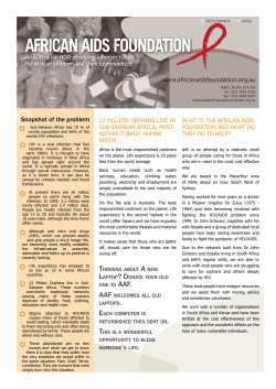


© Copyright 2026