
Poster session 18: Wound healing
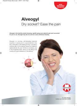
Poster session 18: Wound healing
P18.01
The role of human amniotic membrane in healing diabetic foot ulcer
Tauqeer Ahmed Malik, King Fahad Armed Forces Hospital, Jeddah, Saudi Arabia
Nashat Ghandoura, King Fahad General Hospital, Jeddah, Saudi Arabia
Hussam Itani, King Fahad General Hospital, Jeddah, Saudi Arabia
Mohammad Kareemuddin Majed, King Fahad Armed Forces Hospital, Jeddah, Saudi Arabia
Samia Faraj, King Fahad General Hospital, Jeddah, Saudi Arabia
Aim: The Role of Human Amniotic Membrane in Healing Diabetic Foot Ulcer
Methods: Human Amniotic Membrane (HAM) is extracted under strict sterile conditions
during an elective C section in an operating room. Written consent is obtained from HIV1,
HIV2, HTLV, HBV, HCV and Syphilis negative mothers. HAM is washed with normal saline
to remove blood clots and then preserved in normal saline in a sterile container with 160 mg
of Gentamicin. Patients are screened for seronegativity. Patients with Diabetic foot ulcers,
pressure ulcers and acute wounds are selected. Swabs for culture and sensitivity are taken
from wounds and from the HAM before application. Written consent is obtained from
recipients. HAM is applied as a primary dressing with non adherent silicon dressing to
protect the membrane. Dressing is done as an OPD under sterile protocols. Dressing is
changed one to two times a week. Photographs are taken at all stages of wound treatment.
Results: 39 patients with diabetic foot ulcers were treated with HAM. Among these location
of ulcer was: dorsum (19), plantar (5), malleoli (7), post transmetatarsal amputation (5), and
others (3). Complete healing was achieved in 95 percent of patients
Conclusions: With HAM healing was faster with no adverse affects. HAM is HLA negative,
donates epithelium, has many growth factors, antibacterial properties and has minimal
stroma. It is readily available, easy to harvest, preserve and easy to apply. It is cost effective
as compared to advanced dressings. It reduces patient's daily visits to hospital. We are
including more patients from other hospitals in this study.
Diabetic Foot Ulcer treated with HAM
www.diabeticfoot.nl
Page 1 of 10
P18.03
Complete healing of diabetic foot ulcers with autologous fibroblasts: 3 years followup
George Topakas, ‘G. Gennimatas’ General Hospital, Athens, Greece
Irene Karchilaki, ‘G. Gennimatas’ General Hospital, Athens, Greece
Ιoannis Dontas, ‘G. Gennimatas’ General Hospital, Athens, Greece
Panagiotis Pachantouris, ‘G. Gennimatas’ General Hospital, Athens, Greece
Eleftherios D Voyatzoglou, ‘Demetrios Voyatzoglou’ Diabetic Foot Clinic, ‘A. Fleming’
General Hospital, Athens, Greece
Penelope Sotiriou, National and Kapodistrian University of Athens, Athens, Greece
Andriana Donou, ‘Demetrios Voyatzoglou’ Diabetic Foot Clinic, ‘A. Fleming’ General
Hospital, Athens, Greece
Aliki Iniotaki, ‘G. Gennimatas’ General Hospital, Athens, Greece
Paraskevas Theodossiou, ‘G. Gennimatas’ General Hospital, Athens, Greece
Chariclia V Loupa, ‘Demetrios Voyatzoglou’ Diabetic Foot Clinic, ‘A. Fleming’ General
Hospital, Athens, Greece
Aim: Two cases of long-standing (9 5 and 8 5 months, despite conservative treatment)
diabetic foot (DF), ulcers that healed completely with autologous fibroblast infusion are
described. Both cases had a 3-year follow-up period.
Patients & Methods: Autologous skin fibroblasts from small split-thickness skin biopsies
were cultured in high concentration of fetal bovine serum., No additives with growth factor
activity were used. After 20 to 30 days, they were dispersed in patients΄ own serum, and
injected subcutaneously into the surrounding tissue of uninfected neuropathic DF foot ulcers
of two women, age 64 and 66 years, with well-controlled type 2 DM and without peripheral
angiopathy. There were neither systemic diseases affecting healing, nor corticosteroid
/immunosuppressant use. The estimated cost was 400 euros for 1st session and 200 euros
for each additional injection.
Results: Patient 1: Ulcer of 1st toe (plantar surface), diameter 1 cm. Two fibroblast
infusions, with an interval of 7 weeks. Ulcer was completely healed on week 11. There was
no recurrent ulceration in the 3-year follow-up period., Patient is periodically undergoing
debridement for local callus formation. Patient 2: Heel ulcer, diameter 4 cm. Three sessions
performed. For the 1st and 3rd sessions early cultured cells were used, while for the 2nd
frozen cells were employed. Interestingly, there was no improvement of the healing process
after this 2nd injection. Third infusion (from fresh skin biopsy) was followed by a steady
healing process. Wound closed completely after 27 weeks. After 3 years, there is no ulcer
but local callus formation. Pathology and cytology of specimens showed only keratinocytes
and keratine masses. In both patients, healing was complete, without scars.
Conclusions: Although studies with large samples are needed, it seems that early,
autologous skin fibroblasts consist a promising method to effectively treat persistent DF
ulcers. Healing was permanent, as we had a 3-years follow-up period, and relatively
inexpensive, compared to other additional therapies (e.g. growth factors, VAC). The
avoidance of use of growth factor-like additives in cultures increases the method’s safety
regarding malignancy formation, and tissue biopsy at 3 years was reassuring.
www.diabeticfoot.nl
Page 2 of 10
P18.04
Can oral autovaccination improve wound healing in patients with the diabetic foot?
Vladimira Fejfarova, Institute for Clinical and Experimental Medicine, Prague, Czech
Republic
Alexandra Jirkovska, Institute for Clinical and Experimental Medicine, Prague, Czech
Republic
Michal Dubsky, Institute for Clinical and Experimental Medicine, Prague, Czech Republic
Jana Vydlakova, Institute for Clinical and Experimental Medicine, Prague, Czech Republic
Alena Sekerkova, Institute for Clinical and Experimental Medicine, Prague, Czech Republic
Alena Tomesova, Department of Clinical Microbiology and Autovaccines, Prague, Czech
Republic
Jana Franekova, Institute for Clinical and Experimental Medicine, Prague, Czech Republic
Robert Bem, Institute for Clinical and Experimental Medicine, Prague, Czech Republic
Veronika Woskova, Institute for Clinical and Experimental Medicine, Prague, Czech Republic
Monika Kucerova, Institute for Clinical and Experimental Medicine, Prague, Czech Republic
Jelena Skibova, Institute for Clinical and Experimental Medicine, Prague, Czech Republic
Autovaccination could potentially enhance the immune system and therefore it may play a
role in the healing of infected wounds especially of the diabetic foot ulcers (DFU). The aim of
our double blind placebo controlled study was to evaluate the potential effect of oral
autovaccination on the healing of chronically infected DFU, an elimination of microbes from
these wounds and adverse events (AE) of such therapy. Methods: We consecutively
included into our study 30 patients with chronically infected DFU of Texas IIB-IIIB (mean age
57.2±9 years, DFU duration 13.4±11.7 months and HbA1c 68±21 mmol/mol). Study subjects
were randomized into 2 groups – A group treated by autovaccines (n=14) and P group used
placebo (n=16). Autovacinnes were prepared from microbial isolates found in DFU. All study
subjects were followed for the parameters of wound healing (Δarea, Δdepth, PUSH score
calculated by wound area, secretion and type of wound tissue, percentages of different types
of tissue in DFU) and for the incidence of new DFU, healing and development of
osteomyelitis, amputation rates and an eliminations of microbes during the treating period of
16 weeks. Results: Both study groups did not differ in basal characteristics including DFU of
Texas IIIB (29% in A vs. 25% in P group; NS). A significant reduction of PUSH score (2.8vs.-0.37; p=0.04), increased percentages of epitelisation tissue (21.8%vs.8.1%; p=0.07)
have been detected after the study period in A group in comparison to P group. As the
number of healed (7.1% from A vs. 6.3% of patients from P group; NS) or improved DFU
(57.1% from A vs. 56.3% of patients from P group; NS) as the incidence of new DFU and
healed or newly developed osteomyelitis did not differ significantly between the study
groups. Positive trends in the reduction of minor amputation rates (7.1% vs. 31.3% of
patients; p=0.3) and total microbes eliminations from DFU (in 50% vs. 31.3% of patients;
p=0.23) were found in group A vs. group P. No AE were recorded during the study period.
Conclusion: The application of autovaccines performed from microorganisms present in DFU
is safe for patients and could potentially improve the healing of the DFU.,
Supported by the project 00023001 (IKEM, Prague, Czech Republic) – Institutional support
www.diabeticfoot.nl
Page 3 of 10
P18.05
Efficacy and safety of a novel superoxidized solution in the management of postsurgical lesions of the diabetic foot
Silvia Macchiarini, University of Pisa, Pisa, Italy
Chiara Mattaliano, University of Pisa, Pisa, Italy
Chiara Goretti, University of Pisa, Pisa, Italy
Elisabetta Iacopi, University of Pisa, Pisa, Italy
Alberto Coppelli, University of Pisa, Pisa, Italy
Alberto Piaggesi, University of Pisa, Pisa, Italy
Aim: We investigated if a novel superoxidized solution (SOS) characterized by free chlorine
species with stabilized hypochlorous acid (HClO) in high concentration (>95%) combined
with acidic (pH<3) and oxidizing features (RedOx 1100mV)*, on top of standard treatment,
is safe and effective in reducing re-infections in post-surgical DFU.
Methods: We studied 25 consecutive DF outpatients (Group A; 22 Type 2/3 Type 1; Age
67.3±12.1 yrs, Duration of diabetes 14.7±9.9 yrs; HbA1c 7.9±1.1%) recently dismissed by
our department with a post-surgical, non-ischemic and non infected lesions (area 8.9±6.5
cm2) left to heal by secondary intent. Patients were followed on an outpatient basis, with
monthly control visits. All the patients were instructed to deliver one spray of SOS every 2
cm2 of the DFU’s area at any dressing change.
Patients, compared to a saline-managed group, (Group B), were followed up to complete reepithelisation or for a maximum of 6 months and the number of re-infections (RI) was taken
as primary endpoint. Secondary endpoints were number of debridement procedures (DP),
healing rate (HR) and healing time (HT).
Results:, All patients completed the study. Group A had significantly less RI (3 vs 12;
p<0.05), less DP (1 vs 10; p<0.05), and a faster HT (64.9±43.8 vs 147.4±88.8 days; p<0.01)
than Group B; no difference in HR and adverse events was observed.
Conclusions: The novel SOS solution provided an effective protection against re-infections
in DFU patients, reducing the necessity of DP and possibly fastening, their HT, with a safety
profile similar to saline solution.
*Nexodyn spray (APR s.a., Balerna, CH)
www.diabeticfoot.nl
Page 4 of 10
P18.06
Cold atmospheric pressure plasma as a novel treatment modality in diabetic foot
ulcers: a pilot study
Rimke Lagrand, VU University Medical Center, Amsterdam, Netherlands
Paulien Smits, Eindhoven University of Technology, Eindhoven, Netherlands
Guus Pemen, Eindhoven University of Technology, Eindhoven, Netherlands
Louise Sabelis, VU University Medical Center, Amsterdam, Netherlands
Ana Sobota, Eindhoven University of Technology, Eindhoven, Netherlands
Bas Zeper, Plasmacure, Eindhoven, Netherlands
Edgar Peters, VU University Medical Center, Amsterdam, Netherlands
Aim: Plasma medicine is an innovative field of research with a high potential but with little
clinical evidence to its support. Cold atmospheric plasma (CAP) devices generate an ionized
gas with a cocktail of highly reactive species and UV light. They can be used for application
on living tissue because these operate at normal ambient air pressure and temperature1 2 3.
CAP treatment has advantages over antiseptic or antimicrobial infection prevention and
control, e.g. efficient, painless, instant disinfection without chance of developing antimicrobial
resistance, but with stimulation of fibroblast proliferation and migration contributing to wound
healing. Current evidence for CAP consists of in-vitro studies, animal studies, and studies in
patient groups such as those with burn wounds. Our novel type of CAP device4 is simple to
use and can be applied by a podiatrist and even at a patient’s home. This is a proposal for a
pilot study to safety and efficacy of the technology.
Methods: 20 patients with foot ulcers will be treated on an outpatient basis with daily CAP
for 10 days in 2 weeks. Bacterial load will be measured by culture and molecular technique
of deep tissue swab at days 1, 7 and 14, and directly before and after CAP application.
Standard protocols for wound treatment will be deployed, including proper offloading.
Results: Primary endpoint will be occurrence of adverse events. Safety will be defined as ≤
10% of patients experience serious adverse events other than infection, and ≤ 60% of
patients develop infection. Secondary endpoints will be healing, bacterial load, incidence of
clinical infection during and after treatment5, and clinical outcomes 3 months after
enrolment. The microbiological effect will be considered significant if bacterial load is
reduced with 50% at day 14 compared with day 1, or if a 50% reduction in bacterial load is
achieved directly after CAP application compared with pre-application on days 1, 7 and 14.
Conclusions: We hypothesise that CAP is a safe treatment that reduces bacterial load on
the wound surface and promotes wound healing.
References
1.Von Woedtke, Phys Rep 2013; 530(4)
2.Isbary, Clin Plasma Med 2013; 1(1)
3.Arndt, Plos One 2013; 8(11)
4.Plasmacure©, Eindhoven, The Netherlands
5.Lipsky, Clin Infect Dis, 2012; 54(12)
www.diabeticfoot.nl
Page 5 of 10
P18.07
Coffee for the management of diabetic foot wounds. a new paradigm
Hendro Sudjono Yuwono, Division Vascular Surgery, Padjadjaran University, Bandung,
Indonesia
Aim: Describe the new paradigm in the treatment of diabetic foot wounds using coffee
powder as it has strong inhibition against methicilline-resistant Staphylococcus aureus,
autolytic debridement, kept dry, and fewer dressing-change.1 2,
Methods: Diabetic foot patients with glucose levels less than 200 mg%, not ischemic and
not agree for skin grafting. Coffee arabica used was newly made from a local shop., Topical
coffee treatment were together with blood sugar regulation. After debridement the wound
covered by a large amount of coffee powder and gauze with adhesive-tape to keep the
coffee in place. Average 6 cm wound-diameter and 0.5 cm depth, class, 1, 2 and 3 of
Wagner, without osteomyelitis. Wounds kept dry for 4 weeks, new coffee powder added
without removing the coffee attached onto the wound. The healing of diabetic wounds using
coffee powder was compared to diabetic wounds treated by saline which must be changed
daily. The first healing sign is dry wound surface.
Results: A total 96 patients were collected from January 1, 2003 until May 31, 2014.
Patients did not feel pain during dressing change, wound fluid absorbed by the coffee giving
coffee scent and not invaded by flies. Only 41 of 96 (42.7%) patients needed broadspectrum antibiotics for 1 week. 72 of 96 (75%) patients were able to continue treatment at
home and every 4 weeks came to the hospital for replacing wound dressing. The difference
of time for dry wound surface between coffee (n=84) and saline (n=80) is statistically
significant (p=0.000).
Conclusion: Coffee powder has a better capability in the management of diabetic wound,
with fewer wound dressing changes, kept the wound dry, has autolytic debridement, without
redebridement needed, painless dressing replacement, many antioxidant activity,
deodorized, capillary hemostatic. This raises a new paradigm in the management of diabetic
wounds.
References:
1.
Hendro Sudjono Yuwono. Journal of Surgery, vol.2 no.2 (2014):25-29
2.
Lepelley M, et al.BMC Plant Biology 2012 12:2-16
www.diabeticfoot.nl
Page 6 of 10
P18.08
Safety and effectiveness of therapeutic magnetic resonance (tmr) in the management
of wide post-surgical lesion of the diabetic foot
Lorenza Abbruzzese, University of Pisa, Pisa, Italy
Giovanni Bonino, University of Pisa, Pisa, Italy
Chiara Mattaliano, University of Pisa, Pisa, Italy
Elisabetta Iacopi, University of Pisa, Pisa, Italy
Alberto Piaggesi, University of Pisa, Pisa, Italy
Silvia Macchiarini, University of Pisa, Pisa, Italy
Aim: A number of technologies based on phisical, rather than bio-chemical, approach to
wound repair have been recently proposed for the managemet of a wide range f chronic
ulceration, among which magnetic fields. we designed this study, to evaluate the safety and
effectiveness of therapeutic magnetic resonance (TMR) in the management of the diabetic
foot (DF).
Method: We treated a group of consecutive type 2 diabetic inpatients with wide post-surgical
lesions (Group A - N. 10; age 67.7±18.9 yrs, duration of diabetes 22.3±6.6 yrs, 8.1±1.1%,
BMI 29.4±2.1 kg/m2), for two consecutive weeks, while admitted, with a low-intensity
magnetic resonance equipment*, on top of standard treatment. Patients, compared with a
matched control group with the same clinical characteristics (Group B), were then followed
monthly for 6 months to evaluate healing rate (HR), healing time (HT), rate of granulation
tissue (GT) at three months, and adverse events.
Results / Discussion: HR was of 90% in Group A and 30% in Group B (p<0.05); GT was
73.7±13.2% in Group A vs 51.84±18.77% in Group B (p<0.05). HT in Group A was
84.46±54.38 days vs 148.54±78.96 days in Group B (p<0.01)., No difference in adverse
events (5 in Group A and 6 in Group B) was observed throughout the study period.
Conclusion: TMR proved to be effective and safe, on top of standard treatment in the
management of the wide post-surgical lesions of the diabetic foot.
*TMR-9 (Thereson, Milano, I)
www.diabeticfoot.nl
Page 7 of 10
P18.09
Retrospective evaluation of Maggot Debridement Therapy (MDT) in Lower Limb
Wounds
Adriaan Erasmus, National University Hosipital, Singapore, Singapore
Cheryl Zhang, National University Hospital, Singapore, Singapore
Aims: Maggot debridement therapy (MDT) is well established as a wound debridement
modality for the removal of necrotic tissue and slough. However, research that supports its
benefits is limited. This retrospective study aims to evaluate the therapeutic effects of MDT
on patient outcomes. The secondary aim is to identify specific patient characteristics and
wound characteristics that can influence patient outcomes or indicate patient prognosis.
Methods: Patients who presented to National University Hospital Vascular Department with
advanced vascular disease, high amputation risk and non-healing sloughy/necrotic wounds
were deemed suitable for MDT. Treatment was provided in an inpatient setting, with
management of concomitant medical issues according to accepted clinical practice. Data
was gathered by reviewing the medical records of those who underwent MDT from January
2011 to April 2014. This included patient demographics, medical history, vascular status,
wound location, previous surgical intervention, the number and frequency of MDT
applications and the outcome of care. The treatment outcomes were classified into
successful or unsuccessful categories. Statistical analyses were carried out to compare
between the two groups.
Results: Approval from the local ethics review board was sought. A total of 80 subjects with
varying severity of peripheral arterial disease were included in this study. Exemption for
obtaining informed consent due to the retrospective nature of the study. A mean of 3.5 MDT
applications were completed per subject. Approximately 1.5 vials of free-range maggots
were used at each application. Treatment that led to limb salvage and wound healing was
considered successful. Patients who required major amputation were grouped into the
unsuccessful category.
Conclusion: Through this study, the efficacy of MDT can be evaluated in a local context as
local data is not currently available. This is to provide evidence to support clinical use.
Specific patient and wound factors that may significantly influence outcome can be identified,
so as to guide patient selection for MDT. This may in turn improve clinical outcomes and
reduce the cost of healthcare.
www.diabeticfoot.nl
Page 8 of 10
P18.10
A wound care strategy to increase cost-effectiveness and adherence to recommended
dressings.
Gunilla Larsson, Skane University Hospital, Lund, Sweden
Ulrika Biörklund Brogren, Skane University Hospital, Lund, Sweden
Åsa Asmundsson, Skane University Hospital, Lund, Sweden
Per Katzman, Skane University Hospital, Lund, Sweden
Katarina Fagher, Skane University Hospital, Lund, Sweden
Mirja Ruonakoski Ley, Skane University Hospital, Lund, Sweden
Magnus Londahl, Skane University Hospital, Lund, Sweden
Aim: Although nursing time counts for a larger portion than material costs in the field of the
diabetic foot ulcer care, the total cost for dressing materials is high. In our health care
system community nurses take care of a majority of the dressing changes but all the costs of
the dressing materials are added on the Diabetic Foot Clinic (DFC). Until recently, our DFC
recommended a dressing, which the community nurse units bought from the manufacturers
and then invoiced the DFC., At present, the DFC recommends a dressing, has it in stock and
supplies a sufficient amount of the dressing to the patient, who brings it to a community
nurse. Further, to secure an appropriate initial dressing of acute ulcers in patients seeking
primary care, the general instructions from the DFC have been simplified to only include
three dressings which also are supplied by the DFC; Sorbact ®for dry ulcers, Sorbact
Absorption® for wet non-infected ulcers and Iodosorb ® for wet infected ulcers., The aim of
this study was to compare material costs before and after this change, and to evaluate the
opinion of community nurses regarding this change.
Method: We compared the out patient dressing material cost per patient-week, one year
before vs. two years after the change. Opinion of the community nurses was gathered by
telephone interviews.
Results: Out patient care dressing costs per patient-week in the DFC decreased by 28% by
this change. The community nurses appreciated the new approach and pointed out the
simplicity of always having the recommended dressing available, without having a large
storage at the Primary Care clinic and the usefulness of an easy recommendation when
treating new ulcers. The DFC observed a significantly higher adherence to the
recommended dressings. No adverse event related to this change has been identified.
Conclusion: Giving out a recommended dressing to secure the supply to the next visit to a
DFC seems to be cost-effective and increase adherence to the recommendation. Only a few
different dressings needs to be included in the armamentarium of acute diabetic foot ulcers
treatment in primary care.
www.diabeticfoot.nl
Page 9 of 10
P18.11
First treatment of skin ulcer lower limb in diabetic patient complicated by new
automated CelluTome epidermal harvesting system
Roberto De Giglio, Azienda Ospedaliera Legnano, Abbiategrasso, Italy
Teresa Mondello, Azienda Ospedaliera Legnano, Abbiategrasso, Italy
Sara Lodigiani, Azienda Ospedaliera Legnano, Abbiategrasso, Italy
Ilaria Formenti, Azienda Ospedaliera Legnano, Abbiategrasso, Italy
Giacoma Di Vieste, Azienda Ospedaliera Legnano, Abbiategrasso, Italy
Paola Cavaiani, Azienda Ospedaliera Legnano, Abbiategrasso, Italy
Lorena Barbierato, Azienda Ospedaliera Legnano, Abbiategrasso, Italy
Gianmario Balduzzi, Azienda Ospedaliera Legnano, Abbiategrasso, Italy
Viviana Zoppini, Azienda OspedalieraLegnano, Abbiategrasso, Italy
Barbara Musto, Azienda Ospedaliera Legnano, Abbiategrasso, Italy
The development happened in the last fifteen years in the field of tissue repair with a number
of techniques to cover biological and medical devices, focused to increase the speed of
closure of a cutaneous ulcer lesion of any origin, has enabled the creation of a new
technique for the removal of the epidermis aimed autograft.
Traditionally, the procedure requires the use of a dermatome, the need for an operating
room, anesthesia and the creation of a second skin lesion from lead to healing at the level of
the donor site.
In this case report a patient has been treated type 2 diabetic of 83 years, complicated,
affected by more than two months from ulcerative lesion in the distal third of the right leg
about the size of 10 x 10 cm, superficial, without clinical signs of infection.
We proceeded for the first time at the Diabetic Foot Unit - Hospital of Abbiategrasso (AO
Legnano) a withdrawal of epidermal tissue 5 x 5 cm using a new sampling system
CelluTome from the ipsilateral thigh and grafting at the center of the ulcer. This system,
automated, without the need to perform anesthesia, eliminating the binding of the operating
room, simplifying the procedure and reducing costs, has allowed the removal of micrografts
epidermal exploiting the suction and heat, thus arriving to produce sections constants of thin
epidermal skin that have been applied on the bed of the ulcer.
After 8 weeks, the clinical picture was characterized by a net reduction of lesion size by over
50% with re-epithelialization of the graft in the seat and extension of the reparative process
to adjacent areas.
This article describes the new methodology, analyzed the strengths and the weaknesses
and a comparison is made with the Split-skin graft technique.
www.diabeticfoot.nl
Page 10 of 10


© Copyright 2026