
Enrichment and Characterization of Neural Crest
UNIVERSITÄTSKLINIKUM HAMBURG-EPPENDORF Kopf- und Neurozentrum Klinik und Poliklinik für Mund-, Kiefer- und Gesichtschirurgie Director: Prof. Dr. med. Dr. med. dent. Max Heiland Enrichment and Characterization of Neural Crest - derived Dental Pulp Stem Cells from Human Dental Pulp Dissertation zur Erlangung des Grades eines Doktors Zahnmedizin an der Medizinischen Fakultät der Universität Hamburg. vorgelegt von: Heba Al-Zer aus Jordanien Hamburg 2014 1 Angenommen von der Medizinischen Fakultät der Universität Hamburg am: Veröffentlicht mit Genehmigung der Medizinischen Fakultät der Universität Hamburg. Prüfungsausschuss, der/die Vorsitzende: Prof.Dr.Dr. Ralf Smeets Prüfungsausschuss, zweite/r Gutachter/in: PD Dr. Florian Barvencik To my Mother For as you’ve always believed in me and been by my side, giving me the strength to proceed in life unconditionally. Contents I. Introduction ................................................................................................................................................... 1 I.1 Tissue Engineering in Regenerative Medicine .......................................................................... 1 I.1.1 Overview .......................................................................................................................................... 1 I.1.2 Cellular Component ..................................................................................................................... 1 I.1.3 Acellular Component (Scaffolds) ........................................................................................... 2 I.1.4 Challenges ....................................................................................................................................... 3 I.2 Dental Pulp Stem Cells (DPSCs) ...................................................................................................... 4 I.2.1 Stem Cells ........................................................................................................................................ 4 I.2.2 Dental Pulp ..................................................................................................................................... 5 I.2.3 Dental Pulp Stem Cells (DPSCs) .............................................................................................. 7 I.3 Dental Pulp Stem Cells for Tissue Engineering and Regenerative Medicine .............. 11 I.3.1 Dentine Regeneration and Novel Therapeutics. ........................................................... 11 I.3.2 Pulp Tissue Engineering and Regeneration .................................................................... 11 I.3.3 Treatment of Neurological Diseases .................................................................................. 12 I.3.4 Vasculogenesis after Ischemia ............................................................................................. 13 I.3.5 Bone Defect Repair ................................................................................................................... 14 I.3.6 Miscellaneous ............................................................................................................................. 15 I.4 Hypothesis and Aims ........................................................................................................................ 16 II. Materials ..................................................................................................................................................... 17 II.1 Teeth and Patient Data................................................................................................................... 17 II.2 Equipment........................................................................................................................................... 17 II.3 Reagents .............................................................................................................................................. 18 III. Methods ..................................................................................................................................................... 21 III.1 Culture and Handling of DPSCs ................................................................................................. 21 III.1.1 Enzymatic Digestion Method ............................................................................................. 21 III.1.1.1 Adherent Standard Conditions ................................................................................. 22 III.1.1.2 Non-adherent Serum-free conditions (Sphere Forming Conditions) ....... 23 III.1.2 Explant Method ....................................................................................................................... 24 III.2Enrichment of the Neural Crest-derived DPSCs .................................................................. 25 III.2.1 Magnetic Cell Sorting after Expansion ........................................................................... 25 III.2.2 Conditioned Culture .............................................................................................................. 26 III.3 Cryoconservation and Storage of DPSCs ............................................................................... 27 III.4 Induction of Differentiation in DPSCs ..................................................................................... 28 III.4.1 Osteogenic Differentiation.................................................................................................. 28 III.4.2 Glial Differentiation ............................................................................................................... 28 III.4.3 Melanocytic Differentiation ............................................................................................... 30 III.5 Immunophenotyping ................................................................................................................. 31 III.5.1 Fluorescent Immunocytochemistry ................................................................................ 31 III.5.2 Substrate Immunocytochemistry .................................................................................... 31 III.5.2.1 Alzarin Red-Calcium Deposits Detection .............................................................. 31 III.5.2.2 Alkaline Phosphatase-Phosphatase Activity Detection .................................. 32 III.5.2.3 L-DOPA staining ............................................................................................................. 32 III.5.3 Immunohistochemistry ....................................................................................................... 32 III.5.4 Immunophenotypic analysis-Summary ........................................................................ 34 IV. Results ........................................................................................................................................................ 35 IV.1 Culturing of DPSCs.......................................................................................................................... 35 IV.1.1 Enzymatic Digestion and Adherent Culturing Method ............................................ 35 IV.1.2 Enzymatic Digestion and Sphere Culturing Method ................................................. 35 IV.1.3 Explant Method ....................................................................................................................... 36 IV.2 Characterization of the Enriched Neural Crest-derived DPSCs .................................... 39 IV.2.1 Immunophenotyping ............................................................................................................ 39 IV.2.2 Sphere Formation Assays .................................................................................................... 41 IV.3 Induction of Differentiation ........................................................................................................ 42 IV.3.1 Osteogenic Differentiation .................................................................................................. 42 IV.3.2 Glial Differentiation ............................................................................................................... 45 IV.3.3 Melanocytic Differentiation ................................................................................................ 46 IV.4 Summary of Morphological Characterization of DSPCs and Induced Cells .......... 47 IV.5 Summary of the Results ............................................................................................................ 49 V. Discussion ................................................................................................................................................... 50 V.1. Characteristics of the Enriched DPSCs .................................................................................... 50 V.1.1 Overview ..................................................................................................................................... 50 V.1.2 Culture and Expansion Methods ........................................................................................ 52 V.1.2.1 Extracellular Matrix (ECM) .......................................................................................... 52 V.1.2.2 Conditioned Medium ...................................................................................................... 54 V.1.2.2 Expansion ........................................................................................................................... 54 V.1.3 Sphere Formation .................................................................................................................... 55 V.1.4 Stem Cells Markers .................................................................................................................. 56 V.1.4.1 Nestin ................................................................................................................................... 56 V.1.4.2 CD271 ................................................................................................................................... 57 V.1.4.3 Sox10 .................................................................................................................................... 57 V.1.5 Multipotentiality ...................................................................................................................... 58 V.1.6 Comparison of the Characteristics of the Neural Crest-derived DPSCs with the Published Literature. ......................................................................................................................... 58 V.2 Phenotype of Differentiated Cells Induced from Neural crest-derived DPSCs and Comparison with the Published Literature. .................................................................................. 59 V.2.1 Molecular Control of NCSC Differentiation .................................................................... 59 V.2.2 Osteogenic Differentiation ................................................................................................... 60 V.2.2.1 Induction ............................................................................................................................. 60 V.2.2.2 Characterization............................................................................................................... 61 V.2.3 Glial Differentiation................................................................................................................. 62 V.2.3.1 Induction ............................................................................................................................. 64 V.2.3.2 Characterization............................................................................................................... 66 V.2.4 Melanocytic Differentiation ................................................................................................. 67 V.2.4.1 Induction ............................................................................................................................. 68 V.2.4.2 Characterization............................................................................................................... 68 VI Summary .................................................................................................................................................... 70 VII Abbreviations .......................................................................................................................................... 71 VIII Literature ................................................................................................................................................ 73 IX Acknowledgements ................................................................................................................................ 84 Curriculum Vitae ........................................................................................................................................... 86 Eidesstattliche Versicherung ................................................................................................................... 89 INTRODUCTION I. Introduction I.1 Tissue Engineering in Regenerative Medicine I.1.1 Overview Regenerative medicine according to the National Institute of Health is the process of creating living, functional tissues to repair or replace a tissue or an organ function lost due to age, disease, damage, or congenital defects. This field holds the promise of regenerating damaged tissues and organs in the body by stimulating previously irreparable organs to heal themselves or by growing tissues and organs in the laboratory and safely implant them when the body cannot heal itself [1]. Tissue engineering on the other hand, is agreed to be the application of the principles and methods of engineering and life sciences toward the fundamental understanding of structure-function relationships in normal and pathologic mammalian tissue and the development of biological substitutes to restore, maintain, or improve function [2]. In conclusion tissue engineering is one of the medical sciences which is used to accomplish regeneration as a final result. Tissue engineering is based on two main components; the cellular and the acellular (scaffolds). I.1.2 Cellular Component It is a fact in the scientific society today that the cells, which are able to regenerate a tissue in our body after a disease or a trauma are not simply the differentiated cells of the damaged tissue, but the stem cell populations lying within that tissue, as a result studying stem cells properties and the methods of their isolation has been taking over the medical research community for decades now. This project is an example of this new direction of research as it aims to isolate stem cells out of an adult tissue. 1 INTRODUCTION As will be discussed in (I.2.1 Stem Cells), stem cells have different levels of stemness; which means different capabilities of self-renewal and differentiation, a fact that should be considered along with the knowledge of appropriate methods of isolation, expansion, survival support and differentiation of the designated stem cell population , a population which should be able to multiply in adequate numbers and acquire the ability to differentiate into the specific kind of cells needed for therapy in the corrupted tissue. I.1.3 Acellular Component (Scaffolds) Regenerating tissue is not a process of easily filling the damaged area with the right type of stem cells which are able to regenerate the tissue; a carrier should be used to support the survival of stem cells in many cases because tissue is a complex 3D structure owing a special cell hierarchy which has a specific and a complicated net of interactions between them and the extracellular matrix, also the tissue has its specific network of blood, nerve and immune supply. In vitro studies of scaffolds biocompatibility show different results when they were repeated in vivo [3]. Thus, many factors should be considered to choose a scaffold for a certain treatment, appropriate scaffolds should be made of a biological absorbable inert material which has mechanical properties enabling the build-up of a well-controlled 3D stable structure in the Nano-scale out of it [4]. This 3D structure of the scaffold should enable cell spreading uniformly upon culturing and cell infiltration after implantation enabling proliferation of the seeded cells and new networks of blood and innervation to grow through. Many biomaterials have been used for the construction of scaffolds [5], but the details is beyond the scope of this study. 2 INTRODUCTION I.1.4 Challenges Regenerative medicine and tissue engineering are being considered a leap into a new generation of medicine. However, it is important to understand their current limitations as well as the scientific and the non-scientific challenges they are facing. Both concepts are based on a multidisciplinary approach bringing together various scientific fields such as biochemistry, pharmacology, material science, cell biology, engineering and medical disciplines, which makes progressing from laboratory studies to clinical studies a long and expensive process. The success of transplantation medicine is still to a great extent dependent on the immune state of the graft and the host. The science of immunomodulation and immunosuppression is therefore still a critical aspect in all tissue engineering and regenerative medicine applications if non-autologous cells are used [6] . The expectations must be aligned not only by scientific challenges, but more importantly, ethical considerations and fears. Given that the use of stem cells is a mandatory aspect of regenerative medicine, a special focus is given to the ethical as well as the theological considerations. Both can delay, limit and perhaps prevent the actual application of many intended therapies [7]. 3 INTRODUCTION I.2 Dental Pulp Stem Cells (DPSCs) In order to clarify what dental pulp stem cells (DPSCs) are, the expressions; stem cell and dental pulp should be clarified first. I.2.1 Stem Cells The ideal stem cell is the fertilized oocyte, which is able to give rise to all and each type of cell we know in the body. This miraculous cell divides thoroughly going through many stages of segregation and differentiation creating a whole human being with a noncountable number of cells. In this process the new daughter cells subsequently lose the ability of their mother cell and acquire more specific characteristics leading them to be differentiated [8]. This loss of stemness is not complete but gradual, leaving in each tissue a hierarchy of cells varying in their stemness from being totipotent to finally be completely differentiated cells (Fig. 1). These processes are marked by losing and gaining a specific morphology, gene expression, protein expression and other features used to distinguish stem cells from differentiated cells [9, 10]. Figure 1: Hierarchy of stem cells in the body: All cells of the fertilized oocyte are totipotent (able to differentiate into all human cell types and extra-embryonic tissues) then they lose some potency to become pluripotent (able to differentiate into any cell type of the three germ layers excluding extra-embryonic tissues) and subsequently multipotent (can give rise to several types of cells but all belong to the same lineage). Finally unipotent cells develop (can give rise to one type of cells) which is the last level of stemness before cells become fully differentiated. 4 INTRODUCTION A stem cell in the simplest definition is a cell which can self-renew and give rise to a differentiated cell progeny. These abilities are dependent upon the cell potential, as well as upon microenvironmental signals, including: cell–matrix interactions, cell–cell interactions, growth factors, hormones, and circulation gradients [11]. Interactions within the stem cell niche, a specialized microenvironment composed of mesenchymal cells and extracellular matrix (ECM), are particularly crucial to the self-renewal and differentiation processes (Fig. 2) [11]. This aspect of stem cell biology will be important in the isolation process of DPSCs in this study. Figure 2: Stem cells and niche: Niche cells (green) underlie a basement membrane signal to stem cells (red) to regulate differentiation and self-renewal. When a lineage mechanism prevails (lower mitotic cell), the stem cell divides such that one daughter retains its connections to the niche, while the other (yellow) begins to differentiate. When a self-renew mechanism prevails (upper mitotic cell), symmetric stem cell division occur, both are determined by local factors. ECM, extracellular matrix [11]. I.2.2 Dental Pulp Other than being surrounded by odontoblasts, the dental pulp is a typical loose connective tissue with an abundant extracellular matrix composed mainly of fibronectin, laminin, collagen, elastin, hyaluronic acid, dermatan sulfate and chondroitin sulfate, in which lies the cellular component of mainly fibroblasts, DPSCs and of course neural, vascular and immunological cells [12] (Fig. 3). 5 INTRODUCTION A B [ [ T T y Figure 3: Dental Pulp. A) The main structures of the tooth showing dental pulp position (black box). B) y p odontoblasts (arrows) lining dentin and the pulp consisting of Histological p section of a dental pulp showing e e loose connective tissue [13]. a a q q Dental pulp u mesenchyme is derived u from the neural crest of the ectoderm [14, 15]. o it is called Ectomesenchyme, o Accordingly which suggest that DPSCs may possess t t different characteristics from the eordinary mesenchymal stem cells, akin to those of e f cells. Therefore, special f attention should be given to understand the origin neural crest r r of the stem o cells in this tissue, as that o will help in isolation and recruiting these cells in m the future.m t t h h the Swiss embryologist Wilhelm His in 1868, as a Neural Crest (NC) was identified by e e group of dcells originating in the vertebrate embryo ectoderm at the margins of the d o localized in between theo neural tube and the epidermis (Fig. 4) [16]. NC cells neural tube c c are initially integrated within the u u neuroepithelium where they are morphologically m m indistinguishable from the other neural epithelial cells. Upon induction by signals from e e contact-mediated tissue interactions n n between the neural plate and the surface ectoderm, t t NC cells delaminate through an epithelial-to-mesenchymal transition and start migrating o o extensively in the embryo where they contribute to a r to several different locations r t diverse array of different t tissue types including the dental pulp [17, 18]. The remarkably h h neural crest structure and has been considered by many e is a transient embryonic e scientists sas the fourth germ layer sconsidering the diverse and large contribution of its u u cells in many m tissues and organs [16] m (Fig. 4). m m a a r r y y o o f f a a 6 n n i i n n INTRODUCTION Figure 4: The Neural Crest, a group of cells originating in the vertebrate embryo ectoderm at the margins of the neural tube localized in between the neural tube and the epidermis. Upon induction by signals that come from contact-mediated tissue interactions between the neural plate and the surface ectoderm, NC cells delaminate through an epithelial-to-mesenchymal transition and start migrating extensively to several different locations in the embryo where they contribute to a remarkably diverse array of different tissue types. I.2.3 Dental Pulp Stem Cells (DPSCs) Dental pulp regeneration capacity after harmful stimulus and the formation of secondary and tertiary dentin indicate that highly regenerative cells exist within the pulp, but it was until year 2000 when Gronthos et al. reported for the first time the isolation of a population of DPSCs. 7 INTRODUCTION He described the population as clonogenic, highly proliferative fibroblastic-like shaped cells derived from enzymatically digested adult human dental pulp. When compared to the mesenchymal human bone marrow stem cells (BMSC), DPSCs showed higher frequency of colony-forming and higher number of proliferating cells. Characterization of the immunophenotype of DPSCs showed the mesenchymal nature of these cells and revealed heterogeneity in the culture. Differentiation of DPSCs into osteoblasts but not adipocytes was possible back then [19-21]. Further work showed that in addition to DPSCs dentinogenic potential, subpopulations of human DPSCs also possess adipogenic and neurogenic differentiation capacities [22]. DPSCs were also found to undergo osteogenic, chondrogenic and myogenic differentiation in vitro [23, 24]. Since then attempts to isolate the different populations of DPSCs by different techniques, characterize them and show their potentials are still in progress (Table 1) [25]. The table shows the success of isolating populations of DPSCs which can be described as mesenchymal as their differentiation abilities show. Searching for neural crest stem cells (NCSCs) in the dental pulp has also taken place after mesenchymal DPSCs demonstrated neurogenic and adipogenic differentiation potentials in vitro [22], showed label retaining and neurosphere forming abilities [26, 27] , as these are considered characteristics of neural crest ontogeny (Table 2). In conclusion, it has been suggested that dental pulp comprises of two distinct stem cell populations, with one possessing embryonic neural crest cell markers [28] while the other is mesenchymal ones [29]. 8 INTRODUCTION Table 1: Isolation and Characterization of DPSCs Populations. Group Isolation Growth medium Gronthos Digestion αMEM, 20 % FCS, 100 et al. [19] 3 mg/ml mM L-ascorbic acid 2-P, collagenaseI, 4 2 mM L-glutamine Selection Differentiation osteogenic mg/ml dispase Liu et Digestion αMEM, 20 % FCS, 100 MACS osteogenic, al.[30] 3 mg/ml mM L-ascorbic acid 2-P, STRO-1+ , adipogenic, collagenase I, 4 2 mM L-glutamine, 3G5+, neurogenic mg/ml dispase Honda et Tissue explants al. [31] CC9+ αMEM, 10% FBS, 1x FACS Gluta-max, 50 mg/ml, L- Hoechst osteogenic ascorbic acid-P, 1 mg salt 33342 n–hydrate. Spath et al. Trypsin Mega Cell DMEM osteogenic, [32] pretreated ,10 % FCS, 2 mM chondrogenic, explants L -glutamine, 0.1 mM myogenic ß-mercaptoethanol Ishizaka et Specific enzyme DMEM, 10 % FBS FACS angiogenic, al. [33] is not identified CD31- neurogenic 9 INTRODUCTION Table 2: Isolation and Characterization of Neural Crest-DPSCs Populations. Group Isolation Growth medium Selection Differentiation Paino et al. [34] Digestion 3 mg/ml collagenaseI, 4 mg/ml dispase Mega Cell MEM, 10% FBS, 100 µM L-ascorbic acid 2-P, 2 mM L-glutamine. Stevens et al.[27] Digestion 3 mg/ml collagenaseI, 4 mg/ml dispase DMEM, 20% FCS, 100 µM L-ascorbic acid 2-P. MACS/FACS CD34-, CD45-, LNGFR+ osteogenic, adipogenic, chondrogenic melanocytic Janebodin et al. [35] Digestion 1.2units/ml dispase II, 2 mg/ml collagenaseIV DMEM, 40% MCDB201, 2% FCS, insulin-transferrin selenium, linoleic acid-BSA, 10⁻⁹M dexamethasone, 10⁻⁴ M ascorbic acid 2-P, 1000 units/ml leukemia-inhibitory factor, 10 ng/ml EGF , 10 ng/ml PDGF-BB vWF-, CD31-, Tie1-,Tie2, Cadherin osteogenic, adipogenic, chondrogenic neurogenic, smooth muscle Waddington et al.[29] Digestion 4 mg/ml collagenase /dispase αMEM, 10 % FCS Fibronectin adherence over 20 min, MACS LNFGR+ osteogenic, chondrogenic, adipogenic, Abe et al. [36] Tissue explants αMEM, 10 % FCS, 2 mM L-glutamine melanocytic osteogenic, chondrogenic, adipogenic, neurogenic, smooth muscle 10 INTRODUCTION I.3 Dental Pulp Stem Cells for Tissue Engineering and Regenerative Medicine Oral stem cells-based therapy for the regeneration and engineering of tissues has been a much-discussed subject and it is considered a promising mode of future medicine [21, 30, 37]. Considering DPSCs, many studies tried to reveal the potential regenerative therapies possible for this stem cell entity, of which some are mentioned below. I.3.1 Dentine Regeneration and Novel Therapeutics. The recruitment of dental pulp stem cells to differentiate into a new generation of odontoblast-like cells, leading to reparative dentinogenesis, is a natural reparative response of the dentine–pulp complex when the vitality of the existing odontoblasts is compromised by a carious injury or a trauma. This indicates that if the specific chemotactic signals for these stem cells can be determined, this process may be exploited clinically both by maximizing the migration of stem cells to areas of injury or disease and also through influencing the nature of the stem cell populations recruited, to provide a greater specificity and control to the tissue response [38], such migration occurs in response to pulp capping procedures (stimulated by calcium hydroxide), with subsequent reparative dentinogenesis and dentine bridge formation [39]. I.3.2 Pulp Tissue Engineering and Regeneration Many studies reported the recruitment of DPSCs in dental pulp tissue engineering and regeneration, among them are the following studies. In a tooth slice model Cordeiro et al. showed that DPSCs of deciduous teeth seeded onto synthetic scaffolds seated into the pulp chamber space formed odontoblast-like cells that localized against the existing dentin surface. However, no regeneration of pulp-like tissues in the pulp space has been reported with this approach. One concern is that implanting stem cells/scaffolds into root canals that have a blood supply only from the 11 INTRODUCTION apical end may compromise vascularization to support the vitality of the implanted cells in the scaffolds. It has been proposed that, because of the concern over vascularization, a stepwise insertion of engineered pulp may have to be implemented clinically to achieve the desired pulp tissue regeneration [40]. In an experimental model of amputated canine tooth autogenous transplantation of CD31-/CD146- DPSCs, CD105+ DPSCs together with type I and type III collagen as a scaffold resulted in complete regeneration of pulp tissue with capillaries and neuronal processes. The transplanted cells were in the vicinity of the newly formed vasculature and expressed proangiogenic factors, implying trophic actions on endothelial cells. The regenerated pulp tissue in the cavity of the amputated pulp was not changed into mineralized tissue even 3 months after transplantation [41]. DPSCs generate pulp-dentin complex when transplanted subcutaneously in mouse [19]. I.3.3 Treatment of Neurological Diseases Many studies reported the recruitment of DPSCs in treatment of neurological diseases, among them are the following studies. After transplantation of adult human DPSCs that could differentiate in vitro into functionally active neurons into an avian embryonic model system, implanted cells induced endogenous axon guidance [42]. In a cerebral ischemic model, transplantation of CD31- / CD146- or CD105+ DPSCs into adult rat striatum accelerated neovascularization of the ischemic zone and enhanced subsequent neuronal regeneration; neuronal progenitor cells migrated to the penumbra, differentiated into neuron and survived, localizing in the proximity to the transplanted cells. The volume of the ischemic region was reduced and the functional outcome was improved. Neurotrophic factors were expressed by the transplanted cells in the ischemic boundary zone, suggesting indirect involvement in the survival and the differentiation of the neuroprogenitor cells into neuron [41]. 12 INTRODUCTION Grafted undifferentiated, untreated DPSCs which express Nanog, Rex-1, Oct-4 from the dental pulp of rhesus macaques into the hippocampus of immune-suppressed mice stimulated proliferation of endogenous neural cells and resulted in the recruitment of pre-existing Nestin+, ß-tubulin-III+ neural progenitor cells and mature neurons to the site of the graft. Additionally, many cells born during the first 7 days after implantation proliferated, forming neural progenitor cells and neurons, and, to a lesser extent, underwent astrogliosis by 30 days after implantation. Although the DPSC graft itself was short term, it had long-term effects by promoting growth factor signaling as implanted DPSCs enhanced the expression of ciliary neurotrophic factor, vascular endothelial growth factor, and fibroblast growth factor for up to 30 days after implantation. In conclusion, grafted DPSCs promoted proliferation, cell recruitment, and maturation of endogenous stem/progenitor cells by modulating the local microenvironment [43]. DPSCs produced neurotrophic factors and interacted with trigeminal neurons in vitro, they also rescued motorneurons after spinal cord injury and differentiated into neurons in vitro [44-46]. These findings provide evidence that DPSCs may induce neuroplasticity within a receptive host nervous system and stimulate neurogenesis during tissue regeneration. Therefore, those cells can be a potential and an important cell source for the treatment of neurological diseases. I.3.4 Vasculogenesis after Ischemia Many studies reported the recruitment of DPSCs in vasculogenesis after ischemia, among them are the following studies. DPSCs were able to repair damaged myocardium when implanted in a rat infarction model and this was associated with an increase in the number of vessels and a reduction in the infarct size, probably because of DPSCs ability to secrete proangiogenic, antiapoptotic and cardio protective factors [47]. 13 INTRODUCTION In models of mouse hind limb ischemia, local transplantation of human CD31- /CD146and CD105+ DPSCs, resulted in an increase in the blood flow including high density of capillary formation. The transplanted cells were in proximity of the newly formed vasculature without incorporating into vessels, and expressed several proangiogenic factors, these results suggested a potential utility of these subfractions of human DPSCs to stimulate angiogenesis/vasculogenesis during tissue regeneration [48]. I.3.5 Bone Defect Repair Many studies reported the recruitment of DPSCs in bone defect repair, among them are the following studies. Dental pulp could be considered as an interesting and potentially important source of autologous stem cells that are ready to be used for therapeutic purposes, such as the repair/regeneration of craniofacial bones, as they showed great in vitro and in vivo results [49]. DPSCs seeded onto collagen scaffolds repaired alveolar defects of the mandible produced after extraction of impacted third molars in humans. The autografts produced fast regeneration of bone, which was of optimal quality and quantity when compared to the standard techniques commonly used for guided bone regeneration and bone grafts of various origins [50]. With the osteo-inductive potential, Seo et al. demonstrated that DPSCs of deciduous teeth can repair critical sized calvarial defects in mice with substantial bone formation [51]. 14 INTRODUCTION I.3.6 Miscellaneous Immunomodulatory Functions of DPSCs: Because BMSC expanded in vitro exerted a therapeutic effect in patients with steroid resistant severe graft-versus-host disease [21] researchers decided to study DPSCs immunosuppressive activity and to compare this activity to BMSC, and DPSCs are still under investigation [52]. Liver Regeneration: DPSCs prevented the progression of liver fibrosis and contributed to the restoration of liver function in rats [53]. Corneal Regeneration: DPSCs reconstructed the corneal epithelium in a model of total limbal stem cell deficiency [54]. Muscle Regeneration: DPSCs had migrated, engrafted and displayed myogenic potential when injected into golden retriever muscular dystrophy affected dogs [55]. 15 INTRODUCTION I.4 Hypothesis and Aims Hypothesis: The adult human dental pulp contains stem cells of neural crest origin. Aims: The aim of the study is to identify a population of dental pulp stem cells DPSCs which resembles the neural crest cells and demonstrates neural crest differentiation potentials. Specific Aim 1: Demonstrate the presence of neural crest-derived DPSCs in the adult human permanent teeth. Specific Aim 2: Establish the appropriate conditions to culture and enrich the neural crest-derived DPSCs. Specific Aim 3: Establish induction protocols to differentiate neural crestderived DPSCs into osteogenic, glial and melanocytic lineages. Specific Aim 4: Compare the enriched neural crest-derived DPSCs characteristics to the published literature. Specific Aim 5: Compare the characteristics of the induced cells from neural crest-derived DPSCs to the published literature. 16 MATERIALS II. Materials II.1 Teeth and Patient Data Fifty-four human wisdom teeth from twenty-six patients aged between sixteen and twenty-six were collected at the University Medical Center Hamburg-Eppendorf. The samples from twenty-two patients out of these were used to establish appropriate culture and differentiation conditions and then all the experiments were done in triplicates or tetrads on the left four samples. As a standard procedure, teeth-extraction is being routinely carried out in our Department of Oral and Maxillofacial Surgery. The extracted teeth have been usually discarded as biological material. They can now be preserved anonymously in our Biobank for research purposes. The corresponding Hamburg authority for privacy protection is informed about this Biobank of teeth. No approval is required from the local ethical board. Nevertheless, the patients were informed about the study and asked for their consent for preserving the extracted teeth in our Biobank. Only teeth from patients who have given their written consent were used for the study. II.2 Equipment 8-well chambers (Sarstedt) 5% CO₂ cell culture incubator (Heraeus) Cell culture flasks (T25 and T75, Sarstedt) Cell culture 6 well plate (Sarstedt) Cell culture low attachment 6 well plate (Corning) Cell culture low attachment 96 well plate (Corning) Cell filters 100 µm (Partec) Cell filters 70 µm (BD) Centrifuge (Hettich Rotanta) Centrifuge 96 well plates (Kendro) 17 MATERIALS Cryotubes (Sarstedt) Cryobox (Nalgene Cryo 1°C Freezing Container) Eppendorf tubes 1 ml (Sarstedt) Falcons 15 ml (Sarstedt) Falcons 50 ml (Cell star) Falcons 50 ml brown (Cell star) Forceps (BD) Gauze (Hartman) Hematocytometer (Carl Roth) Image J software MACS® Cell Separation, Midi and Mini (Miltenyi Biotec) Microscope (Nikon Eclipse TS 100) PH meter (Hanna instruments) Pipettes (pipetus-akku) Pasteur pipette (Roth) Surgical disposable Scalpels (B/Braun) II.3 Reagents αMEM 1x (Sigma) Accutase (Sigma) Alcohol (Walter CMP) Alizarin Red S (Sigma) All trans-retinoic acid (Sigma) Alkaline Phosphatase (SIGMA FAST BCIP/NBT tablet, Sigma) Ascorbic acid (2-phospho-l-ascorbic acid trisodium salt, Sigma) B-27 Supplement without vitamin A 1x (Gibco) Bovine serum albumin (Sigma) Collagenase (Clostridium histolyticum, Typ 1, Sigma) CD271 (LNGFR) microbead kit (Miltenyi Biotec) CD90 microbead kit (Miltenyi Biotec) Dexamethasone (sigma) Dispase (Bacillus Polymyxa, Gibco) 18 MATERIALS Distilled water (Baxter) DMSO (Sigma) Dulbecco’s PBS, w/o Ca2⁺/ Mg2⁺, 1x (Gibco) EDTA (Sigma) Epidermal Growth Factor (R&D) Fetal calf serum (Biochrom) Fibroblast Growth Factor-basic (PeproTech) Fibronectin, Human (BD) Formalin (Busing and fasch) Forskolin (Sigma) Fungizone (Gibco) Goat serum (Dako) Glycin (sigma) HBSS 1x ( Gibco) Heparin-Natrium-25,000 (Ratiopharm) Heregulin (recombinant human neuregulin-ß1 or GGF2) (kindly provided by Dr. S. Carroll, Division of Neuropathology, Department of Pathology, University of Alabama at Birmingham, Birmingham, Alabama) Human Melanocyte Growth Supplement, HMGS (Gibco) Insulin, from bovin pancreas (Sigma) Laminin (Natural-Mouse Laminin, Invitrogen) L-Glutamine (Sigma) L-DOPA (Sigma) 2-Mercaptoethanol (Sigma) Methanol (J.T.Baker) Medium 254, Melanocyte supporting medium (Gibco) Mouse Anti-Human CD90 antibody, monoclonal (Dianova) Mouse Anti-Human Nestin antibody, monoclonal (Millipore) Mouse Anti-Human Sox10 antibody, monoclonal (Sigma) Mouse anti-human NGFR antibody, monoclonal (BD Pharmingen) Mounting Medium prolong gold antifade reagent (Invitrogen) N2 Supplement (Gibco) 19 MATERIALS Neurobasal medium (Gibco) Paraformaldehyde (Sigma) Penicillin (Gibco) Poly-L-Lysine (1 mg/ml; 0.01% Solution; Sigma) Potassium phosphate monobasic (Sigma) Platelet Derived Growth Factor-BB (PeproTech) 2-Propanol (Sigma) Rabbit anti-human S100 antibody, polyclonal (Dako) Secondary antibodies (Alexa Fluor 488 Goat anti-Rabbit IgG, Alexa Fluor 488 Goat Anti-Mouse IgG, Invitrogen) Scraber (Sarstedt) Streptomycin (Gibco) Trypsin-EDTA 0.05% (Gibco) Triton x-100 (Sigma) Trypan blue (sigma) Tris-EDTA buffer (Sigma) TWEEN®20 (Sigma) 20 METHODS III. Methods III.1 Culture and Handling of DPSCs Human wisdom teeth were transported in HBSS or standard medium (Table 3) to the laboratory and processed within 24 hours (h). Teeth were cleaned from gingival and periodontal tissues using a scalpel, immersed in 80% alcohol for a couple of minutes then cracked to reveal the pulp. Two different culturing protocols were followed afterwards. Table 3: Standard Medium Components Reagent Final concentration/Quantity αMEM 1x Fetal Calf Serum (FCS) 20% Fungizone 2.5 µg/ml L-Glutamine 2 mM Penicillin 100 U/ml Streptomycin 100 µg/ml III.1.1 Enzymatic Digestion Method Pulp tissue was minced into small pieces, placed in a 1 ml Eppendorf tube and covered with digestion solution (Table 4) for 1-1.30 h at 37°C in 5% CO₂ incubator. Pieces were pipetted every 15 minutes (min) using 1 ml pipette [19]. 21 METHODS Table 4: Digestion Solution Reagent Final concentration/Quantity Standard medium 1x Collagenase 3 mg/ml Dispase 4 mg/ml The resulting cell suspension was then filtered through a 70 µm filter and cultured in two different conditions. III.1.1.1 Adherent Standard Conditions Cells were cultured in T25 cell culture flasks supplemented with standard medium at 37°C and 5% CO₂. Passage into T75 flasks was done after a cell confluence of 70-80 % was reached. Passaging is done by aspirating the culture medium using a sterile Pasteur pipette, washing cells with phosphate buffered saline (PBS) then 0.05% trypsin was added to the flask, cells were incubated for 2-3 min in the cell incubator at 37°C, then the flask was gently tapped to detach the adherent cells from the flask bottom and finally fetal calf serum (FCS) was added to deactivate trypsin; 150 µl FCS for each 1ml trypsin. Cell suspension was transferred to a 15 ml falcon and centrifuged at 1200 rpm for 5-10 min. The supernatant was aspirated with a sterile Pasteur pipette, and the pellet is suspended in standard medium and plated in T75 flask. When 70-80 % confluence in T75 flask was reached, Magnetic Cell Sorting (MACS) for CD271 was done, separated cells were cultured on fibronectin (FN) coated T25 Flasks supplemented with stem cell medium (Table 5) or in standard medium. 22 METHODS Table 5: Stem Cell Medium Reagent Final concentration/Quantity Neurobasal medium 1x B-27 Supplement without vitamin A 1% Epidermal Growth Factor (EGF) 20 ng/ml Fibroblast Growth Factor-basic (bFGF) 20 ng/ml Fungizone 2.5 µg/ml Heregulin 10 nM Insulin 2.5 µM L-Glutamine 2 mM Penicillin 100 U/ml Streptomycin 100 µg/ml III.1.1.2 Non-adherent Serum-free conditions (Sphere Forming Conditions) For the growth of cells in spheres, cells were cultured in ultra-low attachment 6 well plates under sphere forming conditions (Table 6). A supply of fresh medium was provided twice a week by adding 1 ml medium each time. Passaging of the spheres was carried out by transferring the medium from the wells into a 15 ml tube. Medium was centrifuged at 1000 rpm for 5-10 min and the supernatant was aspirated. The pellet was then dissolved in 1-2 ml stem cell medium and mechanically dissociated by pipetting up and down using 200 µl tip followed by 50 µl tip; as chemical dissociation of spheres in 0.05% trypsin or in accutase caused death of the cells. Cells were then filtered through 30 µl filter and plated clonally in 96 well ultra-low attachment plates or at low density in 6 well low attachment plates. 23 METHODS Table 6: Sphere Forming Medium Reagent Final concentration/Quantity Neurobasal medium 1x B-27 Supplement without vitamin A 1% EGF 20 ng/ml bFGF 40 ng/ml Fungizone 2.5 µg/ml Heparin 32 IE/ml L-Glutamine 2 mM N2 Supplement 1% Penicillin 100 U/ml Streptomycin 100 µg/ml III.1.2 Explant Method Pulp tissue was extracted (Fig. 5) and cut down into small pieces then cultured as explants on 6 well plates previously coated with 2.5 µg/cm² human fibronectin and supplemented with stem cell medium. A non-coated well has been kept as a control. Medium was changed 2-3 times a week. For passaging, cells were trypsinized, and then cultured in 40-50% confluence. To test sphere formation ability, cells were cultured in densities between clonal and even leaving the whole explant in the sphere forming conditions described before. The pre-coated plates were not commercially acquired, but coated in the laboratory by incubating the plates with fibronectin solution for 1 h at room temperature (RT) then plates were washed with deionized water (dH₂O), plates could be then used up to three months if dried and kept refrigerated 2°−8°C. 24 METHODS The fibronectin solution was prepared also in the laboratory by dissolving fibronectin powder in dH₂O 1:1 for 30 min then diluting the solution to the appropriate concentration by addition of PBS without Ca2⁺ and Mg2⁺. Figure 5: Explant Method, pulp tissue extracted from the tooth. Figure 21 in section (IV.5 Summary of the Results) shows a flowchart for the project, the culturing methods can be tracked there for an overall look. III.2Enrichment of the Neural Crest-derived DPSCs Enrichment of neural crest-derived DPSCs in the dental pulp cells cultures was done by two different strategies: III.2.1 Magnetic Cell Sorting after Expansion Following expansion of dental pulp cells by enzymatic digestion method, neural crestderived DPSCs were separated by MACS for CD271+ cells after depletion of fibroblasts by MACS using anti-CD90 microbeads. The principle of the MACS can be described as follows: first, the CD271+ cells are magnetically labeled with anti-CD271 microbeads. Then, the cell suspension is loaded 25 METHODS onto a MACS column, which is placed in the magnetic field of a MACS separator. The magnetically labeled CD271+ cells are retained within the column. The unlabeled cells run through. After removing the column from the magnetic field, the magnetically retained CD271+ cells can be eluted as the positively selected cell fraction. CD90+ cells were depleted from the culture in the same principle before CD271+ cells enrichment. Procedure: Cells were trypsinized to a single cell suspension which was centrifuged at 300×g for 10 min, supernatant was aspirated completely and the cell pellet was suspended in buffer (PBS, pH 7.2, 0.5% BSA and 2 mM EDTA ) then the blocking reagent and the anti-CD271 microbeads were added, mixed and incubated for 15 min in the refrigerator 2°−8°C. Cells then were washed by buffer and centrifuged at 300×g for 10 min, supernatant was aspirated completely and the cells were suspended in buffer. Cell suspension was then applied onto the separation column and hanged on the magnetic separator allowing the negative fraction to be flushed out by washing three times with the buffer, column is then removed from the separator and is placed on a suitable collection tube and filled with buffer. By firmly pushing the plunger into the column, magnetically labeled cells are flushed out into the collection tube. Afterwards CD271+ cells were cultured on fibronectin coated T25 flasks in stem cell medium or on non-coated flasks in standard medium or under sphere forming conditions (Table 6) in densities between clonal and 10,000. III.2.2 Conditioned Culture Dental pulp tissue was cultured as explants on fibronectin coated surface in a serum-free conditioned stem cell media (Table 5) providing the conditions which favor NCSCs survival and proliferation, the factors which preserve the NCSCs stemness ex. EGF, FGF, heregulin and eliminating the factors which causes spontaneous differentiation ex. serum, vitamin A, chicken embryo extract. This formulation was inspired from former published work [56, 57]. 26 METHODS III.3 Cryoconservation and Storage of DPSCs When cryoconservation of cells was intended, trypsin 0.05% was used to detach cells from the cultureware, the cell suspension was centrifuged at 1500 rpm for 5-10 min then the supernatant was aspirated. The cell pellet was suspended in 1 ml standard medium and transferred into 2 ml cryogenic tube then 0.6 ml freezing medium was added carefully (standard medium containing 10% DMSO). The freezing tube was closed and transferred into a slow freezing container containing 2–propanol and stored at 80°C freezer [58]. Next day cells were taken out of the slow freezing container and stored in a box in the -80°C freezer. 27 METHODS III.4 Induction of Differentiation in DPSCs III.4.1 Osteogenic Differentiation Neural crest-derived DPSCs were incubated in 8-well chambers till confluence before changing the medium into osteogenic differentiation induction medium (Table 7) [59, 60]. Medium was changed twice a week for three weeks before performing osteoblasts detection assays; alizarin red and alkaline phosphatase. Table 7: Osteogenic Differentiation Induction Medium Reagent Final concentration/Quantity αMEM 1x Ascorbic acid 0.1 mM Dexamethasone 0.01 µM FCS 15% Fungizone 2.5 µg/ml L-Glutamine 2 mM Penicillin 100 U/ml Potassium phosphate monobasic 1.8 mM Streptomycin 100 µg/ml III.4.2 Glial Differentiation Neural crest-derived DPSCs were cultured in a density of 10,000 cell/well on 8-well laminated chambers and were supplemented with 1 mM β-mercaptoethanol αMEM without serum for 24 h, for the next three days the cells were incubated in 20% FCS αMEM with 35 ng/ml retinoic acid. For the next three weeks the cells were 28 METHODS supplemented with glial differentiation induction medium (Table 8), which is inspired by former reports of Schwann cell differentiation induction out of different stem cell sources other than DPSCs [61-63]. After four weeks of differentiation induction, detection of the Schwann cell marker s100β by immunocytochemistry was performed. Table 8: Glial Differentiation Induction Medium Reagent Final concentration/Quantity αMEM 1x bFGF 10 ng/ml FCS 20% Fungizone 2.5 µg/ml Forscolin 5 µM Heregulin 200 ng/ml L-Glutamine 2 mM Platelet Derived Growth Factor BB (PDGF-BB) 5 ng/ml Penicillin 100 U/ml Streptomycin 100 µg/ml The laminated 8-well chambers were not commercially acquired but they were coated in the laboratory. First, 0.5 ml of poly-L- lysine solution was introduced into the chambers for 5 min, chambers were rinsed 3 times with dH2O, water was aspirated, and the chambers were left to dry overnight, in the following day laminin diluted in 1× PBS to a final concentration of 4 µg/ml was introduced into the chambers then the chambers were incubated at 37°C in the incubator for at least 2 h, laminin was aspirated, chambers were washed three times with 1× PBS. Storage of the chambers half -filled with PBS for a maximum duration of 2 weeks at 4°C was possible. 29 METHODS III.4.3 Melanocytic Differentiation Neural crest-derived DPSCs were cultured at low density 500-1000 cell/well in 8-well chambers and were supplemented with melanocyte supporting medium for 5 days (Table 9) [27, 34]. Afterwards tyrosinase activity detection by L-DOPA staining was performed. Table 9: Melanocyte Supporting Medium Reagent Final concentration/Quantity Medium 254 1x Human Melanocyte Growth Supplement, HMGS 5 ml Fungizone 2.5 µg/ml Penicillin 100 U/ml Streptomycin 100 µg/ml The differentiation induction medium was commercially prepared and it contains 500ml medium with the following melanocyte differentiation/survival supporting components: 0.2% bovine pituitary extract, 0.5% fetal bovine serum, 5 µg/ml bovine insulin, 5 µg/ml bovine transferrin, 3 ng/ml basic fibroblast growth factor, 0.18 µg/ml hydrocortisone , 3 µg/ml heparin, 10 ng/ml phorbol 12-myristate 13-acetate, 200 μM calcium chloride. 30 METHODS III.5 Immunophenotyping III.5.1 Fluorescent Immunocytochemistry Antibodies used: CD271, s100β, Sox10, Nestin, CD90. The cells which were seeded in 8-well chambers previously, were washed 3x with PBS then fixed with 4% paraformaldehyde (PFA) for 10 min at RT. Cells were then washed 3x with PBS and blocked with PBS containing 10% goat serum, 1% bovine serum albumin, 2% glycine for 30 min; to prevent nonspecific binding of the antibodies. Icecold methanol fixation for 5 minutes was performed for the permeabilization of the cytoplasmic proteins s100β and nestin just before the blocking step, while permeabilization of the nuclear located Sox10 protein was done by adding 0.3% triton x100 to the blocking step. Afterwards the cells were incubated overnight at 4°C with the primary antibodies in the following dilutions: s100β 1:500, Nestin 1:250, CD271 1:200, Sox10 1:2000, CD90 1:100. Next day the wells were washed 3x with PBS then incubated with the secondary antibody 1:1000 for 2 h at RT then washed 3x with PBS and mounted with mounting medium prolong gold antifade reagent, a cover glass was applied and the evaluation of the staining was performed on Nikon fluorescence microscope. III.5.2 Substrate Immunocytochemistry III.5.2.1 Alzarin Red-Calcium Deposits Detection Cells were washed with PBS without Ca2⁺/ Mg2⁺, fixed with 4% PFA for 30-45 min then washed with distilled water, enough alizarin red staining solution was added to cover the cellular monolayer (1 g Alizarin Red S in 50 ml distilled water, pH 4.1 - 4.3). Cells were incubated at RT in the dark for 45 min, washed 4 times with dH₂O then dH₂O was replaced with PBS. 31 METHODS III.5.2.2 Alkaline Phosphatase-Phosphatase Activity Detection Cells were washed with PBS, fixed with 4% PFA for 15 min then washed with a washing buffer (PBS without Ca2⁺/ Mg2⁺ containing 0.05% Tween 20), enough alkaline phosphatase substrate solution was added to cover the cellular monolayer (one BCIP/NBT tablet in 10 ml dH₂O). Cells were incubated at RT in the dark for 5-10 minutes then washed with the washing buffer, the washing buffer was then replaced with PBS. III.5.2.3 L-DOPA staining Cells were washed with PBS twice, incubated in 10 mM L-DOPA, PH 6.8, 37ᵒC in the dark for 18-24 h then washed with distilled water [34, 64]. III.5.3 Immunohistochemistry Immunohistochemistry was used to stain paraffin sections of the osteogenically differentiated cells sheets. The cell sheets were collected by a scraper and fixed in formalin (Fig. 6). Figure 6: Cell sheets of osteogenic differentiated stem cells were collected from the culture ware by a scraper. 32 METHODS The tissue piece was mounted in a metal mold filled with warm paraffin, when the paraffin cools down to be hard, 4 µm sections of the tissue was made by cutting the paraffin block with the microtome, sections were pressed into a glass slide, washed quickly with warm water to dissolve paraffin then stained with hematoxylin and eosin (H&E) staining as follows: Sections were incubated shortly in xylol then incubated in hematoxylin solution for 5 minutes, washed and then immersed in 1% acid alcohol for 30 seconds then sections were washed with water for 1 minute, followed by counterstaining with eosin solution for 1 minute. After dehydration through 100% alcohol and incubation in xylol for 2 to 5 minutes, sections were mounted and examined by microscope. 33 METHODS III.5.4 Immunophenotypic Analysis-Summary As discussed earlier different markers were used to characterize neural crest-derived DPSCs and the cells induced from them, the markers are summarized in (Table 10). Table 10: Markers used to characterize neural crest-derived DPSCs and the cells induced from neural crest-derived DPSCs. Marker Alkaline phosphatase Description Detect activity of alkaline Location Cell membrane phosphatase enzyme Alizarin red Detect calcium deposition Monoclonal anti-CD90 Fibroblasts marker Cell membrane Monoclonal anti-CD271 Neural crest cells marker Cell membrane L-DOPA Cytoplasm Detect tyrosinase enzymes activity Monoclonal anti-Nestin Neural stem cells marker Cytoplasm Polyclonal anti-s100β Schwann cell marker Cytoplasm Monoclonal anti-Sox10 Neural crest marker Nuclear 34 RESULTS IV. Results IV.1 Culturing of DPSCs As discussed earlier (III.1 Culture and Handling of DPSCs, III.2 Enrichment of neural crest-derived DPSCs), different methods were used to culture and enrich DPSCs, and the results are shown for each. IV.1.1 Enzymatic Digestion and Adherent Culturing Method This method, which is the original established method to culture DPSCs [19], was used to expand DPSCs in standard medium before MACS separation of the targeted neural crestderived DPSCs using anti-CD271 microbeads kit and culturing CD271+ cells on FN coated T25 Flasks supplemented with stem cell media or in standard medium. Isolated CD271+ cells did not survive in absence of serum despite of the FN coating presence. Therefore, they could not be expanded under serum-free conditions. Isolated CD271+ cells that were cultured and expanded in presence of serum; in standard medium, failed to differentiate into Schwann cells (see section IV.3.2 Glial Differentiation). IV.1.2 Enzymatic Digestion and Sphere Culturing Method Isolated CD271+ cells were cultured in low density or clonally in ultra-low attachment plates and supplemented with sphere forming medium. Spheres were observed within one week only when cells were cultured in low density and secondary spheres were also observed only when passaged spheres were cultured in low density (Fig. 7, A). Cloning of these spheres after passaging never succeeded; as a result all the spheres died throughout serial dilution and cloning attempts. That was confirmed after the cells were re-cultured in T25 flasks with standard medium and no cellular growth was observed. 35 RESULTS Even primary cultures of enzymatically digested dental pulp cells under sphere forming conditions directly after the digestion yielded cell clusters which appeared similar to spheres but they yielded no spheres after clonal passaging (Fig. 7, B). A Q B Figure 7: Enzymatic Digestion and Sphere Culturing Method. A) A sphere formed out of CD271+ cells measuring 90 µm after the cells were seeded in low density under sphere forming conditions for one week, 20x. B) Cell clusters looking like spheres were observed when the enzymatically digested dental pulp cells were cultured in sphere forming conditions directly after the digestion, 20x. Cloning of these spheres after passaging never succeeded; as a result all the spheres died throughout serial dilution and cloning attempts. IV.1.3 Explant Method Human dental pulp was minced into small pieces and cultured on fibronectin coated 6 well plates. Cells started migrating 3-10 days after culturing but only on fibronectin coated surfaces (Fig. 8). Cells migrated faster from smaller explants. A concentration of fibronectin lower than 2.5 µg/cm² did not support the adherence of explants to the plate, delaying or preventing cell migration while a higher concentration prevented cells from migrating easily on the plate surface. 36 RESULTS A B [ T Figure 8: Migration of neural crest-derived DPSCs. A)y Migration of neural crest-derived DPSCs can be p observed only on fibronectin coated wells starting as early as 3 days after culturing, 10x. B) Few cells can be e observed on a non-coated well, 10x. a q u 4-8 weeks, the cells were passaged (1:2) When the cell cultures reached confluence after o and seeded out into two new wells. When the t cells were seeded out in lower density e after passaging, the cells ceased to proliferate gradually and eventually died. f r o To determine the proliferation rate, the number of wells after each passaging was m documented. Figure 9 shows the proliferation of neural crest-derived DPSCs starting t primary culturing. The passaged cells from passage 1 which was done 4-8 weeks after h doubled every 4-5 days and the passage was e done at confluence. The cells were d cryoconserved after 6 passages. Neural crest-derived DPSCs survived in culture for an o approximate duration of three months before cryoconservation. Explants survived c u through five months in culture and were discarded afterwards despite cells were still m migrating out of them. e n t o r t h e s u m m a r 37 y o f RESULTS 70 60 Number of wells 50 40 30 20 10 0 60 65 70 75 80 85 Days Figure 9: Proliferation of neural crest-derived DPSCs. Dental pulp explants were cultured on fibronectin coated 6 well plates. First passage was done after 4-8 weeks of primary culturing. Passage was done by halving the cells number, in other words doubling the wells number. Cells doubled every 4-5 days since the first passage, this pattern was observed in the four different cultures. Cells were cryoconserved after the sixth passage. In contrast to the cells expanded by the enzymatic digestion method, the cells that were expanded by the explant method survived and proliferated under restricted stem cell conditions. Therefore these cells were further expanded, characterized and induced to differentiate into multiple lineages, while the cells expanded by the enzymatic digestion method were not subjected to further experiments. 38 RESULTS IV.2 Characterization of the Enriched Neural Crest-derived DPSCs IV.2.1 Immunophenotyping Immunocytochemical staining of DPSCs resulting from the explant method for the neural crest markers CD271, Sox10 and Nestin was positive (Fig. 10), but a decrease in the stem cell-fibroblast ratio became evident with increasing passage number as the number of CD90 positive cells increased during the experiment (Fig. 11). CD271, nestin and Sox10 negative cells were observed throughout the culture in low numbers, their number was relative to the number of CD90+ fibroblasts detected by CD90 staining. The morphology of the CD271+ and nestin+ cells detected by staining with CD271 and nestin antibodies is obviously different from the morphology of CD90+ cells, the former are thin elongated cells, the latters are large flattened and spread widely on the chamber surface (Fig. 10, 11). 39 RESULTS Anti CD271-Alexa fluor 488 and DAPI counterstaining A [ T y p e a q u o t e f C r o m Anti Nestin-Alexa fluor 488 and DAPI counterstaining Anti Sox 10-Alexa fluor 488 Negative control showing only DAPI counterstaining B [ T y p e a q u o t e f D r o m Negative control showing only DAPI counterstaining DAPI counterstaining t t h h e e d d o o c c u u m m Ee eF n n t t Figure 10: Expression of neural crest stem cell markers in neural crest-derived DPSCs culture. Cells were o o cultured in 8-wells chamber for one week and were then subjected to immunofluorescent staining using the r r antibodies denoted in the captions and DAPI-counterstaining was performed where denoted. Representative t t stainings are shown. A) CD271 positive cells show green h h fluorescence, nuclei are clearly visible exhibiting brighteblue DAPI-fluorescence, some CD271- cells are marked by arrows, 20x. B) Negative control for CD271 e s showss only DAPI counterstaining, green dots are artifacts. C) Nestine positive cells showing green u u fluorescence, nuclei are clearly visible exhibiting bright blue DAPI-fluorescence, some nestin negative cells m by arrows, 10x. D) Negative control for Nestin mshows only DAPI counterstaining, 20x. E) Positive are marked m m Sox10 cells show nuclei of cells expressing the nuclear protein visible in green fluorescence. F) DAPI staining a a for the last figure E, some Sox 10- cells are marked with a circle in F, as can be observed they are not visible in r r E. y y o o 40 f f a a n n i i RESULTS Anti CD90 - Alexa fluor 488 and DAPI counterstaining Anti CD90 - Alexa fluor 488 and DAPI counterstaining Figure 11: Fibroblasts detection in neural crest-derived DPSCs cultures. Anti-CD90 antibody was used to detect fibroblasts in the culture. A) CD90+ fibroblasts are scattered throughout the culture growing on the top of the stem cells which their nuclei can be seen ,counterstained with DAPI and unfocused in the back, as can be seen the number of fibroblasts are low in this early passage (passage number 1) culture, 20x. B) Ratio of fibroblasts dramatically increased in the culture with passaging, as observed number of CD90+ cells became more than the CD90- cells in a late passage (passage number 5). IV.2.2 Sphere Formation Assays Sphere forming ability was tested by seeding the cells in ultra-low attachment plates at low density, clonally or by simply leaving the explant in the plate. Spheres were observed within one week only when the cells were cultured in low density or when explants were left in the well (Fig. 12). Clonal cultures or clonally passaged spheres never yielded secondary spheres. Secondary spheres were observed only after passaging the cells in low density. 41 RESULTS A B Figure 12: Sphere formation assay. The ability to form spheres was tested each time the cells were passaged, (cell cultures from four patients were tested and each was tested 5-6 times). A) Sphere measuring 1 mm in diameter was observed within one week when cells from the explant culture were seeded in low density under sphere forming conditions. The smaller dots are single or double cells, 10x. B) Multiple spheres were observed when the explant was left in the low attachment plate at the end of the experiment, 10x. All the resulting spheres yielded secondary spheres only when passaged in low density, they could not be cloned. IV.3 Induction of Differentiation IV.3.1 Osteogenic Differentiation Neural crest-derived DPSCs were successfully induced to differentiate into osteoblasts in vitro by culturing them in a medium containing dexamethasone, inorganic phosphate, and l-ascorbic acid. The differentiation was confirmed by osteoblastic and mineralization markers detection (See below). Cells formed a confluent layer or sheet which could be detached by a scraper without breaking the sheet (Fig. 13). 42 RESULTS A B Figure 13: Osteogenically differentiated cells exhibited a “sheet-like” appearance. A) Sheet of osteogenically differentiated cells detached from the culture ware by a scraper. B) Microscopic image of the sheet shows confluent cells in parallel alignment. 10x phase contrast objective. Histochemical staining of sections from the osteogenic sheets with hematoxylin and eosin H&E showed secretion of collagen by a large number of cells and an arrangement of other cells in in lacuna-like structure (Fig. 14). A B Figure 14: Histochemical staining of osteogenically differentiated cells. After culturing the confluent layer of neural crest-derived DPSCs in osteogeic differentiation induction medium for three weeks, the resulting tissue sheet was scraped, fixed in formalin and stained in H&E. A) H&E staining of a section from the osteogenic sheet. Secretion of collagen is evidenced by a light pink color at the periphery (white arrow), while the inner cells are arranged in small spaces resembling lacunae (black arrow), 40x. B) Negative control. Neural crest-derived DPSCs were cultured in standard medium for the same duration and in the same density as in (A). Few viable cells with nuclei stained blue by hematoxylin are visible, suggesting that most cells died during the culturing period. Within a few weeks after induction, the differentiated osteoblasts showed high phosphatase activity detected after alkaline phosphatase staining. Moreover, mineralized nodules were observed under the microscope as a result of calcium 43 RESULTS accumulation that was detected after alizarin red staining. The alkaline phosphatase assay showed a scattered positive dark violet staining throughout the sheet with a strength that differs from one location to another, while alizarin red staining showed spots of red calcifications scattering in the sheets with different sizes (Fig. 15). Patient 1 Patient 2 Patient 3 A B C Figure 15: Alkaline phosphatase and alizarin red staining of neural crest-derived DPSCs induced osteoblasts. A) 8-well chamber show staining of neural crest-derived DPSCs derived osteoblasts. Positive alizarin red staining results in yellow-orange color (left) and positive alkaline phosphatase staining results in dark violet color (right). Each assay was performed three times with cells from three different patients as denoted in the figure. The lower-most 2 chambers are non-induced cells used as negative controls. B) Alkaline phosphatase assy. The intensity of the staining varies throughout the sheet. C) Alizarin red staining. Red calcification spots with different sizes and strengths were observed in the sheet. The white appearance of the calcified thickened areas could be observed with the naked eye two months after the induction on the flask bottom (Fig. 16). Figure 16: The white appearance of the calcified thickened areas could be observed with the naked eye on the flask bottom two months after the induction of osteogenic differentiation in neural crest-derived DPSCs (arrow). 44 RESULTS IV.3.2 Glial Differentiation Neural crest-derived DPSCs were further induced to differentiate into Schwann cells. During the induced differentiation, neural crest-derived DPSCs changed their morphology from a flat bulky fibroblastic shape into a spindle-like bipolar morphology. This change in morphology was followed by the expression of the Schwann cell marker s100β that was observed after immunocytochemical staining (Fig. 17). Induced Schwann cells were cultured for three passages before they were cryoconserved for further investigation. Glial differentition induction was also performed on CD271+ DPSCs that were expanded in standared medium (IV.1.1 Enzymatic Digestion and Adherent Culturing Method), but the cells did not respond to the induction and gave –ve s100β staining. Anti s100β - Alexa fluor 488 and DAPI counterstaining Anti s100β - Alexa fluor 488 and DAPI counterstaining Figure 17: Neural crest-derived DPSCs induced into Schwann cells. After glial induction of neural crestderived DPSCs for four weeks immunofluorescence staining for Schwann cell marker s100β was performed, the figure shows the result from two different patients showing positive s100β green fluorescence and a bipolar shape, both are characteristics of Schwann cells. Some s100β negative cells are marked by arrows, 20x. 45 RESULTS IV.3.3 Melanocytic Differentiation The differentiation towards the melanocytic lineage was induced with the media described previously (III.4.3 melanocytic induction). The cells changed their shape from a fibroblastic morphology to morphology with multiple processes and stained positive for L-DOPA 5 days after induction. These changes were not seen in induced fibroblasts which were used as a negative control (Fig. 18). A C B D E Figure 8: Melanocytic differentiation of neural crest-derived DPSCs. A) and B) Differentiated melanocytes induced from DPSCs of two different patients show positive brown L-DOPA staining and multiple processes. Some negative cells which did not differentiate are marked with arrows, 20x. C) Negative control. Fibroblasts that were cultured in the same differentiation induction medium do not show any change in morphology and a negative L-DOPA staining. D) and E) Changes in morphology observed from fibroblastic-like neural crestderived DPSCs cultured in stem cells medium (D) into showing multiple long processes (E) after the differentiation. 46 RESULTS IV.4 Summary of Morphological Characterization of DSPCs and Induced Cells Neural crest-derived DPSCs cultured on fibronectin in stem cell medium are bulkier in the center showing thinner and longer peripheries in comparison to the cells cultured under serum conditions, but still can be described as fibroblast-like (Fig. 19). During differentiation this shape changed dramatically to feature the new differentiated cell morphology (Fig. 20). Figure 19: Morphological comparison between DPSCs cultured in standard medium and in serum-free stem cell medium. Cells showed typical fibroblastic morphology described previously for DPSCs when they were cultured with standard medium (left) versus a cell shaped more bulky in the center with a thinner and longer peripheries observed when the cells were cultured by the explant method in stem cell medium, still cells can be described as fibroblastic like (right). Cells induced to differentiate into osteoblasts formed multilayered sheet with cells elongated and aligned parallel to each other (Fig. 20, B). Cells induced to differentiate into melanocytes showed multiple long processes emerging far away from the cell body (Fig. 20, C). Cells induced to differentiate into Schwan cells showed a bipolar spindle-like shape with an elongated nucleus located along with the cell access (Fig. 20, D). 47 RESULTS A B C D Figure 20: Morphological change observed in neural crest-derived DPSCs after differentiation into three different lineages. A) Neural crest-derived DPSCs before differentiation show fibroblast-like morphology. B) Osteogenic differentiation. Cells induced to differentiate into osteoblasts formed multilayered sheet with cells elongated and aligned parallel to each other. C) Melanocytic differentiation. Cells induced to differentiate into melanocytes showed multiple long processes emerging far away from the cell body. D) Glial differentiation. Cells induced to differentiate into Schwann cells showed a bipolar spindle-like shape with an elongated nucleus located along with the cell access. 48 RESULTS IV.5 Summary of the Results Figure 21 illustrates the results of the experiments that were performed in the course of this study. Dental Pulp Enzymatic digestion Culture on uncoated flasks in standard medium CD271 MACS Sphere conditions Sphere conditions Failed to proliferate, died during serial dilution Culture on FN coated flasks in stem cell medium Failed to proliferate, died during serial dilution Failed to proliferate Explant culture Culture on uncoated plate in stem cell medium Failed to migrate Culture in standard medium Failed to differentiate into Schwann cells Culture on FN coated plate in stem cell medium Sphere conditions Failed to proliferate, died during serial dilution Immunocytochemistry, CD271,Nestin,Sox 10 Osteogenic differentiation Glial differentiation Melanocytic differentiation Positive differentiation Positive alkaline phosphatase & alizarin red assays Positive differentiation Positive differentiation Positive s100β staining Positive LDOPA staining Figure 21: Schematic outline of the experiments performed in this study. Experiments are shown in open boxes while outcomes are shown in solid boxes. 49 DISCUSSION V. Discussion V.1. Characteristics of the Enriched DPSCs V.1.1 Overview The aim of this study was the enrichment of a population of DPSCs under culture conditions favoring the survival of neural crest stem cells (NCSCs). The resulting DPSCs expressed NCSCs markers and differentiated in vitro into three different lineages that are described to be neural crest derived in the literature [18]. As outlined in the introduction, dental pulp mesenchyme in the embryo descends from the neural crest (NC), specifically the cranial segment of the neural crest. However, neural crest contributes to the formation of the majority of the cell types in the dental pulp proper [14], intrapulpal blood vessels are the source for the cells of mesodermal origin [65]. This observation led to the development of the hypothesis that the dental pulp contains stem cells of both the NC [14, 27, 66] and the mesoderm [65]. Therefore, special attention should be given to the isolation and culture methods of DPSCs, as culturing of DPSCs by different methods leads to the isolation of variant stem cell populations [67, 68]. This fact was interpreted and supported by the observation seen in this study as different isolation methods acquired two populations one of them was unable to differentiate into Schwann cells, leading to a conclusion that this population is originally from the vascular mesenchyme and not from the NC. Table 2 in the introduction shows earlier isolation attempts of NCSCs from the dental pulp in the literature. Unfortunately, when this former work which always used serum in the composition of the medium was followed, the isolated stem cell population could not be differentiated into the glial lineage. Therefore, the isolation of neural crest-derived DPSCs in this study was carried out in accordance with formerly reported isolation attempts of NCSCs from embryological tissues and other adult tissues summarized in table 11. This table will be used to compare neural crest-derived DPSCs isolated here to other isolated NCSCs. 50 DISCUSSION Table 11: Isolation and Characterization of NCSCs from different sources. Group Isolation Source Growth medium Marker Differentiation Widera et Enzymetic al.[56] digestionSpheres Wistar rats adult palatum DMEM/F12, bFGF/EGF each 20 ng/ml, B27 supplement Nestin, Sox2, CD271 neural, glial Aquino et al. [69] Enzymetic digestionAttachedmatrigel boundary cap NCSCs, mouse DMEM/F12, N2 & B27 supplements, bFGF/ EGF each 20 ng/ml Sox2, Sox10 neural, glial Stemple and Anderson [70] pretreated explantsFN rats embryos L-15 CO₂ medium, 10% chicken embryo extract, additions* CD271, Nestin peripheral, neurons, Schwann cells Lee et al. [71] AttachedFN human embryoni c cells N2 medium, FGF2/EGF each 20 ng/ml CD271, HNK1, CD49d adipogenic, chondrogenic, osteogenic, smooth muscle, neuronal, Schwann cell Krejci and Grim [72] Explantcollagen human hair follicle αMEM, 10% FBS, 5% chicken embryo extract Sox10, Nestin, Nanog neurons Schwann cell smooth muscle Hauser et al. [73] Enzymetic digestionSpheres human adult inferior turbinate DMEM/F-12, bFGF 40 ng/ml, EGF 20 ng/ml, heparin 2 mg/ml, B27 supplement, 10% human blood plasma nestin, p75NTR, s100, Sox10, Slug chondrogenic, osteogenic, smooth muscle, neuronal, adipogenic This study Explantfibronectin Adult human dental pulp Neurobasal medium, B27 without vitamin A, EGF/bFGF each 20 ng/ml, Heregulin 10 nM, insulin 2,5 µm, lgltamin 2 mM CD271, Sox10, nestin osteogenic, melanocytic, Schwann cells *100 µg/ml transferrin, 5 µg/ml insulin, 16 µg/ml putrescine, 20 nM progesterone, 30 nM selenious acid, 1 mg/ml BVA, 39 pg/ml dexamethasone, 35 ng/ml Retinoic acid, 5 µg/ml α-d-l-toccpherol, 63 µg/ml β-hydroxybutyrate , 25 ng/ml cobalt chloride, 1 µg/ml biotin, 10 ng/ml oleic acid, 3.6 mg/ml glycerol, 100 ng/ml α-melanocyte-stimulating Hormone, 10 ng/ml prostaglandin E1, 67.5 ng/ml triiodothyronine, 100 ng/ml EGF, 4 ng/ml bFGF and 20 ng/ml 2.5 S nerve growth factor. 51 DISCUSSION V.1.2 Culture and Expansion Methods Conventional cell culture methods for DPSCs did not succeed in preserving the stemness of NCSCs population existed in the dental pulp, possibly due to reasons like serum existence in the medium and the enzymatic digestion process which probably causes loss of some stem cells [71, 74]. This notion is supported by former experiences [66]. Culture and expansion of DPSCs under serum conditions in this study led to the extinction of the CD271+ population as reported previously by Anderson’t who reported that in addition to neurons and Schwann cells, neural crest clones also contained some flat cells that did not express any of the markers tested, including CD271. He called these cells 0 cells or “other” cells. Approximately 10% of the CD271+ neural crest founder cells gave rise to clones consisting purely of 0 cells. In serum containing medium, multipotent CD271+ founder cells produced clones consisting purely of 0 cells which generated neither neurons nor Schwann cells, and re-expression of CD271 was never observed which proves that addition of fetal bovine serum to NCSCs CD271+ cultures resulted in the loss of CD271 expression [70, 71]. On the other hand, dental pulp is a small-sized tissue and expanding the low-numbered cells in primary cultures without serum is a great challenge. As a result, establishing a culture method that does not require serum was necessary. The established culture method has been thoroughly studied taking care of two important components necessary in establishing serum and feeder-free cultures; extracellular matrixes together with a conditioned medium [75]. V.1.2.1 Extracellular Matrix (ECM) Substrate molecules can control the fate of a neural crest cell, therefore, the coating material should be chosen carefully to achieve certain results. In this study fibronectin (FN) was chosen due to the following reasons: 52 DISCUSSION 1. Understanding the original nature of NC cells, these cells are ECM dependent; they need ECM to survive and migrate [18, 76, 77]. 2. FN favors cell migration of NCSCs and prevents premature neuronal differentiation, which is necessary to preserve the glial potential in NCSCs [70]. 3. In vivo, FN is present on the routes followed by migrating, undifferentiated neural crest cells [78]. 4. Lee et al. reported migration rate on FN as one of the functional features that distinguish early peripheral nervous system and central nervous system precursors, time-lapse microscopy at 6 and 24 h after FACS purification revealed that the migration rates of CD271+ cells were more than threefold higher than those of CD271– cells [71]. In this study, FN has been used as a coating material on the culture-ware surface, due to the absence of serum, which contains FN and because optimal concentration needed for migration and survival of neural crest-derived DPSCs has to be determined. Using a concentration of FN less than 2.5 µg/cm² did not support the adherence of explants to the plate, delaying or preventing cell migration while a higher concentration prevented cells from migrating easily on the plate surface. In both cases when the explants were returned to 2.5 µg/cm² FN coated plates the migration was re-established. Special attention was also dedicated to the normal niche neural crest-derived DPSCs already existed in. Interactions within the stem cell niche, a specialized environment composed of mesenchymal cells and extracellular matrix, are also crucial to the selfrenewal and differentiation processes in NCSCs. Using the explant culture method preserved this niche cells and molecules for the NCSCs and allowed them to migrate out of the explants free of the stress of enzymatic digestion, which disturbs the pulp ECM needed for the survival and migration of NCSCs, allowing the natural cell-cell interactions found in the body to take place. 53 DISCUSSION V.1.2.2 Conditioned Medium A serum free medium formulation which supports NCSCs survival was investigated [79]. The key factors for NCSCs survival were provided in the formulated stem cell medium as described earlier (Table 7) according to the following explanations: Medium: Neurobasal medium with its supplement B27 are formulated for culturing neural stem cells. B27 supplement was chosen without vitamin A to prevent any mitogenic effects possible of vitamin A. EGF and bFGF: Epidermal Growth Factor EGF and basic Fibroblasts Growth Factor bFGF were added to preserve stemness [71, 76, 80]. Significant increase in CD271+ putative neural crest precursors was observed upon exposure of human embryonic stem cells hESCs –derived neural rosettes to bFGF [71]. bFGF has been shown to be required for the routine culture of hESCs in serum free conditions; in serum free medium not substituted with bFGF the cells differentiate, while its addition enables prolonged undifferentiated culturing and enhances the cloning efficiency of the cells [81]. Neuregulin-1: Neuregulin-1 (known as heregulin or GGF2) comprises a large family of EGF-like signaling molecules that are involved in cell–cell communication during development as well as in the adult. Receptor–ligand interaction activates intracellular signaling cascades that induce cellular responses including proliferation, migration, differentiation, and survival or apoptosis [82, 83]. Heregulin has been reported to support NCSCs only in ECM existence. Insulin, which is a component in the stem cell medium used, enables NCSCs to use heregulin [76, 84]. V.1.2.2 Expansion Passaging neural crest-derived DPSCs in low density or without coating prevented cells from proliferation and eventually caused their death; the cells detached and the 54 DISCUSSION proliferation ceased. When appropriate FN coating and stem cell medium were used, the cells proliferated with a doubling time of 76-90 hours which is similar to the previously characterized human NC cell lines [85]. This doubling time did not change throughout the experiment duration of three month. V.1.3 Sphere Formation Sphere formation or the so-called neurosphere assay system [86], has been used to study neural stem cells of the central nervous system. Cells are cultured under nonadherent conditions at a clonal density in the presence of mitogens such as EGF and bFGF. Under these conditions, presumable stem cells generate clonal aggregates called neurospheres. The multipotency of the original stem cell is then assessed by allowing cells derived from a single sphere to differentiate. If progeny of all three neural cell types, i.e. astrocytes, oligodendrocytes and neurons, is produced, this is taken as an evidence for multipotency of the cell of origin. Likewise, when a neurosphere can give rise to secondary neurospheres upon dissociation, this is considered as proof of selfrenewal capacity of the founder cell of the primary neurosphere. Neural crest-derived DPSCs did not grow clonally under the previous sphere forming conditions. However this does not disprove neural crest-derived DPSCs stemness or demean their potential, as the neurosphere assay is not accredited as a stemness evidence for several reasons: 1. It does not apply on all neural stem cells and it gives different results on different neural stem cells [87, 88]. 2. Some non-stem cells can be cultured as spheres [89]. 3. Cloning, in general and cloning on non-adherent conditions, both were not always necessary to achieve successful culturing of the neural stem cells populations [27, 56, 72, 90, 91] and cloning was not possible in every species [9294]. 4. bFGF/EGF is not considered sufficient for long-term self-renewal of human NCSCs spheres in vitro [71]. 55 DISCUSSION 5. The results of in vitro neurosphere assay were postulated not to reflect the in vivo situation [95, 96]. The previous reports indicate that the potential to form neurospheres does not necessarily reflect the presence of stem cells or their behavior in vivo. Taken together, the neurosphere assay cannot be considered to be a conclusive assay to prove or to disprove neural cells stemness. In light of the above mentioned explanations, the results accomplished in this study are in good agreement with the earlier reports as in this project neural crest-derived DPSCs sphere forming ability was diminished, possible reasons could be, cells are ECMdependent, ecto-mesenchymal transition of these cells before leaving the neural crest, the sphere forming conditions were not optimal or simply neural crest-derived DPSCs do not generate spheres. V.1.4 Stem Cells Markers Three markers were chosen to characterize neural crest-derived DPSCs based on former reports of NCSCs characteriztion: CD271 [27, 29, 41, 70, 84], Sox10 [84, 97, 98] and Nestin [56, 73, 99], moreover, these markers were used in the studies mentioned previously in (Table 11). V.1.4.1 Nestin Nestin is an embryonic intermediate filament that is involved in organization and maintenance of the cell shape. Nestin is expressed by the neuroepithelial stem cells of the neural tube and is downregulated when the neural stem cells differentiate [99]. After spinal cord injury in the rat, gray and white matter astrocytes which upregulate nestin expression, are able to clonally expand and show trilineage differentiation in vitro [100]. 56 DISCUSSION V.1.4.2 CD271 CD271, also known as LNGFR (low-affinity nerve growth factor receptor, or p75NTR) belongs to the low-affinity neurotrophin receptor and the tumor necrosis factor receptor superfamily. CD271 was initially described to be expressed on cells of the central and peripheral nervous systems and was suggested to be involved in the development, survival, and differentiation of neural cells [101]. In bone marrow, colony-forming unit fibroblast activity was found only in the CD271+ cell fraction, and not in the CD271– population. Isolated CD271+ cells have higher proliferative capacity and significantly higher secretion of growth factors [101, 102]. V.1.4.3 Sox10 The transcription factor Sox10 is a member of the high-mobility group gene family, Sox 10 is expressed in virtually all migratory and postmigratory NCSCs and then is maintained in the glial lineage where it is required for the differentiation of peripheral glial cells, while downregulated in other NC-derived tissues [103, 104]. Sox10- NCSCs are not able to differentiate into Schwann cells in vitro or in vivo [69, 84]. Sox 10 plays multiple roles in NC development and it is suggested that it may be especially important for NCSCs survival and maintenance, rather, it regulates multipotency and glial fate decision of NCSCs in a context-dependent manner by altering their responsiveness to extracellular cues [94, 97, 98]. Sox10 supports survival of a subpopulation of multipotent neural crest cells, and this function appears to be exerted by regulating the responsiveness of the cells to the survival-promoting activity of NRG1 [94]. In conclusion CD271, Sox10 and Nestin were chosen as well-known neural crest stem cell markers and the neural crest-derived DPSCs expressed all three of them (see results). 57 DISCUSSION V.1.5 Multipotentiality In their target tissues, the uncommitted neural crest cells differentiate into cells of both mesodermal and ectodermal type, giving the neural crest its description as a probable fourth germ layer (Fig. 4). In this study, neural crest-derived DPSCs showed these differentiation abilities, they have differentiated into mesenchymal (osteogenic) and ectodermal (Schwann cells and melanocytes) lineages and the corresponding differentiation assays were positive in each. Differentiation will be discussed in section (V.2 Phenotype of Differentiated Cells Induced from Neural crest-derived DPSCs and Comparison with the Published Literature). V.1.6 Comparison of the Characteristics of the Neural Crest-derived DPSCs with the Published Literature. The enriched neural crest-derived DPSCs and NCSCs can be compared according to many characteristics as shown in (Table 11). Self-renewing multipotent NCSCs were originally isolated by Stemple and Anderson from premigratory neural crest cells by taking advantage of CD271 and nestin expression in a limited population of cells. These cells underwent self-renewal and were multipotent after prolonged time of culture in vitro [70, 80]. While these initial experiments demonstrated that premigratory NC contained multipotent progenitors, later work by Sean Morrison in Andersons laboratory showed that the postmigratory embryonic and adult NC also contained progenitors with similar potential and reveals that NCSCs from different sources respond differently to growth factors [80, 105]. Many culture methods have been used to culture, isolate and characterize post migratory NCSCs, as described in Table 11, in most cases a coated cultureware was used, EGF and FGF were added to the medium, at least one of the stem cell markers used in this study was investigated and similar differentiation potentials were reported. All the previous observations are in agreement with the characteristics of the DPSCs population isolated in this study, which showed CD271, nestin and Sox10 expression, similar multipotency after long serum-free culturing, and responded to the growth factors which cause differentiation in NCSCs. 58 DISCUSSION V.2 Phenotype of Differentiated Cells Induced from Neural crest-derived DPSCs and Comparison with the Published Literature. V.2.1 Molecular Control of NCSC Differentiation As conclusive and exclusive markers for NCSCs cannot be ruled out, differentiation into multiple lineages is strong evidence for stemness. Many studies stated that multiple factors affect either the self-renewal and/or the multipotency of NCSCs; addition of serum, containing numerous uncharacterized growth factors, causes NCSCs to differentiate. Alterations in the substrate molecules fibronectin, laminin and poly-Dlysine influence NCSCs fate [18, 80]. The addition of different mitogens to the medium can lead NCSCs into different differentiation fates, e.g. bone morphogenic proteins 2 and 4 induce neurogenesis, transforming growth factor β promotes smooth muscle differentiation, and neuregulin I induces glial fate [70, 103, 106]. A frequent observation is that differentiation can be induced by a multitude of reagents that do not necessarily need to be specific differentiation factors, such as dimethylsulfoxide, ethanol, methanol, sodium butyrate, retinoic acid (RA), cyclic adenosine monophosphate (cAMP) or ions, they can induce the same terminal differentiation as do physiological signals [9]. It becomes more and more evident that during neural crest development, a complex network of signaling cascades rather than individual signaling pathways controls cell proliferation, lineage specification, and differentiation, which makes it a challenge for the researcher to identify the crucial knots in the signaling network that determine neural crest cell fates. Two lineages were chosen in this study: ectodermal (Schwann cells, Melanocytes) and mesodermal (Osteoblasts). These are the lineages NC cells differentiate into naturally [107]. The differentiation process into these cell types were thoroughly studied in order to apply the right conditions needed for each differentiation process. 59 DISCUSSION V.2.2 Osteogenic Differentiation Osteoblasts are specialized cells that develop from mesenchymal precursors. They secrete and mineralize the bone matrix, osteoblast differentiation in vitro and in vivo can be characterized in three stages; proliferation, extracellular maturation and mineralization [108]. During proliferation, several extra-cellular matrix proteins like collagen can be detected. The matrix maturation phase is characterized by maximal expression of alkaline phosphatase. Finally, mineralization, and once mineralization is completed, calcium deposition can be visualized using adequate staining methods [109]. As discussed below (V.2.2.2 Characterization), these features were present in osteoblasts induced from neural crest-derived DPSCs. V.2.2.1 Induction The standard procedure for the osteogenic differentiation of multipotent stem cells is the treatment of a confluent monolayer with a cocktail of dexamethasone, ascorbic acid and a source of organic phosphate [110, 111]. This protocol was followed as mentioned in (III.4.1 Osteogenic differentiation). Dexamethasone: Dexamethasone is a potent synthetic member of the glucocorticoid class of steroid drugs. Frequently, it has been used to enhance osteogenic, chondrogenic and adipogenic differentiation of mesenchymal stem cells, a low dose of dexamethasone 10⁻⁶ to 10⁻⁹ M favors mesenchymal stem cells expansion in vitro, and protects against apoptosis [112]. Dexamethasone activates Runx2 expression leading the stem cells to enter into osteogenic differentiation [110]. Dexamethasone enhanced the proportion of alkaline phosphatase positive colony forming units and is critical for mineral deposition [113, 114]. L-Ascorbate: Ascorbate, an essential nutrient in humans is involved in many cellular functions, it modulates cell growth and differentiation of several mesenchyme-derived cell types, however, only L-ascorbate (Not isoascorbate nor D-ascorbate) is able to 60 DISCUSSION stimulate cell growth [115]. Ascorbic acid increases proliferation and the number of colony forming units [114]. When Ascorbic acid is added to preosteoblast cultures, collagen synthesis increases two to three days before any induction of osteoblast marker mRNA is seen [110, 115, 116]. Ascorbic acid is not stable in aqueous solutions, though a more stable derivative; sodium ascorbyl phosphate was used in this project. Potassium phosphate monobasic: used as an external source of non-organic phosphate. V.2.2.2 Characterization Alkaline Phosphatase: Alkaline phosphatase is used as a marker of osteoblast phenotype. It has been implicated in the initiation of mineralization, but in cultured cells, only a fraction of the cells stain positively, even in clonal lines because only a proportion of cells undergo maturation in culture and because alkaline phosphatase activity in human bone cell cultures is dependent on cell cycle distribution, cell density and length of time in culture [117]. Production of alkaline phosphatase by human osteoblasts is detectable from approximately day 4 onwards, rising to a peak from day 10 onwards [109]. The former description of alkaline phosphatase distribution and expression is compatible with that observed in osteoblasts monolayer induced from neural crestderived DPSCs as the alkaline phosphatase was detectable from day 10 of induction and was distributed unevenly throughout the culture. Collagen: Collagen, the most abundant bone matrix protein, is an indicator of matrix synthesis as mentioned above. In H&E staining collagen can be seen as pink substance surrounding the secreting cells, this was seen in the histological sections done for the differentiated sheet from neural crest-derived DPSCs (Fig. 22). Calcium deposition: Human osteoblasts in a monolayer spontaneously form nodules with calcium deposition after approximately 21 days in culture [109]. These calcium nodules could be detected by alizarin red staining and they were observed in the differentiated osteoblasts monolayer induced from neural crest-derived DPSCs. 61 DISCUSSION As a conclusion neural crest-derived DPSCs differentiated successfully into osteoblasts after 3 weeks of differentiation. The morphological (Fig. 22) and immunophenotypic characteristics of normal osteoblasts can be applied on osteoblasts induced from neural crest-derived DPSCs and not to the negative non-induced neural crest-derived DPSCs (see results). A B C Figure 22: Comparison of osteoblasts induced from neural crest-derived DPSCs and normal osteoblasts. A) Osteoblasts induced from neural crest-derived DPSCs. H&E staining of a section from the osteogenic sheet, secretion of collagen is evidenced by a light pink color at the periphery (white arrow) while the inner cells are arranged in small spaces resembling lacunae (black arrow) 40x. B) H&E slide showing normal bone cells morphology with cells settling in lacunae (black arrow) which is also seen in A [118]. C) H&E slide showing normal bone cells morphology with the surface cells secreting collagen, white arrow, which is also seen in A [118]. V.2.3 Glial Differentiation Schwann cells (SC) are the principal glia supporting neurons of the peripheral nervous system. Schwann cells originate from the NC, their development goes through different stages in which the intermediate cell types survival requirements are different and critical (Fig. 23, 24) [119, 120]. This fact makes the in vitro NCSCs differentiation into Schwann cells a difficult and a sensitive task. 62 DISCUSSION Figure 23: Stages of differentiation of a neural crest cell into a Schwann cell. A neural crest cell gives rise to a Schwann cell precursor SCP, in a next stage a SCP matures into an immature Schwann cell iSch and finally, a Schwann cell SC which becomes of the myelinating type if it associates with an axon. NC cell, Schwann cell precursor (SCP), immature schwann cell (iSch) and SC are separate and different cell entities, each requires specific survival cues and respond differently to growth factors and differentiation signals [103]. As illustrated in (Fig. 24) NC cell is ECM dependent, heregulin is not enough for a neural crest cell survival unless ECM is provided for these cells, in contrast, heregulin survival support is not ECM dependent for SCP or iSch [103]. Schwann cells can support their own survival, when plated at high densities, whereas SCPs do not possess such autocrine survival circuits [97]. Survival support was provided in this project to these cell types leading to an in vitro successful differentiation of neural crest-derived DPSCs into Schwann cells, neural crestderived DPSCs were cultured for two months in FGF and EGF before they can be differentiated into Schwann cells, an observation reported before in NCSCs [71]. In this time FN is essential to support the population able to give Schwann cell progeny, FN and heregulin worked together to inhibit the neural differentiation of the NCSCs, which is critical to preserve the population undifferentiated and to support its survival, these observations were also reported before in embryonic NCSCs [70]. 63 DISCUSSION Figure 24: Different survival conditions are needed for Schwann cell ancestors. Neural crest cell, Schwann cell precursor and immature Schwann cell, each require different survival conditions which should be considered during differentiation process. ECM: Extracellular matrix, NRG1: Neurogulin-1 V.2.3.1 Induction For differentiation, induction of at least four weeks should be considered, using the components reported to induce NC cells differentiation into Schwann cells. Laminin: Has been shown to be important for SC survival and/or proliferation [103]. β-Mercaptethanol: As discussed in (V.2.1 Molecular Control of NCSC Differentiation) βMercaptethanol is a non-specific mitogen which promotes differentiation to the designated pre-programmed fate in a cell. Retinoic acid: Retinoic acid or vitamin A, an essential dietary supplement, is a potent differentiation agent in many cell types, including neuronal cells [121, 122]. In vitro, RA induces differentiation of embryonic cells into specific cell types including glial lineage in a time and concentration dependent manner [123]. PDGF-BB: Platelet-derived growth factor was first identified as a factor in platelets that allowed the growth of fibroblasts in vitro. However, subsequent studies have shown that PDGF is synthesized by a number of different cell types. PDGF has important roles in the 64 DISCUSSION regulation of growth and differentiation of various mesenchymal cell types during embryonic development [124], it is a potent mitogen for all cells of mesenchymal origin, including glial cells [124, 125]. PDGF has 5 isoforms AA, AB, BB, CC and DD. On cell surface exists three different receptors for the five isoforms but only PDGF-BB is able to bind them all (Fig. 25). Despite many studies conducted to rule out each isoform capabilities, differences in function are still vague, thus, PDGF-BB was chosen as a mitogen due to the fact that PDGF-BB can activate all receptors (Fig. 25), and because it was reported to be important for Schwann cell proliferation along with bFGF [126, 127]. Figure 25: Platelet Derived Growth Factor (PDGF) isoforms. Five isoforms of PDGF are expressed in the human body. They have different affinities to three different cell receptors, and only isoform BB can activate all the three receptors. Neurogulin-1: Neuregulin-1 comprises a large family of EGF-like signaling molecules that are involved in cell–cell communication during development as well as in the adult. Receptor–ligand interaction activates intracellular signaling cascades that induce cellular responses including proliferation, migration, differentiation, and survival or apoptosis [128]. Neurogulin-1 signals regulate survival, proliferation, and differentiation of the Schwann cell lineage and this function appears to be exerted by regulating the responsiveness of the cells to the NRG1 through sox10 [94]. 65 DISCUSSION Boundary NCSCs failed to differentiate into SCs in vitro when they were cultured in a defined media and in vivo when they were grafted into adult rat sciatic nerves. However, in the presence of neurogulins, during long-term cultures, the majority of boundary NCSCs differentiated into SCs [69]. Neurogulin-1 inhibits neuronal early differentiation of neural crest stem cells, therefore, promoting glial fate [129]. Insulin: A progressive factor which accelerates the expression of differentiation markers [10]. Forscolin: Forskolin is a small molecule activator of cAMP. Many features of Schwann cell differentiation and maturation require intimate association with axonal membranes [130] and the need for such contact can be mimicked by agents that elevate intracellular cAMP [131]. PDGF, bFGF and insulin-like growth factor, all promote Schwann cell growth, but only when they are added together with an ectopic source of cAMP or an agent that increases cAMP levels in the cells, such as forskolin [127]. V.2.3.2 Characterization Morphology: Schwann cell morphology in vitro was defined as thin and very elongated cells, with 2 or 3 main processes and an oval blunt-ended nuclei oriented longitudinally relative to the cell main long axis, cells are more frequently grouped in clusters parallel to each other, in a pavement-like arrangements [69], this description matches the morphology of Schwann cells induced from neural crest-derived DPSCs (Fig. 26, F). S100: Cytoplasmic antibody s100 was identified as a SC marker in 1986 [132]. Strong s100β expression was observed in the Schwann cells induced from neural crest-derived DPSCs, the differentiated Schwann cells has survived and multiplied for three passages then they were cryoconserved. Many studies tried to differentiate stem cells out of different sources into Schwann cells and to stain them with s100, the results accomplished are compared to the results of this 66 DISCUSSION study in (Fig. 26). As can be noticed Schwann cells achieved in this study are at least as convincing as earlier results. A B C D E F Figure 26: Comparison of SCs induced from neural crest-derived DPSCs to former differentiation attempts of different stem cells into Schwann cells. A) SCs induced from neural stem cells [133]. B) SCs induced from boundary NCSCs [69]. C) SCs induced from hair follicle NCSCs [72]. D) SCs induced from embryonic NCSCs [71]. E) SCs induced from BMSC [134]. F) SCs induced from neural crest-derived DPSCs generated in this study. All figures show s100 staining of the differentiated Schwann cells. Schwann cells generated in this study show sharp and strong s100β staining and a morphology resembling normal Schwann cells. V.2.4 Melanocytic Differentiation Melanocytes originate from the neural crest, they are melanin-producing cells located primarily in the bottom layer of the epidermis. Melanin is the pigment primarily responsible for the skin color [135]. Differentiated melanocytes are characterized by three properties: [136]. 1) Black pigment or melanin accumulation. 2) Melanin trafficking within organelles called melanosomes. 3) Dendritic morphology. 67 DISCUSSION V.2.4.1 Induction The melanocytes supporting medium used in the project provided the factors which support differentiation and survival of melanocytes basically by bovine pituitary extracts and other additions: Peptide growth factors: including bFGF, insulin , EGF , endothelins [137]. Calcium: which is important for melanogenesis [138]. Enhancers of intracellular levels of cAMP: including α-melanocyte stimulating hormone, follicle stimulating hormone [139]. Activators of protein kinase C: such as 12-O-tetradecanoylphorbol-13-acetate and phorbol compounds, which activate protein kinase C and stimulate melanocyte proliferation [139]. V.2.4.2 Characterization Melanocytes induced from neural crest-derived DPSCs showed a change in morphology from fibroblastic into showing multiple cell processes described above and a positive LDOPA staining. Earlier attempts to differentiate DPSCs into melanocytes are compared to the results achieved in this study (Fig. 27). The strength of the L-DOPA staining and the morphology change in melanocytes induced from NC-DPSCs are at least as convincing as earlier reports. 68 DISCUSSION A B C Figure 27: Comparison of melanocytes induced from neural crest-derived DPSCs to former differentiation attempts of DPSCs into melanocytes. A) L-DOPA staining [34]. B) Melanocyte like morphology [27]. C) Melanocytes induced from NC-DPSCs. In conclusion, the enriched neural crest-derived DPSCs showed great resemblance to the former isolated NCSCs, they are migratory cells which depend on ECM to survive and to preserve stemness. Neural crest-derived DPSCs are highly sensitive to serum and growth factors existence in the medium, both cause rapid differentiation in neural crest-derived DPSCs cultures, neural crest-derived DPSCs expressed Nestin, CD271, Sox10 three of the most famous NCSCs markers, furthermore they had successfully differentiated into neural crest derivatives; osteoblasts, melanocytes and Schwann cells. 69 SUMMARY VI Summary Dental pulp is a soft connective tissue that originates from the neural crest. It has been investigated for the existence of stem cells in the last 14 years. Mesenchymal type of stem cells in the dental pulp was reported by Gronthos et al. [19] who cultured adult human dental pulp cells in serum containing medium after enzymatic digestion of the pulp. In this study the aim was to elucidate whether stem cells with neural crest origin are present in the adult human permanent dental tooth pulp. This question was addressed by characterizing cells for the expression of the neural crest stem cell markers Nestin, Sox10 and CD271, and their ability to differentiate into neural crest derived lineages. The Dental Pulp Stem Cells (DPSCs) identified in this study were cultured and enriched under culture conditions reported to be appropriate for neural crest stem cells culture and expansion. Under serum-free neural stem cell culture conditions, in presence of growth factors such as EGF and FGF, heregulin and a fibronectin coating, neural crestderived DPSCs survived and their expansion without spontaneous differentiation was possible. Neural crest-derived DPSCs expressed the neural crest stem cell markers, CD271, Nestin and Sox10 and displayed limited sphere formation ability. These cells showed a mean doubling time of 4-5 days in long-term culture and were expanded for 6 passages in vitro before cryoconservation. Their differentiation potential towards two different lineages that are considered as neural crest derivatives was determined: the mesenchymal lineage (osteoblasts) and ectodermal lineage (melanocytes and Schwann cells). Osteoblasts induced from neural crest-derived DPSCs showed compact multilayer morphology with parallel aligned cells that were alkaline phosphatase and alizarin red positive. Schwann cells induced from neural crest-derived DPSCs showed the morphology of an elongated bipolar cell with an elongated oval centered nucleus and the cell cytoplasm stained positive for s100β. Melanocytes induced from neural crestderived DPSCs showed multiprocessors and stained positive for L-DOPA. In conclusion, the isolated neural crest-derived DPSCs showed resemblance to their original source, the neural crest, as well as stem cell properties. With their easy accessibility, neural crest-derived DPSCs hold potential as a prime candidate for an autologous stem cell source for the regeneration of dental, skeletal and neural tissues. 70 ABBREVIATIONS VII Abbreviations %..............................Percent °C..............................Degree Celsius µg..............................Microgram µl...............................Microliter µm............................Micrometer µM............................Micromolar BMSC......................Bone marrow stem cells BSA..........................Bovine serum albumin cAMP……………...cyclic adenosine monophosphate DAPI........................4‘,6-Diamidin-2-phenylindol Dihydrochlorid dH2O.......................Deionized water DNA.........................Deoxy ribonucleic acid αMEM.....................Minimum Essential Medium Eagle Alpha Modification DMSO......................Dimethylsulfoxid DPSCs......................Dental Pulp Stem Cells EDTA........................Ethylen diamino tetraacetat EGF............................Epidermal growth factor EMT...........................Epithelial-Mesenchymal Transition FACS………………….Fluorescence-activated cell sorting FBS.............................Fetal bovine serum FCS.............................Fetal calf serum FGF............................Fibroblast Growth Factor Fig…………………...Figure FN..............................Fibronectin h.................................Hour HBSS ........................Hank's Balanced Salt Solution H&E………………….Hematoxylin and Eosin hESCs………………..Human embryonic stem cells IE.................................International unit iSch.............................Immature Schwann cell L-DOPA.....................L-3,4-dihydroxyphenylalanine 71 ABBREVIATIONS MACS..........................Magnetic cell sorting min..............................Minutes NC ................................Neural Crest NCSCs.........................Neural Crest Stem Cells ng..................................Nano Gramm NGFR...........................Nerve Growth Factor Receptor P....................................Passage PBS...............................Phosphate Buffered Saline PDGF-BB....................Platelet Derived Growth Factor-BB PFA ..............................Paraformaldehyde RA……………………...Retinoic acid rpm…………………….round per minute RT .................................Room temperature SC...................................Schwann cell SCP ...............................Schwann cell precursor 72 LITERATURE VIII Literature 1. Health, N.I.o. Regenerative Medicine . NIH Fact sheet 2006. 2. Meyer, U., The History of Tissue Engineering and Regenerative Medicine in Perspective, in Fundamentals of Tissue Engineering and Regenerative Medicine, U. Meyer, et al., Editors. 2009, Springer Berlin Heidelberg. p. 5-12. 3. Seo, Y.K., et al., Correlation between scaffold in vivo biocompatibility and in vitro cell compatibility using mesenchymal and mononuclear cell cultures. Cell Biol Toxicol, 2009. 25(5): p. 513-22. 4. Wiesmann, H.P. and L. Lammers, Scaffold Structure and Fabrication, in Fundamentals of Tissue Engineering and Regenerative Medicine, U. Meyer, et al., Editors. 2009, Springer Berlin Heidelberg. p. 539-549. 5. Willerth, S.M. and S.E. Sakiyama-Elbert, Combining stem cells and biomaterial scaffolds for constructing tissues and cell delivery, in StemBook. 2008, Harvard Stem Cell Institute Copyright: (c) 2008 Stephanie M. Willerth and Shelly E. Sakiyama-Elbert: Cambridge (MA). 6. Gelhaus, P., Ethical Issues in Tissue Engineering, in Fundamentals of Tissue Engineering and Regenerative Medicine, U. Meyer, et al., Editors. 2009, Springer Berlin Heidelberg. p. 23-35. 7. Koch, V., et al., Contemporary Ethical Issues in Stem Cell Research, in Stem Cells Handbook, S. Sell, Editor. 2013, Springer New York. p. 29-37. 8. Burgess, R., Introduction to Stem Cells, in Stem Cells Handbook, S. Sell, Editor. 2013, Springer New York. p. 1-27. 9. Huang, S. and D. Ingber, From Stem Cells to Functional Tissue Architecture, in Stem Cells Handbook, S. Sell, Editor. 2004, Humana Press. p. 45-56. 10. Young, H.E. and A.C. Black, Jr., Adult stem cells. Anat Rec A Discov Mol Cell Evol Biol, 2004. 276(1): p. 75-102. 11. Morrison, S.J., N.M. Shah, and D.J. Anderson, Regulatory mechanisms in stem cell biology. Cell, 1997. 88(3): p. 287-98. 12. Yildirim, S., Dental Pulp Is a Connective Tissue, in Dental Pulp Stem Cells. 2013, Springer New York. p. 17-24. 73 LITERATURE 13. Balogh, K. and L. Pantanowitz, Mouth, Nose, and Paranasal Sinuses, in Histology for Pathologists, S. Mills, Editor. 2007, Lippincott Williams & Wilkins. 14. Chai, Y., et al., Fate of the mammalian cranial neural crest during tooth and mandibular morphogenesis. Development, 2000. 127(8): p. 1671-9. 15. Miletich, I. and P.T. Sharpe, Neural crest contribution to mammalian tooth formation. Birth Defects Res C Embryo Today, 2004. 72(2): p. 200-12. 16. Hall, B., Discovery, in The Neural Crest and Neural Crest Cells in Vertebrate Development and Evolution, B. Hall, Editor. 2009, Springer US. p. 1-22. 17. Dupin, E. and L. Sommer, Neural crest progenitors and stem cells: from early development to adulthood. Dev Biol, 2012. 366(1): p. 83-95. 18. Hall, B., Embryological Origins and the Identification of Neural Crest Cells, in The Neural Crest and Neural Crest Cells in Vertebrate Development and Evolution, B. Hall, Editor. 2009, Springer US. p. 23-61. 19. Gronthos, S., et al., Postnatal human dental pulp stem cells (DPSCs) in vitro and in vivo. Proc Natl Acad Sci U S A, 2000. 97(25): p. 13625-30. 20. Gronthos, S., et al., Molecular and cellular characterisation of highly purified stromal stem cells derived from human bone marrow. J Cell Sci, 2003. 116(Pt 9): p. 1827-35. 21. Huang, G.T., S. Gronthos, and S. Shi, Mesenchymal stem cells derived from dental tissues vs. those from other sources: their biology and role in regenerative medicine. J Dent Res, 2009. 88(9): p. 792-806. 22. Gronthos, S., et al., Stem Cell Properties of Human Dental Pulp Stem Cells. Journal of Dental Research, 2002. 81(8): p. 531-535. 23. Laino, G., et al., A New Population of Human Adult Dental Pulp Stem Cells: A Useful Source of Living Autologous Fibrous Bone Tissue (LAB). Journal of Bone and Mineral Research, 2005. 20(8): p. 1394-1402. 24. Yang, X., et al., Multilineage potential of STRO-1+ rat dental pulp cells in vitro. J Tissue Eng Regen Med, 2007. 1(2): p. 128-35. 25. Yildirim, S., Isolation Methods of Dental Pulp Stem Cells, in Dental Pulp Stem Cells. 2013, Springer New York. p. 41-51. 26. Sasaki, R., et al., Neurosphere generation from dental pulp of adult rat incisor. Eur J Neurosci, 2008. 27(3): p. 538-48. 74 LITERATURE 27. Stevens, A., et al., Human dental pulp stem cells differentiate into neural crestderived melanocytes and have label-retaining and sphere-forming abilities. Stem Cells Dev, 2008. 17(6): p. 1175-84. 28. Lombardi, T. and G. Fiore-Donno, Distribution of neural and neural-crest-related antigens in human dental pulp by immunohistochemistry. Acta Anat (Basel), 1993. 147(1): p. 35-9. 29. Waddington, R.J., et al., Isolation of distinct progenitor stem cell populations from dental pulp. Cells Tissues Organs, 2009. 189(1-4): p. 268-74. 30. Liu, H., S. Gronthos, and S. Shi, Dental pulp stem cells. Methods Enzymol, 2006. 419: p. 99-113. 31. Honda, M.J., et al., Side population cells expressing ABCG2 in human adult dental pulp tissue. Int Endod J, 2007. 40(12): p. 949-58. 32. Spath, L., et al., Explant-derived human dental pulp stem cells enhance differentiation and proliferation potentials. J Cell Mol Med, 2010. 14(6b): p. 163544. 33. Ishizaka, R., et al., Stimulation of angiogenesis, neurogenesis and regeneration by side population cells from dental pulp. Biomaterials, 2013. 34(8): p. 1888-97. 34. Paino, F., et al., Ecto-mesenchymal stem cells from dental pulp are committed to differentiate into active melanocytes. Eur Cell Mater, 2010. 20: p. 295-305. 35. Janebodin, K., et al., Isolation and characterization of neural crest-derived stem cells from dental pulp of neonatal mice. PLoS One, 2011. 6(11): p. e27526. 36. Abe, S., et al., Neural crest stem cell property of apical pulp cells derived from human developing tooth. Cell Biol Int, 2012. 36(10): p. 927-36. 37. Yen, A.H. and P.T. Sharpe, Stem cells and tooth tissue engineering. Cell Tissue Res, 2008. 331(1): p. 359-72. 38. Sloan, A.J. and R.J. Waddington, Dental pulp stem cells: what, where, how? Int J Paediatr Dent, 2009. 19(1): p. 61-70. 39. Lu, Y., et al., Histologic evaluation of direct pulp capping with a self-etching adhesive and calcium hydroxide in beagles. Oral Surg Oral Med Oral Pathol Oral Radiol Endod, 2006. 102(4): p. e78-84. 40. Sakai, V.T., et al., Tooth slice/scaffold model of dental pulp tissue engineering. Adv Dent Res, 2011. 23(3): p. 325-32. 75 LITERATURE 41. Nakashima, M., K. Iohara, and M. Sugiyama, Human dental pulp stem cells with highly angiogenic and neurogenic potential for possible use in pulp regeneration. Cytokine Growth Factor Rev, 2009. 20(5-6): p. 435-40. 42. Arthur, A., et al., Implanted adult human dental pulp stem cells induce endogenous axon guidance. Stem Cells, 2009. 27(9): p. 2229-37. 43. Huang, A.H., et al., Putative dental pulp-derived stem/stromal cells promote proliferation and differentiation of endogenous neural cells in the hippocampus of mice. Stem Cells, 2008. 26(10): p. 2654-63. 44. Nosrat, I.V., et al., Dental pulp cells produce neurotrophic factors, interact with trigeminal neurons in vitro, and rescue motoneurons after spinal cord injury. Dev Biol, 2001. 238(1): p. 120-32. 45. Nosrat, I.V., et al., Dental pulp cells provide neurotrophic support for dopaminergic neurons and differentiate into neurons in vitro; implications for tissue engineering and repair in the nervous system. Eur J Neurosci, 2004. 19(9): p. 2388-98. 46. Kiraly, M., et al., Integration of neuronally predifferentiated human dental pulp stem cells into rat brain in vivo. Neurochem Int, 2011. 59(3): p. 371-81. 47. Gandia, C., et al., Human dental pulp stem cells improve left ventricular function, induce angiogenesis, and reduce infarct size in rats with acute myocardial infarction. Stem Cells, 2008. 26(3): p. 638-45. 48. Iohara, K., et al., A novel stem cell source for vasculogenesis in ischemia: subfraction of side population cells from dental pulp. Stem Cells, 2008. 26(9): p. 2408-18. 49. d'Aquino, R., et al., Human postnatal dental pulp cells co-differentiate into osteoblasts and endotheliocytes: a pivotal synergy leading to adult bone tissue formation. Cell Death Differ, 2007. 14(6): p. 1162-71. 50. d'Aquino, R., et al., Human mandible bone defect repair by the grafting of dental pulp stem/progenitor cells and collagen sponge biocomplexes. Eur Cell Mater, 2009. 18: p. 75-83. 51. Seo, B.M., et al., SHED repair critical-size calvarial defects in mice. Oral Dis, 2008. 14(5): p. 428-34. 52. Pierdomenico, L., et al., Multipotent mesenchymal stem cells with immunosuppressive activity can be easily isolated from dental pulp. Transplantation, 2005. 80(6): p. 836-42. 53. Ikeda, E., et al., Multipotent cells from the human third molar: feasibility of cellbased therapy for liver disease. Differentiation, 2008. 76(5): p. 495-505. 76 LITERATURE 54. Gomes, J.A., et al., Corneal reconstruction with tissue-engineered cell sheets composed of human immature dental pulp stem cells. Invest Ophthalmol Vis Sci, 2010. 51(3): p. 1408-14. 55. Kerkis, I., et al., Early transplantation of human immature dental pulp stem cells from baby teeth to golden retriever muscular dystrophy (GRMD) dogs: Local or systemic? J Transl Med, 2008. 6: p. 35. 56. Widera, D., et al., Adult palatum as a novel source of neural crest-related stem cells. Stem Cells, 2009. 27(8): p. 1899-910. 57. Spyra, M., et al., Cancer stem cell-like cells derived from malignant peripheral nerve sheath tumors. PLoS One, 2011. 6(6): p. e21099. 58. Perry, B.C., et al., Collection, cryopreservation, and characterization of human dental pulp-derived mesenchymal stem cells for banking and clinical use. Tissue Eng Part C Methods, 2008. 14(2): p. 149-56. 59. Sonoyama, W., et al., Multipotent Stem Cells in Dental Pulp, in Culture of Human Stem Cells. 2007, John Wiley & Sons, Inc. p. 187-206. 60. Gronthos, S., et al., A method to isolate and culture expand human dental pulp stem cells. Methods Mol Biol, 2011. 698: p. 107-21. 61. Li, X., P. Gong, and D. Liao, In vitro neural/glial differentiation potential of periodontal ligament stem cells. Arch Med Sci, 2010. 6(5): p. 678-85. 62. Brohlin, M., et al., Characterisation of human mesenchymal stem cells following differentiation into Schwann cell-like cells. Neurosci Res, 2009. 64(1): p. 41-9. 63. Honkanen, H., et al., Isolation, purification and expansion of myelinationcompetent, neonatal mouse Schwann cells. Eur J Neurosci, 2007. 26(4): p. 953-64. 64. Bai, R., et al., Validation of Methods for Isolation and Culture of Alpaca Melanocytes: A Novel Tool for In vitro Studies of Mechanisms Controlling Coat Color. AJAS, 2010. 23(4): p. 430-436. 65. Cho, S.W., et al., Lineage of non-cranial neural crest cell in the dental mesenchyme: using a lacZ reporter gene during early tooth development. J Electron Microsc (Tokyo), 2003. 52(6): p. 567-71. 66. Yu, J., et al., Differentiation potential of STRO-1+ dental pulp stem cells changes during cell passaging. BMC Cell Biol, 2010. 11: p. 32. 77 LITERATURE 67. Bakopoulou, A., et al., Assessment of the impact of two different isolation methods on the osteo/odontogenic differentiation potential of human dental stem cells derived from deciduous teeth. Calcif Tissue Int, 2011. 88(2): p. 130-41. 68. Suchanek, J., et al., Human dental pulp stem cells--isolation and long term cultivation. Acta Medica (Hradec Kralove), 2007. 50(3): p. 195-201. 69. Aquino, J.B., et al., In vitro and in vivo differentiation of boundary cap neural crest stem cells into mature Schwann cells. Experimental Neurology, 2006. 198(2): p. 438-449. 70. Stemple, D.L. and D.J. Anderson, Isolation of a stem cell for neurons and glia from the mammalian neural crest. Cell, 1992. 71(6): p. 973-985. 71. Lee, G., et al., Isolation and directed differentiation of neural crest stem cells derived from human embryonic stem cells. Nat Biotechnol, 2007. 25(12): p. 1468-75. 72. Krejci, E. and M. Grim, Isolation and characterization of neural crest stem cells from adult human hair follicles. Folia Biol (Praha), 2010. 56(4): p. 149-57. 73. Hauser, S., et al., Isolation of novel multipotent neural crest-derived stem cells from adult human inferior turbinate. Stem Cells Dev, 2012. 21(5): p. 742-56. 74. Kerkis, I. and A.I. Caplan, Stem cells in dental pulp of deciduous teeth. Tissue Eng Part B Rev, 2012. 18(2): p. 129-38. 75. Xu, C., et al., Feeder-free growth of undifferentiated human embryonic stem cells. Nat Biotechnol, 2001. 19(10): p. 971-4. 76. Woodhoo, A., et al., The trunk neural crest and its early glial derivatives: a study of survival responses, developmental schedules and autocrine mechanisms. Mol Cell Neurosci, 2004. 25(1): p. 30-41. 77. McKeown, S.J., A.S. Wallace, and R.B. Anderson, Expression and function of cell adhesion molecules during neural crest migration. Developmental Biology, 2013. 373(2): p. 244-257. 78. Newgreen, D. and J.P. Thiery, Fibronectin in early avian embryos: synthesis and distribution along the migration pathways of neural crest cells. Cell Tissue Res, 1980. 211(2): p. 269-91. 79. Persaud, S.J., Cell and Tissue Culture: Laboratory Procedures in Biotechnology. A. Doyle and J. B. Griffiths (Eds), John Wiley & Sons Ltd, 332 pp, ISBN 0 471 98255-5, £55.00, (1999). Cell Biochemistry and Function, 1999. 17(4): p. 289-289. 78 LITERATURE 80. Teng, L. and P. Labosky, Neural Crest Stem Cells, in Neural Crest Induction and Differentiation, J.-P. Saint-Jeannet, Editor. 2006, Springer US. p. 206-212. 81. Amit, M., et al., Clonally derived human embryonic stem cell lines maintain pluripotency and proliferative potential for prolonged periods of culture. Dev Biol, 2000. 227(2): p. 271-8. 82. Burden, S. and Y. Yarden, Neuregulins and their receptors: a versatile signaling module in organogenesis and oncogenesis. Neuron, 1997. 18(6): p. 847-55. 83. Adlkofer, K. and C. Lai, Role of neuregulins in glial cell development. Glia, 2000. 29(2): p. 104-11. 84. Jessen, K.R. and R. Mirsky, The origin and development of glial cells in peripheral nerves. Nat Rev Neurosci, 2005. 6(9): p. 671-82. 85. Thomas, S., et al., Human neural crest cells display molecular and phenotypic hallmarks of stem cells. Hum Mol Genet, 2008. 17(21): p. 3411-25. 86. Reynolds, B.A. and S. Weiss, Generation of neurons and astrocytes from isolated cells of the adult mammalian central nervous system. Science, 1992. 255(5052): p. 1707-10. 87. Rietze, R.L., et al., Purification of a pluripotent neural stem cell from the adult mouse brain. Nature, 2001. 412(6848): p. 736-9. 88. Berninger, B., M.A. Hack, and M. Götz, Neural Stem Cells: On Where They Hide, in Which Disguise, and How We May Lure Them Out, in Stem Cells, A. Wobus and K. Boheler, Editors. 2006, Springer Berlin Heidelberg. p. 319-360. 89. Weiss, S., et al., Multipotent CNS stem cells are present in the adult mammalian spinal cord and ventricular neuroaxis. J Neurosci, 1996. 16(23): p. 7599-609. 90. Hutton, S.R. and L.H. Pevny, Isolation, culture, and differentiation of progenitor cells from the central nervous system. CSH Protoc, 2008. 2008: p. pdb prot5077. 91. Martin, I., et al., Generation of Schwann cell-derived multipotent neurospheres isolated from intact sciatic nerve. Stem Cell Rev, 2012. 8(4): p. 1178-87. 92. Lo, L. and D.J. Anderson, Postmigratory neural crest cells expressing c-RET display restricted developmental and proliferative capacities. Neuron, 1995. 15(3): p. 52739. 93. Morrison, S.J., et al., Prospective identification, isolation by flow cytometry, and in vivo self-renewal of multipotent mammalian neural crest stem cells. Cell, 1999. 96(5): p. 737-49. 79 LITERATURE 94. Paratore, C., et al., Survival and glial fate acquisition of neural crest cells are regulated by an interplay between the transcription factor Sox10 and extrinsic combinatorial signaling. Development, 2001. 128(20): p. 3949-61. 95. Stiles, C.D., Lost in space: misregulated positional cues create tripotent neural progenitors in cell culture. Neuron, 2003. 40(3): p. 447-9. 96. Koutmani, Y., et al., BM88 is an early marker of proliferating precursor cells that will differentiate into the neuronal lineage. Eur J Neurosci, 2004. 20(10): p. 250923. 97. Woodhoo, A. and L. Sommer, Development of the Schwann cell lineage: from the neural crest to the myelinated nerve. Glia, 2008. 56(14): p. 1481-90. 98. Kim, J., et al., SOX10 maintains multipotency and inhibits neuronal differentiation of neural crest stem cells. Neuron, 2003. 38(1): p. 17-31. 99. Lendahl, U., L.B. Zimmerman, and R.D. McKay, CNS stem cells express a new class of intermediate filament protein. Cell, 1990. 60(4): p. 585-95. 100. Lang, B., et al., Astrocytes in injured adult rat spinal cord may acquire the potential of neural stem cells. Neuroscience, 2004. 128(4): p. 775-83. 101. Rogers, M.L., et al., CD 271 (P75 neurotrophin receptor). J Biol Regul Homeost Agents, 2008. 22(1): p. 1-6. 102. Kuci, S., et al., CD271 antigen defines a subset of multipotent stromal cells with immunosuppressive and lymphohematopoietic engraftment-promoting properties. Haematologica, 2010. 95(4): p. 651-9. 103. Jessen, K.R. and R. Mirsky, Chapter 40 - Signaling Pathways that Regulate Glial Development and Early Migration – Schwann Cells, in Patterning and Cell Type Specification in the Developing Cns and Pns, J.L.R. Rubenstein and P. Rakic, Editors. 2013, Academic Press: Oxford. p. 787-801. 104. Wakamatsu, Y., Understanding glial differentiation in vertebrate nervous system development. Tohoku J Exp Med, 2004. 203(4): p. 233-40. 105. Gonçalves Trentin, A. and G. Wosgrau Calloni, Chapter 9 - The Neural Crest and the Stem Cells of Neural Crest, in Resident Stem Cells and Regenerative Therapy, R.C.d.S.G.C.C.d. Carvalho, Editor. 2013, Academic Press. p. 157-176. 106. Sommer, L., Growth Factors Regulating Neural Crest Cell Fate Decisions, in Neural Crest Induction and Differentiation, J.-P. Saint-Jeannet, Editor. 2006, Springer US. p. 197-205. 80 LITERATURE 107. Crane, J.F. and P.A. Trainor, Neural crest stem and progenitor cells. Annu Rev Cell Dev Biol, 2006. 22: p. 267-86. 108. Lian, J.B. and G.S. Stein, Concepts of osteoblast growth and differentiation: basis for modulation of bone cell development and tissue formation. Crit Rev Oral Biol Med, 1992. 3(3): p. 269-305. 109. Di-Silvio, L. and N. Gurav, Osteoblasts, in Human Cell Culture, M. Koller, B. Palsson, and J.W. Masters, Editors. 2001, Springer Netherlands. p. 221-241. 110. Langenbach, F. and J.R. Handschel, Effects of dexamethasone, ascorbic acid and ?glycerophosphate on the osteogenic differentiation of stem cells in vitro. Stem Cell Res Ther, 2013. 4(5): p. 117. 111. Bellows, C.G., et al., Mineralized bone nodules formed in vitro from enzymatically released rat calvaria cell populations. Calcif Tissue Int, 1986. 38(3): p. 143-54. 112. Wang, H., et al., Dexamethasone has variable effects on mesenchymal stromal cells. Cytotherapy, 2012. 14(4): p. 423-30. 113. Yamanouchi, K., Y. Gotoh, and M. Nagayama, Dexamethasone enhances differentiation of human osteoblastic cells in vitro. Journal of Bone and Mineral Metabolism, 1997. 15(1): p. 23-29. 114. Fiorentini, E., et al., Effects of osteogenic differentiation inducers on in vitro expanded adult mesenchymal stromal cells. Int J Artif Organs, 2011. 34(10): p. 998-1011. 115. Alcaín, F. and M.I. Burón, Ascorbate on cell growth and differentiation. Journal of Bioenergetics and Biomembranes, 1994. 26(4): p. 393-398. 116. Englard, S. and S. Seifter, The biochemical functions of ascorbic acid. Annu Rev Nutr, 1986. 6: p. 365-406. 117. Matsuyama, T., K.H. Lau, and J.E. Wergedal, Monolayer cultures of normal human bone cells contain multiple subpopulations of alkaline phosphatase positive cells. Calcif Tissue Int, 1990. 47(5): p. 276-83. 118. University of Arkansas for Medical Sciences, c.f.o.r. http://www.cor.uams.edu/histology.htm 119. Kaplan, S., et al., Chapter 2 Development of the Peripheral Nerve, in International Review of Neurobiology, G. Stefano, T. Pierluigi, and B. Bruno, Editors. 2009, Academic Press. p. 9-26. 81 LITERATURE 120. Mirsky, R. and K.R. Jessen, Schwann Cell Development, in Encyclopedia of Neuroscience, L.R. Squire, Editor. 2009, Academic Press: Oxford. p. 463-473. 121. Breitman, T.R., S.E. Selonick, and S.J. Collins, Induction of differentiation of the human promyelocytic leukemia cell line (HL-60) by retinoic acid. Proc Natl Acad Sci U S A, 1980. 77(5): p. 2936-40. 122. Aneskievich, B.J. and E. Fuchs, Terminal differentiation in keratinocytes involves positive as well as negative regulation by retinoic acid receptors and retinoid X receptors at retinoid response elements. Mol Cell Biol, 1992. 12(11): p. 4862-71. 123. Rohwedel, J., K. Guan, and A.M. Wobus, Induction of cellular differentiation by retinoic acid in vitro. Cells Tissues Organs, 1999. 165(3-4): p. 190-202. 124. Sjöblom, T., et al., Platelet-Derived Growth Factor, in Protein Tyrosine Kinases, D. Fabbro and F. McCormick, Editors. 2006, Humana Press. p. 161-186. 125. Sauer, M.K. and D.J. Donoghue, Identification of nonessential disulfide bonds and altered conformations in the v-sis protein, a homolog of the B chain of plateletderived growth factor. Mol Cell Biol, 1988. 8(3): p. 1011-8. 126. Lobsiger, C.S., et al., Platelet-derived growth factor-BB supports the survival of cultured rat Schwann cell precursors in synergy with neurotrophin-3. Glia, 2000. 30(3): p. 290-300. 127. Kim, H.A., et al., Schwann cell proliferative responses to cAMP and Nf1 are mediated by cyclin D1. J Neurosci, 2001. 21(4): p. 1110-6. 128. S.Britsch, Biology of the Neuregulin/ErbB Signaling Network, in The NeuregulinI/ErbB Signaling System in Development and Disease. 2007, Springer Berlin Heidelberg. p. 3-46. 129. Shah, N.M., et al., Glial growth factor restricts mammalian neural crest stem cells to a glial fate. Cell, 1994. 77(3): p. 349-60. 130. Mirsky, R., et al., 04 and A007-sulfatide antibodies bind to embryonic Schwann cells prior to the appearance of galactocerebroside; regulation of the antigen by axonSchwann cell signals and cyclic AMP. Development, 1990. 109(1): p. 105-16. 131. Lemke, G. and M. Chao, Axons regulate Schwann cell expression of the major myelin and NGF receptor genes. Development, 1988. 102(3): p. 499-504. 132. Scarpini, E., et al., S-100 protein and laminin: immunocytochemical markers for human Schwann cells in vitro. Exp Neurol, 1986. 93(1): p. 77-83. 82 LITERATURE 133. Tong, L., et al., Differentiation of neural stem cells into Schwann-like cells in vitro. Biochemical and Biophysical Research Communications, 2010. 401(4): p. 592597. 134. Kashani, I.R., et al., Schwann-like cell differentiation from rat bone marrow stem cells. Arch Med Sci, 2011. 7(1): p. 45-52. 135. Hall, B., Pigment Cells (Chromatophores), in The Neural Crest and Neural Crest Cells in Vertebrate Development and Evolution, B. Hall, Editor. 2009, Springer US. p. 159-177. 136. Cooper, C.D. and D.W. Raible, Mechanisms for reaching the differentiated state: Insights from neural crest-derived melanocytes. Semin Cell Dev Biol, 2009. 20(1): p. 105-10. 137. Ernfors, P., Cellular origin and developmental mechanisms during the formation of skin melanocytes. Experimental Cell Research, 2010. 316(8): p. 1397-1407. 138. Stanisz, H., et al., ORAI1 Ca(2+) channels control endothelin-1-induced mitogenesis and melanogenesis in primary human melanocytes. J Invest Dermatol, 2012. 132(5): p. 1443-51. 139. Hsu, M.Y., L. Li, and M. Herlyn, Cultivation of normal human epidermal melanocytes in the absence of phorbol esters. Methods Mol Med, 2005. 107: p. 13-28. 83 ACKNOWLEDGEMENT IX Acknowledgements Special thanks for all medical professions and scientists who supported and facilitated this study. Maxillofacial Surgery Department of University of Hamburg Educational Hospital. Director: Univ.-Prof. Dr. med. Dr. med. dent. Max Heiland. Director of research: Univ.-Prof. Dr. med. Dr. med. dent. Ralf Smeets. PD Dr. Dr. Eichhorn Wolfgang Dr. Michael Hartmann. Dr. Tim Hanke. Dr. Marcus Freytag. Dr. Med. Dr. med. Dent. Henning Hanken. and all the nursing crew especially Chief Nurse, Maren. Laboratory of Tumor Genetics. Director: Dr. rer. nat. habil. Lan Kluwe. Dr. rer. nat. Melanie Spyra. Dr. rer. nat Philip Hartjen Wei Jiang, Ellen Drews, Ina Alster , Katarina Schulz, Jessica Knoblauch. Institute of Neuropathology. Vice Chair: Prof. Dr. med. Christian Hagel and all the scientists working in the laboratory. Hans Dietrich Hermann Laboratory for Brain tumor biology. Director: Prof. Dr. med. Katrin Lamszus. Svenja Zapf, Annegret Kathagen, Martin Zamykal. Department of Tumor Biology. Director: Prof. Dr. Steven A. Johnsen. Ph.D. Zeynab Najafova. 84 ACKNOWLEDGEMENTS And not to forget: Prof. Dr. med. dent. Christian Apel, senior doctor of the Department of Restorative Dentistry, Periodontology and Preventive Dentistry at the University Hospital RWTH Aachen. Dr. rer. nat., Dipl.-Biochemiker. Florian Wegwitz. Dr.rer.nat. Xuejun Chai. 85 CURRICULUM VITAE Curriculum Vitae Omitted for privacy reasons Personal Data: Name: Place and Date of Birth: Nationality: E-mail Address: Qualifications: July, 2004 June, 2009 Appointments: Current Appointment: May, 2011Previous Appointments: April, 2010 – April, 2011 Jan, 2010 - Mar, 2010 July, 2009- Jan ,2010 86 CURRICULUM VITAE Courses and Conferences: Jun, 2008. Jun, 2009. Jun, 2008. Jun, 2009. Mar, 2012. Aug, 2012. Apr, 2013. Apr, 2013. Mar, 2013. Sep, 2013. Feb, 2013. 87 CURRICULUM VITAE Languages: Miscellaneous: . 88 EIDESSTATTLICHE VERSICHERUNG Eidesstattliche Versicherung Ich versichere ausdrücklich, dass ich die Arbeit selbständig und ohne fremde Hilfe verfasst, andere als die von mir angegebenen Quellen und Hilfsmittel nicht benutzt und die aus den benutzten Werken wörtlich oder inhaltlich entnommenen Stellen einzeln nach Ausgabe (Auflage und Jahr des Erscheinens), Band und Seite des benutzten Werkes kenntlich gemacht habe. Ferner versichere ich, dass ich die Dissertation bisher nicht einem Fachvertreter an einer anderen Hochschule zur Überprüfung vorgelegt oder mich anderweitig um Zulassung zur Promotion beworben habe. Ich erkläre mich einverstanden, dass meine Dissertation vom Dekanat der Medizinischen Fakultät mit einer gängigen Software zur Erkennung von Plagiaten überprüft werden kann. Unterschrift: ...................................................................... 89

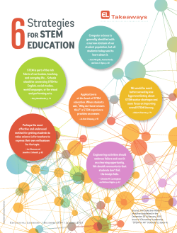


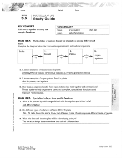

© Copyright 2026