
- European Medical Journal
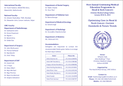
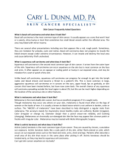
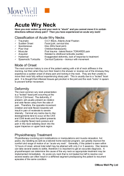
FIELD CANCERISATION OF THE UPPER AERODIGESTIVE
TRACT: SCREENING FOR SECOND PRIMARY CANCERS
OF THE OESOPHAGUS IN CANCER SURVIVORS
*Hans Scherübl,1 Güllü Cataldegirmen,2 Jan Eick,1 Wanda Ring,1
Christoph Schwertner,1 Joachim Steinberg,1 Hermann Herbst3
1. Klinik für Gastroenterologie, GI Onkologie und Infektiologie, Vivantes Klinikum Am Urban,
Berlin, Germany
2. Klinik für Viszeral- und Gefäßchirurgie, Vivantes Auguste-Viktoria-Klinikum, Berlin, Germany
3. Institut für Pathologie, Vivantes – Netzwerk für Gesundheit, Berlin, Germany
*Correspondence to [email protected]
Disclosure: No potential conflict of interest.
Received: 26.09.14 Accepted: 08.01.15
Citation: EMJ Oncol. 2015;3[1]:21-28.
ABSTRACT
Tobacco, alcohol, and betel quid are the main causes of squamous cell cancers of the upper aerodigestive
tract. These substances can cause multifocal carcinogenesis leading to multiple synchronous or
metachronous cancers of the oesophagus, head and neck region, and lungs (‘field cancerisation’).
Globally there are several million people who have survived either head and neck squamous cell cancer
(HNSCC) or lung cancer (LC). HNSCC and LC survivors are at increased risk of developing second primary
malignancies, including second primary cancers of the oesophagus. The risk of second primary oesophageal
squamous cell cancer (OSCC) ranges from 8-30% in HNSCC patients. LC and HNSCC survivors should
be offered endoscopic surveillance of the oesophagus. Lugol chromoendoscopy is the traditional
and best evaluated screening method to detect early squamous cell neoplasias of the oesophagus.
More recently, narrow band imaging combined with magnifying endoscopy has been established as an
alternative screening method in Asia. Low-dose chest computed tomography (CT) is the best evidencebased screening technique to detect (second primary) LC and to reduce LC-related mortality. Low-dose
chest CT screening is therefore recommended in OSCC, HNSCC, and LC survivors. In addition, OSCC
survivors should undergo periodic pharyngolaryngoscopy for early detection of second primary HNSCC.
Secondary prevention aims at quitting smoking, betel quid chewing, and alcohol consumption. As field
cancerisation involves the oesophagus, the bronchi, and the head and neck region, the patients at risk are
best surveilled and managed by an interdisciplinary team.
Keywords: Squamous cell carcinoma, second malignancy, lung, head and neck, endoscopy, surveillance,
tobacco, alcohol, betel, neoplasm, tumour, computed tomography.
EPIDEMIOLOGY AND INCREASED
CANCER SURVIVORSHIP
Oesophageal cancer (OEC) is the eighth-most
common cancer globally with approximately
456,000 new cases per year. Globally the
incidence of oesophageal squamous cell cancer
(OSCC) clearly outweighs that of oesophageal
adenocarcinoma
but
there
are
marked
epidemiological differences between Western
countries and Central Asia and China. Head and
ONCOLOGY • March 2015 neck squamous cell cancer (HNSCC) accounts
for approximately 600,000 new cases annually
worldwide. With almost 1,825,000 new cases
annually, lung cancer (LC) is the most common
cancer in the world.1 The topic of cancer
survivorship is becoming increasingly important in
current cancer management. Both HNSCC and LC
survivors are at risk of developing second primary
cancers, including OSCC.2-5 Long-term survivors
of HNSCC or LC are increasing and may amount
to 3-5 million persons globally. OSCC survivors
EMJ EUROPEAN MEDICAL JOURNAL
21
are increasing too; they are at increased risk of
second primary HNSCC or LC.6-8 This review
addresses the OSCC risk of people who survived
either head and neck cancer (HNC) or LC and gives
recommendations for surveillance.
RISK FACTORS: TOBACCO, ALCOHOL,
AND BETEL QUID
Tobacco and Alcohol
Smoking and alcohol are well-known risk factors
not only of OSCC but also of HNC;4,8-11 tobacco
being the main culprit of LC. Tobacco and
alcohol use can cause ‘field cancerisation’ of
the upper aerodigestive tract (UADT) and the
lungs.12 The development of multiple primary
squamous cell cancers and widespread epithelial
oncogenic alterations, including carcinoma in
situ, dysplasia, and hyperkeratosis, have long
been recognised as the field cancerisation
phenomenon.8,12 Field cancerisation can lead to
multiple
synchronous
and/or
metachronous
cancers of the oesophagus, lungs, and head
and neck region (i.e. oral cavity, oropharynx,
hypopharynx, or larynx). 90% of the tumours in
head and neck are squamous cell carcinomas,
and at least 75% of them are attributable to the
combination of tobacco and alcohol consumption.
The odds ratio of OSCC may be as high as
50.1 for those who are both heavy smokers and
heavy drinkers in comparison to people who
neither drink nor smoke.13 It has been estimated
that a history of smoking, alcohol consumption,
and diets low in fruits and vegetables account
for almost 90% of OSCC cases in the USA. Tobacco
and alcohol synergistically increase OSCC risk.8
Betel Quid
In Central, Southern, and Southeastern Asia
chewing of areca nut or betel quid is prevalent.
Unfortunately, the use of areca nut or betel
quid (areca nuts wrapped in betel leaves) is
associated with an increased risk of oral and
oropharyngeal cancer (ORC) as well as of
OSCC. The combination of betel nut chewing
with tobacco smoking synergistically potentiates
the risks of oral, oropharyngeal, or oesophageal
squamous cell cancers.14-16
Interestingly, the cancer risk from mouth,
pharynx, oesophagus, to larynx increases with
alcohol and cigarette consumption, but decreases
with betel consumption. Tobacco, alcohol, and betel
22
ONCOLOGY • March 2015 quid act synergistically in OSCC tumourigenesis
and are independent risk factors for distinct
cancers of the UADT.9 In Taiwanese men the
lifetime risk of UADT cancer was calculated
to be 9.42% versus 1.65% for betel chewers
versus non-chewers, 3.22% versus 1.21% for
cigarette smokers versus non-smokers, and 4.77%
versus 1.85% for alcohol drinkers versus nondrinkers. The lifetime UADT cancer risk reached
17.2% in men who chewed more than 20 betel
quids a day.9
Mutations of the Enzyme Aldehyde
Dehydrogenase (ALDH)
Alcohol drinking results in exposure to
acetaldehyde, derived from the beverage itself
and formed endogenously. Acetaldehyde is a
genotoxic compound that is detoxified by ALDH.
The presence of the ALDH2-2 allele encodes
ALDH2, an inactive enzyme. Carriers of the
ALDH2-2 allele accumulate acetaldehyde and
have higher relative risks of alcohol-related OEC
and HNCs as compared with individuals with
wild-type alleles. The International Agency for
Research of Cancer stated in 2009 that
acetaldehyde derived from alcoholic beverages
could cause cancer and that alcohol consumption,
i.e. ethanol in alcoholic beverages, was classified
as a group 1 carcinogen.17 A strong linkage of
inactive ALDH2 to increased susceptibility to
multiple cancers was reported in male Japanese
drinkers with OEC or ORC. A similar association
between inactive ALDH2 and the risk of multiple
intraoesophageal and OEC accompanied by
oropharyngolaryngeal or stomach cancers (or all)
was described in Japanese male alcoholics. These
reports indicate that inactive ALDH2 plays an
important role in susceptibility of the UADT to
multiple cancers.18-20
INFECTION WITH HUMAN PAPILLOMA
VIRUS (HPV)
The aetiologic factors of HNSCC in patients who
have never used tobacco or consumed alcohol
are not yet well understood. Multiple lines of
evidence indicate that nowadays HPV infection
contributes to tumourigenesis in up to 70% of ORC
in North America and Europe.21 Approximately
30% of all HNSCC patients are infected with HPV,
mostly with high-risk type HPV-16. Interestingly,
oropharyngeal HNSCC patients with HPV infection
show fewer synchronous second primary tumours
EMJ EUROPEAN MEDICAL JOURNAL
compared with HPV-negative HNSCC.22 The
reason appears to be the absence of carcinogeninduced early genetic changes in the epithelium
and the development of multifocal tumours as
known for heavy smokers and alcohol abusers.
About 25% of OSCC cases are HPV-positive. It is
unclear if having HPV alone is sufficient to cause
OEC or if other factors such as tobacco and
alcohol interact with HPV to trigger carcinogenesis.
At present the role of HPV infection in OSCC
carcinogenesis is not well understood.23,24 A
recent study suggests that HPV-16 infection
may be involved in OSCC tumourigenesis in
Xinjiang Kazakh patients in China.25
RISK OF SECOND PRIMARY OEC
12-19% of LCs are diagnosed at tumour Stage 1.4
When screening for LC is done by using low-dose
computed tomography (CT), the percentage of
LC being detected at early stages rises to 47.5%.26
Thanks to curative treatment options the majority
of Stage 1 (non-small-cell) LC patients become
long-term LC survivors. LC survivors carry a
significantly increased risk of developing second
primary OEC (odds ratio 2.29).4 Endoscopic
surveillance of the oesophagus should be
considered in these patients.4,27
HNSCC patients have quite a good outlook:
5-year disease-specific survival of HNSCC patients
now reaches 66% in the USA5 and steps up to
80% or even 90% in patients with Stage 2 or
Stage 1 HNSCC. Second primary malignancies
(SPM) have been recognised as the leading longterm cause of death in patients surviving
HNC.2,3,5,28 SPM in HNSCC survivors mainly develop
in the lungs and oesophagus but also in the head
and neck region itself.28-30 In Western literature,
the overall incidence of SPM in HNSCC patients
has been reported to range from 9.1-19.0%, with an
annual incidence ranging from 3.2-4.0%.5 Globally
HNSCC patients carry a risk of second primary
oesophageal squamous cell neoplasias (OSCN)
of 8.9-30.4%; the odds ratios or excess absolute
risks may be as high as 240.96 or 72.5.28,31-36
Unfortunately, the OSCC prognosis is generally
dismal, with a 5-year survival rate of approximately
10-16% in Western countries.8 Quitting smoking
reduces the risk of SPM.37
100
Survival (%)
75
Without second primary OSCC
50
Asymptomatic second primary OSCC (detected by screening)
25
Symptomatic advanced second primary OSCC
0
0
1
2
3
4
5
6
7
8
9
Time (years)
Figure 1: Perspective relative survival of HNSCC patients with and without second primary oesophageal
squamous cell cancer (OSCC).
Asymptomatic oesophageal squamous cell neoplasias are detected by screening at an early stage (red
line). Symptomatic second primary OSCC is generally diagnosed at advanced stages.
HNSCC: head and neck squamous cell cancer.
With permission from Scherübl et al.38
ONCOLOGY • March 2015 EMJ EUROPEAN MEDICAL JOURNAL
23
ENDOSCOPIC SCREENING FOR
EARLY OSCN
The aim of surveillance is to detect asymptomatic
OSCC at very early stages, where both endoscopic
and surgical resection generally result in longterm survival. However, when symptomatic OSCC
is diagnosed in HNSCC or LC survivors, advanced
OSCC stages are prevalent and the outlook is very
poor. Overall survival of HNSCC or LC survivors
with second primary cancer, in particular second
primary OSCC, is significantly lower (5-year
survival rate of only 6%) than the overall
survival of those without SPM.2,3,5,38 (Figure 1).
The recommendation that HNSCC and LC
survivors undergo periodic endoscopic surveillance
is based upon the assumption that on the one
hand OSCC adversely affects survival and on
the other, surveillance can reduce mortality
by detecting OSCN at a very early stage.38-41
Several lines of evidence suggest that OSCC
is diagnosed in routinely screened HNSCC
patients more commonly than in those not
screened.29,31-36,39-41 In routinely screened HNSCC
patients, OSCC cases are detected at earlier
cancer stages.35,38 Nowadays, OSCC limited to
the upper layers of the mucosa (T1a: m1, m2) can
be treated effectively by endoscopic resection
and thereby with low morbidity and very low
mortality. OSCC invading the lamina muscularis
mucosae (m3) or the upper layer of the
submucosa (<500 µm: sm1) has a higher risk of
lymph node metastases and in Europe is generally
only chosen for endoscopic resection if no further
risk factors are present, such as poor grade of
differentiation, angioinvasion, or a higher grade
of tumour cell dissociation.27 OSCC invading
the deeper layers of submucosa (sm2, sm3)
should be managed surgically and/or by
chemoradiotherapy. In elderly patients with very
significant comorbidities an endoscopic approach
may be considered even in sm2 or sm3 cancers.
Therefore, the aim of surveillance is to detect
second primary oesophageal neoplasias at (very)
early stages, i.e. intraepithelial neoplasias or
m1/m2 intramucosal cancers.
LUGOL CHROMOENDOSCOPY OF
EARLY OSCC
Chromoendoscopy with Lugol’s solution (1-2%)
used to be the traditional and reference procedure
to screen for early OSCC in high-risk patients.
24
ONCOLOGY • March 2015 Multicentric
squamous
neoplasias
of
the
oesophagus can be visualised by Lugol
chromoendoscopy as Lugol-voiding lesions (LVL),
because dysplastic or hyperkeratotic epithelium
does not stain with Lugol iodine solution and
appears white or pink, whereas normal epithelium
is stained brown. Multiple LVL have been
associated with a very high risk of multiple
cancers arising in the oesophagus, as well as in
the head and neck region.7,38,39 The sensitivity and
specificity of Lugol chromoendoscopy to detect
OSCC in high-risk groups amounts to about
80-96% and 63-72%, respectively.29 The French
Ear, Nose and Throat (ENT) Society suggests
using flexible white-light, high-resolution video
oesophagoscopy combined with targeted biopsies
of any suspected oesophageal lesion. In addition,
it recommends applying Lugol chromoendoscopy
as this technique diagnoses more early-stage
preneoplastic and neoplastic lesions with better
definition of local extension of more advanced
OECs.42 (Figure 2).
NARROW-BAND IMAGING (NBI) AND
MAGNIFYING ENDOSCOPY (ME)
NBI is a novel optical technique that enhances
the diagnostic capability of gastrointestinal
endoscopy by highlighting the intraepithelial
papillary capillary loops of the squamous mucosa
by means of light passed through filters that
narrow the spectral bandwidths, incorporated into
a red-green-blue sequential illumination system.
NBI combined with ME has been demonstrated
to further improve the detection rate and
accuracy of early OSCC in HNSCC patients.33 In
a recent study NBI endoscopy with ME was
reported to be the ideal screening tool to
search
for
early
oesophageal
squamous
neoplasias; the respective sensitivity, specificity,
and accuracy amounted to 97.3%, 94.1%, and
96.3%.29 These observations go in line with an
Asian-Pacific consensus conference on earlystage oesophagogastric cancer in 2011; that
consensus conference stated that NBI could
replace chromoendoscopy in routine examination
because it is easy to use and adds much
information to conventional white light imaging,
but it cannot eliminate chromoendoscopy when
we make a final diagnosis for treatment decision
making (Figure 3).43 Both due to unpleasant
side-effects and low specificity of Lugol
chromoendoscopy, high-resolution flexible video
oesophagoscopy with NBI may well become the
EMJ EUROPEAN MEDICAL JOURNAL
preferred routine screening technique for second
primary OSCN in the near future. In most countries
of Western Europe NBI endoscopy is generally
available and widely used.
SCREENING RECOMMENDATIONS OF
NATIONAL HEALTHCARE SOCIETIES
Risks of second primary malignancies differ among
LC or HNSCC survivors of different countries
and regions. Therefore, there are no generally
and worldwide accepted recommendations of
screening for second primary OSCC. The recent
guidelines of the French ENT Society recommend
upper-gastrointestinal endoscopy in the initial
workup of hypopharyngeal squamous cell cancer
and in all chronic alcoholics with HNSCC,42
corresponding to the great majority of HNSCC
patients in France. Similarly, healthcare specialists
in Taiwan pointed out that the odds ratios for
second primary OSCC were 18.41, 40.49, and
240.96 in patients suffering from malignancy of
the oral cavity, oropharynx, and hypopharynx,
respectively.34 They recommend periodic OSCC
screening according to the individual risk
stratification.35
Still,
most
national
ENT,
gastroenterology, and cancer societies have yet to
make up their minds and have to balance possible
survival benefits resulting from screening against
economic restraints. Efforts to reduce heavy
alcohol and tobacco consumption as well as betel
quid chewing are generally recommended and
often supported by national campaigns.
OSCC SURVIVORS: SURVEILLANCE FOR
SECOND PRIMARY CANCERS OF THE
HEAD AND NECK, AND THE LUNGS
Risk of developing a second malignancy should
be anticipated after curative treatment of OSCC.
Common risk factors including lifestyle and
genetic alterations may explain both the pattern
and the increased incidence of second primary
cancers in OSCC survivors. Because of the
high mortality of OEC itself, not much attention
was previously paid to the development of
SPM. Due to promising results of a recent
prospective study of the National Lung Screening
Trial research team, today LC screening has
become the focus of increasing interest in highrisk groups.26
Figure 2: Oesophageal squamous cell cancer (OSCC) in a HNSCC patient.
Left panel: Videoendoscopic image of an OSCC (Stage T1aN0M0) at 25 cm from the incisors. 29 months
ago the patient had been treated for a squamous cell cancer of the oral cavity. Right panel: The same
tumour after staining with Lugol dye solution to delineate the tumour margins.
HNSCC: head and neck squamous cell cancer.
With permission from Scherübl et al.40
ONCOLOGY • March 2015 EMJ EUROPEAN MEDICAL JOURNAL
25
A
B
C
D
E
F
Figure 3: Endoscopic surveillance and management of synchronous high-grade intraepithelial neoplasia
of oesophagus in a laryngeal cancer patient.
A: A flat superficial neoplasia with hyperaemia in white-light imaging system. B: A superficial neoplasia
with brownish discolouration under narrow-band imaging system. C: Lugol-voiding of the neoplasia after
spraying a 1.5% Lugol’s solution. D: Abnormal intraepithelial capillary loops under narrow-band imaging
system with magnifying endoscopy. E: Endoscopic submucosal dissection of the superficial neoplasia. F:
Mucosal cancer invading the lamina propria (main picture: H&E stain, 40x; right bottom: H&E stain, 100x).
H&E: haematoxylin and eosin.
With permission from Chung et al.29
26
ONCOLOGY • March 2015 EMJ EUROPEAN MEDICAL JOURNAL
Superficial HNCs
Lung Cancer
OSCC patients have a risk of 8.3-27.1% of
developing SPM.8 Due to common risk factors
such as tobacco and alcohol, OSCC shows a
particularly high association with LC and HNC.
Matsubara et al.44 reported that OSCC patients
are at very high risk for the development of both
HNC and LC after oesophagectomy and that the
early detection of second cancers allowed less
invasive treatment with favourable outcomes.
LC is the largest single cause of death from
cancer in the world. As the number of long-term
OEC survivors continues to increase worldwide,
the incidence of second primary cancers including
LC will increase. Detecting and treating SPM
appear to be effective in OSCC patients. Thus,
recent evidence suggests similar overall survival
rates in OEC patients with or without SPM.8
Both early asymptomatic LC and superficial
HNSCC are amenable to curative treatment.4,7
Detection of early LC is best achieved by lowdose chest CT. Periodic, low-dose CT screening
leads to a shift to detection of earlier-stage nonsmall-cell LC and thereby reduces LC mortality.26
Nowadays, both HNSCC and OSCC survivors
should be considered for regular screening
for early LC by low-dose chest CT.
Patients with OSCC, particularly alcohol drinkers,
current smokers, and those with the ALDH-2 allele
and multiple LVL of the oesophageal mucosa,
have an increased risk of superficial squamous cell
cancer within the head and neck region. As Lugol
chromoendoscopy is not applicable to the head
and neck region, NBI in combination with ME is
the preferred technique to search for early (i.e.
superficial) HNSCC in OSCC patients.7,8 The ability
to detect a second primary cancer at a (very)
early stage is of benefit for patients at high
risk of superficial HNSCC. However, controlled
prospective studies that provide evidence for
a survival benefit of endoscopic surveillance in
OSCC survivors have yet to be performed.6
CONCLUSION
As field cancerisation involves the oesophagus,
the bronchi, and the head and neck region, the
patients at risk are best surveilled and managed
by an interdisciplinary team.
REFERENCES
1. Stewart BW, Wild CP (eds.), World
Cancer Report (2014), World Health
Organization.
2. Chen MC et al. Impact of second primary
esophageal or lung cancer on survival of
patients with head and neck cancer. Oral
Oncol. 2010;46(4):249-54.
3. Liao LJ et al. The impact of second
primary malignancies on head and neck
cancer survivors: a nationwide cohort
study. PLoS One. 2013;8(4):e62116.
4. Surapaneni R et al. Stage I lung cancer
survivorship: risk of second malignancies
and need for individualized care plan. J
Thorac Oncol. 2012;7(8):1252-6.
5. Baxi SS et al. Causes of death in longterm survivors of head and neck cancer.
Cancer. 2014;120(10):1507-13.
6. Zhu G et al. Risk of second primary
cancer after treatment for esophageal
cancer: a pooled analysis of nine cancer
registries. Dis Esophagus. 2012;25(6):
505-11.
7. Katada C et al. Risk of superficial
squamous cell carcinoma developing in
the head and neck region in patients with
esophageal squamous cell carcinoma.
Laryngoscope. 2012;122(6):1291-6.
8. Nandy N, Dasanu CA. Incidence
ONCOLOGY • March 2015 of second primary malignancies in
patients with esophageal cancer: a
comprehensive review. Curr Med Res
Opin. 2013;29(9):1055-65.
2010;15(2):126-34.
9. Hsu WL et al. Lifetime risk of distinct
upper aerodigestive tract cancers and
consumption of alcohol, betel and
cigarette. Int J Cancer. 2014;135(6):
1480-6.
15. Akhtar S. Areca nut chewing and
esophageal squamous-cell carcinoma
risk in Asians: a meta-analysis of casecontrol studies. Cancer Causes Control.
2013;24(2):257-65.
10. Li Y et al. Alcohol drinking and upper
aerodigestive tract cancer mortality: a
systematic review and meta-analysis. Oral
Oncol. 2014;50(4):269-75.
16. Huang SF et al. Association of HPV
infections with second primary tumors in
early-staged oral cavity cancer. Oral Dis.
2012;18(8):809-15.
11. Druesne-Pecollo N et al. Alcohol
drinking and second primary cancer
risk in patients with upper aerodigestive
tract cancers: a systematic review and
meta-analysis of observational studies.
Cancer Epidemiol Biomarkers Prev.
2014;23(2):324-31.
17. Testino G, Borro P. Alcohol and
gastrointestinal oncology. World J
Gastrointest Oncol. 2010;2(8):322-5.
12. Slaughter DP et al. Field cancerization
in oral stratified squamous epithelium;
clinical implications of multicentric origin.
Cancer. 1953;6(5):963-8.
13. Morita M et al. Alcohol drinking,
cigarette smoking, and the development
of squamous cell carcinoma of the
esophagus:
epidemiology,
clinical
findings, and prevention. Int J Clin Oncol.
14. Sharan RN et al. Association of betel nut
with carcinogenesis: revisit with a clinical
perspective. PLoS One. 2012;7(8):e42759.
18. Lee CH et al. Carcinogenetic impact of
ADH1B and ALDH2 genes on squamous
cell carcinoma risk of the esophagus with
regard to the consumption of alcohol,
tobacco and betel quid. Int J Cancer.
2008;122(6):1347-56.
19. Muto M et al. Risk of multiple
squamous cell carcinomas both in the
esophagus and the head and neck region.
Carcinogenesis. 2005;26(5):1008-12.
20. Duan F et al. [Genetic polymorphisms
of ADH1B and ALDH-2 associated with risk
of esophageal cancer: a meta-analysis].
EMJ EUROPEAN MEDICAL JOURNAL
27
Wei Sheng Yan Jiu. 2012;41(5):723-9.
2013;149(4):579-86.
21. Curado MP, Boyle P. Epidemiology of
head and neck squamous cell carcinoma
not related to tobacco or alcohol. Curr
Opin Oncol. 2013;25(3):229-34.
29. Chung CS et al. Risk factors for second
primary neoplasia of esophagus in newly
diagnosed head and neck cancer patients:
a case-control study. BMC Gastroenterol.
2013;13:154.
22. Jain KS et al. Synchronous cancers
in patients with head and neck cancer:
risks in the era of human papillomavirusassociated oropharyngeal cancer. Cancer.
2013;119(10):1832-7.
23. de Villiers EM et al. Esophageal
squamous cell cancer in patients with
head and neck cancer: prevalence of
human papillomavirus DNA sequences.
Int J Cancer. 2004;109(2):253-8.
24. Hardefeldt HA et al. Association
between human papillomavirus (HPV)
and
oesophageal
squamous
cell
carcinoma: a meta-analysis. Epidemiol
Infect. 2014;142(6):1119-37.
25. Chen WG et al. Gene chip technology
used in the detection of HPV infection in
esophageal cancer of Kazakh Chinese in
Xinjiang Province. J Huazhong Univ Sci
Technolog Med Sci. 2014;34(3):343-7.
26. Aberle DR et al; National Lung
Screening Trial Research Team. Results
of the two incidence screenings in the
National Lung Screening Trial. N Engl J
Med. 2013;369(10):920-31.
27. Pech O et al. Endoscopic resection
of superficial esophageal squamous-cell
carcinomas: Western experience. Am J
Gastroenterol. 2004;99:1226-32.
28. Lee DH et al. Second cancer
incidence, risk factor, and specific
mortality in head and neck squamous cell
carcinoma. Otolaryngol Head Neck Surg.
28
ONCOLOGY • March 2015 30. Morris LG et al. Anatomic sites at
elevated risk of second primary cancer
after an index head and neck cancer.
Cancer Causes Control. 2011;22(5):671-9.
31. Scherübl H, Zeitz M. Esophageal
cancer. N Engl J Med. 2004;350(13):
1363-4.
32. Priante AV et al. Second primary
tumors in patients with head and neck
cancer. Curr Oncol Rep. 2011;13(2):132-7.
33. Lee CT et al. Narrow-band imaging
with magnifying endoscopy for the
screening of esophageal cancer in
patients with primary head and neck
cancers. Endoscopy. 2010;42(8):613-9.
34. Hung SH et al. Routine endoscopy
for esophageal cancer is suggestive
for patients with oral, oropharyngeal
and hypopharyngeal cancer. PLoS One.
2013;8(8):e72097.
35. Su YY et al. Effect of routine
esophageal screening in patients with
head and neck cancer. JAMA Otolaryngol
Head Neck Surg. 2013;139(4):350-4.
38. Scherübl H et al. Coincidental
squamous cell cancers of the esophagus,
head, and neck: risk and screening. HNO.
2008;56(6):603-8.
39. Horiuchi M et al. Survival benefit of
screening for early esophageal carcinoma
in head and neck cancer patients.
Digestive Endoscopy. 1998;10:110–5.
40. Scherübl H et al. Screening for
oesophageal neoplasia in patients with
head and neck cancer. Br J Cancer.
2002;86(2):239-43.
41. Wang WL et al. The benefit of
pretreatment
esophageal
screening
with image-enhanced endoscopy on the
survival of patients with hypopharyngeal
cancer. Oral Oncol. 2013;49(8):808-13.
42. de Monès E et al; Socéité Française
de l’Otorhinolaryngologie. Initial staging
of squamous cell carcinoma of the oral
cavity, larynx and pharynx (excluding
nasopharynx). Part 2: Remote extension
assessment and exploration for secondary
synchronous locations outside of the
upper aerodigestive tract. 2012 SFORL
guidelines. Eur Ann Otorhinolaryngol
Head Neck Dis. 2013;130(2):107-12.
36. Scherübl H et al. Head and neck
cancer. N Engl J Med. 2002;346(18):
1416-7.
43. Uedo N et al. Role of narrow band
imaging for diagnosis of early-stage
esophagogastric
cancer:
current
consensus of experienced endoscopists
in Asia-Pacific region. Dig Endosc. 2011;23
Suppl 1:58-71.
37. Tabuchi T et al. Tobacco smoking
and the risk of subsequent primary
cancer among cancer survivors: a
retrospective cohort study. Ann Oncol.
2013;24(10):2699-704.
44. Matsubara T et al. Risk of second
primary malignancy after esophagectomy
for squamous cell carcinoma of the
thoracic esophagus. J Clin Oncol.
2003;21(23):4336-41.
EMJ EUROPEAN MEDICAL JOURNAL


© Copyright 2026