
Biomineralization of Se Nanoshpere by Bacillus Licheniformis
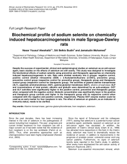
Journal of Earth Science, Vol. 26, No. 2, p. 246–250, April 2015
Printed in China
DOI: 10.1007/s12583-015-0536-9
ISSN 1674-487X
Biomineralization of Se Nanoshpere by Bacillus Licheniformis
Yongqiang Yuan*1, 2, Jianming Zhu3, 1, Congqiang Liu1, Shen Yu2, Lei Lei1
1. State Key Laboratory of Environmental Geochemistry, Institute of Geochemistry, Chinese Academy of Sciences,
Guiyang 550002, China
2. Research Center for Urban Ecological Health and Environmental Safety, Institute of Urban Environment,
Chinese Academy of Sciences, Xiamen 361021, China
3. Insititute of Earth Sciences, China University of Geosciences, Beijing 100083, China
ABSTRACT: Biological dissimilatory reduction of selenite (SeO32-) to elemental selenium (Se0) is common, but the mineral formation and the biogenic process remain uncertain. In this study, we examined
the Se0 formation during the selenite bioreduction by Bacillus licheniformis SeRB-1 through transmission electron microscope (TEM), energy-dispersive spectrometry (EDS) and X-ray absorption fine
structure (XAFS) techniques. Results showed that the reduction process occurred mostly during the
exponential phase and early stationary phase, whilst the elemental selenium was produced in these periods. From the TEM images and polyacrylamide gel electropheresis, it is known that the Se0 granule
formation is a biologically-induced type, and the cell envelopes are the main biomineralization positions,
and particles may go through a process from nucleation to crystallization, under the control of microbes. In fact, the minerals are spherical nanoparticles, occurring as a microcrystal or amorphous
form. It is vital to recognize which kinds of proteins and/or polysaccharides act as a template to direct
nanoparticle nucleation and growth? This should focus for further studies. This study may shed light on
the process of formation of Se(0) nanosphere.
KEY WORDS: biomineralization, genesis, selenite reduction, selenium nanosphere.
0
INTRODUCTION
Biomineralization is a process that forms the minerals under the control of organisms and deposits in the matrix (either
cellular or extracellular) of living organisms (Mann, 2001;
Boskey, 1998). It combines the disciplines of biological,
chemical, and earth sciences together and may shed lights on
new materials biosynthesis (Mann, 1993). Although it has long
been known that microorganisms play a dynamic role in the
biogeochemical cycling of most elements (Ehrlich and
Newman, 2008), only the discipline of ‘geomicrobiology’ developed in the last two decades of 20th century (Chen and Yao,
2005; Knoll, 2003), the full extent of the microbial processes
that can control mineral formation have gained increased interest (Lloyd et al., 2008). In recent decades, metallic minerals
biogenetic is one of the hot spot (Lu, 2007).
Selenium is a redox sensitive element that occurs in four
different oxidation states, -2, 0, +4, and +6, in the environment,
and selenium oxyanions are easy to be reduced to elemental
selenium Se(0). Se(0) is the dominant species of Se in anoxic
sediments (Stolz and Oremland, 1999) whereas the transformation of Se in nature occurs primarily by biotic processes
(Dowdle and Oremland, 1998). Such process is one of the
*Corresponding author: [email protected]
© China University of Geosciences and Springer-Verlag Berlin
Heidelberg 2015
Manuscript received March 18, 2014.
Manuscript accepted February 15, 2015.
major sinks for selenium oxyanions, and studies on selenium
biomineralization have carried out by many researchers (Butler
et al., 2012; Lenz et al., 2011; Kaur et al., 2009). But the mineral formation, dissolution or diagenesis processes remain uncertain. The focus of this study is on the microstructure and
process of formation of biogenic elemental selenium, in order
to elucidate how bacteria generate these novel selenium nanostructures.
1 EXPERIMENTAL SECTION
1.1 Bacterial Isolate and Culture Conditions
The strain SeRB-1 was previously isolated from high-Se
carbonaceous mudstones from Yutangba, Enshi, China, after
enrichment with Na2SeO3 as the electron acceptor and dextrose
as the electron donor. The isolate is a kind of facultative anaerobe and was identified as Baculis licheniformis (Genebank
accession No. JX512417) (Yuan et al., 2014).
SeRB-1 was routinely cultured in medium YEG (yeast extract and glucose) containing 10 g yeast extract, and 10 g of
dextrose per liter of deionized water. The final pH was adjusted
to 7.0 using a diluted HCl or NaOH solution. Facultative anaerobic cultures were incubated at 37 °C on a rotary shaker at
200 rpm.
1.2
Reduction of Selenium Oxyanions by SeRB-1
To determine the ability of SeRB-1 to reduce SeO32- during this growth, a growth experiment was conducted in YEG. In
a typical procedure, 30 mL aliquots of YEG medium with 865
mg/kg sodium selenite (passing through a 0.22 μm filter) were
Yuan, Y. Q., Zhu, J. M., Liu, C. Q., et al., 2015. Biomineralization of Se Nanoshpere by Bacillus Licheniformis. Journal of Earth
Science, 26(2): 246–250. doi:10.1007/s12583-015-0536-9
Biomineralization of Se Nanoshpere by Bacillus Licheniformis
247
added into 50 mL serum bottles, and about 10% of suspended
SeRB-1 culture was inoculated. The bacterium was first pregrown to the exponential growth phase (~24 h). Cell-free controls where selenite was added to medium were also set up to
detect any abiotic transformations. The experiment was done in
triplicate.
1.3
Analytical Methods
Two mL samples were collected after each interval period,
one aliquot was used to measure cell density by viable counting
under fluorescence microscope (Olympus, Japan), and another
mL was centrifuged for 10 min at 10 000 rpm (TCL-18C-C),
the supernatant was used to determine the remaining Se(IV)
concentration in the medium by hydride-generation atomic
fluorescence spectrometry (HG-AFS), while the precipitates/
pellets (including both cells and biominerals) were used for
microscopy and fine structure analysis.
The procedures for the preparation of transmission electron micrograph of cultures have been described elsewhere
(Yuan et al., 2014). The stained ultrathin section (80–100 nm)
was used for ultrastructure analysis by TEM and the unstained
one for mineral composition examination by EDS techniques
(JEM-2000FX II, Japan). Selected-area electron diffraction was
also carried out for crystal type analysis.
Another aliquot of precipitate was added with lysozyme to
break the cell structure, and then it was centrifuged to obtain
biominerals, and the biominerals were dehydrated and freezingdried, and finally were measured at 1W1B-XAFS station in
Beijing Synchrotron Radiation Facility (BSRF).
2 RESULTS
2.1 Selenite Reduction by Strain
Red-colored colonies were present in the cultures grown
in the presence of 5 mM sodium selenite in about 6 hours. This
indicates that Se(IV) was reduced to elemental selenium, while
there was no red color emerging for the cell-free controls. The
effect of time on microbial growth and reduction of selenite by
SeRB-1 was shown in the Fig. 1. The results demonstrated that
the process occurred mainly during the exponential phase. The
concentration of Se(IV) decreased with microbial growth (Fig.
1), while there was little change in Se(IV) concentration in
medium of the controls, or keeping stable at the initial concentration of 390 mg/L. Approximately 50% of selenite was reduced during the experimental period.
2.2
Elemental Analysis of Selenium Precipitates
Electron micrographs of cells grown with selenite were
shown in the Fig. 2. Abundant mineral granules were produced
on the exterior of the cell envelope during cells of SeRB-1 grown
in medium containing selenite for a period of time (~24 h) (Fig.
2a). In addition, TEM images of thin sections also revealed the
common presence of intercellular mineral granules (Fig. 2b).
These internal biomineral accumulations was formed adjacent to
the cell’s periphery and deposited to the contour of the cell envelope. The biominerals appeared as a spherical and were approximately 10–200 nm in diameter. The nano-spheres were not present in the TEM images of the cells at the initial time after strains
Figure 1. Effect of time on microbial growth and reduction
of selenite by Bacillus licheniformis.
inoculated to the Se(IV) medium (<6 h) (Fig. 2c). It showed that
diffraction rings and weak diffraction spots were present from
selected-area electron diffraction (SAD) analysis of the nanoparticles (Fig. 2d, inset).
When the biominerals were analyzed by EDS, the particles
produced specific selenium absorption peak at 1.37 keV (peak
SeLα), 11.22 keV (peak SeKα), and 12.49 keV (peak SeKβ)
(Fig. 2d). This indicates that it is mostly composed of selenium
element, which is consistent with the previous study (Kessi et
al., 1999). The Cu peaks were produced by or resulted from the
TEM grid, the Ca and S peaks might be the component of the
medium and/or the microbes, and the C and O most likely associated with cellular exudates.
2.3
XAFS Analysis
XAFS has very high sensitivity on the absorption of element types and the geometrical structure of the atom, which
can reflect the valence state of elements and their compounds
(Rehr and Ankudinov, 2001). Figure 2 showed the selenium
K-edge XAFS spectra for the biominerals produced by strain
SeRB-1 and three reference chemicals of standard elemental Se,
sodium selenite and sodium selenate. The spectra exhibited
subtle but significant differences between the selenium compounds, and the position of Se K-edge can be used to determine
the oxidation state of Se. The XAFS spectra measured for the
pellets showed the same peak position of 12.658 keV as Se0,
indicating the Se particles might primarily be composed of
elemental selenium.
3
DISCUSSION
Selenite reduction accompanies microbial growth which is
well-known phenomenon. The strain used selenium oxyanions
as the sole electron acceptor, and dextrose as the electron donor.
Finally, selenium oxyanion was reduced to elemental Se, but
the mineral formation and the biogenic process remain uncertain.
The formation of selenium is a kind of redox reaction,
which can be described by a simplified equation (1), according
to Debieux et al. (2011).
SeO32-+4e-+6H+
Se0+3H2O
(1)
It seems to be a simple electron transfer process for equation
248
Yongqiang Yuan, Jianming Zhu, Congqiang Liu, Shen Yu and Lei Lei
Figure 2. Electron micrographs and EDS analysis of the pellets. (a) TEM image of B. licheniformis cells grown facultaive
anaerobically using selenite as the sole electron acceptor after 24 h Se(IV) reduction; (b) thin sections of B. licheniformis cells
grown facultaive anaerobically using selenite as the sole electron acceptor after 24 h Se(IV) reduction; (c) TEM image of
B. licheniformis cells grown facultaive anaerobically using selenite as the sole electron acceptor at the initial time strain
inoculated; (d) EDS and SAD (inset) analysis of electron-dense particles formed by B. licheniformis grown in facultaive
anaerobical cultures using selenite as the sole electron acceptor after 24 h Se(IV) reduction; (e) entire protein group
SDS-PAGE.
(1), but the mechanism of mineral’s biogenesis is a complex
process. It depends on both the microbe species involved in the
reaction and its conditions including electronic donor and extracellular redox state during biomineralization (Butler et al., 2012;
Pearce et al., 2009). In this study, dextrose was used as the
carbon and energy source, and SeO32- was supplied as electronic acceptor, and was reduced to Se(0) through the metabolism of microorganism. Generally, the processes of biominerals
formation can be classified as either biologically-controlled
mineralization (BCM), or biologically-induced mineralization
(BIM). The differences between them are based on whether the
biomineral is used by an organism for a biological function (as
in BCM), or is the byproduct of an organism’s metabolism (as
in BIM) (Weiner and Dove, 2003; Mann, 2001; Lowenstam,
1981). From the comparison TEM images between Fig. 2a and
Fig. 2c, it implies that the bacteria are induced to produce a
mechanism to cope with Se(IV) stress. Polyacrylamide gel
electropheresis (PAGE) also confirmed this deduction process
Biomineralization of Se Nanoshpere by Bacillus Licheniformis
249
oxyanions reacts with glutathione reductase and generates selenodiglutathione (GS-Se-SG), and finally dismutates into elemental selenium (Kessi and Hanselmann, 2004).
The Se nanoparticles may experience nucleation, growth
and cell processing to the phase change process, and finally the
nano-selenium mineral generation. But the Se bionanomineral
phases are composite materials, because some biomolecules
such as proteins and/or polysaccharides may act as a template
to direct nanoparticle nucleation and growth.
Figure 3. Selenium K-edge X-ray absorption spectra of Se
particles precipitated by B. licheniformis.
(Lei, 2010), strain can be induced to produce more selenite
reductase in the presence of selenite. From PAGE, it appeared
several specific bands for bacterial protein compared with before and after addition of Se(IV) (24 h). Therefore, this result
indicate that the process of Se(0) produced by strain SeRB-1 is
a BIM type.
A distinct characteristic of BIM is the formation of extracellular minerals along with occurrence of intracellular
biominerals (Fig. 1b) (Frankel and Bazylinski, 2003; Weiner
and Dove, 2003). It could be a result of metabolic processes of
the organism and subsequent chemical reactions involving
metabolic byproducts. In the medium, the organisms may secrete a metabolic product(s) that reacts with selenite in the surrounding environment. Usually, the structure/composition of
the cell wall and cytomembrane/capsula, such as polypeptide,
prion, lipid, and complexes, makes biomineralization to be
easier. When cell envelopes are involved in the reductive process of selenium oxyanions, and then it is resulting from the
production of mineral particles extracellularly.
Another distinct characteristics of BIM is that the biogenic
elemental selenium generally does not form large crystals, or
lacking specific crystalline morphologies (seen the SAD image
inset in Fig. 2d), but rather spherical nano-particles, having a
broad particle size distribution (Lenz and Lens, 2009;
Oremland et al., 2004). It is likely to be that the mineralization
is an unintended consequence of metabolic activities. A significant discovery is that the longer the biomineralized time,
the larger the nano particles. The granules may go through a
process from small to big, which is under the control of microbes.
Despite, it is common phenomena for elemental selenium
formation during bioreduction, it has remained poorly studied
the biological process of selenium nanosphere generation. Recent articles have suggested that proteins/enzymes may play an
important role in selenium nanosphere assembly (Debieux et al.,
2011; Dobias et al., 2011). For example, four proteins: AdhP,
Idh, OmpC and AceA have identified in strain Escherichia coli,
which are strongly associated with the selenium nanoparticles
formed (Dobias et al., 2011). Whether strain Bacillus licheniformis has similar to proteins or structures, need further study.
In fact, when the cell wall reductive enzymes or soluble secreted enzymes are involved in the reductive process of selenium oxyanions, then it is obvious to find the Se nanoparticles
extracellularly. While, for intracellular Se nanoparticles formation, a possible explanation explanation is that selenium
4
CONCLUSIONS
The results can be summarized below.
Elemental selenium was produced during bioreduction by
Bacillus licheniformis SeRB-1. These Se biominerals were in
nano-scale level and mainly produced and deposited extracellularly.
From the TEM images and PAGE, it infer that the
nano-Se(0) biomineralization is a BIM type.
The biogenic elemental selenium generally does not form
large crystals but rather spherical nanoparticles. These biominerals are likely linked to metabolic product(s) secreted by microorganisms, which need further study.
This study represents a further step toward understanding
the mechanisms underlying the formation of nano-Se.
ACKNOWLEDGMENTS
This study was financially supported by the National
Natural Science Foundation of China (No. 41273029), and
National Basic Research Program of China (No.
2014CB238903).
REFERENCES CITED
Boskey, A. L., 1998. Biomineralization: Conflicts, Challenges,
and Opportunities. Journal of Cellular Biochemistry,
30/31(Suppl.): 83–91
Butler, C. S., Debieux, C. M., Dridge, E. J., et al., 2012.
Biomineralization of Selenium by the Selenate-Respiring
Bacterium Thauera Selenatis. Biochemical Society
Transactions, 40: 1239–1243
Chen, J., Yao, S., 2005. Geomicrobiology and Its Progress.
Geological Journal of China Universities, 11: 154–166 (in
Chinese with English Abstract)
Debieux, C. M., Dridge, E. J., Mueller, C. M., et al., 2011. A
Bacterial Process for Selenium Nanosphere Assembly.
Proceedings of the National Academy of Sciences of the
United States of America, 108: 13480–13485
Dobias, J., Suvorova, E. I., Bernier-Latmani, R., 2011. Role of
Proteins in Controlling Selenium Nanoparticle Size.
Nanotechnology, 22(19): 195605
Dowdle, P. R., Oremland, R. S., 1998. Microbial Oxidation of
Elemental Selenium in Soil Slurries and Bacterial Cultures.
Environmental Science & Technology, 32: 3749–3755
Ehrlich, H. L., Newman, D. K., 2008. Geomicrobiology. CRC
Press: Boca Raton, FL. 656
Frankel, R. B., Bazylinski, D. A., 2003. Biologically Induced
Mineralization by Bacteria. Biomineralization, 54: 95–114
Kaur, G., M., Iqbal, M., Bakshi, M. S., 2009. Biomineralization
of Fine Selenium Crystalline Rods and Amorphous
250
Yongqiang Yuan, Jianming Zhu, Congqiang Liu, Shen Yu and Lei Lei
Spheres. Journal of Physical Chemistry C, 113:
13670–13676
Kessi, J., Hanselmann, K. W., 2004. Similarities between the
Abiotic Reduction of Selenite with Glutathione and the
Dissimilatory Reaction Mediated by Rhodospirillum
Rubrum and Escherichia Coli. Journal of Biological
Chemistry, 279: 50662–50669
Kessi, J., Ramuz, M., Wehrli, E., et al., 1999. Reduction of
Selenite and Detoxification of Elemental Selenium by the
Phototrophic Bacterium Rhodospirillum Rubrum. Applied
and Environmental Microbiology, 65: 4734–4740
Knoll, A. H., 2003. The Geological Consequences of Evolution.
Geobiology, 1: 3–14
Lenz, M., Kolvenbach, B., Gygax, B., et al., 2011. Shedding
Light on Selenium Biomineralization: Proteins Associated
with Bionanominerals. Applied and Environmental
Microbiology, 77: 4676–4680
Lenz, M., Lens, P. N. L., 2009. The Essential Toxin: The
Changing Perception of Selenium in Environmental
Sciences. Science of the Total Environment, 407:
3620–3633
Lei, L., 2010. Microbial Geochemistry of Selenium in Se-Rich
Carbonaceous Mudstone from Yutangba, Enshi of Hubei
Province in China: [Dissertation]. Graduate University
Chinese Academy of Sciences, Beijing (in Chinese with
English Abstract)
Lloyd, J. R., Pearce, C. I., Coker, V. S., et al., 2008.
Biomineralization: Linking the Fossil Record to the
Production of High Value Functional Materials.
Geobiology, 6: 285–297
Lowenstam, H. A., 1981. Minerals Formed by Organisms.
Science, 211: 1126–1131
Lu, A. H., 2007. Mechanisms of Environmental Response to
Biomineralization. Geological Journal of China
Universities, 13: 613–620 (in Chinese with English
Abstract)
Mann, S., 1993. Biomineralization—The Hard Part of
Bioinorganic Chemistry. Journal of the Chemical
Society-Dalton
Transactions,
1–9.
doi:10.1039/DT9930000001
Mann, S., 2001. Biomineralization: Principles and Concepts in
Bioinorganic Materials Chemistry. Oxford University
Press, New York.
Oremland, R. S., Herbel, M. J., Blum, J. S., et al., 2004.
Structural and Spectral Features of Selenium Nanospheres
Produced by Se-Respiring Bacteria. Applied and
Environmental Microbiology, 70: 52–60
Pearce, C. I., Pattrick, R. A. D., Law, N., et al., 2009.
Investigating Different Mechanisms for Biogenic Selenite
Transformations: Geobacter Sulfurreducens, Shewanella
Oneidensis and Veillonella Atypica. Environmental
Technology, 30: 1313–1326
Rehr, J. J., Ankudinov, A. L., 2001. Progress and Challenges in
the Theory and Interpretation of X-Ray Spectra. Journal
of Synchrotron Radiation, 8: 61–65
Stolz, J. F., Oremland, R. S., 1999. Bacterial Respiration of
Arsenic and Selenium. Fems Microbiology Reviews, 23:
615–627
Weiner, S., Dove, P. M., 2003. An Overview of
Biomineralization Processes and the Problem of the Vital
Effect. Biomineralization, 54: 1–29
Yuan, Y. Q., Zhu, J. M., Liu, C., et al., 2014. Three
High-Reducing
Selenite-Tolerance
Bacteria
from
Se-Laden Carbonaceous Mudstone. Earth Science
Frontiers, 21(1): http://www.cnki.net/kcms/detail/11.3370.
P.20140108.20141634.20140002.html (in Chinese with
English Abstract)


© Copyright 2026