
Viral infection induced respiratory distress in childhood
D 1291
OULU 2015
UNIVERSITY OF OUL U P.O. Box 8000 FI-90014 UNIVERSITY OF OULU FINLA ND
U N I V E R S I TAT I S
O U L U E N S I S
ACTA
A C TA
D 1291
ACTA
U N I V E R S I T AT I S O U L U E N S I S
Hannele Pruikkonen
University Lecturer Santeri Palviainen
Postdoctoral research fellow Sanna Taskila
Hannele Pruikkonen
Professor Esa Hohtola
VIRAL INFECTION INDUCED
RESPIRATORY DISTRESS IN
CHILDHOOD
Professor Olli Vuolteenaho
University Lecturer Veli-Matti Ulvinen
Director Sinikka Eskelinen
Professor Jari Juga
University Lecturer Anu Soikkeli
Professor Olli Vuolteenaho
Publications Editor Kirsti Nurkkala
ISBN 978-952-62-0790-2 (Paperback)
ISBN 978-952-62-0791-9 (PDF)
ISSN 0355-3221 (Print)
ISSN 1796-2234 (Online)
UNIVERSITY OF OULU GRADUATE SCHOOL;
UNIVERSITY OF OULU
FACULTY OF MEDICINE;
MEDICAL RESEARCH CENTER OULU;
OULU UNIVERSITY HOSPITAL
D
MEDICA
ACTA UNIVERSITATIS OULUENSIS
D Medica 1291
HANNELE PRUIKKONEN
VIRAL INFECTION INDUCED
RESPIRATORY DISTRESS IN
CHILDHOOD
Academic dissertation to be presented with the assent
of the Doctoral Training Committee of Health and
Biosciences of the University of Oulu for public defence
in Auditorium 12 of the Department of Paediatrics, on
8 May 2015, at 12 noon
U N I VE R S I T Y O F O U L U , O U L U 2 0 1 5
Copyright © 2015
Acta Univ. Oul. D 1291, 2015
Supervised by
Professor Matti Uhari
Docent Marjo Renko
Reviewed by
Professor Ville Peltola
Docent Mikael Kuitunen
ISBN 978-952-62-0790-2 (Paperback)
ISBN 978-952-62-0791-9 (PDF)
ISSN 0355-3221 (Printed)
ISSN 1796-2234 (Online)
Cover Design
Raimo Ahonen
JUVENES PRINT
TAMPERE 2015
Pruikkonen, Hannele, Viral infection induced respiratory distress in childhood.
University of Oulu Graduate School; University of Oulu, Faculty of Medicine; Medical
Research Center Oulu; Oulu University Hospital
Acta Univ. Oul. D 1291, 2015
University of Oulu, P.O. Box 8000, FI-90014 University of Oulu, Finland
Abstract
Dyspnoea associated with respiratory infection is a common symptom in infancy and early
childhood. Inspiratory stridor is the main symptom in cases of croup and expiratory wheezing in
cases of bronchiolitis, obstructive bronchitis and acute asthma exacerbations. Dyspnoea
associated with respiratory infection is a common cause of emergency department visits and
unplanned hospital admissions among infants and preschool children. The assessment of dyspnea
associated with acute childhood respiratory infection is largely subjective, and evidence regarding
the severity of acute dyspnoea is needed in order to target hospital admissions more accurately.
Wheezing associated with respiratory infection in infancy has been recognized as an important
predictor of recurrent wheezing and asthma at school age.
The aims of this study were to determine the risk factors for croup, to evaluate factors that
reliably predict the need for hospitalizing children with acute wheezing and to find out whether
respiratory infection with wheezing during infancy has a positive association with the
development of asthma during childhood. The work included two register-based surveys and one
prospective cohort study.
It is concluded that a family history of croup is an exceptionally strong risk factor for croup and
its recurrence in childhood. The early phase of bronchiolitis is unstable in infants below 6 months
of age. These infants are most likely to need medical interventions in the first 5 days after onset of
the disease. A positive respiratory syncytial -virus test result, a fever of more than 38°C and low
initial oxygen saturation are predictors of the need for hospitalization and medical interventions.
An initial oxygen saturation >93% effectively identifies children aged more than 6 months with
mild wheezing, and this limit can be used to avoid unplanned hospital admissions. There is an
association between early respiratory syncytial -virus infections and subsequent wheezing and
asthma, in that such infections select children who are prone to wheezing and asthma before school
age, but the symptoms tend to decrease with time and an early respiratory syncytial -virus infection
will not permanently alter bronchial reactivity.
Keywords: asthma exacerbation, bronchiolitis, croup, dyspnoea, infection-induced
wheezing, obstructive bronchitis
Pruikkonen, Hannele, Virusinfektion aiheuttama hengitysvaikeus lapsilla.
Oulun yliopiston tutkijakoulu; Oulun yliopisto, Lääketieteellinen tiedekunta; Medical Research
Center Oulu; Oulun yliopistollinen sairaala
Acta Univ. Oul. D 1291, 2015
Oulun yliopisto, PL 8000, 90014 Oulun yliopisto
Tiivistelmä
Hengitysvaikeus on yleinen oire lapsilla virusten aiheuttamien hengitystieinfektioiden yhteydessä. Kurkunpäätulehdukseen liittyy sisäänhengitysvaikeus. Ilmatiehyttulehdukseen, ahtauttavaan
keuhkoputkentulehdukseen ja akuuttiin astmakohtaukseen liittyy uloshengitysvaikeus. Hengitystieinfektioihin liittyvä hengitysvaikeus on yksi yleisimmistä syistä päivystyspoliklinikkakäynteihin ja äkillisiin sairaalahoitojaksoihin lapsipotilailla. Hengitystieinfektioiden taudinkulun tuntemisella ja hengitysvaikeuden vaikeusasteen arvioinnilla on tärkeä merkitys näiden potilaiden
hoidon toteuttamisessa. Hengitystieinfektioon liittyvää hengitysvaikeutta on pidetty riskitekijänä astman kehittymiselle.
Tämän tutkimuksen tarkoituksena oli selvittää kurkunpäätulehduksen riskitekijöitä ja sairaalahoitoon vaikuttavia tekijöitä hengitystieinfektioon liittyvän uloshengitysvaikeuden hoidossa
sekä varhaislapsuudessa sairastetun hengitystieinfektion yhteyttä myöhempään astma- ja allergiasairastavuuteen. Tutkimukseen sisältyi kaksi rekisteriaineistoa ja yksi seurantatutkimusaineisto.
Tutkimuksessa todettiin, että kurkunpäätulehduksen uusiutuminen on erittäin tavallista ja
sisarusten ja vanhempien sairastama kurkunpäätulehdus on merkittävin riskitekijä kurkunpäätulehdukselle ja sen uusiutumiselle. Alle 6 kuukauden ikäisillä lapsilla ilmatiehyttulehduksen taudinkuva on epävakaa ensimmäisen 5 oirepäivän aikana. Kuume, matala happisaturaatioarvo ja
respiratory syncytial -virusinfektio ennustavat osastohoidon ja invasiivisten toimenpiteiden tarvetta ilmatiehyttulehduksen yhteydessä. Yli 6 kuukauden ikäisillä lapsilla happisaturaatioarvo >
93 % ennustaa lievää taudinkuvaa hengitystieinfektioon liittyvän uloshengitysvaikeuden hoidossa. Käyttämällä tätä happisaturaatioarvoa raja-arvona, kun arvioidaan sairaalahoidon tarvetta,
voidaan merkittävästi ja turvallisesti vähentää sairaalahoidon tarvetta lasten hengitystieinfektioon liittyvän uloshengitysvaikeuden hoidossa. Alle 6 kuukauden iässä sairastettu respiratory
syncytial -virusinfektio on riskitekijä varhaislapsuudessa ilmeneville astmaoireille, mutta tämä
riski vähenee iän myötä ja 8 vuoden iässä ei ole havaittavissa eroja astma- ja allergiasairastavuudessa, kun verrataan näitä potilaita muun hengitystieinfektion sairastaneisiin potilaisiin ja terveisiin kontrollipotilaisiin.
Asiasanat: ahtauttava keuhkoputkentulehdus, astma, bronkioliitti, hengitystieinfektioon
liittyvä hengenahdistus, ilmatiehyttulehdus, kurkunpäätulehdus, laryngiitti,
obstruktiivinen bronkiitti
To lifelong learning
8
Acknowledgements
This study was carried out at the Department of Children and Adolescents,
University of Oulu and Oulu University Hospital as thesis, during the years 2009
to 2014.
Proper supervising is needed in early phases of scientific work. Considering
this, I have had the privilege of working with Professor Matti Uhari. I have come
to know him as exceptionally dedicated and inspiring supervisor with
commendable patience. I am also grateful for his valuable advice on clinical
decision-making. My second supervisor, Docent Marjo Renko, deserves gratitude
for her continuous support and caring heart. Her endless optimism has been for
great value during this project and during my clinical work. I would also like to
thank Docent Päivi Tapanainen, Chief Physician of the Department of Children and
Adolescents, Professor Mika Rämet and Professor Riitta Veijola for creating a
positive atmosphere and providing excellent conditions to combine scientific and
clinical work.
I warmly thank Professor Ville Peltola and Docent Mikael Kuitunen, the
official reviewers of this thesis, for their advice and constructive comments on the
thesis.
I present my warmest thanks to co-authors Teija Dunder, PhD and Hanna Juntti,
PhD, without your prior work considering the content of this thesis, this study
would not have been possible.
I am grateful to Tytti Pokka, MSc for her help with statistical analyses and I
would also like to thank Leena Okkonen, RN, for dedicated and competent work
as study nurse in our project. I thank Malcom Hicks, MA, for revising the English
language of this thesis.
The members of our research team, InTeam, deserve my gratitude for inspiring
and supportive company and especially Docent Terhi Tapiainen is appreciated for
being exceptionally clear thinker with endless positive attitude, it is a pleasure to
work with you.
All my colleagues at the Department of Children and Adolescents are greatly
appreciated for their support during this project. I would like to thank the
Avokonttori group for sharing ups and downs of scientific work and as well as for
discussions on other aspects of clinical work and life.
I am deeply appreciative for my friends. Above all, I wish to express my loving
thanks to Nana Nordman-Hytönen for invaluable support and long friendship.
9
Finishing doctoral thesis would be impossible without a solid background. My
parents Marja-Liisa and Leino deserve my deepest gratitude, your attitude
concerning work, education and life itself as well as your encouragement have
made this possible. I wish to express my loving thanks to my sister Eija. I warmly
thank my other close relatives for support, you have balanced and complemented
my life a lot, and you have always offered practical help in everyday life
arrangements.
Finally, I fish to thank my closest loved ones, my dear husband Matti and our
lovely daughter Ella. You know, that a straight line is the shortest path between two
points, but it is not always the most interesting one. I have chosen the most
interesting path and I thank you for understanding and love.
This study was supported financially by the Alma and K.A. Snellman
Foundation, the Research Foundation of Pulmonary Diseases, the National
Graduate School of Clinical Investigation and by a grant from Yliopiston Apteekin
Rahasto which are gratefully acknowledged.
March 2015
10
Hannele Pruikkonen
Abbreviations
CI
CF
CPAP
GINA
HR
hMPV
ICD-10
IgE
IgG
ISAAC
MMI
NIC
OR
PaO2
PCR
PPB
ROC-curve
RR
RSV
SaO2
SCORAD
SD
SND-test
SP-A
SP-D
TLR4
confidence interval
cystic fibrosis
continuous positive airway pressure
Global Initiative for Asthma
hazard ratio
human metapneumovirus
International Classification of Diseases, version 10
immunoglobulin E
immunoglobulin G
International study of Asthma and Allergies in Childhood
major medical intervention
neonatal intensive care
odds ratio
partial pressure of oxygen in arterial blood
polymerase chain reaction
parts per billion
receiver operating characteristic -curve
relative risk
respiratory syncytial virus
oxygen saturation
severity scoring of atopic dermatitis
standard deviation
standard normal deviation -test
surfactant protein A
surfactant protein D
toll-like receptor 4
11
12
List of original publications
This thesis is based on the following publications, which are referred to throughout
the text by their Roman numerals:
I
Pruikkonen H, Dunder T, Renko M, Pokka T & Uhari M (2009) Risk factors for croup
in children with recurrent respiratory infections: a case-control study. Paediatr Perinat
Epidemiol 23(2): 153–9.
II Pruikkonen H, Uhari M, Dunder T, Pokka T & Renko M (2014) Infants under 6 months
with bronchiolitis are most likely to need major medical interventions in the 5 days after
onset. Acta Paediatr 103(10): 1089–93.
III Pruikkonen H, Uhari M, Dunder T, Pokka T & Renko M (2014) Initial oxygen
saturation values can predict the need to hospitalise children with mild wheezing. Acta
Paediatr 103(9): 951–6.
IV Pruikkonen H, Juntti H, Uhari M, Dunder T, Pokka T & Renko M (2014) Early RSV
infection does not permanently alter bronchial reactivity. Manuscript.
13
14
Contents
Abstract
Tiivistelmä
Acknowledgements
9 Abbreviations
11 List of original publications
13 Contents
15 1 Introduction
17 2 Review of the literature
19 2.1 Infection-induced stridor in children ....................................................... 19 2.1.1 Croup ............................................................................................ 19 2.2 Infection-induced wheezing in children .................................................. 22 2.2.1 Terminology and occurrence ........................................................ 22 2.2.2 Etiology ........................................................................................ 25 2.2.3 Bronchiolitis (wheezing in infancy) ............................................. 27 2.2.4 Obstructive bronchitis (preschool wheeze) .................................. 40 2.3 Evaluation of the severity of childhood dyspnoea .................................. 43 2.3.1 Auscultation.................................................................................. 43 2.3.2 Clinical scores .............................................................................. 43 2.3.3 Pulse oximetry .............................................................................. 44 3 Aims of the study
49 4 Risk factors for croup and recurrent croup (I)
51 4.1 Subjects ................................................................................................... 51 4.2 Methods................................................................................................... 53 4.3 Results ..................................................................................................... 54 5 Evaluation of childhood wheezing (II and III)
57 5.1 Subjects ................................................................................................... 57 5.2 Methods................................................................................................... 60 5.2.1 Infants younger than 6 months of age .......................................... 60 5.2.2 Children aged from 6 months to 16 years..................................... 60 5.3 Results ..................................................................................................... 64 5.3.1 Infants younger than 6 months of age .......................................... 64 5.3.2 Children aged from 6 months to 16 years..................................... 66 6 Outcomes after RSV infection in infancy (IV)
69 6.1 Subjects ................................................................................................... 69 6.2 Methods................................................................................................... 71 15
6.3 Results ..................................................................................................... 73 7 Discussion
75 7.1 Risk factors for croup and recurrent croup (I) ......................................... 75 7.2 Evaluation of childhood wheezing (II and III) ........................................ 76 7.2.1 Infants younger than 6 months of age (II) .................................... 76 7.2.2 Children aged from 6 months to 16 years (III) ............................. 78 7.3 Outcomes after RSV infection in infancy (IV)........................................ 79 8 Conclusions
83
References
Original articles
16
1
Introduction
Croup is a common illness responsible for up to 15% of emergency department
visits due to respiratory disease in children (Cherry 2008). It is characterized by a
barking cough, hoarseness and inspiratory stridor as a result of viral infection
(Bjornson & Johnson 2008). Even though croup is a common paediatric infection,
little is known of its predisposing factors or recurrence rate.
Approximately 50% of children experience at least one episode of infectioninduced wheezing before the age of 6 years (Martinez et al. 1995). The initial
episode is most often diagnosed as bronchiolitis in a child <1–2 years old
(Beigelman & Bacharier 2014). The terminology for subsequent episodes or
episodes occurring at >1 years of age varies greatly (Beigelman & Bacharier 2014,
Gotz et al. 2006, Mertsola et al. 1991, Wilson 1989).
Severe bronchiolitis in infancy and recurrent wheezing in cases of viral
infections have been considered important predictors of asthma, which is often
accompanied by infection-induced exacerbations (Beigelman & Bacharier 2014,
Sigurs et al. 2000, Stein et al. 1999). Viral respiratory infections are associated with
almost 85% of asthma exacerbations in childhood (Johnston et al.1995). Infectioninduced wheezing is one of the most common causes of emergency department
visits and unplanned hospital admissions among preschool children (Kuehni et al.
2001). Hospital admission rates among children <4 years of age with infectioninduced wheezing range from 15 to 39 per 1000 per year (Langley et al. 2003,
Martinez et al. 1995). Although the admission rates for asthma among
schoolchildren have decreased considerably during recent decades, admission rates
for wheezing illness in young children have remained high (Wennergren &
Strannegard 2002).
The evaluation of acute childhood wheezing is largely subjective, and it is
difficult to predict the severity of the disease and the need for hospitalization
(Gorelick et al. 2008). Evidence regarding the clinical course and severity of acute
childhood wheezing disease is needed in order to relieve the burden on parents,
hospitals and society at large.
17
18
2
Review of the literature
2.1
Infection-induced stridor in children
2.1.1 Croup
The word “croup” is used to refer to a number of respiratory illnesses that are
characterized by varying degrees of inspiratory stridor, barking cough and
hoarseness due to obstruction in the laryngeal region. The terminology for croup
illnesses has evolved over time, and prior to the 20th century all croup-like illnesses
were thought to be diphtheria (Cherry 2008).
Today, the vast majority of cases of croup are either laryngotracheitis or
spasmodic croup (Bjornson & Johnson 2005). Acute laryngotracheitis and
spasmodic croup entail a sudden onset of inspiratory stridor including mild upper
respiratory infection. Acute laryngotracheitis involves inflammation of the larynx
and trachea, whereas this inflammation is absent in spasmodic croup (Cherry 2008).
Occurrence
Croup is a common illness responsible for up to 15% of emergency department
visits due to respiratory disease in children (Cherry 2008). About 5% of children
have croup during the second year of life, the peak incidence being between 7 and
36 months of age. Croup rarely occurs in children younger than 3 months of age.
The incidence in boys is about 1.5 times that in girls (Denny et al. 1983). Croup
also has seasonal pattern, as the peak incidence typically appears in the autumn
(Denny et al. 1983). Annual patterns are also found, with about 50% more cases
occurring in odd-numbered years (Marx et al. 1997). Reported hospital admission
rates for children with croup have varied from 1% to 8%, and less than 3% of those
admitted receive intubation (Rosychuk et al. 2010, Sofer et al. 1991). A marked
decrease in hospitalization rates was observed in the early 1990s (Segal et al. 2005).
Clinical course
Croup patients frequently present with an abrupt onset of barking cough, hoarse
voice and often inspiratory stridor during the night, sometimes preceded by 12–24
hours of non-specific coughing, rhinorrhoea, coryza and fever (Bjornson &
19
Johnson 2008). Croup is a clinical diagnosis, and less than 1% of children with
acute onset stridor have any another diagnosis (Bjornson & Johnson 2008,
Bjornson & Johnson 2013). Croup symptoms can fluctuate rapidly and are typically
short-lived, with about 60% of children having resolution of the barking cough by
48 hours and less than 2% having symptoms persisting longer than 5 nights
(Bjornson & Johnson 2013). At least two-thirds of children with croup have mild
symptoms (Bjornson & Johnson 2013). Death appears to be rare, as it has been
estimated that it occurs in no more than 1 in 30 000 cases (Bjornson & Johnson
2013).
Pathogenesis
The symptoms of croup result from upper airway obstruction caused by an acute
viral infection, most typically parainfluenza virus types 1 or 3 (Marx et al. 1997).
Other viruses that cause croup include enterovirus, human bocavirus, influenza A
and B viruses, respiratory syncytial virus (RSV), rhinovirus, coronavirus and
adenovirus (Rihkanen et al. 2008). A common respiratory viral genome, as
determined by PCR assays, was found in the nasopharynx of 80% of 144 children
with croup in a recent study (Rihkanen et al. 2008). Influenza viruses cause more
severe croup than parainfluenza viruses, and children with croup caused by
influenza require longer hospitalization and more medication and have a greater
risk of readmission than children whose croup is attributable to parainfluenza
(Peltola et al. 2002). Measles, diphtheria and Mycoplasma pneumoniae are other
occasional causes of croup (Zoorob et al. 2011).
Croup symptoms nearly always become worse during the night and a
physiologically plausible explanation may be circadian fluctuations in endogenous
serum cortisol and epinephrine cycling (Bjornson & Johnson 2008, Weitzman et al.
1971).
Acute croup involves erythema and swelling of the lateral walls of the trachea
just below the vocal cords, resulting in narrowing of the subglottic region of the
trachea (Davison 1959). The affected area becomes edematous histologically, with
cellular infiltration in the lamina propria, submucosa and adventitia, and the
infiltrate contains histiocytes, lymphocytes, plasma cells and neutrophils (Richards
1938). The parainfluenza virus contributes to airway swelling, as it activates
chloride secretion and inhibits sodium absorption across the tracheal epithelium
(Kunzelmann et al. 2004). Generalized airway swelling and inflammation of the
larynx and trachea lead to epithelial necrosis and shedding (Bjornson & Johnson
20
2008). In spasmodic croup there is non-inflammatory swelling in the subglottic
region of the trachea (Davison 1959).
Recurrent croup
Croup episodes occurring frequently (two or more episodes per year) have been
defined as recurrent croup (Kwong et al. 2007). This is characterized by repeated
bouts of barking cough that occur with the onset of upper respiratory tract infections
(Kwong et al. 2007). Recurrent croup was identified in 6.4% of a population of 486
infants born during a 12-month period and followed up for the first 4 years of life
(Hide & Guyer 1985). More than one episode of croup requiring hospitalization
was reported in 23% of 204 children admitted for croup when hospitalizations
during both the preceding and following 3 years were evaluated (Wall et al. 2009).
More than two episodes of croup were observed in 52% of 110 children who were
evaluated for 9 years after hospitalization for croup (Zach et al. 1981).
Recurrent croup can be a manifestation of an underlying intrinsic and/or
extrinsic airway narrowing process (Joshi et al. 2014). A relapsing course and long
duration of episodes should alert the clinician to consider the possibilities of
congenital or traumatic airway abnormalities, a foreign body in the airways, gastrooesophageal reflux, asthma, allergy, bacterial infections and tumors (Joshi et al.
2014).
Risk factors for croup
Croup symptoms vary from child to child depending on host factors such as
immunity and the anatomy of the subglottic space (Zoorob et al. 2011). The peak
incidence of croup at the age of 2 years could be attributable to increased exposure
to viral pathogens combined with the smaller subglottic space found in toddlers,
leaving them at a greater risk of airway narrowing (Bjornson & Johnson 2008).
Parainfluenza virus infections are common in children, as 80% of children are
seropositive for parainfluenza virus types 1, 2 and 3 by the age of 5 years, but croup
develops in only a small percentage of those exposed (Schomacker et al. 2012). It
is possible that primary infection with parainfluenza virus type 3 may lead to
sensitization to the parainfluenza virus group rather than to type 3 alone, setting the
stage for spasmodic croup due to parainfluenza virus types 1 and 2 (Cherry 2008).
Sensitization to viral antigens may also play a role in recurrent croup (Ottolini et
al. 2002).
21
Two-thirds of children with croup in the first 3 years of life have wheezing
during the episode or during an episode of some other lower respiratory infection,
while one-third show no wheezing during respiratory infections (Castro-Rodriguez
et al. 2001). Children with croup accompanied by wheezing have significantly
lower mean levels of small airway function shortly after birth, i.e. before any
respiratory infection, than do infants who have croup without wheezing later in the
first 3 years of life (Castro-Rodriguez et al. 2001). Children with croup without
wheezing have higher inspiratory resistance before any respiratory infection than
infants who do not have lower respiratory infections later in the first 3 years of life
(Castro-Rodriguez et al. 2001).
A family history of asthma has been found to be a risk factor for croup during
the first 2 years of life (Kuiper et al. 2007). A family history of asthma and allergy
has been found to be a risk factor in children hospitalized for croup, but not in
outpatient children, suggesting that it indicates a tendency to develop a more severe
disease (Konig 1978, Van Bever et al. 1999, Zach et al. 1981). The effect of a
positive family history of asthma on respiratory morbidity during childhood is
increased by environmental factors such as parental smoking and exposure to
inhaled house dust (Kuiper et al. 2007). Day care attendance increases the risk of
croup among children with a familial history of atopy in the first year of life
(Celedon et al. 1999). A family history of hay fever, eczema and chronic bronchitis
is associated with croup and recurrent croup (Van Bever et al. 1999). The risk
factors for recurrent croup include a patient history of asthma and wheezy
bronchitis (Cohen & Dunt 1988).
A family history of croup has been found to be a risk factor for recurrent croup
(Cohen & Dunt 1988, Zach et al. 1981).
2.2
Infection-induced wheezing in children
2.2.1 Terminology and occurrence
Wheezing, defined as a continuous high-pitched sound emitted from the chest
during expiration, is the end result of narrowing of the intrathoracic airways and
expiratory flow limitation (Brand et al. 2008).
In the Tucson Children´s Respiratory Study 1246 newborns were followed up
for lower respiratory tract infections and wheezing symptoms during the first 3
years and again at age 6 years (Martinez et al. 1995). Four phenotypes were
22
identified at the latter age: (1) early transient wheezers, the 20% of the children,
who had wheezing during first 3 years of life but no longer at 6 years of age, (2)
persistent wheezers, the 14% in whom wheezing had occurred before the age of 3
years and was still present at the age of 6 years, (3) late-onset wheezers, the 15%
who had wheezing between 3 and 6 years of age, and (4) non-wheezers, the 52%
who had never wheezed (Fig.1) (Martinez et al. 1995, Stein et al. 1997).
The initial episode of infection-induced wheezing is most often diagnosed as
bronchiolitis in a child <1–2 years old (Beigelman & Bacharier 2014). The word
“bronchiolitis” is a pathological description which has come to be used as a clinical
diagnosis since the 1940s (Smyth & Openshaw 2006). Its definitions have varied
between countries, and even between hospitals within the same country (Mecklin
et al. 2014, Smyth & Openshaw 2006). According to current US guideline, the
diagnosis of bronchiolitis includes wheezing in children <24 months of age
whereas the current UK guideline includes wheezing children <12 months age
(AAP 2006, SIGN 2006). In some studies, the definition of bronchiolitis has been
restricted to respiratory syncytial virus (RSV) cases only, or to children with their
first episode of wheezing (Jartti et al. 2009a, Zorc & Hall 2010).
The terminology for subsequent infection-induced wheezing episodes after
bronchiolitis, or for episodes occurring at an age of >1 year of age varies greatly.
The terms that have been used are: acute virus-associated wheezing illness,
infection-induced wheezing, wheezy bronchitis, obstructive bronchitis, acute
bronchial obstruction, asthmatic bronchitis and preschool wheeze (Beigelman &
Bacharier 2014, Ducharme et al. 2014, Gotz et al. 2006, Mecklin et al. 2014,
Mertsola et al. 1991, Paasilta et al. 2011, Wilson 1989).
Recurrent wheezing in cases of viral infection can progress to asthma, which
is often accompanied by infection-induced exacerbations (Fig. 1) (Beigelman &
Bacharier 2014, Sigurs et al. 2000, Stein et al. 1999). Viral respiratory infections
are associated with nearly 85% of asthma exacerbations in childhood (Johnston et
al. 1995).
23
Fig. 1. Hypothetical yearly occurrence of wheezing for the three childhood wheezing
phenotypes.
The dashed lines suggest that wheezing curves can adopt different
shapes due to the many factors involved, including overlapping between the groups
(Stein et al. 1997). Reprinted with permission from BMJ Publishing Group Ltd.
The presentation of children with infection-induced wheezing is influenced by age,
previous respiratory infections, genetic background, sensitization to aeroallergens,
environmental influences and interactions between these factors (Beigelman &
Bacharier 2014). Maternal asthma, maternal smoking, rhinitis apart from colds,
eczema during the first year of life and male sex are risk factors for persistent
wheezing during first 6 years of life (Martinez et al. 1995). One cohort study
including 285 children with a high risk of allergic disease showed sensitization to
an aeroallergen to be a risk factor for wheezing in the first 6 years of life (OR 1.9
95% CI 1.2 to 3.1), with this allergic sensitization preceding wheezing due to a
24
rhinovirus infection (Jackson et al. 2012). Genetic variants in an asthma-associated
gene locus on chromosome 17 (17q21) were associated with future asthma
diagnoses among the children in this same cohort who had wheezing due to
rhinovirus infection in the first 3 years of life (Caliskan et al. 2013).
2.2.2 Aetiology
Evidence of viral infection is found in 80% to 90% of childhood wheezing episodes
(Jackson et al. 2008). The major viral species causing wheezing in children include
RSV, rhinovirus, human metapneumovirus (hMPV) and influenza viruses (Inoue &
Shimojo 2013).
Respiratory syncytial virus (RSV)
Since its identification and isolation from children with pulmonary disease in 1956,
RSV has been documented as the single most important virus causing acute
respiratory infections in children (Simoes 1999). RSV has been the causative agent
in 27–77% of bronchiolitis cases requiring hospitalization (Jartti et al. 2004,
Stempel et al. 2009). RSV affects about 90% of infants and young children by the
age of 2 years, with peak rates of infection in infants aged between 6 weeks and 6
months (Simoes 1999). Repeated infections are common in all age-groups, as
previous infection does not prevent subsequent infections (Simoes 1999). RSV is a
seasonal virus, with winter and spring epidemics occurring annually in temperate
climates (Kim et al. 1973). In Finland RSV epidemics have typically occurred in
two-year cycles, with a minor epidemic in spring preceding a major epidemic in
winter (Waris 1991). RSV has two subtypes, A and B, according variations in the
large envelope glycoproteins (Papadopoulos et al. 2004).
Rhinoviruses
Rhinoviruses have been identified in 8–82% of children requiring hospitalization
for lower respiratory tract infection with wheezing (Hasegawa et al. 2014a, Jartti
et al. 2004). Children hospitalized with rhinovirus infection have been older than
children hospitalized on account of an RSV infection (Jartti et al. 2009b, Korppi et
al. 2004a). Infants with rhinovirus bronchiolitis have been reported to be more
likely to have atopic dermatitis and eosinophilia than infants with RSV
bronchiolitis, and to have higher levels of exhaled nitric oxide and more allergic
25
sensitization to foods (Jartti et al. 2009b, Korppi et al. 2004a). The prevalence of
rhinovirus-associated wheezing increases with age, and it is significantly more
common in children with recurrent wheezing episodes than in first-time wheezers
in the age categories <24 and <36 months (Jartti et al. 2009a). There are over 100
types of human rhinovirus, which are classified into three species (A, B and C) on
phylogenetic sequence criteria. The recently identified human rhinovirus group C
has been shown to be associated with severe wheezing episodes in young children
more frequently than the other rhinovirus groups (Cox et al. 2013).
Human metapneumovirus (hMPV)
The hMPV is a single-stranded RNA virus and a member of the newly discovered
Metapneumovirus genus of the Paramyxoviridae family that infects humans (van
den Hoogen et al. 2001). The clinical course of hMPV infection resembles that of
RSV infection (Jartti et al. 2002). The proportion of hMPV detected in
nasopharyngeal aspirate samples of children with acute wheezing has varied from
5% to 9% (Garcia-Garcia et al. 2010, Jartti et al. 2002, Smuts et al. 2008, Williams
et al. 2005).
A recent prospective population-based surveillance project including over 10 000
children less than 5 years of age with acute respiratory illness detected hMPV in 6%
of children admitted to hospital and 7% of outpatients (Edwards et al. 2013). The
children hospitalized on account of an hMPV infection were older and were more
likely to require supplementary oxygen and to have a longer stay in the intensive
care unit than those hospitalized without an hMPV infection (Edwards et al. 2013).
Other viruses
Influenza viruses, which belong to the Orthomyxoviridae family, cause severe
lower respiratory tract complications such as bronchitis and pneumonia (Inoue &
Shimojo 2013). In addition, influenza is significantly associated with wheezing
during the winter among children younger than 3 years of age and has been detected
in 6–10% of such patients (Mansbach et al. 2008b, Miller et al. 2013).
The parainfluenza viruses, belonging to the Pneumoviridae genus of the
Paramyxoviridae family, are classified into types 1–4 and cause respiratory
illnesses (Weinberg et al. 2009). Parainfluenza virus 3 in particular may cause
bronchiolitis and other lower respiratory tract infections in infants (Weinberg et al.
2009).
26
The human bocavirus, which belongs to the Bocavirus genus of the family
Parvoviridae, has been associated with upper and lower respiratory infections and
is detected all the year around with rates of 0.5–25% in patients with lower
respiratory tract infection (Allander 2008). The pathogenicity of human bocavirus
has been questioned, as it is mainly detected as a co-infection (Allander 2008).
However, more solid data for the case that primary human bocavirus infection is an
important cause of respiratory tract infections in young children have been provided
by use of PCR in serum, by high viral copy numbers in nasopharyngeal samples
and by serology (Peltola et al. 2013).
2.2.3 Bronchiolitis (wheezing in infancy)
Occurrence
More than a third of all children develop bronchiolitis during the first year of life,
and approximately 3% are hospitalized (Smyth & Openshaw 2006, Zorc & Hall
2010). The annual incidence of emergency department visits on account of
bronchiolitis was 36.3 per 1000 person-years in the United States in 2010 within
children aged <2 years, implying total costs of $389 million (Hasegawa et al.
2014b). Hospitalization rates for bronchiolitis doubled from 1980s to 2000s in
United States and Europe (Bjor & Braback 2003, Langley et al. 2003, Shay et al.
1999). The annual incidence of bronchiolitis hospitalization has decreased from
17.9 to 14.9 per 1000 person-years between 2000 and 2009 in the United States,
while the use of mechanical ventilation and also hospital charges increased
significantly over the same period. The total hospital charges for bronchiolitis in
the United States in 2009 were $1.7 billion (Hasegawa et al. 2013). The risk of
hospitalization is high in children aged <6 months with bronchiolitis, and these
children face a high risk of needing mechanical respiratory support with this
condition (Deshpande & Northern 2003, Mansbach et al. 2012). The annual rates
of hospitalization for RSV bronchiolitis are 16.9 per 1000 for infants at –05 months
of age in the United States and 5.1 per 1000 for those aged 6–11 months (Langley
& Anderson 2011).
27
Clinical course
After a brief 1 to 3 day prodrome of upper respiratory symptoms including nasal
obstruction and/or rhinorrhoea with low-grade fever, typical signs of bronchiolitis
occur, characterized by coughing, wheezing, retractions, dyspnoea, respiratory
distress, poor feeding and tachypnoea (>50/min) (AAP 2006, Horst 1994). On
physical examination, diffuse wheezing, prolonged expiration and crackles are
heard in the lungs (Horst 1994). The diagnosis of bronchiolitis is a clinical one
(AAP 2006). The severity of bronchiolitis can vary from mild respiratory symptoms
treated at home to severe respiratory distress and hypoxaemia requiring mechanical
ventilation (Smyth & Openshaw 2006). The symptoms can progress rapidly at the
beginning of the disease, and the first 48 to 72 hours of coughing and dyspnoea are
the most critical in terms of respiratory compromise (Horst 1994). The duration of
the disease varies, with the symptoms resolving within 13 days in 50% of patients,
and within 21 days in 90% (Thompson et al. 2013). Bronchiolitis-associated deaths
are rare, the annual rate reported among children under the age of 1 year in the
United States being 2.0 per 100 000 live births (Holman et al. 2003).
Central apnoea is a serious and well-known complication in infants with
bronchiolitis (Church et al. 1984). Previous studies among hospitalized infants
found rates of apnoea ranging from 16% to 24%, with prematurity, young age and
RSV infection identified as risk factors for its development (Ralston & Hill 2009,
Willwerth et al. 2006). Recent studies have found a <1% incidence of apnoea in
previously healthy term infants, the risk of apnoea being similar for all the major
viral pathogens (Ralston & Hill 2009, Schroeder et al. 2013).
Pathogenesis
The word “bronchiolitis” refers to inflammation of bronchioles due to virus
infection. The bronchioles are filled with necrotic debris and purulent exudative
material and the mucosa and surrounding bronchiolar wall contain a neutrophilic
infiltrate (Allen 2010). Plugs of sloughed necrotic epithelium and fibrin in the
airways cause partial or total obstruction of the airflow, the degree of which may
vary as these areas are cleared, resulting in rapidly changing clinical signs that
confound any accurate assessment of the severity of illness (Zorc & Hall 2010). A
“ball-valve” mechanism can result in the trapping of air in places distal to the
obstructed areas, with subsequent absorption, atelectasis, and a mismatch of
pulmonary ventilation and perfusion that may lead to hypoxaemia (Zorc & Hall
28
2010). Atelectasis may be accelerated by the lack of collateral channels in young
children, and potentially by the administration of a high concentration of
supplementary oxygen, which is absorbed more rapidly than room air. Smoothmuscle constriction seems to have a minor role in the pathological process (Zorc &
Hall 2010).
The number of viruses recognized as causing bronchiolitis has markedly
expanded with the use of molecular amplification techniques. The most common
cause of bronchiolitis is RSV, while other causes include parainfluenza viruses,
influenza, adenovirus, human metapneumovirus (hMPV), human bocavirus,
coronaviruses, rhinovirus and Bordetella pertussis (Miller et al. 2013, Nuolivirta
et al. 2010). Multiple viruses have been found in 10-30% of bronchiolitis cases
(Zorc & Hall 2010).
Risk factors for severe bronchiolitis
Host factors
RSV is recognized by the transmembrane Toll-like receptor 4 (TLR4) and adapter
proteins in the distal respiratory tract, leading to the production of proinflammatory
cytokines and subsequent activation of the adaptive immune response (Rämet et al.
2011). The surfactant proteins A (SP-A) and D (SP-D) are able to bind both RSV
and TLR4, modulating the inflammatory response. Genetic variations in TLR4, SPA and SP-D have been associated with a risk of severe RSV bronchiolitis, but the
results have varied between studies (Rämet et al. 2011).
The majority of severe bronchiolitis cases occur in infants <2 months old and
at that age nearly all cases of bronchiolitis are caused by RSV (Mansbach et al.
2012). These young infants have more than a 4-fould risk of requiring intubation
or mechanical respiratory support during the course of bronchiolitis compared to
infants >1 year of age (OR 4.29 95% CI 1.7 to 11.5) (Mansbach et al. 2012). The
risk factors for severe RSV infection are premature birth, especially with chronic
lung disease of prematurity but also without, congenital heart disease and T-cell
immunodeficiency (Boyce et al. 2000, Hall et al. 1986, MacDonald et al. 1982). In
observational studies 7% to 13% of children born at or before 32 weeks of gestation
were hospitalized for RSV as compared with 2% to 7% of children born after 32
weeks (Langley & Anderson 2011). In addition to a greater likelihood of
hospitalization, premature infants have a longer stay in hospital (6–7 vs. 4 days)
29
and in the intensive care unit (6 vs. 4 days) than healthy infants (Langley &
Anderson 2011). In a recent population-based birth cohort of nearly 300 000
children 7.5% were born prematurely, and prematurity was associated with
hospitalization for RSV infection during the first year of life with a relative risk
(RR) of 2.1 (95% CI 1.8 to 2.0) compared with infants born at term (Murray et al.
2014).
Infants having a low birth weight for their gestational age are more prone to be
admitted to hospital for lower respiratory tract infections in the first year of life
than normal birth weight infants (Barker et al.1991). On the other hand, in a birth
cohort of 298 healthy term newborns, birth weight >4000g proved to be a risk factor
for lower respiratory infection caused by RSV in the first year of life (OR 2.2 95%
CI 1.1 to 4.6) (Houben et al. 2011).
Children with neuromuscular impairment in all age groups are more likely to
require intensive care and mechanical ventilation for RSV infection than children
without neuromuscular impairment, despite their higher median age at diagnosis
(Wilkesmann et al. 2007). Cystic fibrosis (CF) is a risk factor for severe viral
respiratory infections, and RSV is an important cause of early acute respiratory
tract morbidity in young infants with CF (Abman et al.1988). Down´s syndrome is
an independent risk factor for severe RSV infections. In a prospective cohort study
of 215 children with Down´s syndrome, where other high-risk conditions were
excluded, 8% were hospitalized for an RSV infection during the first 2 years of life
(Bloemers et al. 2007).
The month of birth affects an infant´s risk of RSV-related hospitalization
during the entire first year of life, children born between October and February
having the highest risk in the United States (Lloyd et al. 2014). One-month-old
infants born in January have a 10-fold risk of RSV-related hospitalization than those
born in October (RR 9.8 95% CI 7.8 to 12.4) (Lloyd et al. 2014). Male sex is a risk
factor for severe RSV infection, the risk ratio of boys to girls being 1.4:1 (Simoes
2003). Breastfeeding has been reported to reduce the risk of hospitalization for
bronchiolitis during the first year of life (Lanari et al. 2013). In a prospective cohort
study including 1814 newborns with ≥33 gestational weeks, in which 23% had
never been breastfed, in the breastfed group 65% had been exclusively breastfed
and 35% were breastfed but received additional formula, the risk of hospitalization
by 12 months age was significantly higher in those who had never been breastfed
(HR 1.6 95% CI 1.0 to 2.5) (Lanari et al. 2013).
30
Aetiology
The severity of bronchiolitis, defined in terms of the length of hospitalization and
need for supplementary oxygen, intensive care and mechanical ventilation, is
significantly worse in children with an RSV infection than those with a non-RSV
infection (Garcia et al. 2010, Hervas et al. 2012, Marguet et al. 2009) .
RSV subtype A has been reported to cause more severe bronchiolitis than
subtype B (Papadopoulos et al. 2004). RSV shedding durations have been closely
associated with age and the severity of infection, as young infants with severe
bronchiolitis symptoms take longer to shed the virus than those with a milder
infection (Munywoki et al. 2014). Symptomatic RSV infections have an average
virus shedding time of 13.5 days as compared with 7.8 days in asymptomatic
episodes (Munywoki et al. 2014). The level of RSV load has been observed to
correlate positively with the duration of hospitalization and the severity of
bronchiolitis (Scagnolari et al. 2012).
Clinical predictors
Findings at physical examination have been less consistently associated with the
outcomes of bronchiolitis than either individual risk factors or viral findings (AAP
2006).
Tachypnoea, defined as a respiratory rate of 60 or more breaths per minute, is
associated with an increased risk of severe bronchiolitis (Brooks et al. 1999,
Corneli et al. 2012, Parker et al. 2009, Shaw et al. 1991). Severe retractions on
arrival and a history of poor fluid intake have been observed to predict the need for
medical interventions in infants brought to an emergency department with
bronchiolitis (Damore et al. 2008, Mansbach et al. 2012, Parker et al. 2009).
Tachycardia, defined as a heart rate of 150 or more per minute or greater than the
97th percentile for age, has been identified as a risk factor for admission and a
longer stay in hospital (Walsh et al. 2004). On the other hand, it has been reported
that the clinical assessment of respiratory rate, heart rate and the presence of
retractions fails to differentiate between patients with bronchiolitis who are
discharged from the emergency department but return requiring admission and
those who do not return (Roback & Baskin 1997). A fever (>38°C) at the time of
admission to hospital for bronchiolitis is reported to predict a severe course of the
disease (Ricart et al. 2013).
31
Environmental factors
Crowded living conditions, day care attendance and living with siblings are
significant risk factors for severe bronchiolitis (Simoes 2003). Exposure to
environmental tobacco smoke is a universally established risk factor for
susceptibility to bronchiolitis, its severity and a detrimental outcome (Simoes 2003).
In a recent prospective cohort of 378 children admitted to hospital for bronchiolitis,
household tobacco smoking predicted the need for oxygen supplementation on
admission (OR 2.5 95% CI 1.6 to 3.7) and was a significant predictor of a need for
mechanical ventilation during the course of the disease (OR 5.5 95% CI 2.8 to 10.8)
(Semple et al. 2011).
Outcomes after bronchiolitis in infancy
RSV infection in infancy, particularly if severe enough to lead to hospitalization,
has been recognized as an important predictor of recurrent wheezing, airway
hyperresponsiveness, asthma and allergic sensitization in later childhood (Regnier
& Huels 2013, Sigurs et al. 2000, Stein et al. 1999). It is not known whether RSV
infection contributes to the inception of asthma or simply identifies infants who run
an increased risk of subsequent wheezing due to an asthmatic predisposition, an
atopic predisposition or pre-existing abnormal lung function (Martinez et al. 1995,
Singh et al. 2007). The relationships between viral respiratory infections in early
life, clinical bronchiolitis and the development of asthma are complex and it is
likely that both host and environmental factors modify the risk of asthma
(Beigelman & Bacharier 2013).
In the double-blind, placebo-controlled trial 429 otherwise healthy preterm
infants born at gestational age of 33 to 35 weeks were randomly assigned to receive
either monthly palivizumab injections or placebo (Blanken et al. 2013).
Palivizumab treatment resulted in a significant reduction of 61% (95% CI 56 to 65)
in the total number of wheezing days during the first year of life (Blanken et al.
2013). These findings implicate RSV infection as an important mechanism of
wheeze during the first year of life in these infants.
32
Wheezing and asthma
Up to 40% of children with a history of RSV bronchiolitis have wheezing during
the first 5 years of life, compared with 11% of patients without bronchiolitis, but
this difference diminishes after the age of 5 years (Kneyber et al. 2000).
Asthma is a chronic inflammatory disorder of the airways, the typical
symptoms of which are coughing, wheezing and shortness of breath (Lemanske &
Busse 2003). The development of airflow limitation is the result of
bronchoconstriction, airway oedema, mucus secretion and airway remodelling
(Kudo et al. 2013). Generally most asthma starts from childhood in connection with
sensitization to common inhaled allergens (Kudo et al. 2013). Asthma is diagnosed
based on the symptoms observed and measurements of pulmonary function,
revealing variable or reversible airways obstruction (Lemanske & Busse 2003).
According to the International Study of Asthma and Allergies in Childhood
(ISAAC), the global prevalence of frequent or severe asthma symptoms is 4.9%,
and wheezing occurs in 11.5% of children at the age of 6 to 7 years (Lai et al. 2009).
The lifetime prevalence of asthma was estimated to be 4.0% in a non-selected
population-based study of children aged from 7 to 12 years in Finland (Remes et
al. 1996).
Existing clinical studies on the association of RSV infection during early
childhood with asthma or wheezing in relation to the situation in healthy children
are summarized in Table 1. The definition of wheezing as an outcome has mainly
been based on parental reports and has varied from any wheezing during the followup to frequent or recurrent wheezing, i.e. at least three episodes of wheezing either
during the whole follow-up or in the past year. Asthma has usually been defined in
terms of either parentally reported and doctor-diagnosed asthma (with ongoing
maintenance medication in some studies) or recurrent wheezing verified by a doctor.
A positive association between RSV infection and asthma or wheezing has been
found in most studies, but in several series either no association has been found or
the positive association has been lost during follow-up. Two recent reviews and one
meta-analysis concluded that the association between early RSV infection and
subsequent asthma/wheezing was strong but tended to decrease with age at followup (Perez-Yarza et al. 2007, Regnier & Huels 2013). In the Tucson Children´s
Respiratory Study it was found that children with previous RSV infection were no
more likely to have infrequent or frequent wheezing at the age of 13 years than the
controls (Stein et al. 1999).
33
Hospitalization for wheezing caused by non-RSV viruses, especially
rhinovirus, has been found to have an even closer association with the later
development of asthma and wheezing than hospitalization for RSV-induced
wheezing (Jackson et al. 2008). In series of 82 children who had been hospitalized
for wheezing in infancy the risk of asthma at 7 years of age was 4-fold after
rhinovirus wheezing relative to rhinovirus-negative wheezing (KotaniemiSyrjanen et al. 2003). Among children with a non-RSV infection, a history of
wheezing, atopic dermatitis, skin prick test positivity and age more than 12 months
were reported to be significant predictors of asthma 3 years after hospitalization
(Reijonen et al. 2000). Children requiring hospitalization for wheezing during
rhinovirus infection have atopic dermatitis and blood eosinophilia more often than
those with RSV infection and they are older (Jartti et al. 2004, Korppi et al. 2004a).
34
35
Number of RSV Age at the time
Hospitalized/
35/35
130/111
100/200
42/73
47/93
47/93
46/92
46/92
207/369
Pullan & Hey 1982
Mok & Simpson 1982
Murray et al. 1992
Sigurs et al. 1995
Sigurs et al. 2000
Sigurs et al. 2005
Sigurs et al. 2010
Stein et al. 1999
followed up
<3 years
<1 year
<1 year
<1 year
<1 year
<1 year
<1 year
<1 year
Infancy
Outpatients
Hospitalized
Hospitalized
Hospitalized
Hospitalized
Hospitalized
patients/controls of RSV infection outpatients
Sims et al. 1978
References
Any wheezing
OR 10.1 (1.2 to 85.6)
OR 9.3 (3.6 to 24.5)
13 years
11 years
OR 1.9 (0.9 to 4.2)
OR 2.4 (1.3 to 4.6)
Frequent wheezing
OR 1.4 (0.7 to 2.6)
Infrequent wheezing OR 1.4 (0.7 to 2.7)
Frequent wheezing
Infrequent wheezing OR 1.7 (1.0 to 2.9)
Frequent wheezing
Infrequent wheezing OR 2.5 (1.5 to 4.3)
8 years
OR 4.3 (2.2 to 8.7)
Frequent wheezing
recurrent wheezing
32.6% vs. 6.5%, diff. 0.26 (0.13 to 0.41)
39.1% vs. 8.7%, diff. 0.30 (0.16 to 0.46)
Current asthma
Current asthma or
recurrent wheezing
Current asthma or
RR 5.3 (2.2 to 12.5)
OR 10.1 (3.4 to 29.8)
Current asthma
Any wheezing
RR 1.9 (1.3 to 2.7)
RR 12.7 (3.4 to 47.1)
Any wheezing
Asthma
RR 1.8 (0.8 to 3.9)
RR 21.8 (2.9 to 163.6)
Recurrent wheezing
OR 3.8 (1.6 to 9.3)
OR 2.5 (0.8 to 8.4)
OR 1.4 (0.4 to 4.4)
Asthma
year
Wheezing in last
Asthma
year
Wheezing in last
differences in proportions
and 95% confidence intervals (CI) or
Odds Ratio (OR)/ Relative Risk (RR)
6 years
18 years
13 years
7.5 years
3 years
5.5 years
7 years
10 years
8 years
of follow-up
Age at the end Outcome
Table 1. Clinical studies of the association of RSV infection with wheezing/asthma employing healthy controls.
36
Number of RSV Age at the time
Hospitalized/
<1 year
<1 year
<1 year
76/76
36/45
27/86
96/9826
62/7216
73/8039
Juntti et al. 2003
Korppi et al. 2004b
Backman et al. 2014
Henderson et al. 2005
<1 year
90/141
159/549
Mikalsen et al. 2012
Zomer-Kooijker et al. 2014
<13 months
<2 years
32/38
Garcia-Garcia et al. 2007
<1 year
35/64
Fjaerli et al. 2005
<2 years
<2 years
<1 year
<2 years
95/113
<1 year
42/84
Singleton et al. 2003
followed up
Hospitalized
Hospitalized
Hospitalized
Hospitalized
Hospitalized
Hospitalized
Hospitalized
Hospitalized
Hospitalized
patients/controls of RSV infection outpatients
Schauer et al. 2002
References
1 year
6 years
11 years
4 years
7 years
91 months
OR 2.3 (1.3 to 3.9)
Current wheezing
Asthma
75.0% vs. 40.0%, diff. 0.35 (0.10 to
OR 3.2 (1.2 to 8.1)
OR 1.8 (0.8 to 4.1)
0.56)
OR 10.1 (2.5 to 40.1)
Recurrent wheezing
OR 17.7 (5.7 to 55.1)
OR 2.5 (1.4 to 4.3)
OR 3.5 (1.8 to 6.6)
Asthma
Asthma
Asthma
69-81 months Reported wheezing
0.41)
33.3% vs. 12.8%, diff. 0.21 (0.03 to
OR 1.4 (0.4 to 5.4)
Current asthma
Asthma
OR 2.1 (0.6 to 7.6)
OR 1.9 (0.9 to 4.2)
RR 3.1 (1.6 to 6.1)
OR 8.9 (1.4 to 55.9)
differences in proportions
and 95% confidence intervals (CI) or
Odds Ratio (OR)/ Relative Risk (RR)
Previous asthma
Asthma
Asthma
30-42 months Reported wheezing
28-31 years
18-20 years
8 years
5-8 years
Recurrent wheezing
Age at the end Outcome
of follow-up
Atopic allergy
The link between RSV infection in infancy and atopy in later childhood is uncertain,
with some studies supporting an association and others refuting it (Table 2). In
many studies the term atopy has been used solely to indicate positive findings of
sensitization, e.g. in skin-prick tests.
Atopic manifestations after RSV infection other than asthma or atopic
sensitization have seldom been assessed in clinical studies. No difference has been
found in the occurrence of atopic dermatitis after RSV infection in infancy relative
to healthy controls (Garcia-Garcia et al. 2007, Juntti et al. 2003, Schauer et al. 2002,
Sigurs et al. 2000, Sigurs et al. 2005, Sigurs et al. 2010).
37
38
Number of RSV Age at the time
Hospitalized/
32/26
130/111
47/93
47/93
46/92
46/92
207/369
42/84
76/76
36/45
Pullan & Hey 1982
Sigurs et al. 1995
Sigurs et al. 2000
Sigurs et al. 2005
Sigurs et al. 2010
Stein et al. 1999
Schauer et al. 2002
Juntti et al. 2003
Korppi et al. 2004b
followed up
<2 years
<1 year
<1 year
<3 years
<1 year
<1 year
<1 year
<1 year
<1 year
Infancy
Hospitalized
Hospitalized
Hospitalized
Outpatients
Hospitalized
Hospitalized
Hospitalized
patients/controls of RSV infection outpatients
Sims et al. 1978
References
18-20 years
8 years
Skin prick tests
Skin prick tests
antibodies
Serum IgE
Skin prick tests
11 years
1 year
Skin prick tests
Perennial allergens
Animal dander
Animal dander
antibodies
or serum IgE
Skin prick tests
Skin prick tests
Skin prick tests
defined by
6 years
18 years
13 years
7.5 years
3 years
10 years
8 years
of follow-up
Age at the end Sensitization
0.33)
60.0% vs. 47.7% diff. 0.12 (-0.10 to
0.19)
7.8% vs. 43.1%, diff. -0.35 (-0.50 to -
OR 20.7 (3.5 to 120.8)
0.10)
59.3% vs. 58.9%, diff. 0.00 (-0.10 to
diff. -0.02 (-0.12 to 0.07)
37.4% vs. 39.7%,
0.43)
40.0% vs. 14.1%, diff. 0.26 (0.10 to
0.39)
32.5% vs. 10.6%, diff. 0.22 (0.07 to
OR 5.6 (2.2 to 14.4)
RR 2.4 (1.1 to 5.5)
RR 3.6 (1.6 to 8.0)
diff. -0.12 (-0.24 to -0.01)
15.0% vs. 27.5%,
0.22)
20.0% vs. 17.1%, diff. 0.03 (-0.16 to
or difference in proportions
with sensitization; OR/RR (95% CI)
Association of RSV infection
Table 2. Clinical studies of the association of RSV infection with atopic sensitization employing healthy controls
39
Number of RSV Age at the time
Hospitalized/
48/6377
32/38
Garcia-Garcia et al. 2007
followed up
<2 years
<1 year
Hospitalized
Hospitalized
patients/controls of RSV infection outpatients
Henderson et al. 2005
References
4 years
7 years
Skin prick tests
Skin prick tests
defined by
Age at the end Sensitization
of follow-up
Association of RSV infection
0.15)
25.8% vs. 37.5% diff. -0.12 (-0.40 to
OR 0.7 (0.2 to 1.7)
or difference in proportions
with sensitization; OR/RR (95% CI)
2.2.4 Obstructive bronchitis (preschool wheeze)
Occurrence
Obstructive bronchitis is highly prevalent among children aged between 1 and 6
years. It has been shown in a population-based survey that about a third of all
children in Europe and the United States had suffered from recurrent wheezing and
coughing during the recent 6 winter months (Bisgaard & Szefler 2007). A life-time
prevalence of doctor-diagnosed obstructive bronchitis in a population of over
21 000 schoolchildren aged between 7 and 11 years was 12.3% in a cross-sectional
study conducted in Central and Eastern Europe (Leonardi et al. 2002). The
prevalence of obstructive bronchitis episodes among children aged between 1 and
5 years increased in England in the 1990s from 9% to 19% in repeated population
surveys (Kuehni et al. 2001) .
Obstructive bronchitis is one of the most common causes of emergency
department visits and unplanned hospital admissions among preschool children
(Kuehni et al. 2001). Hospital admission rates among children <4 years of age with
infection-induced wheezing, including obstructive bronchitis, ranged from 15 to 39
admissions per 1000 per year (Langley et al. 2003, Martinez et al. 1995). Hospital
admission rates for asthma among schoolchildren have decreased considerably
during recent decades, but admission rates for infection-induced wheezing illness
in young children have remained high (Wennergren & Strannegard 2002).
Clinical course
Obstructive bronchitis is an acute respiratory illness characterised by cough,
rhonchi and expiratory distress (Court 1973). Upper respiratory symptoms such as
nasal discharge are frequent (Court 1973). The expiratory distress experienced
during obstructive bronchitis is characterized by wheezing, shortness of breath,
tachypnoea and a need for and response to inhaled short-acting beta-agonists
(Konstantinou et al. 2013). Obstructive bronchitis is associated with viral
respiratory tract infections, and repeated episodes tend to occur seasonally
(Ducharme et al. 2014). The most common causative agents are rhinovirus, RSV,
coronavirus, hMPV, parainfluenza virus and adenovirus (Brand et al. 2008).
Wheezing was reported to start a mean of 43 hours (SD 7 hours) after the first
symptoms of respiratory infection and to persist for 3.8 days (SD 4.2 days) among
40
54 patients aged from 1 to 6 years with a history of recurrent attacks of obstructive
bronchitis, who were prospectively followed up for 3 months (Mertsola et al. 1991).
Mechanisms of bronchoconstriction
The epithelial cells are a focal point in the pathogenesis of viral respiratory
infections, serving as host cells for viral replication and initiators of innate immune
responses. Damage to the epithelial cells can disturb the airway physiology through
various pathways. Epithelial oedema and shedding, together with mucus
production, can cause airway obstruction and wheezing (Gern 2003). It has been
hypothesized that smooth muscle cells in the airways may become infected during
viral invasion, although this has not been verified in vivo (Grunstein et al. 2001).
Information derived from animals and humans indicates that viruses can
enhance the susceptibility of airways to virus-induced bronchoconstriction (Gern
2003). The mechanisms of virus-induced bronchoconstriction appear to be
multifactorial, and contributing factors are likely to include impairment in the
inactivation of tachykinins, virus effects on nitric oxide production and virusinduced changes is neural control over the airways (Smith et al. 1987). The
tachykinins, which are synthesized by sensory nerves, are potent
bronchoconstrictors and the airway epithelial cells help to regulate their levels
through production of the enzyme neutral endopeptidase, which can be damaged
by viral infection (Gern 2003). Nitric oxide synthesis, which can regulate both
vascular and bronchial tone, is enhanced by viral infections (Gern 2003). Viruses
can also affect airway tone by enhancing vagally mediated reflex
bronchoconstriction, as demonstrated in both human and animal models (Gern
2003). One possible mechanism for this effect is that the M2 muscarinic receptor
is damaged by a virus or virus-induced interferon, leading to enhanced
bronchoconstriction (Bowerfind et al. 2002).
Risk factors for obstructive bronchitis
Maternal smoking has been associated with transient early wheezing, i.e. wheezing
by the age of 3 years but no longer at the age of 6 years (Martinez et al. 1995). By
contrast, late-onset wheezing, i.e. no wheezing at the age of 3 years but wheezing
at age of 6 years, has been associated with maternal asthma, male sex and earlyonset rhinitis (Martinez et al. 1995). Persistent wheezing, i.e. wheezing at the age
of 3 years and at the age of 6 years has been associated with maternal asthma,
41
frequent wheezing and the diagnosis of asthma and atopy in childhood (Martinez
et al. 1995).
It was found in the Tucson Children´s Respiratory Study that infants exposed
to other children at home or in day care experienced more frequent wheezing at an
age of 2 years (OR 1.6 95% CI 1.1 to 2.1) (Taussig et al. 2003). In addition, a
population-based prospective cohort study of 5125 children concluded that
accelerated weight gain from birth to 3 months following normal fetal growth was
associated with an increased risk of wheezing until the age of 4 years (OR 1.4 95%
CI 1.2 to 1.7) (Sonnenschein-van der Voort et al. 2012).
Genome-wide association studies and candidate-gene analyses have identified
several common genetic variants associated with asthma (Ducharme et al. 2014).
The 17q21 locus is the most consistently replicated risk factor for childhood asthma
and recent studies have documented a more pronounced effect of this locus among
children exposed to environmental tobacco smoke in early life and children with
early life respiratory infections, suggesting gene-environment interactions
(Caliskan et al. 2013, Smit et al. 2010).
Outcomes after obstructive bronchitis
Wheezing symptoms and recurrent episodes of obstructive bronchitis during viral
infections are common among preschool children, but the symptoms tend to
decrease with time (Goksor et al. 2006, Wennergren et al. 1992). Thus 50% of
children hospitalized for acute wheezing before the age of 2 years were symptomfree by the age of 5 years and 70% by the age of 10 years (Goksor et al. 2006,
Wennergren et al. 1992). On the other hand, 25% of the children with persistent
asthma in the Tucson Children´s Respiratory Study had started to wheeze by the
age of 6 months and 75% by the age of 3 years (Martinez et al. 1995).
Despite a seemingly benign prognosis regarding symptoms, findings from birth
cohorts have consistently shown that, as a group, preschool children with wheezing
have permanent deficits in lung function at 6 years of age (Grad & Morgan 2012).
Population-based lung function studies have shown adolescents with a history of
transient or persistent wheezing before the age of 3 years to have significantly lower
forced expiratory flows than those without any history of early wheezing (Morgan
et al. 2005). However, in some studies it has been lung function at birth rather than
wheezing symptoms at preschool age that has explained later deficits in lung
function (Haland et al. 2009).
42
2.3
Evaluation of the severity of childhood dyspnoea
2.3.1 Auscultation
The aim of auscultation of the chest is to assess the ratio of expiration to inspiration
and the degree of dyspnoea, whereas the degree of airway narrowing can only be
estimated crudely and indirectly by this means (Brand et al. 2008). One important
aim of auscultation is to identify unusual or atypical wheezing features, that might
suggest some other underlying condition such as a foreign body or an anatomical
abnormality (Brand et al. 2008).
Croup
Inspiratory stridor is the main auscultation finding it the case of croup (Bjornson &
Johnson 2005). Impeding respiratory failure in severe croup includes decreasing
stridor and decreasing breath sounds (Bjornson & Johnson 2005).
Bronchiolitis
Crackles and/or wheezing are the typical auscultation findings in the case of
bronchiolitis and these findings have variability according the state of the disease
(AAP 2006, Horst 1994).
Acute asthma exacerbations
Wheezing and prolonged expiration are the main auscultation findings in the case
of asthma exacerbations and impeding respiratory failure in severe exacerbations
includes decreasing breath sounds (GINA 2014).
2.3.2 Clinical scores
Individual clinical sings such as auscultation findings, respiratory rate or accessory
muscle use do not correlate well with the degree of airway narrowing in acute
dyspnoea (Commey & Levison 1976, Kerem et al. 1991). Many clinical trials have
used clinical scores to describe the degree of acute dyspnoea (Powell et al. 2013,
Wu et al. 2014). These scores have included measurements of respiratory rate, heart
43
rate, respiratory effort and oxygenation in addition to auscultation findings (Bekhof
et al. 2014).
The substantial temporal variability in physical findings and the fact that
clinical scores have not been validated for their clinical predictive value in cases of
croup, bronchiolitis, obstructive bronchitis and acute asthma exacerbations have
not allowed these scores to be used in a clinically meaningful manner, and
comparison between studies is difficult (AAP 2006, Bekhof et al. 2014, Bjornson
& Johnson 2008, Bjornson & Johnson 2013).
2.3.3 Pulse oximetry
Measurement of the partial pressure of oxygen in arterial blood (PaO2) has been the
standard method for evaluating oxygenation in a clinical setting. Pulse oximetry is
a widely available technology that provides an easy, non-invasive and reliable
method of monitoring oxygenation (Schnapp & Cohen 1990). Clinical assessment
of hypoxaemia is unreliable, and arterial blood oxygen saturation ~75% is required
before central cyanosis becomes clinically detectable even under optimal
conditions (Martin & Khalil 1990).
The estimation of arterial haemoglobin oxygen saturation by pulse oximetry is
based on the specific characteristics of oxygenated and deoxygenated haemoglobin
with regard to light absorption in the red and infrared spectra (Fouzas et al. 2011).
Deoxygenated haemoglobin is characterized by greater red light absorption than
oxygenated haemoglobin, whereas oxygenated haemoglobin exhibits higher
absorption in the infrared spectrum (Fouzas et al. 2011). By obtaining the ratio of
light absorption it the red and infrared spectra and then calculating the ratio of these
2 ratios, the percentage of oxygenated haemoglobin can be calculated (Fouzas et
al. 2011).
The estimation of arterial haemoglobin oxygen saturation by pulse oximetry
and PaO2 are related through the oxygenated haemoglobin dissociation curve, and
their relation is not linear. A series of factors can influence the shape of the curve
(Fig.2) (Fouzas et al. 2011).
44
Fig. 2. The oxygenated haemoglobin dissociation curve and factors that influence its
shape. The curve is shifted to the right by increases in the hydrogen ion (H+)
concentration (acidosis), 2,3diphosphoglycerate (DPG), temperature (T°), and the
partial pressure of carbon dioxide (Pco2) and by the presence of haemoglobin S (HbS)
(sickle cell disease). Decreases in H+ (alkalosis), DPG, T°, and Pco2 and the presence of
fetal haemoglobin (HbF) shift the curve to the left (Fouzas et al. 2011). Reprinted with
permission from The American Association of Pediatrics.
Pulse oximetry readings vary with age and altitude, with mean oxygen saturation
values of 97% to 99% (–2SDs: 95%–96%) reported in healthy infants and children
at sea level (Balasubramanian et al. 2006, Mau et al. 2005).
In paediatric practise, pulse oximetry should be available in any situation
associated with hypoxaemia (Fouzas et al. 2011). Oxygen saturation is a sensitive
indicator of disease severity in children with wheezing (Rosen et al. 1989, Sole et
al. 1999). Oxygen saturation is not reliable indicator of disease severity in proximal
airway obstruction such as acute laryngotracheitis, foreign body aspiration or vocal
chord dysfunction (Fouzas et al. 2011).
Croup
Continuous pulse oximetry is indicated in children with severe croup, as they can
have low oxygen saturation as a result of intrapulmonary involvement of their viral
infection, but is not necessary in mild croup cases (Bjornson & Johnson 2008). It
was concluded in a study of 199 patients with a mean age of 2 years requiring
hospitalization for croup that there was no correlation between initial oxygen
saturation and the length of stay (Stoney & Chakrabarti 1991).
45
Bronchiolitis
Pulse oximetry is among the measures that are most closely correlated with the
outcomes of bronchiolitis (Table 3) (Zorc & Hall 2010). However, there is no
consensus on the oxygen saturation thresholds that should be used to admit, treat
and discharge infants with acute bronchiolitis (Fouzas et al. 2011).
The American Academy of Pediatrics guideline recommends the
administration of supplementary oxygen if oxygen saturation values fall to <90%
(AAP 2006). The Scottish Intercollegiate Guidelines Network recommends
admission to hospital for all symptomatic infants with oxygen saturation values of
≤92%, whereas the decision to admit and/or treat patients with an oxygen saturation
of 93% to 94% should be made on an individual basis (SIGN 2006).
In a multicentre prospective study including 1456 bronchiolitis patients aged
<2 years, initial oxygen saturation values ≥94% predicted safe discharge from the
emergency department (Mansbach et al. 2008a). Furthermore, an initial oxygen
saturation of <94% proved to be the best predictor of hospital admission in a recent
multicentre study of 598 infants aged 2 to 12 months with moderate to severe
bronchiolitis (OR 5.5 95% CI 2.9 to 10.2) and served better for this purpose than
did a respiratory rate of 60 or more breaths per minute (Corneli et al. 2012).
46
Table 3. Studies evaluating the threshold for initial oxygen saturation values in relation
to the outcomes of bronchiolitis.
References
Number
Age
Outcome
of
Oxygen saturation value
predicting the outcome
patients
Mulholland et al. 1990
60
≤15 months
Severe disease
<90%
Shaw et al. 1991
213
<13 months
Severe disease
<95%
Brooks et al. 1999
542
<12 months
Deterioration during
<85%
admission
Voets et al. 2006
378
2 weeks-24
Admission
<95%
months
Mansbach et al. 2008a
1456
<24 months
Safe discharge
≥94%
Parker et al. 2009
312
2-23 months
Major medical
≤92%
Marlais et al. 2011
449
<12 months
Admission
<97%
Corneli et al. 2012
598
2-12 months
Length of stay >1 night
<94%
Evans et al. 2012
169
<12 months
Nasal CPAP
<95%
Mansbach et al. 2012
2207
<24 months
CPAP and/or intubation
<85%
intervention
Acute asthma exacerbations
Current guidelines as accepted by the Global Initiative for Asthma (GINA) state
that oxygen saturation should be monitored by pulse oximetry during asthma
exacerbations to assess the severity of the disease and the response to treatment
(GINA 2014). Mild asthma exacerbations are associated with oxygen saturation
values of >95% and severe exacerbations with values of <92% in children ≤5 years
age (GINA 2014). Children aged ≤5 years should be admitted if their acute
exacerbation fails to resolve within 1-2 hours despite repeated doses of short-acting
beta-agonist and their oxygen saturation is <92% when breathing room air (GINA
2014). Mild to moderate asthma exacerbations in children ≥6 years age are
associated with oxygen saturation values of 90% to 95% and severe exacerbations
with values of <90% (GINA 2014). Although it has been suggested that initial
oxygen saturation values predict the outcome of acute asthma exacerbations in
children, some studies have focused on post-treatment values as being predictive
of the need for hospitalization (Table 4). According to the current guidelines,
children with an acute asthma exacerbation can be discharged after initial treatment
if oxygen saturation values of 94% to 98% are maintained while breathing room
air (GINA 2014).
47
Table 4. Studies evaluating the threshold for initial oxygen saturation values in relation
to the outcomes of acute asthma exacerbations.
References
Number
Age
Outcome
of
Oxygen saturation value
predicting the outcome
patients
Bishop & Nolan 1991
100
6 months-17
Admission
<91%
years
Geelhoed et al. 1994
280
1-15 years
Admission
≤91%
Wright et al. 1997
85
1-17 years
Admission
≤91% (post-treatment
Sole et al. 1999
174
4 months-15
Increased severity of
years
acute asthma
value)
Keogh et al. 2001
278
≥1 year
Long duration
<94%
≤92%
(>12 hours)
of bronchodilator therapy
Keahey et al. 2002
1040
2-17 years
Admission rate >55%
≤92%
Mehta et al. 2004
273
1-17 years
Prolonged (>4 hours)
≤91%
frequent
bronchodilator therapy
Boychuk et al. 2006
48
1219
1-18 years
Admission rate >45%
<92%
3
Aims of the research
The specific aims of the research were:
1.
2.
3.
4.
To determine the risk factors for croup and recurrent croup (I).
To examine the need for and timing of major medical interventions in infants
under 6 months of age with bronchiolitis (II).
To identify factors that reliably predict the need to hospitalize children with
mild acute wheezing disease (III).
To evaluate whether RSV infection during infancy has a positive association
with the development of asthma and atopy during childhood (IV).
49
50
4
Risk factors for croup and recurrent croup (I)
4.1
Subjects
All the children who visited the emergency outpatient department at the Paediatric
Department of Oulu University Hospital during 1996-2000 because of acute croup
were included in this survey. At that time the catchment area for the emergency
department at night (10 pm to 8 am) consisted of the city of Oulu, with about 120
000 inhabitants, for which it was the only primary care unit. There were 1752 visits
because of acute croup, bronchiolitis, bronchitis, tonsillitis or upper respiratory
infection, and almost half of these, i.e. 817 visits, were attributable to croup. A total
of 597 patients actually had croup, 67% of whom were boys.
We selected control patients from among those who had had other airway
infections and matched them for age, (±3 months for children ≤2 years, and ±6
months for children >2 years) and sex. Altogether we were able to identify 492
pairs and after excluding eight pairs for which the data for either the croup patient
or the control patient were inadequate, we sent a questionnaire to the remaining 484
pairs in 2003.
We received 308 replies from the parents of the croup patients, giving a
response rate of 64%. After excluding from the set of 283 control patients (58%)
who replied the 85 who were reported to have had croup on another occasion and
those whose questionnaires were incompletely filled in, we had a series of 194
control patients with adequate data, 58% of whom were boys. For the final analysis
we rematched the controls and cases for age (±6 months) and sex and eventually
arrived at 182 pairs of patients representing croup versus other airway infections
(Fig.3) (Table 5). The age of the croup and the control patients was 8.9 years (SD
2.4) at the time when the questionnaire was sent (Table 5).
51
Fig. 3. Study profile (I).
52
Table 5. Demographic data of the 182 croup cases and their age- and sex-matched
controls (I).
Characteristic
Cases
Age at the time of the questionnaire, years, mean (SD)
8.9 (2.4)
8.9 (2.4)
Number of boys (%)
111 (61%)
111 (61%)
Episodes of croup, mean (SD)
6.4 (7.4)
Visits to a physician because of croup, mean (SD)
4.3 (5.5)
Hospitalizations because of croup, mean (SD)
2.1 (3.5)
Controls
Episodes of otitis media, mean (SD)
9.1 (8.9)
9.3 (7.7)
Episodes of bronchitis diagnosed by a physician, mean (SD)
3.1 (4.8)
2.9 (4.1)
Adenoidectomy performed, n (%)
75 (42%)
79 (44%)
Tympanostomy performed, n (%)
35 (20%)
48 (27%)
Mother´s smoking, n (%)
35 (20%)
56 (32%)
4.2
Methods
The questionnaire asked about the number of croup episodes in the patient´s history,
the number of hospitalizations owing to croup episodes and whether the child´s
parents or siblings had suffered from croup episodes and, if so, in how many
episodes. The number of episodes of bronchitis and otitis media were also asked
for, as well as the use of anti-asthmatic medication during respiratory infections.
Physician-diagnosed diseases and medical procedures such as anaesthesia,
adenoidectomy, tympanostomy and intensive neonatal care were recorded. In
addition, there were questions on a tendency to snore or suck their thumb, duration
of breast feeding, use of a dummy and feeding bottle, form of day care, parental
smoking, level of parental education and family pets.
Some of the variables evaluated are known risk factors for respiratory
infections. As we took the controls from children visiting the outpatient clinic
because of respiratory infections, these factors could occur more commonly among
the controls than in a general paediatric population. To exclude this kind of
selection bias we took a second unselected control group from our previous survey.
This unselected population was used to compare the frequency of parental smoking
and having pets among the croup cases and their matched controls.
We used conditional logistic regression analysis for pairs matched for sex and
age to estimate the risk factors for croup and recurrent croup (patients who had
three or more episodes). We performed two case-control analyses, the first
comparing all the patients who had ever suffered from croup with the control
patients who had had some other airway infection, and the second comparing the
53
patients who had suffered from recurrent croup with their controls. The results are
presented in terms of odds ratios (OR) and their 95% confidence intervals (CI). The
level of education of the mother was taken as a confounding factor in the
multivariate risk factor analysis.
4.3
Results
The mean number of croup episodes among croup population was 6.4, the mean
number of episodes requiring a visit to a physician was 4.3 and the mean number
of hospitalizations for croup was 2.1 (Table 5). Boys were over-represented among
the croup patients (67%).
The most significant risk factor for croup appeared to be a history of croup
among the patient´s siblings, with an OR of 3.9 (95% CI 2.2 to 6.9, P<0.01) (Table
6). Another risk factors was a history of croup in the parents (Table 6). Parental
smoking appeared to be more common among the control patients with respiratory
infections than the croup patients or unselected controls (Table 6). A cat as a pet
was more common among both the respiratory controls and the unselected controls,
so that this factor was looked on as reducing the risk of croup (Table 6).
Multivariable analysis adjusted for maternal education gave an OR of 3.2 (95% CI
1.5 to 7.1, P<0.01) for a history of croup in the parents and 4.4 (95% CI 2.1 to 9.2,
P<0.01) for a history of croup in the siblings (Table 6).
Altogether 111 (61%) of the patients with croup had had recurrent episodes.
The risk factors for recurrent croup were similar to those for croup, except that
snoring increased the risk of recurrent croup with an OR of 1.7 (95% CI 1.1 to 2.9,
P=0.03) and a pet cat was equally common among the cases and controls (Table 7).
Multivariate analysis adjusted for the mother´s level of education, gave an OR of
4.1 (95% CI 1.4 to 11.7, P<0.01) for a history of croup in the parents and an OR of
5.5 (95% CI 1.9 to 15.6, P<0.01) for a history of croup in the siblings (Table 7). No
other factors such as asthma, hay fever or atopic dermatitis were associated with
the risk of croup or recurrent croup.
54
Table 6. Risk factors for croup among 182 patients and their age- and sex-matched
controls (I).
Crude OR
Risk factor
N
Multivariate analysis
n1
n2
59
15
OR
(95%CI)
P-value N
2.8
(1.6, 5.1) <0.01
3.9
(2.2, 6.9) <0.01
OR
95%CI
P-value
111
3.2
(1.5, 7.1) <0.01
111
4.4
(2.1, 9.2) <0.01
History of croup
Parents
159
Siblings
146
Snoring
167
1.4
(1.0, 2.0) 0.08
Parental smoking
178
0.8
(0.6, 0.9) 0.01
Number of siblings
166
1.0
(0.8, 1.1) 0.76
Day care
148
4
7
0.6
(0.2, 2.0) 0.37
Breast feeding
174
5
4
1.3
(0.3, 4.7) 0.74
(0.6, 1.5) 0.91
Asthma
176
35
36
1.0
Hay fever
179
32
37
0.9
(0.5, 1.4) 0.55
Atopic dermatitis
177
24
25
1.0
(0.5, 1.7) 0.89
Animal allergy
179
20
26
0.8
(0.4, 1.4) 0.38
Pet dog
172
31
32
1.0
(0.6, 1.6) 0.90
Pet cat
172
10
22
0.5
(0.2, 1.0) 0.04
Pet rodent
172
18
18
1.0
(0.5, 1.9) 1.00
NIC
172
21
15
1.4
(0.7, 2.7) 0.32
Otitis media episodes 166
1.0
(1.0, 1.0) 0.55
Bronchitis episodes
1.0
(0.9, 1.1) 0.98
95
N is the total number of pairs in the analysis, and varies because of missing data, n1 is the number of
pairs in which the cases were exposed and the controls unexposed, and n2 is the number of pairs in
which the cases were unexposed and the controls exposed.
The following variables were included in the multivariate stepwise conditional logistic regression analysis:
history of croup in siblings, parents´ smoking, history of croup in a parent, pet cat, tympanostomy tubes,
child´s snoring and maternal education as a confounding factor, P>0.05 for all variables excluded.
NIC, neonatal intensive care, yes/no
55
Table 7. Risk factors for recurrent croup (three or more episodes) among 111 patients
and their age- and sex-matched controls (I).
Crude OR
Risk factor
N
Multivariate analysis
n1
n2
40
7
OR
(95%CI)
P-value N
OR 95%CI
P-value
3.4
(1.6, 7.2) <0.01
74
4.1
(1.4, 11.7) <0.01
5.7
(2.6,
74
5.5
(1.9, 15.6) <0.01
History of croup
Parents
101
Siblings
89
<0.01
12.8)
Snoring
103
1.7
(1.1, 2.9) 0.03
Parental smoking
110
0.7
(0.6, 0.9) 0.01
Number of siblings
102
1.0
(0.8, 1.2) 0.84
Day care
87
3
6
0.5
(0.1, 2.0) 0.33
Breast feeding
108
4
3
1.3
(0.3, 6.0) 0.71
(0.6, 1.9) 0.68
Asthma
108
27
24
1.1
Hay fever
109
23
24
1.0
(0.5, 1.7) 0.88
Atopic dermatitis
107
17
15
1.1
(0.6, 2.3) 0.72
Animal allergy
109
16
16
1.0
(0.5, 2.0) 1.00
Pet dog
106
22
15
1.5
(0.8, 2.8) 0.25
Pet cat
106
8
10
0.8
(0.3, 2.0) 0.64
Pet rodent
106
8
9
0.9
(0.3, 2.3) 0.81
NIC
106
13
8
1.6
(0.7, 3.9) 0.28
1.0
(1.0, 1.0) 0.51
Otitis media episodes 107
N is the total number of pairs in the analysis, and varies because of missing data, n1 is the number of
pairs in which the cases were exposed and the controls unexposed, and n2 is the number of pairs in
which the cases were unexposed and the controls exposed.
The following variables were included in the multivariate stepwise conditional logistic regression analysis:
history of croup in siblings, parents´ smoking, history of croup in a parent, tympanostomy tubes, child´s
snoring and maternal education as a confounding factor, P>0.05 for all variables excluded.
NIC, neonatal intensive care, yes/no
56
5
Evaluation of childhood wheezing (II and III)
5.1
Subjects
This survey included all the children aged ≤16 years who visited the emergency
outpatient department at the Paediatric Department of Oulu University Hospital
between 1 January 2007 and 31 December 2008 because of wheezing during a
respiratory infection. Oulu University Hospital has the only paediatric department
in the Northern Ostrobothnia Hospital District and served a total population of 384
280 people when the study started in 2007, including 5 650 infants younger than 6
months of age and 91 511 children aged 6 months to 16 years.
We carried out an electronic search of the hospital records for all visits during
the period concerned, using version 10 of the International Classification of
Diseases (ICD-10) codes and accepting all those that showed a primary diagnosis
of `bronchitis´ (J20), ´bronchiolitis´ (J21), ´asthma´ (J45 and J46) or ´obstructive
breathing´(R06.0 and R06.2). Data were collected on each patient´s history, status
and treatment. Initial oxygen saturation (SaO2) values had been measured using the
Nellcor OxiMax N-560-pulse oximetry device in the emergency room without
supplementary oxygen. The professional experience of the physicians in charge of
the emergency room and the ward was assessed, based on how long they had been
working for the Paediatric Department. A fluoroimmunoassay was used to detect
viral antigens in the patient´s nasopharyngeal aspirates based on monoclonal
antibodies for RSV, influenza A and B viruses, parainfluenza viruses of types 1, 2
and 3, adenovirus and metapneumovirus.
We focused on each patient´s first visit during the period and excluded patients
who did not have any symptoms of respiratory infection despite wheezing or had a
confirmed diagnosis of pneumonia or sepsis in addition to wheezing.
We identified 353 patients younger than 6 months of age with the specified
ICD-10 codes and 539 patients aged between 6 months and 16 years (Figs. 4 and
5).
57
Fig. 4. Study profile (II), 353 patients, age <6 months (MMI; major medical intervention).
58
Fig. 5. Study profile (III), 539 patients, age from 6 months to 16 years.
59
5.2
Methods
5.2.1 Infants younger than 6 months of age
We set out to evaluate the progression of bronchiolitis in young infants by analysing
the time course of the disease in order to identify the need for, and timing of,
medical interventions (Fig. 4).
We recorded the need for any major medical intervention (MMI), with MMI
defined as the need for any of the following interventions during admission:
supplementary oxygen, intravenous fluids, intravenous antibiotics or admission to
the intensive care unit. We also recorded all apnoeas occurring in the emergency
room or the ward. Intravenous fluids were routinely used in our clinic at that time
rather than nasogastric tubes if the patient had feeding problems during
bronchiolitis.
We calculated that a sample size of 330 patients would provide 80% power
with a 5% type I error when detecting a factor with a risk ratio of 2.0 or greater for
MMI. The Kaplan-Meier method was used to analyse the timing of the MMIs.
Comparisons between the two groups, MMI versus no MMI, were performed using
the standard normal deviate -test (SND-test) for dichotomous variables and
Student´s t-test for continuous variables. Variables associated with a MMI with a P
value of <0.10 were selected as candidate explanatory variables and further tested
by multivariate forward stepwise logistic regression analysis with adjustment for
age (Table 8).
The local ethical committee said that it did not need to approve the study design,
as it was observational, with no interventions.
5.2.2 Children aged from 6 months to 16 years
In the children who were older than 6 months we explored the parameters that
reliably predict the severity of acute wheezing disease. To analyse the factors that
represent the severity of the disease, we classified the patients into two groups
according to their length of stay in hospital. Those remaining there for less than 48
hours were deemed to have a mild disease and those remaining for at least 48 hours
or taken to the intensive care unit were said to have a severe disease. As it was clear
that most patients with initial SaO2 values below 90% should be hospitalized, we
analysed these cases separately (Fig. 5).
60
We calculated that a sample size of 460 patients would provide 80% power
with a 5% type I error when detecting a factor with a risk ratio of 1.7 or greater for
severe disease. The comparisons between the mild and severe disease groups were
performed using the standard normal deviate -test (SND-test) for dichotomous
variables and Student´s t-test for continuous variables. A receiver operating
characteristic (ROC) curve for the initial SaO2 values between 90-100% was
constructed to predict the length of hospitalization, and an optimum cut-off value
at which both sensitivity and specificity were at their best was calculated. Variables
affecting the severity of the disease with a p-value <0.10 were selected as
explanatory variables to be tested by multivariate forward stepwise logistic
regression analysis (Table 9).
61
Table 8. Characteristics of patients (age <6 months) with and without MMI (major
medical intervention) (II).
Characteristic
No MMI
MMI
P value
N1=286
N2=67
for the
N1 (%)
N2 (%)
difference
Age in days, mean (SD)
82 (45)
63 (41)
0.002
Male
181 (63)
37 (55)
0.213
Prematurity
18 (6)
5 (7)
0.595
Underlying comorbidity
8 (3)
3 (4)
0.230
0-50 km
215 (75)
49 (73)
0.644
50-100 km
16 (6)
3 (4)
0.999
Over 100 km
55 (19)
15 (22)
0.502
Earlier visits on account of wheezing
81 (28)
9 (13)
0.009
Fever
92 (33)
27 (43)
0.112
Fever over 38°C
49 (18)
17 (28)
0.056
Fever
2.0 (1.4)
2.0 (1.3)
0.804
Dyspnoea
1.8 (1.6)
1.6 (1.1)
0.579
Respiratory infection symptoms
4.6 (4.2)
3.9 (2.9)
0.177
Respiratory infection symptoms <5 days
187 (68)
54 (82)
0.018
Background
Distance of hospital from home
Home*
Duration of symptoms in days, mean (SD)
Emergency room
Initial oxygen saturation %, mean (SD)**
97.1 (2.4)
93.3 (5.5)
<0.001
Abnormal auscultation finding
220 (77)
53 (79)
0.635
Treatment in ward
180 (63)
67 (100)
Duration of hospitalization in days, mean (SD)
1.5 (1.0)
3.9 (2.9)
<0.001
178 (71)
58 (92)
<0.001
Hospital
Viral tests
RSV-positive***
*Up to 10 patients with missing values.
** Initial oxygen saturation values available from 241 patients without MMI and 61 with MMI.
*** RSV test performed on 251 patients without MMI and 63 with MMI.
62
Table 9. Characteristics of the patients (age from 6 months to 16 years) according to the
severity of the disease (III).
Characteristic
Mild disease
Severe disease
N1=469
N2=30
P value
for the
N1 (%)
N2 (%)
difference
Background
Age in days, mean (SD)
2.2 (2.0)
2.4 (2.9)
0.673
Male
300 (64)
18 (60)
0.567
Prematurity
25 (5)
5 (17)
0.017
Underlying comorbidity
Disease affecting respiratory tract
8 (2)
1 (3)
0.264
Disease not affecting respiratory tract
14 (3)
1 (3)
0.418
0-50 km
327 (70)
19 (63)
0.423
50-100 km
31 (7)
4 (13)
0.100
Over 100 km
111 (24)
7 (23)
0.999
Earlier visits on account of wheezing
203 (43)
13 (43)
0.999
Earlier visits to the asthma outpatient clinic
35 (7)
4 (13)
0.177
Distance of hospital from home
Home*
Fever
268 (59)
18 (60)
0.999
Fever over 38°C
165 (38)
13 (43)
0.451
Fever
2.1 (1.4)
1.8 (1.3)
0.412
Wheezing
1.7 (1.7)
1.5 (1.0)
0.472
Respiratory infection symptoms
4.3 (4.3)
3.0 (2.7)
0.109
Short-acting inhaled beta-receptor agonist use
266 (57)
17 (57)
0.999
Anti-asthmatic medication use
83 (18)
6 (20)
0.632
Duration of symptoms in days, mean (SD)
Emergency room
Initial oxygen saturation %, mean (SD)**
95.3 (2.6)
92.9 (2.8)
<0.001
Initial oxygen saturation ≤93%
81 (22)
19 (63)
<0.001
Abnormal auscultation finding
412 (88)
30 (100)
0.035
Short-acting inhaled beta-receptor agonist use
380 (81)
29 (97)
0.016
Systemic corticosteroid use
41 (9)
3 (10)
0.532
200 (43)
30 (100)
Hospital
Treatment in ward
*Up to 25 patients with mild disease had missing values. ** Initial oxygen saturation values were available
for 370 patients with mild disease and 30 with severe disease.
63
5.3
Results
5.3.1 Infants younger than 6 months of age
The incidence of visits to the emergency room by children younger than 6 months
with bronchiolitis was 37 per 1 000 per year and the admission rate was high, as
247 (70%) of the 353 patients were hospitalized. The incidence of recorded apnoea
among the study population was 3%, and out of the 314 children (89%) who
underwent viral testing 236 (75%) tested positive for RSV. Just under three-quarters
(74%) of the patients who were admitted had suffered from respiratory infection
symptoms for less than 5 days before admission and the mean duration of
hospitalization was 2.2 days (SD 2.0).
Out of the total of 353 patients, 67 (19%) needed an MMI, while 286 (81%)
did not (Table 8). The risk of a patient needing an MMI continued for up to 5 days
after the onset of the respiratory infection symptoms (Fig. 6). All the recorded
apnoeas occurred within 4 days of the onset of symptoms (Fig. 6). Most of the MMI
cases resolved within 8 days of the onset of the symptoms (Fig. 6). The need for
MMI occurred within the first 2 days after admission, and the apnoeas occurred
within 1 day of admission. Almost all the MMIs resolved within 4 days of
admission.
A positive RSV test predicted an MMI with an OR of 11.5 (95% CI 2.6 to 50.5,
P=0.001) in a logistic regression model adjusted for age, while a fever of over 38°C
predicted an MMI with an OR of 3.5 (95% CI 1.4 to 8.8, P=0.007). There was an
inverse association between the initial SaO2 value and MMI, with each 1% increase
in the initial SaO2 value lowering the OR for an MMI by 0.7 (95% CI 0.6 to 0.8,
P<0.001).
Of the 106 discharged patients, 31 (29%) revisited the emergency room within
two weeks of the first visit, including 21 who had experienced respiratory infection
symptoms for less than 5 days when they paid their first visit. As a result of their
subsequent visit, 17 patients were admitted and 7 needed an MMI.
Physicians with at least 5 years´ professional experience in the Paediatric
Department were more likely to discharge patients from the emergency room than
those with less experience, with a discharge rate 50% versus 28% (P=0.007).
64
Fig. 6. (A) Occurrence of a need for major medical interventions (MMIs) and (B) timing
of their termination in relation to the duration of respiratory infection symptoms (II).
65
5.3.2 Children aged from 6 months to 16 years
The study population included 469 patients (87%) with mild disease, 30 (6%) with
severe disease and 40 (7%) with an initial SaO2 below 90% (Table 9). The area
under the ROC curve for an initial SaO2 of 90–100%, predicting mild disease was
0.75 (95% CI 0.53 to 0.97) (Fig. 7). The cut-off value for optimum sensitivity and
specificity was 93%. An initial SaO2 >93% had a negative predictive value of 93%
and a positive predictive value of 21%. An initial SaO2 of >93% was the only
variable that predicted a mild disease in the stepwise logistic regression analysis,
with an OR of 6.3 (95% CI 2.8 to14.3, P<0.001).
Fig. 7. Receiver operating characteristic (ROC) curve of initial oxygen saturation values
between 90-100% as used to predict mild disease among 400 children visiting the
emergency room on account of wheezing during respiratory infection. The area under
the ROC curve equals 0.75 (III).
The admission rate was high, as 270 (50%) of the patients were hospitalized. The
incidence of emergency room visits by children aged from 6 months to 16 years
because of wheezing during respiratory infection was 440 per 100 000 per year.
The mean age of the study population was 2.3 years (SD 2.1) and 88% of the
patients were aged between 6 months and 4 years. The mean initial SaO2 was 93.3%
66
(SD 3.9) in the patients who were admitted and 95.8% (SD 2.5) in those who were
discharged from the emergency room. If an initial limit of SaO2>93% had been
used when considering discharge, only 140 (26%) of the patients would have
required admission and the admission rate could have been reduced by 48%.
Three-quarters (75%) of the patients who were admitted had been treated with
short-acting inhaled beta-agonists at the referral outpatient clinic and 86% received
this treatment in the emergency room. Just under two-thirds (65%) of the
discharged patients had been treated with short-acting beta-agonists at the referral
outpatient clinic and 78% received this treatment at the emergency room.
The patients who were admitted were hospitalized for a mean of 1.4 days (SD
1.4), and 16 patients of them (6%) were treated in the intensive care unit, although
only one needed mechanical ventilation. Just under half of the admitted patients
(130, 48%) had an initial SaO2 of >93%, but only 14 of these patients (11%)
required oxygen in hospital.
A total of 269 patients were discharged directly from the emergency room,
including 27 (10%) who had an initial SaO2 ≤93%. Of these, 35 (13%) revisited the
emergency room within 48 hours of their first visit, and 13 (5%) were then admitted,
with a mean hospital stay of 1.5 days (SD 1.4).
All the 40 patients (7%) who had an initial SaO2 below 90% were hospitalized
and 6 of them (15%) had a chronic disease of some kind. The mean duration of
hospitalization was 2.0 days (SD 1.8) and 13 of these patients (33%) remained in
hospital for more than 48 hours.
67
68
6
Outcomes after RSV infection in infancy (IV)
6.1
Subjects
The study was carried out in the Department of Paediatrics at Oulu University
Hospital, the only children´s hospital in the area. From August to November 2001,
before the RSV epidemic season, we collected a cord blood sample from each
newborn of at least 37 gestational weeks (n=1084) whose parents provided their
informed consent (Fig. 8). If the parents were interested in participating in the study
we asked them to contact the authors immediately if their child presented with
symptoms of respiratory tract infection before the age of 6 months. In cases of
infection we examined and, if necessary, admitted the child to the hospital within
24 hours. We used an in-house viral antigen-specific enzyme immunoassay method
and PCR (for detection of rhinovirus) to determine the cause of the viral infection
from a nasopharyngeal aspiration sample, after which the samples that still
contained enough nasopharyngeal secretion were stored at -20°C.
Infants with a clinical respiratory infection who tested positive for RSV formed
the initial study group (n=48: 27 hospitalized [1 later excluded due to a
chromosomal abnormality] and 21 treated as outpatients; mean age 3.0 months;
range 3 weeks to 5.5 months) (Fig. 8). Those who tested negative for RSV (n=28,
3 hospitalized; mean age 3.1 months; range 3 weeks to 5.7 months) formed the
control group, which comprised 10 patients with a rhinovirus infection and 18
patients with a negative viral test result (Fig. 8). Later in 2014, we used a multiple
PCR assay (Seeplex RV12; Seegene; Seol, Korea) to analyse five nasopharyngeal
samples, which had tested negative in the initial viral analysis and had then been
stored. Two of these samples tested positive for coronaviruses and two for the
parainfluenza 4 –virus, while one sample tested positive for both influenza A -virus
and rhinovirus. A control group of healthy subjects with no respiratory tract
infections before the age of 6 months (based on parental reports, n=84) and matched
with the above infants for date of birth and sex were selected from those whose
cord blood specimens were available (Fig. 8). None of the patients or control
subjects required respiratory assistance during the neonatal period or had any
concomitant disease such as congenital heart disease or a severe neurological
disease.
69
Fig. 8. Study profile (IV).
Altogether 93% of the subjects were contacted by telephone for a follow-up visit
during the autumn of 2009, when they were 7 to 8 years old. We asked their parents
to complete a questionnaire about their child´s asthma and allergic symptoms. The
children were invited for a clinical examination, lung function tests, skin-prick tests
for 13 common allergens, and blood tests to determine their serum IgE
concentrations, eosinophil count and mononuclear cells. A total of 127 (76%)
families returned the questionnaire, and 96 (60%) subjects visited the outpatient
department during the winter of 2010 (Fig. 8). The mean age of the 121 children
either attending the control visit or returning the questionnaire was 8.3 years (SD
0.1), and 70 (58%) of them were male (Table 10).
70
6.2
Methods
We used the same questionnaire as in the ISAAC, along with some additional
questions about the type of day-care, breastfeeding, number of siblings, occurrence
of otitis media, food allergy, pets and smoking at home. The questionnaire was
checked during the visit, and if the parents were unwilling to participate in the
clinical examinations, they were interviewed by telephone to determine whether
their child had had any visits or examinations on account of symptoms of asthma
or allergy.
Current asthma was considered to be present if the child was on continuous
maintenance medication for asthma at the time of the study. The term “asthma ever”
was used if the child had either a history of doctor-diagnosed asthma or current
asthma. Atopic eczema was clinically assessed with the SCORAD index and graded
as mild (SCORAD index <25), moderate (SCORAD index 25-50) or severe
(SCORAD index >50).
A trained nurse performed the lung function tests, spirometry (Medikro
spirometry software 3.0.3, Medikro, Kuopio, Finland) with a bronchodilator test
and exhaled nitric oxide (Niox Mino, Aerocrine AB, Solna, Sweden), with no
knowledge of the group to which the child belonged. The results of the spirometry
were expressed as percentages of the national reference values. The results of the
exhaled nitric oxide measurements were expressed as ppb (parts per billion), with
values above 10 ppb considered to be elevated.
Skin-prick testing was performed on the volar aspect of the forearm with allergens
produced by ALK (Allergologisk Laboratorium A/S. Horsholm, Denmark). These
included allergens of birch, mugworth, timothy, meadow grass, milk, egg, wheat,
fish, dermatophagoides pteronyssinus, epithelia of dag, cat and horse and latex. We
used a 1-mm, one-peak lancet with a shoulder to prevent deeper penetration.
Histamine dihydrochloride (10mg/ml) served as a positive control. The reactions
were read at 15 min, and considered positive if the mean diameter of the weal was
over 3 mm and/or at least half the size of the histamine reaction. The reactions were
analysed by a trained nurse with no knowledge of the group to which the child
belonged.
We used a chemiluminescence method to determine the concentrations of IgE,
and, according to our laboratory reference values, concentrations above 130 kU/l
for children <8 years of age and concentrations above 320 kU/l for children >8
years of age were considered to be elevated.
71
Table 10. Demographic characteristics of patients with RSV infection and controls (IV).
Characteristic
RSV
Control subjects
RSV vs. Control subjects
N=40
N=81
Difference
Mean/no (Range/%)
Mean/no (Range/%) (95%CI)
P
8.3 (8.1-8.6)
0.01 (-0.03 to 0.06)
0.673
3.3 (1-14)
-0.43 (-1.32 to 0.47) 0.344
Age at the time of the study, 8.3 (8.1-8.6)
years
Number of children in the
3.7 (1-12)
family
Number of siblings
2.8 (0-11)
2.4 (0-13)
-0.38 (-1.48 to 0.72) 0.494
Birth order
2.6 (1-9)
2.4 (1-11)
-0.19 (-1.48 to 0.72) 0.617
Gestational age, weeks
40.0 (37.4-42.3)
39.7 (37.7-41.7)
-0.03 (-0.49 to 0.42) 0.383
Number of acute otitis
7.5 (0-30)
5.6 (0-30)
-1.91 (-4.92 to 1.09) 0.209
Tympanostomy performed
7 (18)
15 (19)
-1 (-14 to 16)
1.0
Adenoidectomy performed
9 (23)
19 (23)
-1 (-16 to 16)
1.0
Duration of breastfeeding,
7.7 (0.0-24.0)
8.1 (0.3-23.0)
0.95 (-1.47 to 3.37)
0.810
Home
17 (43)
29 (36)
7 (-11 to 25)
0.436
Family day-care
4 (10)
14 (17)
-7 (-19 to 7)
0.295
media episodes
months
Type of day-care
At age 3-5 years
Day-care centre
Mother smoked during
19 (48)
38 (47)
1 (-18 to 19)
1.0
7 (18)
9 (11)
6 (-6 to 22)
0.275
13 (33)
28 (35)
-2 (-18 to 16)
1.0
21 (53)
44 (54)
-2 (-20 to 17)
0.850
pregnancy
Smoking in the household
during the past year
Pets at home at the time of
the study
The routine method used in the laboratory served to determine the eosinophil count,
and values above 0.4 (x10E9/l) were considered to be elevated.
The data were analysed with SPSS for Windows version 20.0. The patients
with an RSV infection in infancy were compared with the control patients (Fig. 8)
The patients who tested negative for RSV formed heterogenic group as 10 tested
positive for rhinovirus (7 of these patients participated at control visit) and 18 had
a negative viral test result. Due to this heterogeneity we formed a one combined
control group including those who tested negative for RSV and healthy control
subjects (Fig. 8). The Student´s t-test, the Chi-squared test, Fisher´s exact test, and
the test for a difference in proportions served to calculate the statistical
72
significances of the differences between the groups. We also calculated odds ratios
and 95% confidence intervals.
The Ethics Committee of the Northern Ostrobothnia Hospital District found
the study protocol acceptable. All the patients provided their written informed
consent during the control visit.
6.3
Results
Asthma ever was diagnosed among 10 (25%) patients with an RSV infection in
infancy, whereas 7 (9%) children among the control subjects had ever had asthma
(P=0.014) (Table 11). An RSV infection in infancy predicted asthma ever with an
odds ratio (OR) of 3.5 (95% CI 1.2 to 10.5, P=0.022). The mean age at the time of
asthma diagnosis between the two study groups did not differ, 2.6 years (SD 2.1)
vs. 3.0 years (SD 1.9), (P=0.693).
Current asthma was present in 11 (7%) children: 5 (13%) among the former
RSV patients (4 of whom were hospitalized for RSV infection in infancy) and 6
(7%) of the controls (Table 11). This difference was not statistically significant.
Wheezing symptoms were more common in the history of patients with an
RSV infection in their infancy than in the history of the control patients (48% vs.
27%, P=0.027) (Table 11). An RSV infection in infancy predicted wheezing ever
with an OR of 2.4 (95% CI 1.1 to 5.4, P=0.031). Wheezing symptoms within the
last 12 months and the lung function tests, as measured by spirometry and exhaled
nitric oxide showed no differences between the study groups (Table 11).
We observed atopic eczema, as measured by the Scorad index, more often in
the patients with an RSV infection in infancy than in the controls, but this difference
was not statistically significant (32% vs. 17%, P=0.058) (Table 11).
Symptoms of sneezing, a runny or blocked nose or allergic rhinitis were
reported equally often in patients with an RSV infection in their infancy as in the
control subjects (Table 11). Food intolerance or atopic symptoms reported by the
parents did not differ between the groups (Table 11). Patients with an RSV infection
in their infancy and control subjects showed no differences in family history of
atopy and/or asthma.
The children who had either participated in the control visit or their parents had
completed the questionnaire had had more visits and examinations on account of
their history of allergy and atopy than had the children who did not participate, and
this pattern occurred to an equal extent in both the RSV infection and control groups.
73
Table 11. Asthma and atopy among patients with RSV infection and controls; symptoms
and diagnoses (IV).
Symptoms and diagnoses
Wheezing ever
RSV
Control subjects
N=40
N=81
RSV vs. Control subjects
Difference
N (%)
N (%)
(95%CI)
P
19 (48)
22 (27)
20 (2 to 38)
0.027
Wheezing in the past 12 months
7 (18)
6 (7)
10 (-2 to 25)
0.075
Asthma ever
10 (25)
7 (9)
16 (3 to 33)
0.014
Anti-asthmatic medication in use
5 (13)
6 (7)
6 (-6 to 20)
0.339
11 (28)
27 (33)
-6 (-22 to 12)
0.424
Allergic rhinitis ever
7 (18)
19 (23)
-6 (-20 to 11)
0.371
Atopic eczema ever
20 (50)
36 (44)
6 (-13 to 24)
0.569
Mild eczema (Scorad index<25)
12 (32)
10 (17)
15 (2 to 34)
0.058
2 (5)
7 (12)
-6 (-18 to 7)
0.311
Food intolerance ever
9 (23)
24 (30)
-7 (-22 to 10)
0.398
Food intolerance at the time of
8 (20)
12 (15)
5 (-8 to 21)
0.451
One positive skin-prick test*
10 (27)
5 (8)
19 (4 to 36)
0.012
Totally negative skin-prick tests*
21 (57)
42 (71)
-14 (-34 to 5)
0.132
Blood eosinophil count above
6 (17)
9 (16)
-1 (-14 to 19)
1.0
7 (21)
12 (21)
0 (-17 to 18)
1.0
at the time of study
Symptoms of sneezing, or a
runny or blocked nose
at the time of study*
Moderate eczema (Scorad index
25-50) at the time of study*
study
reference values**
Serum IgE above reference
values***
* Follow-up visit and skin-prick tests were performed on 37 patients with RSV infection and 59 control
subjects.
** Eosinophil counts were available for 35 patients with RSV infection and 56 control subjects.
*** Serum IgE values were available for 34 patients with RSV infection and 56 control subjects.
74
7
Discussion
7.1
Risk factors for croup and recurrent croup (I)
The recurrence rate for croup was significant, as 61% of the croup patients had had
at least three croup episodes during the last 3–7 years after the initial visit to the
emergency outpatient room. Recurrence rates reported in the literature have varied
from 6% to 52%, wide variation that could be due to the fact that previous studies
have employed different definitions of recurrent croup (Hide & Guyer 1985, Wall
et al. 2009, Zach et al. 1981).
A history of croup among the siblings and parents of the index case appeared
to be an exceptionally powerful risk factor for croup and its recurrence, a result
which is in line with earlier observations (Cohen & Dunt 1988, Zach et al. 1981).
One explanation for this could be that there are some families with inherited
structural features in the area of the larynx that lead to an increased risk of croup
during viral respiratory infection in childhood.
The fact that the patients with croup had a cat as a pet less often than the
controls leads us to consider the controversial data that have emerged in earlier
studies regarding the effect of cat exposure on airway diseases. Exposure to a cat
at the age of 2–3 months has been found to reduce the risk of wheezing in case of
that the mother did not have asthma, whereas it increased the risk of wheezing if
the mother had asthma (Celedon et al. 2002). High exposure to a cat can induce
high IgG antibody levels to cat in some children without any IgE-mediated
sensitisation associated with a low risk of wheezing and bronchial hyper-reactivity
(Lau et al. 2005). In families with a history of atopy, high exposure to a cat allergen
in children has been associated with IgE-mediated sensitization, high IgG antibody
levels to cat and an increased risk of asthma (Lau et al. 2005).
Parental smoking is known to be a risk factor for respiratory infections in
childhood (DiFranza et al. 2004). The mothers are less likely to start smoking and
more likely to quit if their child has an airway disease (Meinert et al. 1994).
Nevertheless, the desirable outcomes of environmental tobacco smoke exposure
can lead to situation in which parental smoking is more common in households
with healthy children (DiFranza et al. 2004). This phenomenon can create the
biased impression in case-control studies that environmental tobacco smoke
exposure provides a protective effect (DiFranza et al. 2004). This possibility also
75
arose here, as we found that smoking by both parents was associated with a
decreased occurrence of both croup and recurrent croup.
The strengths of our investigation lie in its design and the fact that the
emergency outpatient room from which the patients were recruited was the only
primary care unit available at night during at the time of the survey. This was a
case-control study and each croup patient had control patient matched for sex and
age. The information on the risk factors for croup and its recurrence can be of value
to the families of affected children, as it enables the prediction of possible recurrent
episodes. Both the controls and the croup patients in this survey had had multiple
respiratory infections, and it is possible that more families in whom respiratory
infections were common returned the questionnaire. The main limitation, however,
is that the questionnaire was sent out 3–7 years after the child´s initial visit to the
emergency outpatient room, so that some parental reporting bias concerning
especially on history of croup is possible.
7.2
Evaluation of childhood wheezing (II and III)
7.2.1 Infants younger than 6 months of age (II)
Altogether 19% of the infants below 6 months of age coming to our emergency
room with bronchiolitis required an MMI and 3% had apnoea. These infants were
most likely to need MMIs in the first 5 days after the disease onset. We identified
three predictors of MMIs: a positive RSV test result, a fever of more than 38°C and
a low initial oxygen saturation value. All the patients with apnoea were under 2
months of age and 40% were born prematurely. Underlying comorbidities did not
predict the need for MMIs in our analysis.
To emphasize the course of bronchiolitis in young infants, we focused on
patients below 6 months of age. These infants belong to the age group most likely
to be hospitalized with bronchiolitis and face a high risk of needing mechanical
respiratory support with this condition (Deshpande & Northern 2003, Mansbach et
al. 2012). Age less than 2 months is the strongest predictor of apnoea during RSV
infection (Kneyber et al. 1998). The definition of bronchiolitis and especially its
upper age limit, have been challenged recently and there has been a call for clinical
studies that only include patients younger than 12 months age (Korppi et al. 2012,
Mecklin et al. 2014, Zorc & Hall 2010). Bronchiolitis is a clinical diagnosis and
the most important characteristics are an unstable early phase of the disease,
76
increased respiratory effort and poor response to bronchodilators (Gadomski &
Scribani 2014, Horst 1994, Mecklin et al. 2014).
RSV infection was the strongest predictor of MMIs in our infants with
bronchiolitis. Although the high-risk groups for severe forms of RSV infection are
traditionally infants younger than 6 weeks, premature infants and those with
immunodeficiency or chronic lung, congenital heart or neurological diseases,
prematurity and underlying chronic illness did not significantly predict the need for
MMIs in our series (Boyce 2000, Hall 1986, Wilkesmann 2007). This is in line
with recent claims that most children with RSV infections in developed countries
have no underlying medical conditions, whereas non-RSV bronchiolitis has been
associated with underlying comorbidities (Garcia et al. 2010, Hall et al. 2009).
Prematurity is a risk factor for apnoea during RSV infection (Ralston & Hill 2009),
this was also the case in our analysis. A fever higher than 38°C predicted the need
for an MMI even when the patients with a confirmed diagnosis of pneumonia and
sepsis were excluded, indicating that patients with severe bronchiolitis can have a
bacterial co-infection, as also reported previously (Ricart et al. 2013). We found
an inverse association between the initial SaO2 value and the need for MMI. This
result is in line with earlier reports in which initial SaO2 values below 95% have
been considered predictive of severe bronchiolitis (Mansbach et al. 2012, Parker et
al. 2009, Shaw et al. 1991).
The strengths of our study lie in its focus on young infants who are at risk of
serious deterioration during bronchiolitis and our use of the need for MMI as a
measure of the severity of the disease. Our findings that the possibility of needing
an MMI continued for up to 5 days after the onset of symptoms of bronchiolitis and
that 19% of the patients eventually needed an MMI provide evidence-based
information for use in developing and implementing guidelines concerning
bronchiolitis. RSV infection, a fever higher than 38°C and a low initial SaO2 value
predict the need for MMIs and infants with these symptoms should be hospitalized.
Discharge is safe when the respiratory symptoms have lasted for at least 5 days, as
the risk of deterioration diminishes once the unstable phase of the disease is over.
We found that length of professional experience has an effect on decision making,
as those doctors who had longer professional experience were more likely to
discharge patients from the emergency room than those with less experience. This
is interesting as it is very common that the decisions to admit patients are often
made by most junior and inexperienced clinical staff. The limitations of our study
were that our data did not include systematic observations of respiratory rate and
heart rate or clinical scoring of the severity of the disease and that our hospital did
77
not systematically check for the rhinovirus infection at the time when it was carried
out.
7.2.2 Children aged from 6 months to 16 years (III)
We found that 87% of the children aged from 6 months to 16 years coming to the
emergency room with wheezing during respiratory infection had a mild disease,
defined as a stay in hospital of less than 48 hours. Altogether 6% of the patients had
a severe disease and 7% had an initial SaO2 value below 90%. An initial SaO2
value >93% effectively identified the children with a mild wheezing disease, and
the use of this figure as the limit for hospitalization would have almost halved the
number of admissions.
There is poor agreement on the terminology and diagnosis of infection-induced
wheezing in children after the initial episode, which is most commonly diagnosed
as bronchiolitis (Ducharme et al. 2014). Bronchiolitis has typical features, whereas
wheezing at preschool age has been defined according the duration of wheezing or
according the triggers of wheezing (Brand et al. 2008). In addition, a diagnosis of
asthma in young children is usually based on a series of clinical criteria, because
the confirmatory lung-function testing has traditionally been available only at 6
years of age (Ducharme et al. 2014). Some task forces even hesitate to recommend
to make a diagnosis of asthma in young children (Brand et al. 2008). Bronchiolitis
has an unstable early phase and the response to bronchodilators is poor, whereas
with obstructive bronchitis the symptoms are worst at the time of admission and
there is a clear response to bronchodilators (Ducharme et al. 2014, Horst 1994,
Reindal & Oymar 2006). There are clear pathophysiological differences between
bronchiolitis and obstructive bronchitis. Virus-induced bronchoconstriction in
obstructive bronchitis is obvious, whereas smooth muscle constriction seems to
have only a minor role in bronchiolitis (Gern 2003, Zorc & Hall 2010).
The decision to admit children because of wheezing is partly determined by
social and behavioural factors that are specific to the patient population, such as
parental compliance, the availability of medication and the distance from the
patient´s home to the hospital (Brand et al. 2008). We believed that the length of
hospitalization would be a more realistic measure of the severity of the wheezing
episode rather than just hospitalization, as hospital stays of less than 48 hours
indicate a relatively mild disease. We analysed patients with an initial SaO2 below
90% separately, because it is evident that most of these patients have a severe
disease and need hospitalization (Keahey et al. 2002).
78
We found that an initial SaO2 value >93% effectively identified children with
mild wheezing disease and the use of this value as a limit for hospitalization would
have almost halved the number of admissions. Pulse oximetry is an accurate, simple
and non-invasive method for measuring arterial oxygen saturation, and it can
reliably detect desaturation in wheezing children (Rosen et al. 1989, Schnapp &
Cohen 1990). Initial SaO2 has been shown to be the dominant predictor of
hospitalization in wheezing children (Boychuk et al. 2006). Although SaO2 has
been evaluated in several studies, there is no consensus on the initial thresholds for
admission, treatment and discharge in case of children with acute wheezing (Fouzas
et al. 2011). A two percentage point difference in initial SaO2 may represent a very
small difference in the partial pressure of arterial oxygen, but this difference can
significantly influence the decision about whether to admit or discharge wheezing
children (Mallory et al. 2003, Schuh et al. 2014). The routine use of pulse oximetry
has led to changes in bronchiolitis management and may have lowered the
hospitalization threshold for patients with bronchiolitis (Schuh et al. 2014). In our
study just under half of the admitted patients (48%) had an initial SaO2 of >93%,
but only 11% of these patients required additional oxygen in hospital. Our study
established a threshold that predicts mild wheezing disease, and we suggest that
children with acute wheezing disease who are at least 6 months old do not need to
be hospitalized if their initial SaO2 is more than 93%. Our finding is in accordance
with earlier studies (Corneli et al. 2012, Geelhoed et al. 1994, Parker et al. 2009).
Most of our patients were male, but male gender as such did not have any effect on
the severity of the disease. Although prematurity was one of the variables
predicting the duration of hospitalization for children younger than 2 years of age
with respiratory infections in earlier study (Weigl et al. 2004), it did not have any
predictive power with respect to the severity of wheezing disease in the present
series.
The strengths of our study are that we used a prolonged stay in hospital as an
indicator of severe wheezing and focused on patients with an initial SaO2 of at least
90%. One limitation was that we collected the data from the hospital´s patient
records and they did not include systematic recording of respiratory rates or clinical
scoring of the severity of the wheezing episode.
7.3
Outcomes after RSV infection in infancy (IV)
We found that patients who had an RSV infection in early infancy more often had
symptoms of asthma and wheezing before school age than did control patients.
79
However, these symptoms decreased with time, and so we found no differences in
current asthma, wheezing symptoms and lung function measurements at the age of
8 years. We found no association between RSV infection in early infancy and
subsequent atopic eczema or allergic sensitization.
In previous post-bronchiolitis studies the prevalence of asthma at the age of 6
to 8 years after an RSV infection in early infancy has varied from 8% to 48%
(Castro et al. 2008, Koponen et al. 2012). We found that the prevalence of asthma
was 13% after an RSV infection in infancy and 7% among the controls. Our study
setting is comparable to that of earlier prospective studies in which the control
subjects were recruited concomitantly with the index children at birth or during
infancy (Korppi et al. 1994, Sigurs et al. 2000, Zomer-Kooijker et al. 2014). These
prospective studies have reported the prevalence of asthma at the age of 6 to 8 years
after hospitalization due to an RSV infection in infancy to be 15–23% and asthma
diagnoses in 2–5% of healthy control subjects, as well as an association between
hospitalization for RSV infection and current asthma during follow-up visits
(Korppi et al. 1994, Sigurs et al. 2000, Zomer-Kooijker et al. 2014). The lack of
this association and the lower prevalence of current asthma after an RSV infection
in infancy in our study could be due to the fact that we recruited children with an
RSV infection who required hospitalization as well as those who were treated as
outpatients. The prevalence of asthma among the controls in our study is close to
4-6% of the asthma prevalence in Finnish children at school-age (Remes et al.
1996).
A severe early RSV infection requiring hospitalization has been considered a
predictor for wheezing and asthma that persists into early adulthood, while in
outpatient RSV population the association steadily subsides after the age of 3 years
suggesting that a dose-response exists between infection severity and the higher
odds of developing childhood asthma (Sigurs et al. 2010, Stein et al. 1999). A
recent meta-analysis found an association between infant RSV hospitalization and
subsequent asthma and wheezing that decreases with age (Regnier & Huels 2013).
Our results are in line with this finding.
Rhinovirus wheezing illness in early life has been identified as a stronger
predictor of the development of asthma than episodes due to RSV infection
(Jackson et al. 2008, Koponen et al. 2012, Kotaniemi-Syrjanen et al. 2003).
Children with a rhinovirus infection in early life are older and more likely to have
a family history of asthma than those who have an RSV infection (Jackson &
Lemanske 2010). Our study population included infants younger than 6 months of
age with respiratory infection symptoms, most of whom had an RSV infection.
80
One recent epidemiological study and two recent birth cohort studies suggest
that severe bronchiolitis in infancy most likely represents a genetic predisposition
to asthma (Chawes et al. 2012, Jackson et al. 2012, Thomsen et al. 2009). In our
previous analysis of this study population, infants who were later hospitalized on
account of their RSV infection differed from their healthy control subjects in their
higher capacity to produce stimulated proinflammatory cytokines at birth (Juntti et
al. 2009). Thus, these results and those published in the recent literature support
the interpretation that RSV infections pick up those children who are prone to
wheezing and asthma before school age, but do not permanently alter their
bronchial reactivity.
Our finding of no association between RSV infection in early infancy and
subsequent atopic eczema is in line with earlier studies (Garcia-Garcia et al. 2007,
Juntti et al. 2003, Schauer et al. 2002, Sigurs et al. 2010). The association between
RSV infection in early infancy and subsequent atopic sensitization as measured by
skin-prick tests remains controversial, and we did not find any such association,
although both positive and negative ones has been found earlier (Juntti et al. 2003,
Sigurs et al. 1995, Sigurs et al. 2000, Sigurs et al. 2005, Sigurs et al. 2010).
The strengths of our study lie in its prospective design and the fact that our
control patients belonged to the same birth cohort as the patients with an RSV
infection. The limitation of our study is that the number of patients with respiratory
infection was low, so the subgroups formed were rather small and in this cohort
study we could not assess the effect of rhinovirus infection in infancy considering
wheezing and atopy at school age. Participation in the study visit was in parents´
interest and the patients with a history of asthma and allergy symptoms were more
likely to participate. Just over three-quarters (76%) of the study population returned
the questionnaire and 60% participated in the study visit. The limitation of our
study is that the number of patients with respiratory infection was low, so the
subgroups formed were rather small.
81
82
8
1.
2.
3.
4.
Conclusions
A family history of croup is an exceptionally strong risk factor for croup and
its recurrence in childhood.
The early phase of bronchiolitis is unstable in infants below 6 months of age.
These infants are most likely to need medical interventions in the first 5 days
after the disease onset. A positive RSV test result, a fever of more than 38°C
and a low initial oxygen saturation value are predictors of a need for medical
interventions.
Mild wheezing disease during respiratory infection is a common cause of
paediatric hospitalization. An initial oxygen saturation >93% effectively
identifies children aged more than 6 months with mild wheezing and this limit
could be used in order to avoid unplanned hospital admissions.
There is an association between early RSV infections and subsequent wheezing
and asthma. Early RSV infections select those children who are prone to wheezing
and asthma before school age, but the symptoms tend to decrease with time and
early RSV infection does not permanently alter bronchial reactivity.
83
84
References
AAP (2006) American Academy of Pediatrics Subcommittee on Diagnosis and Management
of Bronchiolitis. Pediatrics 118(4): 1774–1793.
Abman SH, Ogle JW, Butler-Simon N, Rumack CM & Accurso FJ (1988) Role of
respiratory syncytial virus in early hospitalizations for respiratory distress of young
infants with cystic fibrosis. J Pediatr 113(5): 826–830.
Allander T (2008) Human bocavirus. J Clin Virol 41(1): 29–33.
Allen TC (2010) Pathology of small airways disease. Arch Pathol Lab Med 134(5): 702–
718.
Backman K, Piippo-Savolainen E, Ollikainen H, Koskela H & Korppi M (2014) Adults face
increased asthma risk after infant RSV bronchiolitis and reduced respiratory healthrelated quality of life after RSV pneumonia. Acta Paediatr 103(8): 850–855.
Barker DJ, Godfrey KM, Fall C, Osmond C, Winter PD & Shaheen SO (1991) Relation of
birth weight and childhood respiratory infection to adult lung function and death from
chronic obstructive airways disease. BMJ 303(6804): 671–675.
Beigelman A & Bacharier LB (2013) The role of early life viral bronchiolitis in the inception
of asthma. Curr Opin Allergy Clin Immunol 13(2): 211–216.
Beigelman A & Bacharier LB (2014) Infection-induced wheezing in young children. J
Allergy Clin Immunol 133(2): 603–604.
Bekhof J, Reimink R & Brand PL (2014) Systematic review: insufficient validation of
clinical scores for the assessment of acute dyspnoea in wheezing children. Paediatr
Respir Rev 15(1): 98–112.
Bisgaard H & Szefler S (2007) Prevalence of asthma-like symptoms in young children.
Pediatr Pulmonol 42(8): 723–728.
Bishop J & Nolan T (1991) Pulse oximetry in acute asthma. Arch Dis Child 66(6): 724–725.
Bjor O & Braback L (2003) A retrospective population based trend analysis on hospital
admissions for lower respiratory illness among Swedish children from 1987 to 2000.
BMC Public Health 3: 22.
Bjornson CL & Johnson DW (2005) Croup-treatment update. Pediatr Emerg Care 21(12):
863–70; quiz 871–3.
Bjornson CL & Johnson DW (2008) Croup. Lancet 371(9609): 329–339.
Bjornson CL & Johnson DW (2013) Croup in children. CMAJ 185(15): 1317–1323.
Blanken MO, Rovers MM, Molenaar JM, Winkler-Seinstra PL, Meijer A, Kimpen JL, Bont
L & Dutch RSV Neonatal Network (2013) Respiratory syncytial virus and recurrent
wheeze in healthy preterm infants. N Engl J Med 368(19): 1791–1799.
Bloemers BL, van Furth AM, Weijerman ME, Gemke RJ, Broers CJ, van den Ende K,
Kimpen JL, Strengers JL & Bont LJ (2007) Down syndrome: a novel risk factor for
respiratory syncytial virus bronchiolitis--a prospective birth-cohort study. Pediatrics
120(4): e1076–81.
Bowerfind WM, Fryer AD & Jacoby DB (2002) Double-stranded RNA causes airway
hyperreactivity and neuronal M2 muscarinic receptor dysfunction. J Appl Physiol (1985)
92(4): 1417–1422.
85
Boyce TG, Mellen BG, Mitchel EF,Jr, Wright PF & Griffin MR (2000) Rates of
hospitalization for respiratory syncytial virus infection among children in medicaid. J
Pediatr 137(6): 865–870.
Boychuk RB, Yamamoto LG, DeMesa CJ & Kiyabu KM (2006) Correlation of initial
emergency department pulse oximetry values in asthma severity classes (steps) with the
risk of hospitalization. Am J Emerg Med 24(1): 48–52.
Brand PL, Baraldi E, Bisgaard H, Boner AL, Castro-Rodriguez JA, Custovic A, de Blic J,
de Jongste JC, Eber E, Everard ML, Frey U, Gappa M, Garcia-Marcos L, Grigg J,
Lenney W, Le Souef P, McKenzie S, Merkus PJ, Midulla F, Paton JY, Piacentini G,
Pohunek P, Rossi GA, Seddon P, Silverman M, Sly PD, Stick S, Valiulis A, van
Aalderen WM, Wildhaber JH, Wennergren G, Wilson N, Zivkovic Z & Bush A (2008)
Definition, assessment and treatment of wheezing disorders in preschool children: an
evidence-based approach. Eur Respir J 32(4): 1096–1110.
Brooks AM, McBride JT, McConnochie KM, Aviram M, Long C & Hall CB (1999)
Predicting deterioration in previously healthy infants hospitalized with respiratory
syncytial virus infection. Pediatrics 104(3 Pt 1): 463–467.
Caliskan M, Bochkov YA, Kreiner-Moller E, Bonnelykke K, Stein MM, Du G, Bisgaard H,
Jackson DJ, Gern JE, Lemanske RF,Jr, Nicolae DL & Ober C (2013) Rhinovirus
wheezing illness and genetic risk of childhood-onset asthma. N Engl J Med 368(15):
1398–1407.
Castro M, Schweiger T, Yin-Declue H, Ramkumar TP, Christie C, Zheng J, Cohen R,
Schechtman KB, Strunk R & Bacharier LB (2008) Cytokine response after severe
respiratory syncytial virus bronchiolitis in early life. J Allergy Clin Immunol 122(4):
726–733.e3.
Castro-Rodriguez JA, Holberg CJ, Morgan WJ, Wright AL, Halonen M, Taussig LM &
Martinez FD (2001) Relation of two different subtypes of croup before age three to
wheezing, atopy, and pulmonary function during childhood: a prospective study.
Pediatrics 107(3): 512–518.
Celedon JC, Litonjua AA, Ryan L, Platts-Mills T, Weiss ST & Gold DR (2002) Exposure
to cat allergen, maternal history of asthma, and wheezing in first 5 years of life. Lancet
360(9335): 781–782.
Celedon JC, Litonjua AA, Weiss ST & Gold DR (1999) Day care attendance in the first year
of life and illnesses of the upper and lower respiratory tract in children with a familial
history of atopy. Pediatrics 104(3 Pt 1): 495–500.
Chawes BL, Poorisrisak P, Johnston SL & Bisgaard H (2012) Neonatal bronchial
hyperresponsiveness precedes acute severe viral bronchiolitis in infants. J Allergy Clin
Immunol 130(2): 354–61.e3.
Cherry JD (2008) Clinical practice. Croup. N Engl J Med 358(4): 384–391.
Church NR, Anas NG, Hall CB & Brooks JG (1984) Respiratory syncytial virus-related
apnea in infants. Demographics and outcome. Am J Dis Child 138(3): 247–250.
Cohen B & Dunt D (1988) Recurrent and non-recurrent croup: an epidemiological study.
Aust Paediatr J 24(6): 339–342.
86
Commey JO & Levison H (1976) Physical signs in childhood asthma. Pediatrics 58(4): 537–
541.
Corneli HM, Zorc JJ, Holubkov R, Bregstein JS, Brown KM, Mahajan P, Kuppermann N &
Bronchiolitis Study Group for the Pediatric Emergency Care Applied Research
Network (2012) Bronchiolitis: clinical characteristics associated with hospitalization
and length of stay. Pediatr Emerg Care 28(2): 99–103.
Court SD (1973) The definition of acute respiratory illnesses in children. Postgrad Med J
49(577): 771–776.
Cox DW, Bizzintino J, Ferrari G, Khoo SK, Zhang G, Whelan S, Lee WM, Bochkov YA,
Geelhoed GC, Goldblatt J, Gern JE, Laing IA & Le Souef PN (2013) Human rhinovirus
species C infection in young children with acute wheeze is associated with increased
acute respiratory hospital admissions. Am J Respir Crit Care Med 188(11): 1358–1364.
Damore D, Mansbach JM, Clark S, Ramundo M & Camargo CA,Jr (2008) Prospective
multicenter bronchiolitis study: predicting intensive care unit admissions. Acad Emerg
Med 15(10): 887–894.
Davison FW (1959) Acute laryngeal obstruction in children. J Am Med Assoc 171: 1301–
1305.
Denny FW, Murphy TF, Clyde WA,Jr, Collier AM & Henderson FW (1983) Croup: an 11–
year study in a pediatric practice. Pediatrics 71(6): 871–876.
Deshpande SA & Northern V (2003) The clinical and health economic burden of respiratory
syncytial virus disease among children under 2 years of age in a defined geographical
area. Arch Dis Child 88(12): 1065–1069.
DiFranza JR, Aligne CA & Weitzman M (2004) Prenatal and postnatal environmental
tobacco smoke exposure and children's health. Pediatrics 113(4 Suppl): 1007–1015.
Ducharme FM, Tse SM & Chauhan B (2014) Diagnosis, management, and prognosis of
preschool wheeze. Lancet 383(9928): 1593–1604.
Edwards KM, Zhu Y, Griffin MR, Weinberg GA, Hall CB, Szilagyi PG, Staat MA, Iwane
M, Prill MM, Williams JV & New Vaccine Surveillance Network (2013) Burden of
human metapneumovirus infection in young children. N Engl J Med 368(7): 633–643.
Evans J, Marlais M & Abrahamson E (2012) Clinical predictors of nasal continuous positive
airway pressure requirement in acute bronchiolitis. Pediatr Pulmonol 47(4): 381–385.
Fjaerli HO, Farstad T, Rod G, Ufert GK, Gulbrandsen P & Nakstad B (2005) Acute
bronchiolitis in infancy as risk factor for wheezing and reduced pulmonary function by
seven years in Akershus County, Norway. BMC Pediatr 5: 31.
Fouzas S, Priftis KN & Anthracopoulos MB (2011) Pulse oximetry in pediatric practice.
Pediatrics 128(4): 740–752.
Gadomski AM & Scribani MB (2014) Bronchodilators for bronchiolitis. Cochrane Database
Syst Rev 6: CD001266.
Garcia CG, Bhore R, Soriano-Fallas A, Trost M, Chason R, Ramilo O & Mejias A (2010)
Risk factors in children hospitalized with RSV bronchiolitis versus non-RSV
bronchiolitis. Pediatrics 126(6): e1453–60.
87
Garcia-Garcia ML, Calvo C, Casas I, Bracamonte T, Rellan A, Gozalo F, Tenorio T & PerezBrena P (2007) Human metapneumovirus bronchiolitis in infancy is an important risk
factor for asthma at age 5. Pediatr Pulmonol 42(5): 458–464.
Garcia-Garcia ML, Calvo C, Falcon A, Pozo F, Perez-Brena P, De Cea JM & Casas I (2010)
Role of emerging respiratory viruses in children with severe acute wheezing. Pediatr
Pulmonol 45(6): 585–591.
Geelhoed GC, Landau LI & Le Souef PN (1994) Evaluation of SaO2 as a predictor of
outcome in 280 children presenting with acute asthma. Ann Emerg Med 23(6): 1236–
1241.
Gern JE (2003) Mechanisms of virus-induced asthma. J Pediatr 142(2 Suppl): S9–13;
discussion S13–4.
GINA (2014) Global Initiative for Asthma. Global Strategy for Asthma Management and
Prevention 2014. Available from: www.ginasthma.org.
Goksor E, Amark M, Alm B, Gustafsson PM & Wennergren G (2006) Asthma symptoms in
early childhood--what happens then? Acta Paediatr 95(4): 471–478.
Gorelick M, Scribano PV, Stevens MW, Schultz T & Shults J (2008) Predicting need for
hospitalization in acute pediatric asthma. Pediatr Emerg Care 24(11): 735–744.
Gotz M, Eber E, Frischer T, Horak E, Kurz H, Riedler J, Schmitzberger R & Zach M (2006)
Recurring obstructive bronchitis and asthmatic bronchitis in preschool aged children.
Wien Klin Wochenschr 118(9–10): 302–306.
Grad R & Morgan WJ (2012) Long-term outcomes of early-onset wheeze and asthma. J
Allergy Clin Immunol 130(2): 299–307.
Grunstein MM, Hakonarson H, Whelan R, Yu Z, Grunstein JS & Chuang S (2001)
Rhinovirus elicits proasthmatic changes in airway responsiveness independently of
viral infection. J Allergy Clin Immunol 108(6): 997–1004.
Haland G, Lodrup Carlsen KC, Mowinckel P, Munthe-Kaas MC, Devulapalli CS, Berntsen
S & Carlsen KH (2009) Lung function at 10 yr is not impaired by early childhood lower
respiratory tract infections. Pediatr Allergy Immunol 20(3): 254–260.
Hall CB, Powell KR, MacDonald NE, Gala CL, Menegus ME, Suffin SC & Cohen HJ (1986)
Respiratory syncytial viral infection in children with compromised immune function. N
Engl J Med 315(2): 77–81.
Hall CB, Weinberg GA, Iwane MK, Blumkin AK, Edwards KM, Staat MA, Auinger P,
Griffin MR, Poehling KA, Erdman D, Grijalva CG, Zhu Y & Szilagyi P (2009) The
burden of respiratory syncytial virus infection in young children. N Engl J Med 360(6):
588–598.
Hasegawa K, Mansbach JM, Teach SJ, Fisher ES, Hershey D, Koh JY, Clark S, Piedra PA,
Sullivan AF & Camargo CA,Jr (2014a) Multicenter Study of Viral Etiology and
Relapse in Hospitalized Children with Bronchiolitis. Pediatr Infect Dis J 33(8)809–13.
Hasegawa K, Tsugawa Y, Brown DF, Mansbach JM & Camargo CA,Jr (2013) Trends in
bronchiolitis hospitalizations in the United States, 2000–2009. Pediatrics 132(1): 28–
36.
88
Hasegawa K, Tsugawa Y, Brown DF, Mansbach JM & Camargo CA,Jr (2014b) Temporal
trends in emergency department visits for bronchiolitis in the United States, 2006 to
2010. Pediatr Infect Dis J 33(1): 11–18.
Henderson J, Hilliard TN, Sherriff A, Stalker D, Al Shammari N & Thomas HM (2005)
Hospitalization for RSV bronchiolitis before 12 months of age and subsequent asthma,
atopy and wheeze: a longitudinal birth cohort study. Pediatr Allergy Immunol 16(5):
386–392.
Hervas D, Reina J, Yanez A, del Valle JM, Figuerola J & Hervas JA (2012) Epidemiology
of hospitalization for acute bronchiolitis in children: differences between RSV and nonRSV bronchiolitis. Eur J Clin Microbiol Infect Dis 31(8): 1975–1981.
Hide DW & Guyer BM (1985) Recurrent croup. Arch Dis Child 60(6): 585–586.
Holman RC, Shay DK, Curns AT, Lingappa JR & Anderson LJ (2003) Risk factors for
bronchiolitis-associated deaths among infants in the United States. Pediatr Infect Dis J
22(6): 483–490.
Horst PS (1994) Bronchiolitis. Am Fam Physician 49(6): 1449–53, 1456.
Houben ML, Bont L, Wilbrink B, Belderbos ME, Kimpen JL, Visser GH & Rovers MM
(2011) Clinical prediction rule for RSV bronchiolitis in healthy newborns: prognostic
birth cohort study. Pediatrics 127(1): 35–41.
Inoue Y & Shimojo N (2013) Epidemiology of virus-induced wheezing/asthma in children.
Front Microbiol 4: 391.
Jackson DJ, Evans MD, Gangnon RE, Tisler CJ, Pappas TE, Lee WM, Gern JE & Lemanske
RF,Jr (2012) Evidence for a causal relationship between allergic sensitization and
rhinovirus wheezing in early life. Am J Respir Crit Care Med 185(3): 281–285.
Jackson DJ, Gangnon RE, Evans MD, Roberg KA, Anderson EL, Pappas TE, Printz MC,
Lee WM, Shult PA, Reisdorf E, Carlson-Dakes KT, Salazar LP, DaSilva DF, Tisler CJ,
Gern JE & Lemanske RF,Jr (2008) Wheezing rhinovirus illnesses in early life predict
asthma development in high-risk children. Am J Respir Crit Care Med 178(7): 667–672.
Jackson DJ & Lemanske RF,Jr (2010) The role of respiratory virus infections in childhood
asthma inception. Immunol Allergy Clin North Am 30(4): 513–22, vi.
Jartti T, Lehtinen P, Vuorinen T, Osterback R, van den Hoogen B, Osterhaus AD &
Ruuskanen O (2004) Respiratory picornaviruses and respiratory syncytial virus as
causative agents of acute expiratory wheezing in children. Emerg Infect Dis 10(6):
1095–1101.
Jartti T, Lehtinen P, Vuorinen T & Ruuskanen O (2009a) Bronchiolitis: age and previous
wheezing episodes are linked to viral etiology and atopic characteristics. Pediatr Infect
Dis J 28(4): 311–317.
Jartti T, Paul-Anttila M, Lehtinen P, Parikka V, Vuorinen T, Simell O & Ruuskanen O
(2009b) Systemic T-helper and T-regulatory cell type cytokine responses in rhinovirus
vs. respiratory syncytial virus induced early wheezing: an observational study. Respir
Res 10: 85–9921–10–85.
Jartti T, van den Hoogen B, Garofalo RP, Osterhaus AD & Ruuskanen O (2002)
Metapneumovirus and acute wheezing in children. Lancet 360(9343): 1393–1394.
89
Johnston SL, Pattemore PK, Sanderson G, Smith S, Lampe F, Josephs L, Symington P,
O'Toole S, Myint SH & Tyrrell DA (1995) Community study of role of viral infections
in exacerbations of asthma in 9-11 year old children. BMJ 310(6989): 1225–1229.
Joshi V, Malik V, Mirza O & Kumar BN (2014) Fifteen-minute consultation: structured
approach to management of a child with recurrent croup. Arch Dis Child Educ Pract Ed
99(3): 90–93.
Juntti H, Kokkonen J, Dunder T, Renko M, Niinimaki A & Uhari M (2003) Association of
an early respiratory syncytial virus infection and atopic allergy. Allergy 58(9): 878–884.
Juntti H, Osterlund P, Kokkonen J, Dunder T, Renko M, Pokka T, Julkunen I & Uhari M
(2009) Cytokine responses in cord blood predict the severity of later respiratory
syncytial virus infection. J Allergy Clin Immunol 124(1): 52–58.e1–2.
Keahey L, Bulloch B, Becker AB, Pollack CV,Jr, Clark S, Camargo CA,Jr & Multicenter
Asthma Research Collaboration (MARC) Investigators (2002) Initial oxygen saturation
as a predictor of admission in children presenting to the emergency department with
acute asthma. Ann Emerg Med 40(3): 300–307.
Keogh KA, Macarthur C, Parkin PC, Stephens D, Arseneault R, Tennis O, Bacal L & Schuh
S (2001) Predictors of hospitalization in children with acute asthma. J Pediatr 139(2):
273–277.
Kerem E, Canny G, Tibshirani R, Reisman J, Bentur L, Schuh S & Levison H (1991)
Clinical-physiologic correlations in acute asthma of childhood. Pediatrics 87(4): 481–
486.
Kim HW, Arrobio JO, Brandt CD, Jeffries BC, Pyles G, Reid JL, Chanock RM & Parrott
RH (1973) Epidemiology of respiratory syncytial virus infection in Washington, D.C.
I. Importance of the virus in different respiratory tract disease syndromes and temporal
distribution of infection. Am J Epidemiol 98(3): 216–225.
Kneyber MC, Brandenburg AH, de Groot R, Joosten KF, Rothbarth PH, Ott A & Moll HA
(1998) Risk factors for respiratory syncytial virus associated apnoea. Eur J Pediatr
157(4): 331–335.
Kneyber MCJ, Steyerberg EW, de Groot R & Moll HA (2000) Long-term effects of
respiratory syncytial virus (RSV) bronchiolitis in infants and young children: a
quantitative review. Acta Paediatr 89(6): 654–660.
Konig P (1978) The relationship between croup and asthma. Ann Allergy 41(4): 227–231.
Konstantinou GN, Xepapadaki P, Manousakis E, Makrinioti H, Kouloufakou-Gratsia K,
Saxoni-Papageorgiou P & Papadopoulos NG (2013) Assessment of airflow limitation,
airway inflammation, and symptoms during virus-induced wheezing episodes in 4– to
6–year-old children. J Allergy Clin Immunol 131(1): 87–93.e1–5.
Koponen P, Helminen M, Paassilta M, Luukkaala T & Korppi M (2012) Preschool asthma
after bronchiolitis in infancy. Eur Respir J 39(1): 76–80.
Korppi M, Koponen P & Nuolivirta K (2012) Upper age limit for bronchiolitis: 12 months
or 6 months? Eur Respir J 39(3): 787–8; author reply 788–9.
Korppi M, Kotaniemi-Syrjanen A, Waris M, Vainionpaa R & Reijonen TM (2004a)
Rhinovirus-associated wheezing in infancy: comparison with respiratory syncytial virus
bronchiolitis. Pediatr Infect Dis J 23(11): 995–999.
90
Korppi M, Kuikka L, Reijonen T, Remes K, Juntunen-Backman K & Launiala K (1994)
Bronchial asthma and hyperreactivity after early childhood bronchiolitis or pneumonia.
An 8–year follow-up study. Arch Pediatr Adolesc Med 148(10): 1079–1084.
Korppi M, Piippo-Savolainen E, Korhonen K & Remes S (2004b) Respiratory morbidity 20
years after RSV infection in infancy. Pediatr Pulmonol 38(2): 155–160.
Kotaniemi-Syrjanen A, Vainionpaa R, Reijonen TM, Waris M, Korhonen K & Korppi M
(2003) Rhinovirus-induced wheezing in infancy--the first sign of childhood asthma? J
Allergy Clin Immunol 111(1): 66–71.
Kudo M, Ishigatsubo Y & Aoki I (2013) Pathology of asthma. Front Microbiol 4: 263.
Kuehni CE, Davis A, Brooke AM & Silverman M (2001) Are all wheezing disorders in very
young (preschool) children increasing in prevalence? Lancet 357(9271): 1821–1825.
Kuiper S, Muris JW, Dompeling E, Kester AD, Wesseling G, Knottnerus JA & van Schayck
CP (2007) Interactive effect of family history and environmental factors on respiratory
tract-related morbidity in infancy. J Allergy Clin Immunol 120(2): 388–395.
Kunzelmann K, Konig J, Sun J, Markovich D, King NJ, Karupiah G, Young JA & Cook DI
(2004) Acute effects of parainfluenza virus on epithelial electrolyte transport. J Biol
Chem 279(47): 48760–48766.
Kwong K, Hoa M & Coticchia JM (2007) Recurrent croup presentation, diagnosis, and
management. Am J Otolaryngol 28(6): 401–407.
Lai CK, Beasley R, Crane J, Foliaki S, Shah J, Weiland S & International Study of Asthma
and Allergies in Childhood Phase Three Study Group (2009) Global variation in the
prevalence and severity of asthma symptoms: phase three of the International Study of
Asthma and Allergies in Childhood (ISAAC). Thorax 64(6): 476–483.
Lanari M, Prinelli F, Adorni F, Di Santo S, Faldella G, Silvestri M, Musicco M & Italian
Neonatology Study Group on RSV Infections (2013) Maternal milk protects infants
against bronchiolitis during the first year of life. Results from an Italian cohort of
newborns. Early Hum Dev 89 Suppl 1: S51–7.
Langley GF & Anderson LJ (2011) Epidemiology and prevention of respiratory syncytial
virus infections among infants and young children. Pediatr Infect Dis J 30(6): 510–517.
Langley JM, LeBlanc JC, Smith B & Wang EE (2003) Increasing incidence of
hospitalization for bronchiolitis among Canadian children, 1980–2000. J Infect Dis
188(11): 1764–1767.
Lau S, Illi S, Platts-Mills TA, Riposo D, Nickel R, Gruber C, Niggemann B, Wahn U &
Multicentre Allergy Study Group (2005) Longitudinal study on the relationship
between cat allergen and endotoxin exposure, sensitization, cat-specific IgG and
development of asthma in childhood--report of the German Multicentre Allergy Study
(MAS 90). Allergy 60(6): 766–773.
Lemanske RF,Jr & Busse WW (2003) 6. Asthma. J Allergy Clin Immunol 111(2 Suppl):
S50–219.
Leonardi GS, Houthuijs D, Nikiforov B, Volf J, Rudnai P, Zejda J, Gurzau E, Fabianova E,
Fletcher T & Brunekreef B (2002) Respiratory symptoms, bronchitis and asthma in
children of Central and Eastern Europe. Eur Respir J 20(4): 890–898.
91
Lloyd PC, May L, Hoffman D, Riegelman R & Simonsen L (2014) The effect of birth month
on the risk of respiratory syncytial virus hospitalization in the first year of life in the
United States. Pediatr Infect Dis J 33(6): e135–40.
MacDonald NE, Hall CB, Suffin SC, Alexson C, Harris PJ & Manning JA (1982)
Respiratory syncytial viral infection in infants with congenital heart disease. N Engl J
Med 307(7): 397–400.
Mallory MD, Shay DK, Garrett J & Bordley WC (2003) Bronchiolitis management
preferences and the influence of pulse oximetry and respiratory rate on the decision to
admit. Pediatrics 111(1): e45–51.
Mansbach JM, Clark S, Christopher NC, LoVecchio F, Kunz S, Acholonu U & Camargo
CA,Jr (2008a) Prospective multicenter study of bronchiolitis: predicting safe discharges
from the emergency department. Pediatrics 121(4): 680–688.
Mansbach JM, McAdam AJ, Clark S, Hain PD, Flood RG, Acholonu U & Camargo CA,Jr
(2008b) Prospective multicenter study of the viral etiology of bronchiolitis in the
emergency department. Acad Emerg Med 15(2): 111–118.
Mansbach JM, Piedra PA, Stevenson MD, Sullivan AF, Forgey TF, Clark S, Espinola JA,
Camargo CA,Jr & MARC-30 Investigators (2012) Prospective multicenter study of
children with bronchiolitis requiring mechanical ventilation. Pediatrics 130(3): e492–
500.
Marguet C, Lubrano M, Gueudin M, Le Roux P, Deschildre A, Forget C, Couderc L, Siret
D, Donnou MD, Bubenheim M, Vabret A & Freymuth F (2009) In very young infants
severity of acute bronchiolitis depends on carried viruses. PLoS One 4(2): e4596.
Marlais M, Evans J & Abrahamson E (2011) Clinical predictors of admission in infants with
acute bronchiolitis. Arch Dis Child 96(7): 648–652.
Martin L & Khalil H (1990) How much reduced hemoglobin is necessary to generate central
cyanosis? Chest 97(1): 182–185.
Martinez FD, Wright AL, Taussig LM, Holberg CJ, Halonen M & Morgan WJ (1995)
Asthma and wheezing in the first six years of life. The Group Health Medical Associates.
N Engl J Med 332(3): 133–138.
Marx A, Torok TJ, Holman RC, Clarke MJ & Anderson LJ (1997) Pediatric hospitalizations
for croup (laryngotracheobronchitis): biennial increases associated with human
parainfluenza virus 1 epidemics. J Infect Dis 176(6): 1423–1427.
Mecklin M, Hesselmar B, Qvist E, Wennergren G & Korppi M (2014) Diagnosis and
treatment of bronchiolitis in Finnish and Swedish children's hospitals. Acta Paediatr
103(9): 946–950.
Mehta SV, Parkin PC, Stephens D, Keogh KA & Schuh S (2004) Oxygen saturation as a
predictor of prolonged, frequent bronchodilator therapy in children with acute asthma.
J Pediatr 145(5): 641–645.
Meinert R, Frischer T & Kuehr J (1994) The "healthy passive smoker": relationship between
bronchial hyper-reactivity in school children and maternal smoking. J Epidemiol
Community Health 48(3): 325–326.
Mertsola J, Ziegler T, Ruuskanen O, Vanto T, Koivikko A & Halonen P (1991) Recurrent
wheezy bronchitis and viral respiratory infections. Arch Dis Child 66(1): 124–129.
92
Mikalsen IB, Halvorsen T & Oymar K (2012) The outcome after severe bronchiolitis is
related to gender and virus. Pediatr Allergy Immunol 23(4): 391–398.
Miller EK, Gebretsadik T, Carroll KN, Dupont WD, Mohamed YA, Morin LL, Heil L,
Minton PA, Woodward K, Liu Z, Hartert TV & Williams JV (2013) Viral etiologies of
infant bronchiolitis, croup and upper respiratory illness during 4 consecutive years.
Pediatr Infect Dis J 32(9): 950–955.
Mok JY & Simpson H (1982) Outcome of acute lower respiratory tract infection in infants:
preliminary report of seven-year follow-up study. Br Med J (Clin Res Ed) 285(6338):
333–337.
Morgan WJ, Stern DA, Sherrill DL, Guerra S, Holberg CJ, Guilbert TW, Taussig LM,
Wright AL & Martinez FD (2005) Outcome of asthma and wheezing in the first 6 years
of life: follow-up through adolescence. Am J Respir Crit Care Med 172(10): 1253–1258.
Mulholland EK, Olinsky A & Shann FA (1990) Clinical findings and severity of acute
bronchiolitis. Lancet 335(8700): 1259–1261.
Munywoki PK, Koech DC, Agoti CN, Kibirige N, Kipkoech J, Cane PA, Medley GF &
Nokes DJ (2014) Influence of age, severity of infection, and co-infection on the duration
of respiratory syncytial virus (RSV) shedding. Epidemiol Infect : 1–9.
Murray J, Bottle A, Sharland M, Modi N, Aylin P, Majeed A, Saxena S & Medicines for
Neonates Investigator Group (2014) Risk factors for hospital admission with RSV
bronchiolitis in England: a population-based birth cohort study. PLoS One 9(2): e89186.
Murray M, Webb MS, O'Callaghan C, Swarbrick AS & Milner AD (1992) Respiratory status
and allergy after bronchiolitis. Arch Dis Child 67(4): 482–487.
Nuolivirta K, Koponen P, He Q, Halkosalo A, Korppi M, Vesikari T & Helminen M (2010)
Bordetella pertussis infection is common in nonvaccinated infants admitted for
bronchiolitis. Pediatr Infect Dis J 29(11): 1013–1015.
Ottolini MG, Porter DD, Blanco JC & Prince GA (2002) A cotton rat model of human
parainfluenza 3 laryngotracheitis: virus growth, pathology, and therapy. J Infect Dis
186(12): 1713–1717.
Paasilta M, Mecklin M & Korppi M (2011) Acute bronchial obstruction in preschool
children: change from nebulizers to metered dose inhalers with spacers. Pediatr
Pulmonol 46(8): 832–3; author reply 834.
Papadopoulos NG, Gourgiotis D, Javadyan A, Bossios A, Kallergi K, Psarras S, Tsolia MN
& Kafetzis D (2004) Does respiratory syncytial virus subtype influences the severity of
acute bronchiolitis in hospitalized infants? Respir Med 98(9): 879–882.
Parker MJ, Allen U, Stephens D, Lalani A & Schuh S (2009) Predictors of major intervention
in infants with bronchiolitis. Pediatr Pulmonol 44(4): 3583–63.
Peltola V, Heikkinen T & Ruuskanen O (2002) Clinical courses of croup caused by influenza
and parainfluenza viruses. Pediatr Infect Dis J 21(1): 76–78.
Peltola V, Soderlund-Venermo M & Jartti T (2013) Human bocavirus infections. Pediatr
Infect Dis J 32(2): 178–179.
Perez-Yarza EG, Moreno A, Lazaro P, Mejias A & Ramilo O (2007) The association
between respiratory syncytial virus infection and the development of childhood asthma:
a systematic review of the literature. Pediatr Infect Dis J 26(8): 733–739.
93
Powell C, Kolamunnage-Dona R, Lowe J, Boland A, Petrou S, Doull I, Hood K, Williamson
P & MAGNETIC study group (2013) Magnesium sulphate in acute severe asthma in
children (MAGNETIC): a randomised, placebo-controlled trial. Lancet Respir Med
1(4): 301–308.
Pullan CR & Hey EN (1982) Wheezing, asthma, and pulmonary dysfunction 10 years after
infection with respiratory syncytial virus in infancy. Br Med J (Clin Res Ed) 284(6330):
1665–1669.
Ralston S & Hill V (2009) Incidence of apnea in infants hospitalized with respiratory
syncytial virus bronchiolitis: a systematic review. J Pediatr 155(5): 728–733.
Rämet M, Korppi M & Hallman M (2011) Pattern recognition receptors and genetic risk for
rsv infection: value for clinical decision-making? Pediatr Pulmonol 46(2): 101–110.
Regnier SA & Huels J (2013) Association between respiratory syncytial virus
hospitalizations in infants and respiratory sequelae: systematic review and metaanalysis. Pediatr Infect Dis J 32(8): 820–826.
Reijonen TM, Kotaniemi-Syrjanen A, Korhonen K & Korppi M (2000) Predictors of asthma
three years after hospital admission for wheezing in infancy. Pediatrics 106(6): 1406–
1412.
Reindal L & Oymar K (2006) Hospital admissions for wheezing and asthma in childhood-are they avoidable? J Asthma 43(10): 801–806.
Remes ST, Korppi M, Remes K & Pekkanen J (1996) Prevalence of asthma at school age: a
clinical population-based study in eastern Finland. Acta Paediatr 85(1): 59–63.
Ricart S, Marcos MA, Sarda M, Anton A, Munoz-Almagro C, Pumarola T, Pons M &
Garcia-Garcia JJ (2013) Clinical risk factors are more relevant than respiratory viruses
in predicting bronchiolitis severity. Pediatr Pulmonol 48(5): 456–463.
Richards L (1938) A further study of the pathology of acute laryngo-tracheobronchitis in
children. Ann Otol Rhinol Larygol (47): 326–41.
Rihkanen H, Ronkko E, Nieminen T, Komsi KL, Raty R, Saxen H, Ziegler T, Roivainen M,
Soderlund-Venermo M, Beng AL, Hovi T & Pitkaranta A (2008) Respiratory viruses
in laryngeal croup of young children. J Pediatr 152(5): 661–665.
Roback MG & Baskin MN (1997) Failure of oxygen saturation and clinical assessment to
predict which patients with bronchiolitis discharged from the emergency department
will return requiring admission. Pediatr Emerg Care 13(1): 9–11.
Rosen LM, Yamamoto LG & Wiebe RA (1989) Pulse oximetry to identify a high-risk group
of children with wheezing. Am J Emerg Med 7(6): 567–570.
Rosychuk RJ, Klassen TP, Metes D, Voaklander DC, Senthilselvan A & Rowe BH (2010)
Croup presentations to emergency departments in Alberta, Canada: a large populationbased study. Pediatr Pulmonol 45(1): 83–91.
Scagnolari C, Midulla F, Selvaggi C, Monteleone K, Bonci E, Papoff P, Cangiano G, Di
Marco P, Moretti C, Pierangeli A & Antonelli G (2012) Evaluation of viral load in
infants hospitalized with bronchiolitis caused by respiratory syncytial virus. Med
Microbiol Immunol 201(3): 311–317.
94
Schauer U, Hoffjan S, Bittscheidt J, Kochling A, Hemmis S, Bongartz S & Stephan V (2002)
RSV bronchiolitis and risk of wheeze and allergic sensitisation in the first year of life.
Eur Respir J 20(5): 12771–283.
Schnapp LM & Cohen NH (1990) Pulse oximetry. Uses and abuses. Chest 98(5): 1244–
1250.
Schomacker H, Schaap-Nutt A, Collins PL & Schmidt AC (2012) Pathogenesis of acute
respiratory illness caused by human parainfluenza viruses. Curr Opin Virol 2(3): 294–
299.
Schroeder AR, Mansbach JM, Stevenson M, Macias CG, Fisher ES, Barcega B, Sullivan AF,
Espinola JA, Piedra PA & Camargo CA,Jr (2013) Apnea in children hospitalized with
bronchiolitis. Pediatrics 132(5): e1194–201.
Schuh S, Freedman S, Coates A, Allen U, Parkin PC, Stephens D, Ungar W, DaSilva Z &
Willan AR (2014) Effect of oximetry on hospitalization in bronchiolitis: a randomized
clinical trial. JAMA 312(7): 712–718.
Segal AO, Crighton EJ, Moineddin R, Mamdani M & Upshur RE (2005) Croup
hospitalizations in Ontario: a 14-year time-series analysis. Pediatrics 116(1): 51–55.
Semple MG, Taylor-Robinson DC, Lane S & Smyth RL (2011) Household tobacco smoke
and admission weight predict severe bronchiolitis in infants independent of deprivation:
prospective cohort study. PLoS One 6(7): e22425.
Shaw KN, Bell LM & Sherman NH (1991) Outpatient assessment of infants with
bronchiolitis. Am J Dis Child 145(2): 151–155.
Shay DK, Holman RC, Newman RD, Liu LL, Stout JW & Anderson LJ (1999) Bronchiolitisassociated hospitalizations among US children, 1980–1996. JAMA 282(15): 1440–
1446.
SIGN (2006) Scottish Intercollegiate Guidelines Network. A national clinical guideline.
Available at: www.sign.ac.uk/pdf/sign91.pdf.
Sigurs N, Aljassim F, Kjellman B, Robinson PD, Sigurbergsson F, Bjarnason R &
Gustafsson PM (2010) Asthma and allergy patterns over 18 years after severe RSV
bronchiolitis in the first year of life. Thorax 65(12): 1045–1052.
Sigurs N, Bjarnason R, Sigurbergsson F & Kjellman B (2000) Respiratory syncytial virus
bronchiolitis in infancy is an important risk factor for asthma and allergy at age 7. Am
J Respir Crit Care Med 161(5): 1501–1507.
Sigurs N, Bjarnason R, Sigurbergsson F, Kjellman B & Bjorksten B (1995) Asthma and
immunoglobulin E antibodies after respiratory syncytial virus bronchiolitis: a
prospective cohort study with matched controls. Pediatrics 95(4): 500–505.
Sigurs N, Gustafsson PM, Bjarnason R, Lundberg F, Schmidt S, Sigurbergsson F &
Kjellman B (2005) Severe respiratory syncytial virus bronchiolitis in infancy and
asthma and allergy at age 13. Am J Respir Crit Care Med 171(2): 137–141.
Simoes EA (1999) Respiratory syncytial virus infection. Lancet 354(9181): 847–852.
Simoes EA (2003) Environmental and demographic risk factors for respiratory syncytial
virus lower respiratory tract disease. J Pediatr 143(5 Suppl): S118–26.
95
Sims DG, Downham MA, Gardner PS, Webb JK & Weightman D (1978) Study of 8–yearold children with a history of respiratory syncytial virus bronchiolitis in infancy. Br
Med J 1(6104): 11–14.
Singh AM, Moore PE, Gern JE, Lemanske RF,Jr & Hartert TV (2007) Bronchiolitis to
asthma: a review and call for studies of gene-virus interactions in asthma causation. Am
J Respir Crit Care Med 175(2): 108–119.
Singleton RJ, Redding GJ, Lewis TC, Martinez P, Bulkow L, Morray B, Peters H, Gove J,
Jones C, Stamey D, Talkington DF, DeMain J, Bernert JT & Butler JC (2003) Sequelae
of severe respiratory syncytial virus infection in infancy and early childhood among
Alaska Native children. Pediatrics 112(2): 285–290.
Smit LA, Bouzigon E, Pin I, Siroux V, Monier F, Aschard H, Bousquet J, Gormand F, Just
J, Le Moual N, Nadif R, Scheinmann P, Vervloet D, Lathrop M, Demenais F,
Kauffmann F & EGEA Cooperative Group (2010) 17q21 Variants Modify the
Association between Early Respiratory Infections and Asthma. Eur Respir J 36(1): 57–
64.
Smith JJ, Lemen RJ & Taussig LM (1987) Mechanisms of viral-induced lower airway
obstruction. Pediatr Infect Dis J 6(9): 837–842.
Smuts H, Workman L & Zar HJ (2008) Role of human metapneumovirus, human
coronavirus NL63 and human bocavirus in infants and young children with acute
wheezing. J Med Virol 80(5): 906–912.
Smyth RL & Openshaw PJ (2006) Bronchiolitis. Lancet 368(9532): 312–322.
Sofer S, Dagan R & Tal A (1991) The need for intubation in serious upper respiratory tract
infection in pediatric patients (a retrospective study). Infection 19(3): 131–134.
Sole D, Komatsu MK, Carvalho KV & Naspitz CK (1999) Pulse oximetry in the evaluation
of the severity of acute asthma and/or wheezing in children. J Asthma 36(4): 32–7333.
Sonnenschein-van der Voort AM, Jaddoe VW, Raat H, Moll HA, Hofman A, de Jongste JC
& Duijts L (2012) Fetal and infant growth and asthma symptoms in preschool children:
the Generation R Study. Am J Respir Crit Care Med 185(7): 731–737.
Stein RT, Holberg CJ, Morgan WJ, Wright AL, Lombardi E, Taussig L & Martinez FD
(1997) Peak flow variability, methacholine responsiveness and atopy as markers for
detecting different wheezing phenotypes in childhood. Thorax 52(11): 946–952.
Stein RT, Sherrill D, Morgan WJ, Holberg CJ, Halonen M, Taussig LM, Wright AL &
Martinez FD (1999) Respiratory syncytial virus in early life and risk of wheeze and
allergy by age 13 years. Lancet 354(9178): 541–545.
Stempel HE, Martin ET, Kuypers J, Englund JA & Zerr DM (2009) Multiple viral respiratory
pathogens in children with bronchiolitis. Acta Paediatr 98(1): 123–126.
Stoney PJ & Chakrabarti MK (1991) Experience of pulse oximetry in children with croup. J
Laryngol Otol 105(4): 295–298.
Taussig LM, Wright AL, Holberg CJ, Halonen M, Morgan WJ & Martinez FD (2003)
Tucson Children's Respiratory Study: 1980 to present. J Allergy Clin Immunol 111(4):
661–75; quiz 676.
96
Thompson M, Vodicka TA, Blair PS, Buckley DI, Heneghan C, Hay AD & TARGET
Programme Team (2013) Duration of symptoms of respiratory tract infections in
children: systematic review. BMJ 347: f7027.
Thomsen SF, van der Sluis S, Stensballe LG, Posthuma D, Skytthe A, Kyvik KO, Duffy DL,
Backer V & Bisgaard H (2009) Exploring the association between severe respiratory
syncytial virus infection and asthma: a registry-based twin study. Am J Respir Crit Care
Med 179(12): 1091–1097.
Van Bever HP, Wieringa MH, Weyler JJ, Nelen VJ, Fortuin M & Vermeire PA (1999) Croup
and recurrent croup: their association with asthma and allergy. An epidemiological
study on 5–8-year-old children. Eur J Pediatr 158(3): 253–257.
van den Hoogen BG, de Jong JC, Groen J, Kuiken T, de Groot R, Fouchier RA & Osterhaus
AD (2001) A newly discovered human pneumovirus isolated from young children with
respiratory tract disease. Nat Med 7(6): 719–724.
Voets S, van Berlaer G & Hachimi-Idrissi S (2006) Clinical predictors of the severity of
bronchiolitis. Eur J Emerg Med 13(3): 134–138.
Wall SR, Wat D, Spiller OB, Gelder CM, Kotecha S & Doull IJ (2009) The viral aetiology
of croup and recurrent croup. Arch Dis Child 94(5): 359–360.
Walsh P, Rothenberg SJ, O'Doherty S, Hoey H & Healy R (2004) A validated clinical model
to predict the need for admission and length of stay in children with acute bronchiolitis.
Eur J Emerg Med 11(5): 265–272.
Waris M (1991) Pattern of respiratory syncytial virus epidemics in Finland: two-year cycles
with alternating prevalence of groups A and B. J Infect Dis 163(3): 464–469.
Weigl JA, Puppe W & Schmitt HJ (2004) Variables explaining the duration of
hospitalization in children under two years of age admitted with acute airway infections:
does respiratory syncytial virus have a direct impact? Klin Padiatr 216(1): 7–15.
Weinberg GA, Hall CB, Iwane MK, Poehling KA, Edwards KM, Griffin MR, Staat MA,
Curns AT, Erdman DD, Szilagyi PG & New Vaccine Surveillance Network (2009)
Parainfluenza virus infection of young children: estimates of the population-based
burden of hospitalization. J Pediatr 154(5): 694–699.
Weitzman ED, Fukushima D, Nogeire C, Roffwarg H, Gallagher TF & Hellman L (1971)
Twenty-four hour pattern of the episodic secretion of cortisol in normal subjects. J Clin
Endocrinol Metab 33(1): 14–22.
Wennergren G, Hansson S, Engstrom I, Jodal U, Amark M, Brolin I & Juto P (1992)
Characteristics and prognosis of hospital-treated obstructive bronchitis in children aged
less than two years. Acta Paediatr 81(1): 40–45.
Wennergren G & Strannegard IL (2002) Asthma hospitalizations continue to decrease in
schoolchildren but hospitalization rates for wheezing illnesses remain high in young
children. Acta Paediatr 91(11): 1239–1245.
Wilkesmann A, Ammann RA, Schildgen O, Eis-Hubinger AM, Muller A, Seidenberg J,
Stephan V, Rieger C, Herting E, Wygold T, Hornschuh F, Groothuis JR, Simon A &
DSM RSV Ped Study Group (2007) Hospitalized children with respiratory syncytial
virus infection and neuromuscular impairment face an increased risk of a complicated
course. Pediatr Infect Dis J 26(6): 485–491.
97
Williams JV, Tollefson SJ, Heymann PW, Carper HT, Patrie J & Crowe JE (2005) Human
metapneumovirus infection in children hospitalized for wheezing. J Allergy Clin
Immunol 115(6): 1311–1312.
Willwerth BM, Harper MB & Greenes DS (2006) Identifying hospitalized infants who have
bronchiolitis and are at high risk for apnea. Ann Emerg Med 48(4): 441–447.
Wilson NM (1989) Wheezy bronchitis revisited. Arch Dis Child 64(8): 1194-1199.
Wright RO, Santucci KA, Jay GD & Steele DW (1997) Evaluation of pre- and posttreatment
pulse oximetry in acute childhood asthma. Acad Emerg Med 4(2): 114–117.
Wu S, Baker C, Lang ME, Schrager SM, Liley FF, Papa C, Mira V, Balkian A & Mason
WH (2014) Nebulized hypertonic saline for bronchiolitis: a randomized clinical trial.
JAMA Pediatr 168(7): 657–663.
Zach M, Erben A & Olinsky A (1981) Croup, recurrent group, allergy, and airways hyperreactivity. Arch Dis Child 56(5): 336–341.
Zomer-Kooijker K, van der Ent CK, Ermers MJ, Uiterwaal CS, Rovers MM, Bont LJ &
RSV Corticosteroid Study Group (2014) Increased risk of wheeze and decreased lung
function after respiratory syncytial virus infection. PLoS One 9(1): e87162.
Zoorob R, Sidani M & Murray J (2011) Croup: an overview. Am Fam Physician 83(9):
1067–1073.
Zorc JJ & Hall CB (2010) Bronchiolitis: recent evidence on diagnosis and management.
Pediatrics 125(2): 342–349.
98
Original articles
I
Pruikkonen H, Dunder T, Renko M, Pokka T & Uhari M (2009) Risk factors for croup
in children with recurrent respiratory infections: a case-control study. Paediatr Perinat
Epidemiol 23(2): 153–9.
II Pruikkonen H, Uhari M, Dunder T, Pokka T & Renko M (2014) Infants under 6 months
with bronchiolitis are most likely to need major medical interventions in the 5 days after
onset. Acta Paediatr 103(10): 1089–93.
III Pruikkonen H, Uhari M, Dunder T, Pokka T & Renko M (2014) Initial oxygen
saturation values can predict the need to hospitalise children with mild wheezing. Acta
Paediatr 103(9): 951–6.
IV Pruikkonen H, Juntti H, Uhari M, Dunder T, Pokka T & Renko M (2014) Early RSV
infection does not permanently alter bronchial reactivity. Manuscript.
The original articles are reprinted with permission from John Wiley and Sons (I, II
and III). Original publications are not included in the electronic version of the
dissertation.
99
100
ACTA UNIVERSITATIS OULUENSIS
SERIES D MEDICA
1276. Savela, Salla (2014) Physical activity in midlife and health-related quality of life,
frailty, telomere length and mortality in old age
1277. Laitala, Anu (2014) Hypoxia-inducible factor prolyl 4-hydroxylases regulating
erythropoiesis, and hypoxia-inducible lysyl oxidase regulating skeletal muscle
development during embryogenesis
1278. Persson, Maria (2014) 3D woven scaffolds for bone tissue engineering
1279. Kallankari, Hanna (2014) Perinatal factors as predictors of brain damage and
neurodevelopmental outcome : study of children born very preterm
1280. Pietilä, Ilkka (2015) The role of Dkk1 and Wnt5a in mammalian kidney
development and disease
1281. Komulainen, Tuomas (2015) Disturbances in mitochondrial DNA maintenance in
neuromuscular disorders and valproate-induced liver toxicity
1282. Nguyen, Van Dat (2015) Mechanisms and applications of disulfide bond formation
1283. Orajärvi, Marko (2015) Effect of estrogen and dietary loading on rat condylar
cartilage
1284. Bujtár, Péter (2015) Biomechanical investigation of the mandible, a related donor
site and reconstructions for optimal load-bearing
1285. Ylimäki, Eeva-Leena (2015) Ohjausintervention vaikuttavuus elintapoihin ja
elintapamuutokseen sitoutumiseen
1286. Rautiainen, Jari (2015) Novel magnetic resonance imaging techniques for articular
cartilage and subchondral bone : studies on MRI Relaxometry and short echo
time imaging
1287. Juola, Pauliina (2015) Outcomes and their predictors in schizophrenia in the
Northern Finland Birth Cohort 1966
1288. Karsikas, Sara (2015) Hypoxia-inducible factor prolyl 4-hydroxylase-2 in cardiac
and skeletal muscle ischemia and metabolism
1289. Takatalo, Jani (2015) Degenerative findings on MRI of the lumbar spine :
Prevalence, environmental determinants and association with low back symptoms
1290. Pesonen, Hanna-Mari (2015) Managing life with a memory disorder : the mutual
processes of those with memory disorders and their family caregivers following a
diagnosis
Book orders:
Granum: Virtual book store
http://granum.uta.fi/granum/
D 1291
OULU 2015
UNIVERSITY OF OUL U P.O. Box 8000 FI-90014 UNIVERSITY OF OULU FINLA ND
U N I V E R S I TAT I S
O U L U E N S I S
ACTA
A C TA
D 1291
ACTA
U N I V E R S I T AT I S O U L U E N S I S
Hannele Pruikkonen
University Lecturer Santeri Palviainen
Postdoctoral research fellow Sanna Taskila
Hannele Pruikkonen
Professor Esa Hohtola
VIRAL INFECTION INDUCED
RESPIRATORY DISTRESS IN
CHILDHOOD
Professor Olli Vuolteenaho
University Lecturer Veli-Matti Ulvinen
Director Sinikka Eskelinen
Professor Jari Juga
University Lecturer Anu Soikkeli
Professor Olli Vuolteenaho
Publications Editor Kirsti Nurkkala
ISBN 978-952-62-0790-2 (Paperback)
ISBN 978-952-62-0791-9 (PDF)
ISSN 0355-3221 (Print)
ISSN 1796-2234 (Online)
UNIVERSITY OF OULU GRADUATE SCHOOL;
UNIVERSITY OF OULU
FACULTY OF MEDICINE;
MEDICAL RESEARCH CENTER OULU;
OULU UNIVERSITY HOSPITAL
D
MEDICA


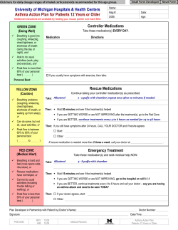


© Copyright 2026