
Staying alive Department of Science
Staying
alive
Department of Science
IES Ramon Casas i Carbó
3r ESO
This material has been elaborated by Rosa M. López Casas, a teacher in the
Department of Science of the IES Ramon Casas i Carbó (Palau-solità i
Plegamans), as her project for a paid study leave (C modality) which took
place in Scotland in the school year 2005-2006♣.
♣
Resolució EDC/1011/2005, de 6 d’abril, DOGC 4364 de 15 d’abril de 2005
Contents
UNIT 1. LIFE’S BUILDING BLOCKS ............................................................................................. 5
ORIGIN AND CELL THEORY ................................................................................................................. 7
MANY DIFFERENT CELLS .................................................................................................................... 9
CELLS: STRUCTURE & FUNCTION ..................................................................................................... 10
Plant ........................................................................................................................................... 16
Animal......................................................................................................................................... 16
CELLULAR FUNCTIONS ..................................................................................................................... 17
Function of nutrition................................................................................................................... 17
Function of relation .................................................................................................................... 18
Function of reproduction............................................................................................................ 18
GETTING ORGANISED .................................................................................................................... 21
NOW IT´S YOUR TURN....................................................................................................................... 23
LET’S LOOK TO WHAT WE’VE LEARNT .............................................................................................. 24
UNIT 2. NUTRITION........................................................................................................................ 25
THE HUMAN DIGESTIVE SYSTEM ..................................................................................................... 27
Digestive system glossary. .......................................................................................................... 27
Digestion..................................................................................................................................... 29
THE HUMAN RESPIRATORY SYSTEM ................................................................................................ 33
Our breathing system.................................................................................................................. 33
And now a few words about breathing. Inhalation and exhalation. ........................................... 34
A change or air. .......................................................................................................................... 35
Why do we have to breathe?....................................................................................................... 35
LIVING LIQUID. THE HUMAN CIRCULATORY SYSTEM ........................................................................ 37
Your Blood Stream...................................................................................................................... 38
Heartbeat.................................................................................................................................... 39
Blood circulation. ....................................................................................................................... 40
What's blood, anyway? ............................................................................................................... 41
EXCRETORY SYSTEM........................................................................................................................ 42
THE URINARY SYSTEM...................................................................................................................... 42
How does the urinary system work? ........................................................................................... 42
LET’S LOOK TO WHAT WE’VE LEARNT.............................................................................................. 43
UNIT 3. IN TOUCH WITH THE WORLD..................................................................................... 45
THE RELATIONSHIP WITH THE WORLD .............................................................................................. 47
Receptors: Sense organs and cells.............................................................................................. 47
COORDINATION: THE NERVOUS SYSTEM ........................................................................................... 48
The nervous system ..................................................................................................................... 48
The functions of the brain ........................................................................................................... 48
Composition of the brain ............................................................................................................ 49
Nerve cells .................................................................................................................................. 50
Synapses ..................................................................................................................................... 51
MOVE YOUR BODY 1: MUSCULAR SYSTEM ....................................................................................... 52
Muscle structure ......................................................................................................................... 52
How do muscles move?............................................................................................................... 53
Types of muscle tissue................................................................................................................. 53
Muscles by Function ................................................................................................................... 54
MOVE YOUR BODY 2: THE SKELETON .............................................................................................. 56
What Are Bones Made Of? ......................................................................................................... 56
How do my bones move? ............................................................................................................ 57
Your Joints.................................................................................................................................. 57
LET’S LOOK TO WHAT WE’VE LEARNT.............................................................................................. 59
ORIGIN OF IMAGES ....................................................................................................................... 60
Caution!
This machine has no brain
use your own!
If you don’t understand
ASK
Unit 1. Life’s
building blocks
"Where a cell exists, there must have been a pre-existing
cell, just as the animal arises only from an animal and a
plant only from a plant." "Omnis cellula e cellula" or "All cells
from cells." - Rudolf Virchow (1855).
WALT: What am I learning today?
By the end of this unit I will know:
• What a cell is
• Living beings are made up of cells
• The cell theory premises
• The structure of cells
• Levels of organization
• How a microscope works
All organisms are made up of cells a cell is the simplest collection of matter
that can live. Most cells are very small, so
we need to use a microscope to see them.
Each cell can live alone, doing
everything it needs, or it can live together
with other cells by forming many-celled
organisms like humans, other animals, and
plants.
Our bodies consist of more than a
billion cells, with each type of cell having
its own special function. All the different
cells communicate and cooperate with each
other to accomplish all the functions that
our bodies need. In contrast, there are
organisms called Protists that are singlecelled organisms and do all the different
functions that are needed to live.
STAYING ALIVE
Origin and cell theory
Anton van Leeuwenhoek made the first optic microscope; he observed
blood cells and rainwater.
Robert Hooke, in 1665, was the first scientist to
give the name cells. He did it after looking at a
laminates of cork and see their hexagonal shapes
that reminded him of a honeycomb made by bees.
Hooke saw that the cork seemed to be made up of
little boxes, little boxes that Hooke called cells.
Two centuries later Scheleiden (in plants) and
Schwann (in animals) discovered that all living
beings are made of cells.
In 1855, Rudolph Virchow established the premises
of the CELL THEORY.
Slice of cork as it was
seen by Hooke.
The CELL THEORY, or cell doctrine, states that all organisms are composed
of similar units of organization, called cells.
The premises of the Cell Theory include:
1. All known living things are made up of cells.
2. All cells come from pre-existing cells by division. (Spontaneous
Generation does not occur).
3. The cell is the structural & functional unit of all living things.
3.1.
Cells
contain
hereditary
information which is passed
from cell to cell during cell
division.
3.2.
All cells are basically the same in
chemical composition.
3.3.
All energy flow (metabolism &
biochemistry) of life occurs within
cells.
R. Virchow.
Life’s building blocks
Page 7
IES Ramon Casas i Carbó
Department of Science
Activity 1. Cell theory. Historical time line activity.
Your teacher will give you a paper with a list of
scientists and their contributions to the
What is a cell?
cell theory.
1.1. Open your jotter and:
A) Draw a timeline showing the
chronological order of these
scientists and their contributions.
Cells are the smallest
units of living beings.
B) Label the timeline with the dates of
the scientists' discoveries.
C) The earliest date should be on the left of the timeline and the most
recent date on the right.
D) Label each date with the corresponding scientist's name and
contribution(s) in an organized and readable manner.
E) Be sure your spacing shows a reasonable approximation of the
amount of time elapsed between dates.
1.2. Once you have finished, answer questions 1 to 5 in full sentences:
Questions:
1. What theory did these scientists provide evidence for?
2. What instrument was necessary before the cell theory could be
developed?
3. Which three scientists directly contributed evidence for the cell theory?
4. How did the earlier scientists and their contributions directly affect the
discoveries of later scientists (see #2)? For example, what had to come first?
5. List the three parts of the cell theory. (Write them down in English and in
Catalan)
It’s a fact!
The human body contains about 60 billion
cells. 50 millions die every second and are
replaced.
Some cells in the intestine only last 36 hours.
Blood white cells live about 13 days, red blood
cells about 120.
Nerve cells can live 100 years, but are
irreplaceable. After the age of 18, the brain
loses about 1000 cells every day.
Page 8
STAYING ALIVE
Many different cells
A free cell tends to have a round shape, but, in multicellular beings cells have
different shapes according to the tissue they belong to.
Scanning electron
micrograph of human
red blood cells
Ovum surrounded by
spermatozoa.
Drawing of a neuron
by Santiago Ramón y
Cajal.
We can find cells of very different
measures. Some are small, like red blood
cells (7 µm) others are bigger.
A micrometer
(µm) is a unit used
to measure cells.
1 µm is 10-6 m.
Life’s building blocks
Page 9
IES Ramon Casas i Carbó
Department of Science
Cells: Structure & function
Humans are multicellular beings, because they are made up of more than
one cell.
Cells can be classified into two different types: cells that have a nucleus and
cells that don’t have a nucleus.
Eukaryotic cells are cells which have a nucleus. (Algae, protozoa, fungi,
animals and plants).
Prokaryotic cells are cells which have no nucleus and are very simple cells
(bacteria).
Human cells are Eukaryotic cells because
they have a nucleus.
The three main parts of a eukaryotic cell
are:
• The cell membrane. It protects and
supports the cell.
• The nucleus. It is a large and oval
structure. It contains genetic material.
• The cytoplasm. It is a thick, similar to jelly
substance filling the space between the
nucleus and the cell membrane. It contains
the cell organelles.
Eukaryotic cell.
There are two main eukaryotic cells: Animal cells and plant cells.
Activity 2. The structure of the cell
In your jotter, copy these sentences and complete them with the appropriate
word:
1. Humans are ________________ beings
2. Eukaryotic cells are cells which have a _______________
3. Prokaryotic cells are cells which have no _________________
4. Human cells are __________________ cells
5. The three main parts of a eukaryotic cell are: ___________________ ,
________________ and __________________
Page 10
STAYING ALIVE
Let’s have a look at the structure of a cell.
The plant cell wall is made up of cellulose. Cellulose provides a stiff and rigid
environment for the cell to live in. The cell wall is what mainly distinguishes
plant cells from animal cells. The cell wall provides the cells with mechanical
protection and a chemically buffered (protected) environment. The cell wall is
a wall that allows for the circulation and distribution of water, minerals, and
other small nutrient molecules into and out of the cell. It provides rigid
building blocks which can produce stable structures such as leaves and
stems.
The cell membrane is found in both plant and animal cells. It is the
outermost layer in the animal cell and is found just inside the cell wall in the
plant cell.
The animal cell membrane
contains cholesterol, but the
plant cell does not.
It has pores and is
selectively
permeable
allowing the movement of
certain substances into and
out of the cell. This helps
maintain homeostasis in the
cell.
The cell membrane also
protects and supports the
cell.
Cell membrane
The cytoplasm is a thick, jellylike substance found in both plant and animal
cells filling the space between the nucleus and the cell membrane.
The cytoplasm is constantly in motion thanks to the cytoskeleton, a system
of molecules which provides them with shape, internal spatial organization,
and motility. Red blood cells, for instance, would be spherical instead of flat if
it were not for their cytoskeleton
It contains
organelles.
and
supports
the
cell
The nucleus is a large, oval structure
found in both plant and animal cells. It’s
the biggest organelle. It controls and
regulates all cell activities. It contains
genetic material.
Life’s building blocks
Page 11
IES Ramon Casas i Carbó
Department of Science
The nucleus is formed by:
• The nuclear membrane. It’s a double membrane with pores that allows the
entrance and exit of molecules.
• The nucleoplasm, a substance similar to jelly.
• The chromosomes
The mitochondrion is a bean-shaped
structure found in both plant and animal
cells. It has inner membranes or cristae.
Cells with high activity levels contain more
mitochondria, for example muscle cells.
The mitochondria (plural) break down
sugars and release energy for use by the
cell. As shown:
Glucose + O2
CO2 + H2O + Energy (ATP)
Respiration
The vacuole is a round, fluid-filled sac found in both plant and animal cells.
Plants usually have a few large vacuoles, while animal cells have smaller
vacuoles, if any. It stores food, water, and other materials for use by the cell.
Wastes may also be stored in vacuoles.
The lysosome is a small, round structure common in animal cells, but not
usually found in plant cells. The lysosome is involved in digestive activities,
including breaking down large food molecules into smaller ones and the
digestion of old cell parts. It is round, surrounded by a membrane and
contains enzymes.
Ribosomes are small and similar to a dot. The only function of ribosomes is
to build proteins
Ribosomes are made up of proteins and ribonucleic acid (RNA). These
molecules are arranged into two subunits called the large and small subunits.
These subunits are attached to each other and together they form the entire
ribosome. When we see them through a light microscope the ribosomes
appear as dots, they are very small and the subunits can not be seen.
Page 12
STAYING ALIVE
Endoplasmic reticulum is a network of membranes that extends through
the cytoplasm to the nuclear membrane. The membranes of the ER enclose
a series of tubes and flattened membranous areas.
There are two types of ER, smooth ER which has no ribosomes associated
with it and rough ER which has large numbers of ribosomes.
The basic function of rough ER is to transport the proteins produced by the
ribosomes to regions of the cell where they are needed or to the Golgi
apparatus.
Smooth ER is associated with regions of the cytoplasm involved in
detoxification of poisons and lipid synthesis.
Nucleus, endoplasmic reticulum and Golgi apparatus working together
The Golgi apparatus (sometimes called the Golgi body) consists of sacs
(with a single membrane) piled up. Near the edges of the flattened sacs you
will see vesicles.
The functions of the Golgi apparatus are:
• Modification of lipids and proteins.
• Storage and packaging of materials that will be exported from the cell. The
Golgi apparatus is often called the "shipping department" of the cell. The
vesicles that pinch off from the Golgi apparatus move to the cell membrane
and the material in the vesicle is released to the outside of the cell. Some of
these pinched off vesicles also become lysosomes.
The centrosome, is an area in the animal cell where microtubules are
produced. As a part of the cell centrosome there is a pair of small organelles,
the centrioles, each made up of a ring of nine groups of microtubules. The
two centrioles are arranged in such a way that one is perpendicular to the
other.
Life’s building blocks
Page 13
IES Ramon Casas i Carbó
Department of Science
During animal cell division, the centrosome
divides and the centrioles replicate (make new
copies). The result is two centrosomes, each
with its own pair of centrioles. The two
centrosomes move to opposite ends of the
nucleus, and from each centrosome,
microtubules grow into a "spindle" which is
responsible
for
separating
replicated
chromosomes into the two daughter cells.
Cilia and flagella are formed from the
centrioles of the cell.
Cell in a phase of division
showing two centrosomes, one at
each side of the cell
Plastids are organelles that only exist in plant
cells. They are found in the cytoplasm of the cell and have a double
membrane surrounding them. The number of plastids in a cell varies. This
number depends on the changing of environmental conditions that the plant
encounters and how the plant adjusts to these changes. The number of
plastids in a cell also depends on the type of species the plant is.
The primary function of plastids is to store molecules. One molecule that
they store is pigment; pigments give fruits and vegetables an orange or red
colour. Plastids also store photosynthetic products for plants that continue to
grow year after year. These products are obtained during the summer, and
they are stored in the plastid for the winter and spring. Potatoes, for example,
have many plastids in their cells containing starch.
The chloroplast is a large, complex double membraned organelle that
performs the function of photosynthesis within plant cells and contains the
substance chlorophyll that is essential for this process. All reactions of
photosynthesis occur in this organelle, and in addition, the chloroplasts also
create sugar from the sun for the cell and make all the food for other
organelles. The chloroplasts use photosynthetic chlorophyll pigment and take
in sunlight, water and carbon dioxide to produce glucose and oxygen. This is
the process of photosynthesis.
Chloroplasts are one
of the most typical
plant cell organelle
Page 14
STAYING ALIVE
Here is a hypothetical cell showing all the organelle and parts.
Remember that this cell doesn’t exists at all
Activity 3. Differences between animal and plant cells
Let’s see what the most important differences
between animal cells and plant cells are.
Open your jotter and draw a table similar to the
example to show the distinctions between plant
and animal cells (check previous pages). Add as
many rows as necessary.
Example:
Structure
Typical Plant
Eukaryotic Cell
Cell Part or
Is It Found In A Plant
Organelle
Cell?
Cell Wall
Yes
Typical Animal
Eukaryotic Cell
Is It Found In An
Animal Cell?
No
…
Then answer those questions:
1. What cell parts do Animal cells have that Plant cells do not have?
2. What cell parts do Plant cells have that Animal cells do not have?
3. Why do Plant cells have cell walls and Animal cells do not?
4. Why do you think Plant cells have bigger vacuoles than Animal
cells?
Life’s building blocks
Page 15
IES Ramon Casas i Carbó
Department of Science
Activity 4. Is it a plant cell or is it an animal cell?
The photographs below illustrate the basic comparison of plant and animal
cells.
Plant
Animal
Here you can see two microscope images of animal and plant cells
Look at them carefully and then:
1. Copy these two pictures in your jotter
2. Write, in your jotter, five sentences that show the main differences
between animal cells and plant cells. To do that you just have to joint
sentences from the list below.
Plant cells generally have a more but animal cells do not
rectangular shape because the
cell wall is more rigid.
Animal cells have centrosome
but animal cells do not
Plants cells usually have one or animal cells have a round or
more large vacuole(s)
irregular shape because they do
not have a cell wall.
Plant cells have chloroplasts
while animal cells have smaller
vacuoles, if any are present.
Plant cells have a cell wall
but plant cells do not
Activity 5. Drawing a cell.
In your jotter draw an animal cell. Make sure to draw and label all of the parts
listed below. Identify each part by colouring it the colour indicated in the box.
Nucleus
Blue
Endoplasmic reticulum
Light brown
Cell Membrane
Brown
Golgi apparatus
Orange
Mitochondrion
Red
Centrosome
Yellow
Lysosome
Pink
Vacuole
No colour
Ribosome
Black
Cytoplasm
Light green
Page 16
STAYING ALIVE
Activity 6. Cell organelle table.
To help you remember all cell organelle and their
functions, let’s make a table.
In your jotter draw a table similar to the example and
fill it with all the information you can (search previous
pages from 6 to 9)
Example:
LOCATION
DESCRIPTION
Plant or animal
What’s my name?
What am I like?
or both?
ORGANELLE
plant, not
animal
Cell wall
Cell membrane
both
plant/animal
*stiff and rigid,
*made up of
cellulose
FUNCTION
What do I do?
*support (grow tall)
*protection
*allows circulation and
distribution of nutrients
*plant - inside *support
cell
wall *protection
*animal - outer *controls movement of
layer;
materials in/out of cell
*barrier between cell
cholesterol
and its environment
*selectively
permeable
Cellular functions
Every cell does three kinds of functions: nutrition, relation and reproduction.
Function of nutrition
Nutrition is all the processes that a cell does in order to obtain energy and
material.
Cells are divided into two groups according to their form of nutrition:
-
Autotrophic. The cell transforms inorganic material into its own
organic material.
-
Heterotrophic. The cell transforms organic material from other beings
into its own organic material.
As all the animal cells, human cells are heterotrophic. They need to take
nutrients from other beings. Those nutrients must be digested inside the cell,
by lysosomes and there they will be used by mitochondria to provide energy
to the cell in a process called Respiration.
Life’s building blocks
Page 17
IES Ramon Casas i Carbó
Glucose + O2
Department of Science
CO2 + H2O + Energy (ATP)
Respiration
All the chemical reactions that take place in a cell are called Metabolism.
Most of the reactions take place in the cytoplasm and others in organelle
such as mitochondrion or chloroplast. There are two main types of
metabolism.
Catabolism is all the chemical reactions that destroy molecules in order to
obtain energy.
Anabolism is all the chemical reactions that build up complex organic
molecules from small ones.
Function of relation
All cells interact with the environment, that’s called the function of relation.
One way of interaction with the environment is movement. Some cells can
move in different ways, three of them are:
The amoeboid movement, which takes place thanks to pseudopodia. The
typical example is the amoeba.
The vibrate movement, which takes place thanks to cilia and flagella, one
example is the human spermatozoa.
The contractile movement, which takes place when cells contract and relax
the example is the muscle cells.
Function of reproduction
The cellular reproduction is the process that gives new cells starting from an
original cell. Thanks to this process life can go on.
During cellular reproduction it is very important to make sure that all the new
cells inherit the genetic material. An important structure to guarantee this is
chromosomes.
A chromosome is a piece of the DNA chain which is very packed in order to
reduce its length.
Every species has a characteristic number of chromosomes. For example,
human cells have 46 chromosomes, while frog cells have only 26. This
number is called diploid and it is symbolised as 2n. Ovule and spermatozoa
have half of the genetic material, so they are haploid (n). When an ovule and
a spermatozoa join together they form the zygote which is 2n (diploid) again.
There are to main types of cellular reproduction:
Mitosis, which consists in obtaining two new cells with the same genetic
information that the mother cell (original cell).
Meiosis, which is a process that gives four new cells with half of the genetic
material of the mother cell (original cell). This process is used to obtain
gametes.
Page 18
STAYING ALIVE
Mitosis
Prophase
Telophase
Metaphase
Anaphase
The mitosis process is divided in four phases: Prophase, metaphase,
anaphase and telophase.
During the interphase, before the mitosis starts, cells duplicate all the genetic
material so every piece of DNA (every chromosome) has an identical copy.
Chromosomes have two arms called chromatid.
Chromatids remain together by the centromeres.
Let’s see what happens during the four phases of mitosis
Prophase - the phase of mitosis in which the duplicated chromosomes
condense, the nuclear membrane dissolves, and centrioles divide and move
to opposite ends of the cell. Centriols remain “in touch” thanks to spindle
fibres that go across the cell.
Metaphase - the phase of mitosis in which the chromosomes line up at the
equator (the central plane) of the cell. The spindle fibres attach to the
centromere of every chromosome.
Anaphase - the phase of mitosis in which the chromosomes begin to
separate. The chromatids are pulled by the spindle fibres right to the poles.
Telophase - the last phase of mitosis, when the chromosomes migrate to
opposite ends of the cell, two new nuclear membranes form, and the
chromosomes uncoil. The cytoplasm divides.
Life’s building blocks
Page 19
IES Ramon Casas i Carbó
Department of Science
Activity 7. Animal cell mitosis.
Your teacher will give you a diagram of all the phases of the mitosis in an
animal cell. Glue it in your jotter and label it with those terms:
Anaphase
Centrioles
Centromeres
Chromosome
Interphase
Microtubules
Metaphase
Prophase
Telophase
Activity 8. About cells:
Open your jotter and write down these sentences, then complete them:
1. Human cells are heterotrophic because they need __________________
2. All the chemicals reactions that take place in a cell are called __________
3. Catabolism is all the chemicals reactions that ______________________
4. Anabolism is all the chemicals reactions that _______________________
5. Some cells can move in different ways, three of them are: _____________
6. A cell can perform three functions: _______________________________
7. The four phases of mitosis are: __________________________________
(Look at pages 13 to 15 to find out the answers)
Activity 9. About mitosis.
In your jotter write a table to summarise all the mitosis phases. It must have
two columns and 5 rows. Then fill it in with the appropriate sentences from
the list below. The first two sentences are already done for you.
You will have a table similar to these one:
Mitosis phases
Events at these phase
Prophase
The chromosomes start to condense strands. They are
made of two chromatides.
Here are the seven sentences you have to put in the table:
• The cytoplasm divides.
• The chromosomes line up along the equator.
• The pairs of identical chromatids are pulled apart and move to the
opposite poles.
• The nuclear membrane reappears.
• Two daughter cells are formed. Each has the same number of
chromosomes and is identical to the parent cell.
• Spindle fibres attach to the centromeres of the chromosomes.
• The nuclear membrane disappears.
Page 20
STAYING ALIVE
Getting ORGANised
In unicellular (single-celled) organisms, the single cell performs all life
functions. It functions independently. However, multicellular (many celled)
organisms have various levels of organisation within them. Individual cells
may perform specific functions and also work together for the good of the
entire organism. The cells become dependent on one another.
Multicellular organisms have the following 5 levels of organisation ranging
from simplest to most complex:
LEVEL 1 - Cells
• Are the basic unit of structure and function in living things
• May serve a specific function within the organism
• Examples- blood cells, nerve cells, bone cells, etc.
• There are many types of cells, each one with a special function All the cells
in an animal make up the level of cellular organisation.
LEVEL 2 - Tissues
• Made up of cells that are similar in structure and function
and which work together to perform a specific activity
• Examples - blood, nerve, bone, etc. Humans have 4 basic
tissues: connective, epithelial, muscle, and nerve.
• Tissues: A tissue is a group of cells of the same type that perform the same
function.
LEVEL 3 - Organs
• Made up of tissues that work together to perform a specific activity
• Examples - heart, brain, skin, etc.
• Tissues come together to make organs. An organ is made up
of various different types of tissues which work in coordination.
Heart, liver, muscle, etc. are organs.
LEVEL4 - Systems or apparatus
• Groups of two or more tissues that work together to perform a specific
function for the organism.
• Examples - circulatory system, nervous system, skeletal system, etc.
• The Human body has 11 organ systems - circulatory, digestive,
endocrine, excretory (urinary), immune(lymphatic), integumentary,
muscular, nervous, reproductive, respiratory, and skeletal.
• Organs are grouped together to form systems and apparatus. A
system is a set of organs that act in coordination in order to perform a
complex function. The digestive apparatus is formed by oesophagus,
stomach, liver, etc.
Life’s building blocks
Page 21
IES Ramon Casas i Carbó
Department of Science
LEVEL 5 - Organisms
• Entire living things that can carry out all basic life processes. Meaning they
can take in materials, release energy from food, release wastes, grow,
respond to the environment, and reproduce.
• Usually made up of organ systems, but an organism may be
made up of only one cell such as bacteria or protist.
• Examples - bacteria, amoeba, mushroom, sunflower, human
The levels of organisation in the correct order then are:
cells --> tissues --> organs --> organ systems -->
organisms
Now let's compare the Properties of Higher Levels
of Organisation with the Properties of the parts of
the organism.
An organ, such as the heart, is made up of groups
of tissues that work together to perform a specific
function. The heart is a pump that keeps blood
flowing throughout the body. The heart is primarily
made up of muscle tissue, but also contains
connective and nervous tissue. However, each of
these individual types of tissues has its own
primary function, which differs from the basic
function of the heart.
Activity 10. Levels of organization.
In your jotter write down correct phrases by joining one sentence from each
box.
BOX 2
BOX 1
→An organ is made up of
→The organs
together to form
are
grouped
→A system is a set of organs
that act in coordination
→All the cells in an animal make
up
→A tissue is a group of cells of
the same type
Page 22
→ in order to perform a complex
function
→ various different types of tissues
which work in coordination.
→ the level of cellular organization.
→ that perform the same function.
→ systems and apparatus.
STAYING ALIVE
Now it´s your turn
Now you are going to the laboratory and there you
will learn how to use a microscope in order to see
cells.
Going to the
lab is a lot
of fun!!
Remember that:
Cells can be seen under a microscope. This makes
the object appear larger.
When looking at cells under a microscope you stain them (colour them). This
makes the cell’s structure more visible but some structures can be seen
without using a stain.
If you are looking at any tissue under the microscope, you must make sure
that the tissue is very thin so the light can shine through it.
Don’t forget the rules about using a microscope!!
Life’s building blocks
Page 23
IES Ramon Casas i Carbó
Department of Science
Let’s look to what we’ve learnt
Open your jotter and copy those sentences, complete each space with the
correct word.
All living beings are composed of one or more _______, which are the basic
units of life.
Plant and animal cells have a nucleus, ____________ and ______________.
Only plant cells have a _____________, and a large permanent
__________. Green plants also have __________________
Animal cells have _______________ but plant cells do not have.
Proteins are made in ________________.
Respiration takes place in the ____________________.
The general reaction for respiration is:
A cell can reproduce and give two new cells with the same genetic
information, this is called ____________.
Mitosis phases are: prophase, _____________, anaphase and __________.
All cell activities are controlled by the _______________, which contains
___________.
All the chemical reactions that take place in cells are called _____________.
The correct order of levels of organisation is:
Cells, ___________, ____________, ______________ and organisms.
If you are looking at any tissue under the ________________ you must make
sure the tissue is very thin so the light can shine through it.
Page 24
Unit 2. Nutrition
Hello
Respiratory!
Human Respiratory System
Hi! How are
you Digestive?
I can’t stop
beating!
I can’t stop
beating!
Heart makes
blood go
round!
Somebody
has to do the
dirty job!
WALT: What am I learning today?
By the end of this unit I will know:
• Why we eat
• Why we breathe
• How the digestion works
• What the parts of the digestive system are
• What the parts of the breathing system are
• What the parts of the circulatory system are
• How the heart beats
• What the parts of the excretory system are
Multicellular livings had to “invent” a
system to bring nutrients to all the cells in
the body and, at the same time, to take
away waste materials.
So all multicellular livings have some
systems specialised in the function of
nutrition. Those systems are:
Digestive system,
processes food
the
Respiratory system, the
allows the gas exchange
system
that
system
that
Circulatory system, the system that carries
gas, nutrients and waste to and from all
cells.
Excretory system,
excretes waste.
the
system
In this unit we will learn how they work.
that
STAYING ALIVE
The Human Digestive System
The human digestive system is a complex series of organs and glands that
processes food.
In order to use the food
body has to break the
into smaller molecules
process; it also has
waste.
we eat, our
food down
that it can
to excrete
The human digestive
system is a tube about
12 metres long
Most of the digestive organs (like
the stomach and intestines) are like
a tube and contain the food as it
makes its way through the body.
The digestive system is essentially a long, twisting tube that runs from the
mouth to the anus, plus a few other organs (like the liver and pancreas) that
produce or store digestive chemicals.
Growth
Movement
Food
Tissue
repair
Body
heat
Fighting
disease
What do we need food for?
Digestive system glossary.
Let’s have a look at all the parts of the digestive system.
The mouth is the first part of the digestive system, where food enters the
body.
Chewing and salivary enzymes in the mouth are the beginning of the
digestive process (breaking down the food).
The salivary glands are glands located in the mouth that produce saliva.
Saliva contains enzymes that break down carbohydrates (starch) into smaller
molecules.
The epiglottis is a flap at the back of the tongue that keeps chewed food
from going down the windpipe to the lungs.
Nutrition
Page 27
IES Ramon Casas i Carbó
Department of Science
When you swallow, the epiglottis automatically closes. When you breathe,
the epiglottis opens so that air can go in and out of the windpipe.
The oesophagus or gullet is
the long tube between the
mouth and the stomach. It
uses
rhythmic
muscle
movements (called peristalsis)
to force food from the throat
into the stomach.
The stomach is a sack-like,
muscular
organ
that
is
attached to the oesophagus.
Both chemical and mechanical
digestion takes place in the
stomach. When food enters
the stomach, it is churned in a
bath of acids and enzymes.
Chyme is the name of food in
the stomach that is partly
digested and mixed with
stomach acids. Chyme goes
on to the small intestine for
further digestion.
Activity 1. Let’s try peristalsis movement!
Peristalsis is a rhythmic muscle movement that forces food in the
oesophagus from the throat into the stomach.
Peristalsis is involuntary -you cannot control it. It is also what allows you to
eat and drink while upside-down.
Don’t you believe it? Why don’t you try it?
Let’s try to chew and swallow down a biscuit while you are upside-down.
The small intestine is a large, thin and curved tube, divided into three parts:
• Duodenum - the first part of the small intestine; it is C-shaped and runs
from the stomach to the jejunum.
• Jejunum - the long, coiled mid-section of the small intestine; it is
between the duodenum and the ileum.
• Ileum - the last part of the small intestine before the large intestine
begins.
At the beginning of the small intestine there are two main glands that produce
enzymes to help in digestion. Those glands are:
Page 28
STAYING ALIVE
Liver - a large organ located above and in front of the stomach. It filters
toxins from the blood, and makes bile (which breaks down fats).
Gall bladder is a small, sac-like organ located by the duodenum. It
stores and releases bile (a digestive chemical which is produced in
the liver) into the small intestine.
Sometimes it fills with solids called “stones” and has to be removed.
Pancreas - an enzyme-producing gland located below the stomach
and above the intestines. Enzymes from the pancreas help in the
digestion of carbohydrates, fats and proteins in the small intestine.
The large intestine is not as curved as the small intestine but it is wider. It is
also divided into three parts:
cecum - the first part of the large
• Ascending colon - the
intestine; the appendix is connected to
part of the large intestine
the cecum.
that runs upwards; it is
located after the cecum.
Appendix - a small sac located on the
cecum. When the appendix is infected it
• Transverse colon - the
has to be removed in an operation
part of the large intestine
called appendicitis.
that
runs
horizontally
across the abdomen.
• Descending colon - the part of the large intestine that runs downwards
after the transverse colon and before the sigmoid colon.
The rectum is the lower part of the large intestine, where feces are stored
before they are excreted.
The anus is the opening at the end of the digestive system from which feces
(waste) exits the body.
Digestion.
Before our bodies can use the food that we eat it must be digested.
When food is digested it is broken into very small molecules: There are
special digestive juices in our body.
What happens to your food
They contain enzymes that digest
when you swallow?
large molecules into small ones. They
act like scissors to cut up molecules.
It enters a tube that starts with
Enzymes can be very particular. For
your mouth and ends at your
example the enzyme in saliva only
anus. The whole of this food
digests starch into glucose.
tube is called your gut.
It takes a different enzyme to digest
Your gut is about 9-12 metres
proteins into amino acids and another
long!
to digest fat into fatty acids.
Nutrition
Page 29
IES Ramon Casas i Carbó
Department of Science
Types of digestive enzymes
Each of which works on a specific substrate
Example(s)
Type of
enzyme
Amylase
Type of food
Digested (substrate)
End products of
digestion
Salivary
amylase
Starch
Maltose
Pancreatic
amylase
Starch
Maltose
Protein
Peptides
Pepsin
(In gastric
juice)
Peptides
Trypsin
Protease
(In pancreatic
juice)
Protein
(and
acids)
Amino
Peptidase
(In intestinal
juice)
Peptides
Amino acids
Pancreatic
lipase
Lipase
Fat
Glycerol and
Fatty acids
Why digest?
Food must be broken down and made soluble in your body.
This process takes place so that nutrients can pass through your gut wall into
your blood. Your blood then carries the digested food all around your body.
To think about:
Why can’t undigested food pass through your gut wall into your blood?
Which parts of your body need the food?
Fibre cannot be digested so it isn’t broken down.
Fibre adds a solid shape to your food so that it can
be pushed along your gut.
Plants have lots of fibre.
Page 30
STAYING ALIVE
The digestive process.
The start of the process - the mouth: The digestive process begins in the
mouth.
Food is partly broken down by the process of chewing and by the chemical
action of salivary enzymes (these enzymes are produces by the salivary
glands and break down starch into smaller molecules).
On the way to the stomach: the oesophagus: After being chewed and
swallowed, the food enters the oesophagus.
The oesophagus is a long tube that runs from the mouth to the stomach. It
uses rhythmic, wave-like muscle movements (called peristalsis) to force food
from the throat into the stomach. This muscle movement gives us the ability
to eat or drink even when we're upside-down.
In the stomach: The stomach is a large, sack-like organ that churns the food
and bathes it in a very strong acid (gastric acid). The food in the stomach that
is partly digested and mixed with stomach acids is called chyme.
In the small intestine: After being in
the stomach, food enters the
duodenum, the first part of the small
intestine. It then enters the jejunum
and then the ileum (the final part of the
small intestine).
In the small intestine, bile (produced in
the liver and stored in the Gall
bladder), pancreatic enzymes, and
other digestive enzymes produced by
the inner wall of the small intestine
help in the breakdown of food.
In the large intestine: After passing through the small intestine, food passes
into the large intestine.
In the large intestine, some of the water and electrolytes (chemicals like
sodium) are removed from the food.
Many microbes (bacteria like Bacteroides, Lactobacillus acidophilus,
Escherichia coli, and Klebsiella) in the large intestine help in the digestion
process.
The first part of the large intestine is called the cecum (the appendix is
connected to the cecum). Food then travels upward in the ascending colon.
The food travels across the abdomen in the transverse colon, goes back
down the other side of the body in the descending colon, and then through
the sigmoid colon.
The end of the process: Solid waste is then stored in the rectum until it is
excreted via the anus.
Nutrition
Page 31
IES Ramon Casas i Carbó
Department of Science
Activity 2. Down the tube.
Look at the diagram of the human gut on page 27. Follow the path your food
goes down. There are lots of twists and turns.
Here you have a list of 5 parts that food passes through and the explanation
of what happens. Open your jotter and write down every part with the right
explanation.
Parts of the gut:
Large intestine; Stomach; Mouth; Gullet or oesophagus; Small intestine
Explanations:
More juices are added from Only food that can not be digested (like
your liver and your pancreas.
fibre) reaches here.
These complete digestion. A lot of water passes back into your body.
Then food passes through into
your blood. This is called This leaves solid waste to pass through
your anus.
absorption.
(up to 24 hours)
(About 5 hours)
Food chewed and mixed
A straight, muscular with saliva.
tube leading to your
Then you swallow it (gulp!)
stomach.
(Food is here for 20
(10 seconds)
seconds)
The
acid
bath!
Digestive juices and
acid are added to food
here. The mixture is
churned up
(2 to 6 hours)
Now answer the following questions (in your jotter, of course!)
1. In which part of your gut does food stay the longest?
2. Proteins are digested in your stomach. What are conditions like here?
3. How long does it take food to pass down the whole length of your gut?
Activity 3. Copy and complete:
Open your jotter and copy and complete the following text with words from
the box:
Digestion is the ______ down of food into ________ molecules by chemicals
called ______. Food has to be digested so that it can pass through the
______ wall into ______.Starch is digested to _______ but _______ cannot
be digested.
blood, enzymes, fibre, break, gut, very small, glucose
Page 32
STAYING ALIVE
The Human Respiratory System
Take a nice deep breath. Inhale through the nose, exhale through your
mouth.
Aaaaahh........... Inhale, exhale. Inhale, exhale. Inhale, exhale.
Hey --- wake up! Time to learn about the system of organs that's responsible
for taking in oxygen and getting rid of carbon dioxide: the respiratory system.
Activity 4. There must be an order!
First, allow me to list the structures and organs
that together make up the respiratory system.
Your job is to write them down in the order that
air passes through them as it is inhaled. OK?
Write the answer in your jotter.
Alveoli, bronchiole, bronchus, larynx, lung,
pharynx, nasal cavity, nostril, trachea
Our breathing system.
Let’s take a look at what the respiratory (or breathing) structures look like.
Nose/nasal cavity. Warms and
filters air as it is inhaled.
Pharynx (throat). Passageway
for air that leads to trachea.
Larynx. The voice box, where
vocal chords are located.
Trachea (windpipe). It’s a tube
from pharynx to bronchi. It got
rings of cartilage to keep the
windpipe open. The trachea has
fine hairs called “cilia” that filter
air before it reaches the lungs.
Bronchi (singular bronchus). Two
tubes at the end of the trachea,
each lead to a lung.
Bronchioles. A network of
smaller tubes leading from the
bronchi into the lung and, finally,
to air sacs
Nutrition
Page 33
IES Ramon Casas i Carbó
Department of Science
Alveoli. The functional respiratory units in the lung, where gases (oxygen and
carbon dioxide) are exchanged.
Let’s take a closer look at the air sacs (alveoli) in the lungs.
O2
C
As the bronchioles branch out into smaller & smaller & smaller & smaller &
smaller tubes, they lead to microscopic groups of alveoli, also called air sacs.
You can think of the air sacs as a bunch of grapes, with each individual grape
representing a single alveolus.
The red structures represent blood vessels leading to & from the air sacs.
The wall of an alveolus is only one cell thick. This allows gases to diffuse into
& out of the alveoli.
The alveoli are surrounded by capillaries so that oxygen and carbon dioxide
can be exchanged between the lungs & the blood.
Oxygen in the alveolus can diffuse into the bloodstream (& be transported
throughout the body) and carbon dioxide in the bloodstream can enter the
alveoli (& then be exhaled).
And now a few words about breathing. Inhalation and exhalation.
There are no muscles in your lungs.
They do not actively pump air in & out, in &
out. The muscle responsible for breathing
actually lies below the lungs. It is like a rubber
sheet that separates your chest cavity & your
abdominal cavity. Its name is diaphragm.
When you inhale (breath in), the ribs move out
and up and the diaphragm contracts & moves
down.
So the space inside your chest cavity gets
bigger & lets air get into the lungs.
When you exhale, the ribs move down and in
and the diaphragm relaxes & moves up,
forcing air out of the lungs.
Breathing involves the lungs, the ribs, the
diaphragm and the space inside your chest.
Page 34
STAYING ALIVE
A change or air.
Air breathed in and breathed out are different.
Air breathed in has more oxygen and less
carbon dioxide than air breathed out.
The air contains:
But there are other differences:
20% Oxygen
The air breathed out is cleaner because your
air passages are recovered with a mucus that
traps dust and germs. There are also millions of
microscopic hairs that carry the mucus up to
your nose and throat.
1% other gases: argon,
carbon dioxide (CO2),
water vapour, etc.
79% nitrogen
The air breathe out is warmer because it comes from the inside of your body.
Activity 5. Air breathed in and breathed out.
Open your jotter and copy and complete the following table:
Air breathed in
Air breathed out
It contains more O2
It contains
It contains
It contains more CO2
It contains the same
It contains the same
It contains less
It contains more
It is less
It is
It is
It is
Use words from the list:
cleaner; less O2; water vapour; less CO2; nitrogen; warmer; colder
Why do we have to breathe?
All cells in your body need energy to stay alive. Can you remember where
you get your energy?
You get energy out of your food in respiration:
Glucose + O2
Glucose + oxygen
CO2 + H2O + Energy (ATP)
Carbon dioxide + water + energy
So oxygen is needed for respiration to happen.
When sugar is burnt in oxygen, it gives out energy for your cells to use. You
get oxygen into your body by breathing it in.
Nutrition
Page 35
IES Ramon Casas i Carbó
Department of Science
Activity 6. Puffing and panting.
We call “Breathing rate” the number of times we breathe per minute.
Do you think that you change your breathing rate depending on your activity?
Let’s see it.
Open your jotter and get ready to follow next step:
1. Sit still and count how many times you breathe out in 1 minute. Try to
make “normal” breathings. That will be your breathing rate at rest.
2. Copy this table and fill in your breathing rate at rest
Breathing rate at rest
(breathing per minute)
Breathing rate after
light exercise
(breaths per minute)
Breathing rate after
heavy exercise
(breaths per minute)
3. Now do step-ups for 1 minute (light exercise). As soon as you have
finished, sit down and measure your breathing rate. Write it down in your
table.
4. Now do step-ups as quickly as you can for 3 minutes (heavy exercise). As
soon as you have finished, sit down and measure your breathing rate.
Write it down in your table.
5. Draw a bar diagram to show your breathing rate at rest, after light exercise
and after heavy exercise
6. Now copy and answer those questions in your jotter:
A) After exercising your breathing rate: increased or decreased?
B) What happens to the size of each breath in the investigation?
C) What do you think affects your breathing rate and your depth of
breathing?
Activity 7. Copy and complete:
Open your jotter and copy and complete the following text with words from
the box:
We breathe in air containing nitrogen, __________ and some carbon dioxide.
The air that we breathe ______ contains the same amount of___________,
less _________ and __________carbon dioxide. The air we breathe out also
contains more _____________ vapour, it is at a __________ temperature
and it is ___________.
more; nitrogen; cleaner; water; out; oxygen; oxygen; higher;
Page 36
STAYING ALIVE
Living liquid. The human circulatory system
What does blood make you think of?
Horror movies, vampires, wars?
It’s flowing around your body all the time. But what is it for?
It's a big name for one of the most important systems in the body. Made up of
the heart, blood and blood vessels, the circulatory system is your body's
delivery system. Blood moving from the heart, delivers oxygen and nutrients
to every part of the body. On the return trip, the blood picks up waste
products so that your body can get rid of them.
Your already know that the cells of your body need food and oxygen to give
you energy in respiration.
Your cells make waste chemicals too
Your kidneys get rid of these waste chemicals.
Activity 8. The circle of life.
Look at the diagram and answer the questions:
HEART
f)
KIDNEYS
g)
INTESTINES
d)
e)
CELLS IN
THE BODY
a)
b)
c)
LUNGS
a)
b)
c)
d)
e)
f)
g)
What does blood collect when it is here?
What does blood collect when it is here?
What does blood drop off here?
Name 2 things blood drops off here and that the cells use to live.
What does blood take away from the cells?
What does blood drop off at the kidneys?
What keeps blood moving round and round?
So your blood carries many things around your body. It is rather like a railway
system, where trains pick things up at one place and deliver them to another
How do you thing blood is kept on the move?
Yes, thanks to the heart!
Nutrition
Page 37
IES Ramon Casas i Carbó
Department of Science
Your Blood Stream
Your blood travels through a rubbery
pipeline with many branches, both big
and small.
The blood is carried around your body
in tubes called blood vessels.
Strung together end
to end, your blood
vessels could circle
the globe 2 1/2 times!
There are three main blood vessels:
•
Arteries are tubes that carry blood away
from the heart
•
Veins are tubes that return blood to the
heart
•
Capillaries connect arteries and veins.
They are tiny tubes that exchange food,
oxygen and wastes between blood and
body cells.
The tubes that carry blood away from your heart
are called arteries. They carry blood pumped
under high pressure to smaller and smaller
branched tubes called capillaries. The tubes that
drain back to the heart are veins.
Differences between arteries and veins
Arteries
Veins
Thick muscular wall
Small inside space
Blood under high pressure
Has a “pulse”
No valves
Thin muscular wall
Large inside space
Blood under low pressure
No “pulse”
Has valves
Activity 9. Blood vessels in the human
body.
Your teacher will give you a diagram of the
main human blood vessels to label.
Label it, make sure it is correct and glue it
in your jotter.
Once finished translate all the words into
your mother tongue.
Page 38
STAYING ALIVE
Heartbeat
What do you think is the strongest muscle in your body?
It is the heart! About the size of your
fist, your heart is a muscle.
It contracts and relaxes some 70 or so
times a minute at rest -- more if you
are exercising -- and squeezes and
pumps blood through its chambers to
all parts of the body.
Oxygen-poor blood enters the right
atrium of the heart (via veins called the
inferior vena cava and the superior
vena cava).
The blood is then pumped into the right ventricle and then through the
pulmonary artery to the lungs, where the blood is enriched with oxygen (and
loses carbon dioxide).
The oxygen-rich (oxygenated) blood is then carried back to the left atrium of
the heart via the pulmonary vein.
The blood is pumped to the left ventricle, and then the blood is pumped
through the aorta and to the rest of the body.
Every day, the
heart pumps
about
7,600
litres of blood,
beating about
100,000 times
Activity 10. Around and around.
Do you know how to take your pulse? When you take your pulse you feel an
artery. Blood flows through your arteries. This is the pulse that you feel.
Search for arteries around your body: under your ear, on your waist.
Once you find a good one, relax, look at your watch and count.
How many beats do you feel? This is your hear rate at rest.
Nutrition
Page 39
IES Ramon Casas i Carbó
Department of Science
The heart rate is the number of beats a heart has per minute.
Open your jotter and write down your heart rate at rest.
Your heart beats 60 minutes per hour and 24 hours a day, to keep you alive.
Use a calculator to work out how many times your heart beats:
a) per hour
c) in a year
b) per day
d) since you were born
Blood circulation.
The human blood circulation is double,
because blood passes twice through
the heart, and complete, because
oxygenated blood never gets mixed
with deoxygenated blood. Blood
vessels and heart form two main
circuits:
•
•
Pulmonary circulation is the
movement of blood between the
heart and lungs.
Systemic circulation is the
movement of blood between the
heart and the rest of the body.
We can consider a third circuit:
Coronary circulation is the movement
of blood from within the heart
chambers to the heart tissues
themselves.
Activity 11. Copy and complete.
Open your jotter and copy and complete the following text with words from
the box:
Blood is pumped around my body by my_______. Blood travels away from
my heart in ________ and back to my heart in_______. The tiniest blood
vessels are called ________ and these have very _______walls so things
can pass in and out. When I feel my pulse I am touching an ________.
The heart is made out of ______. The blood on the left-side contains ______
oxygen than the blood on the _____-side. This is because the blood has just
come back from the _____. The left-side of the heart pumps blood all around
the _____. The heart has _____ to stop the blood from flowing backwards.
Arteries; body; capillaries; lungs; veins; thin; artery; muscle; more; right;
lungs; valves; heart;
Page 40
STAYING ALIVE
What's blood, anyway?
Most of your blood (55 %) is a colourless liquid called plasma. Flouting in
this plasma there are cells:
Red blood cells make the blood look red and deliver oxygen to the cells in
the body and carry back waste gases in exchange.
White blood cells are part of your body's defence against disease. Some
attack and kill germs by gobbling them up; others by manufacturing chemical
warfare agents that attack.
Platelets are other cells that help your body repair itself after injury.
Red blood cells
White blood cells
platelets
Diagram
Plasma
How is it? What’s it Job?
It’s transparent No nucleus (to
help with more
Transports
space)
cells
Has
Carries:
haemoglobin
Food
Small+flexible to
Heat
squeeze through
capillaries
Hormones
Has nucleus
Small
Fights disease by: pieces of
cells.
1. Engulfing and
Clot your
breaking down
blood.
2. Sending out an
antibody.
Carries oxygen
…
Activity 12. Mind map.
Open your jotter. Make up a mind map about the circulatory system, make
sure you put the following information:
Blood vessels
Heart
Blood
Blood circulation
Arteries
Heartbeat
Plasma
Pulmonary
Veins
Blood flood
through the heart
Cells (red,
white,…)
Systemic
Capillaries
Parts of the heart
You can make some drawings to improve your mind map.
Once finished show it to your teacher to get approbation.
Nutrition
Page 41
IES Ramon Casas i Carbó
Department of Science
Excretory system.
The job of the excretory system consists in eliminating all waste substances
(carbon dioxide, which is eliminated through lungs) that cells produce.
Humans have two ways to eliminate waste:
The urinary system and the skin
The urinary system
Your urinary system filters out excess fluid and other substances from your
bloodstream. Some fluid gets reabsorbed by your body but most gets
expelled as urine. If your body wasn’t clever enough to get rid of some of this
stuff, you'd get sick!
How does the urinary system work?
Your body makes chemical waste products it can't use. They go into your
bloodstream, then pass into your kidneys where the excess fluid and
chemical is filtered out.
As your kidneys do this work, they also make sure that your blood is just the
right combination -- not too thin or too thick, not too salty, or with excess
vitamins and minerals or wastes made by other parts of the body.
The kidneys work as they do because they contain millions of infinitesimal
filters. Hundreds of times a day the blood in your body gets filtered through
these filters and the liquid and wastes are removed.
The wastes go to the centre of your kidneys, and then down through tubes
called the ureters into a bag. This bag is your bladder and it can stretch
enough to hold more than a litre of urine!
When your bladder becomes too full, it sends a message to your brain. You
feel the need to pee and start looking for a place to do it. When the time and
place are right, your brain orders the muscles around your bladder to start
squeezing and for the circle of muscles at the bottom of your bladder to open.
Pee squirts out through the urethral
opening in your body. Hopefully, the pee
meets the water of a toilet. Now that
spells relief!
Activity 13. Draw and Label!
Open your jotter, copy the picture on the
right and name all its parts.
Use the following words:
Bladder, ureters, urethra, cava vein,
aorta artery, renal artery, renal vein,
kidney.
Page 42
STAYING ALIVE
Let’s look to what we’ve learnt.
Read this text carefully. Then open your jotter and translate it into your
mother tongue.
“Food is digested from large insoluble molecules into small soluble molecules
thanks to enzymes. The food particles enter the digestive system through the
mouth and then go to the oesophagus, stomach and intestine. Digested food
particles pass through villi in the walls of the small intestine.
The small intestine absorbs the maximum amount of food, as it is very long,
and has a large surface area. The large intestine absorbs water from food.
When you inhale (breath in) the ribs move out and up and the diaphragm
contracts & moves down. You breathe in air and send it down to your lungs
until the alveoli.
Blood is pumped from the heart to your lungs, where oxygen from the air
you've breathed in gets mixed with haemoglobin in the red cells.
That oxygen-rich blood then
travels back to the heart where it
is pumped through arteries and
capillaries to the whole body,
delivering oxygen to all the cells
in the body -- including bones,
skin and other organs.
Blood picks up carbon dioxide
and other waste from cells in the
body. Veins then carry the
deoxygenate and waste blood
back to the heart for another ride.
Carbon dioxide travels to the
alveoli where it is exhaled. The
rest of the wastes are transport to
the Urinary system.”
Nutrition
Page 43
When you want what
you’ve never had
you have to do
what you’ve never done
Unit 3. In touch
with the world
Build a body
Want to know more?
Just follow me!
Your brain:
Use it or lose it!
WALT: What am I learning today?
By the end of this unit I will know:
• How we communicate with the outside world.
• How the nervous system works.
• A special kind of cell: The neuron.
• What a muscle is and how it contracts.
• What a bone is made up of.
• The main parts of the muscular and skeleton
systems.
All organs and body systems are co-ordinated.
They cooperate with each other to work
together. This brings about the functions that
are vital to a healthy life. For example, if you
exercise hard, your heart beat increases, and
you breathe faster to take in more oxygen.
The sense organs are also co-ordinated. If
you are playing a ball game, your eyes see
the ball, they pass signals to the brain, and
central nervous system (CNS), which then
helps you to react and hit or catch the ball.
All of these functions are co-ordinated by the
brain and CNS. Messages from your eyes and
ears are passed to the brain, which in turn
sends nerve impulses down your spinal cord,
and then to the muscles you need to move.
The nerve impulses also make your heart beat
faster and make your diaphragm contract
faster so you take in more air for the extra
oxygen you need for the increased energy you
use for all physical activities.
STAYING ALIVE
The relationship with the world
The relationship with the outside world consists in the detection of
information from the environment, the analyses of this information and,
finally, making a decision of what to do.
Your sense organs detect information from your surroundings. This
information is the stimulus which triggers a nerve impulse.
The impulse is carried along a sensory nerve cell to the CNS (Central
Nervous System), where the information is sorted out. Another impulse is
sent along a motor nerve to make, for instance, muscles contract, this is
called a response.
Stimulus
Sense organs
Co-ordination
systems:
Environment
Nerve system &
endocrine
Response
Effectors
Activity 1. Quick reaction.
Open your jotter and try to write some sentences about how the relationship
with the world works in this example: “Your hand touches a very hot object”,
use this words:
Stimulus, response, skin receptors, CNS, arm muscles, impulse
That’s called a Reflex action because the response is unconscious. That
means that the brain doesn’t participate in the co-ordination.
Receptors: Sense organs and cells.
Detection of stimulus consists in transforming all the environment changes
into electrical impulses that will travel inside our body. Those cells and
organs able to do this are called sense organs.
There are four main groups of sense organs, read about them on next page.
In touch with the world
Page 47
IES Ramon Casas i Carbó
Department of Science
Mechanoreceptors. They detect pressure, for example skin receptors.
Thermo receptors. They detect changes in temperature.
Chemical receptors. They detect chemicals.
Photoreceptors. They detect light.
Activity 2. Senses and receptors.
Open your jotter and copy and complete the following table.
Sense
Touch
Hearing
Balance
Smell
Taste
Sight
Kind of receptor
Why? Or example of how it works
Coordination: the nervous system
The nervous system
The brain and spinal cord make up the central nervous system (CNS).
The brain is connected to the spinal cord, which runs from the neck to the hip
area. The spinal cord carries nerve messages between the brain and the
body.
The nerves that connect the CNS to the rest of the body are called the
peripheral nervous system.
The autonomic nervous system controls our life support systems which we
don't consciously control, like breathing, digesting food, blood circulation, etc.
The functions of the brain
The human brain is a complex
organ that allows us to think,
move, feel, see, hear, taste, and
smell. It controls our body,
receives, analyzes and stores
information (our memories).
The brain produces electrical
signals, which, together with
chemical reactions, let the parts
of the body communicate.
Nerves send these signals
throughout the body.
Page 48
STAYING ALIVE
Nourishment of the brain
Although the brain is only 2% of the body's weight, it uses 20% of the oxygen
supply and gets 20% of the blood flow. Blood vessels (arteries, capillaries,
and veins) supply the brain with oxygen and nourishment, and take away
wastes. If brain cells do not get oxygen for 3 to 5 minutes, they begin to die.
Cerebrospinal fluid (CSF) surrounds the brain.
Size of the human brain
The average human brain
weighs about 1300-1400 g.
At birth, the human brain
weighs about 350-400 g. As a
child grows, the number of cell
remains relatively stable, but
the cells grow in size and the
number
of
connections
increases. The human brain
reaches its full size at about 6
years of age.
Composition of the brain
The brain consists of grey
matter (40%) and white matter
(60%) contained within the
skull. Brain cells include
neurons and glial cells.
The brain has three main
parts: the cerebrum, the
cerebellum, and the medulla
(brain stem).
Protection
The cells of the nervous system are quite fragile and need extensive
protection from being crushed, being infected by disease organisms, and
other harm.
The brain and spinal cord are covered by a tough, translucent membrane,
called the dura matter. Cerebrospinal fluid (CSF) is a clear, watery liquid that
surrounds the brain and spinal cord, and is also found throughout the
ventricle (brain cavities and tunnels).
The cranium (the top of the skull) surrounds and protects the brain. The
spinal cord is surrounded by vertebrae (hollow spinal bones). Also, some
muscles serve to pad and support the spine.
In touch with the world
Page 49
IES Ramon Casas i Carbó
Department of Science
Activity 3. What does my brain do?
Open your jotter and copy and complete the following table:
Part of the brain
Cerebrum
Cerebellum
Medulla
Function. What do I control?
Then read carefully the information about the nervous system and answer
these questions:
1. What is the central nervous system (CNS) made up of?
2. What is the name of the nerves that connect the CNS to the rest of the
body?
3. What does the autonomic nervous system do?
4. How long can a brain cell live without oxygen?
5. How is the nervous system protected?
6. Can a nerve cell reproduce?
Nerve cells
The brain and spinal cord are made up of many cells, including neurons and
glial cells. Neurons are cells that send and receive electro-chemical signals
to and from the brain and nervous system. There are about 100 billion
neurons in the brain. There are many more glial cells; they provide support
functions for the neurons, and are far more numerous than neurons.
There are many types of neurons. They vary in size from 4 microns (0.004
mm) to 100 microns (0.1 mm) in diameter. Their length varies from
centimetres to meters.
Neurons are nerve
cells that transmit nerve
signals to and from the
brain at up to 400 km/h.
The neuron consists of
a cell body (or soma)
with dendrites (signal
receivers)
and
a
projection called an
axon, which conducts
the nerve signal. At the
other end of the axon,
the axon terminals
transmit the electrochemical signal across a synapse.
The cell body (soma) contains the neuron's nucleus (with DNA and typical
nuclear organelles). Dendrites bring information to the cell body.
Page 50
STAYING ALIVE
The axon is a long extension of a nerve cell, and takes information away
from the cell body. Nerves are groups of axons (also called tracts or
pathways).
Myelin sheath covers and insulates the axon (except for periodic breaks
called nodes of Ranvier), increasing transmission speed along the axon.
Myelin is manufactured by Schwann's
cells, and consists of 70-80% lipids
(fat) and 20-30% protein.
There are different types of neurons.
They all carry electro-chemical nerve
signals but differ in structure and are
found in different parts of the body.
The word "neuron" was coined by
the German scientist Heinrich
Wilhelm Gottfried von WaldeyerHartz in 1891 (he also coined the
term "chromosome").
Unlike most other cells, neurons cannot regrow after damage (except
neurons from the hippocampus). Fortunately, there are about 100 billion
neurons in the brain.
Glial cells make up 90 percent of the brain's cells. Glial cells are nerve cells
that don't carry nerve impulses. Glial means “glue”. The glial cells perform
many important functions, including:
digestion of parts of dead neurons
manufacturing myelin for neurons
providing physical and nutritional support for neurons,
Types of glial cells include Schwann's Cells, Satellite Cells, Microglia,
Oligodendroglia, and Astroglia.
Synapses
Information from one neuron flows to another neuron across a synapse. The
synapse is a small gap separating neurons. A synapse is a gap between the
axon terminal of a neuron and the dendrites of the receiving cell.
The synapse consists of:
1. a presynaptic ending (in an axon
terminal) that contains neurotransmitters,
2. a postsynaptic ending (dendrites of the
postsynaptic neurone) that contains
receptor sites for neurotransmitters,
3. a synaptic space between the
presynaptic and postsynaptic endings.
To communicate between neurons an
electrical impulse must travel down an
axon to the synaptic terminal.
Chemicals, called neurotransmitters, are
released from one neuron at the
presynaptic
nerve
terminal.
Neurotransmitters then cross the synapse where they may be accepted by
the next neuron at a specialized site called a receptor.
In touch with the world
Page 51
IES Ramon Casas i Carbó
Department of Science
Move your body 1: Muscular system
Nerve impulses are carried along motor nerve cells until an effectors cell,
which can be a gland or a muscle.
The muscular system is the largest system in the body. Muscles are located
in practically every region. And control virtually all movement. Extremities are
almost entirely made up of muscles. There are over forty muscles located in
the skull. Muscles are unique and very useful because they can contract.
There are three different types of muscles tissue, cardiac, skeletal, and
smooth.
Muscles move you! Without muscles you wouldn’t be able to open your
mouth, speak, shake hands, walk, talk, or move your food through your
digestive system. There would be no smiling, blinking, breathing. You
wouldn’t be able to move anything inside or outside yourself. The fact is,
without muscles, you wouldn't be alive for very long!
Do I have lots of muscles?
Indeed. On average, probably 40% of your body weight is in muscles. You
have over 630 muscles that move you. Muscles can't push. They pull. You
may ask yourself, if muscles can't push how can you move your fingers in
both directions, back and forth, back and forth?
The answer?
Muscles often work in pairs so that they can pull in different or opposite
directions.
Muscle structure
A muscle fibre is a single, multinucleated muscle cell. A muscle may be
made up of hundreds or even thousands of muscle fibres, depending on the
muscles size. Muscle is attached to bone by tendons.
Active muscles use a lot of energy and require a continuous supply of oxygen
and nutrients, which are supplied by arteries. Muscles produce large
amounts of metabolic waste that must be removed by Veins.
Page 52
STAYING ALIVE
which work together. Those proteins form a series of light and dark bands
inside the muscle fibre. There are two types of protein filaments- thick ones
and thin ones.
The thick filaments are made up of a protein called MYOSIN.
The thin filaments are made up of a protein called ACTIN.
How do muscles move?
When muscle contracts the light and dark bands contained in muscle fibres
get closer together. This happens because when a muscle contracts myosin
filaments and actin filaments interact to shorten the length of the cytoplasm.
Types of muscle tissue
There are three types of muscle tissue, or Muscles: SKELETAL, SMOOTH,
AND CARDIAC.
Each type has a different structure and plays a different role in the body.
SKELETAL MUSCLE
Skeletal muscle is responsible for moving
parts of the body. Skeletal muscles are
generally attached to bones by tendons
and are at work every time we make a
move.
Skeletal muscles are responsible for
voluntary (conscious) movement. Most
skeletal
muscles
are
consciously
controlled by the central nervous system
(CNS).
Skeleton Muscle Cells are large and have
more than one nucleus. They vary in
length from 1mm to 30 to 60 cm.
When viewed under a microscope,
skeletal muscles appear to have striations
In touch with the world
Page 53
IES Ramon Casas i Carbó
Department of Science
(bands or stripes). This gives skeleton muscle the name of voluntary or
striated muscle.
SMOOTH MUSCLES
Smooth muscles are usually
not under voluntary control.
Smooth muscle cells have a
single nucleus and are not
striated. Smooth muscles are
found in many internal organs,
stomach, intestines, and in the
walls of blood vessels.
Eye muscles are the
busiest muscles in the
body. Scientists estimate
they may move more than
100,000 times a day!
Smooth muscle fibres do not
have tendons. Most smooth muscle cells can contract without nervous
stimulation.
Smooth muscle is called
involuntary muscle because most of its
You have over 30 facial
movements
cannot
be
consciously
muscles which create
controlled.
looks
like
surprise,
The contractions in smooth muscles move
happiness, sadness, and
food through our digestive tube, control the
frowning.
way blood flows through the circulatory
system, and decrease the size of the pupils
of our eyes in bright light.
CARDIAC MUSCLE
The only place in the body where cardiac muscle is found is in the heart.
Cardiac cells are striated, but they are not under voluntary control.
Cardiac muscle contracts without direct
stimulation of the nervous system. A group
of specialized muscle cells in the upper part
of the heart sends electrical signals through
cardiac muscle tissue, causing the heart to
rhythmically contract and pump blood
through the body.
The largest muscle in
the body is the gluteus
maximus muscle in the
bottom.
The cardiac muscle cell contains one nucleus located near the centre.
Muscles by Function
Each muscle has its own special name. Muscles, however, are also
described by their function.
Muscles that bend an extremity are flexors; those which straighten an
extremity are extensors (e.g. elbow flexors and elbow extensors.)
Muscles which move an extremity to the side, away from the body, are
abductors; those which move an extremity sideways toward the body are
adductors (e.g. hip abductors and hip adductors.)
Other functional groups are elevators, depressors, rotators, doriflexors,
planar flexors, and palmar flexors.
Page 54
STAYING ALIVE
Activity 4. The three types of muscles.
Open your jotter and copy and complete the following table with words and
sentences from the list below:
Type of muscle Smooth muscle
Cardiac muscle
Skeletal muscle
Appearance
Voluntary
or
involuntary
Function
It controls movement of internal
organs.
Voluntary
Involuntary
Smooth
It controls contraction of the heart.
Striated
It moves bones. It works in pairs:
when one contracts, the other
relaxes. It is attached to bone by
bands of tissue called tendons.
Activity 5. Labelling the muscular system.
Your teacher will give you a diagram
of the muscular system to label.
Label the diagram, make sure it is
correct and glue it in your jotter.
To help you find the names of the
muscles your teacher will give you
different pieces of paper with
drawings and explanations of the
following muscular system parts:
• Muscles of the head and neck
• Muscles of the trunk
• Muscles of the upper extremity
• Muscles of the lower extremity
Use them to complete your diagram
of the muscular system.
Activity 6. Fast questions and answers.
Open your jotter and copy and answer the following questions. You can find
the answers through the last two pages.
A. Which is the largest muscle in the body?
B. Who are the busiest muscles in the body? How many times do they
move?
C. About how many facial muscles do you have?
D. A muscle fibre has two proteins filaments. What are their names?
In touch with the world
Page 55
IES Ramon Casas i Carbó
Department of Science
Move your body 2: The Skeleton
Every single person has a skeleton made up of many
The human hand
bones. These bones give your body structure, let you
has 27 bones;
move in many ways, protect your internal organs, and
your face has 14!
more. It's time to look at all your bones - the adult
human body has 206 of them! We are actually born
with more bones (about 300), but many fuse together as a child grows up.
Bones contain a lot of calcium (an element found in milk, broccoli, and other
foods). Bones manufacture blood cells and store important minerals.
What would happen if humans didn't have bones?
Could you stand up? Forget it. Could you walk? No way. Without bones you
would be just a mixture of skin and guts on the floor.
Bones have two functions:
• Some, like your backbone, provide the structure which enables you to
stand erect instead of lying on the floor.
• Other bones protect the delicate, and sometimes soft, insides of your
body. Your skull, a series of fused bones, acts like a hard protective
helmet for your brain. The bones, or vertebrae, of your spinal column
surround your spinal cord. Imagine what could happen to your heart and
lungs without the protection of your rib cage!
What Are Bones Made Of?
The bones that make up your skeleton are all very much alive, growing and
changing all the time like other parts of your body.
Which is the longest bone in
your body? Your thigh bone,
the femur -- it's about 1/4 of
your height. The smallest is
the stirrup bone in the ear
which can measure 1/4 cm.
Bone consists of flexible protein fibres
and hard minerals (mainly Calcium).
The fibres are made by living cells
which need a good blood supply for
food and oxygen. Without the
minerals the "bone" becomes bendy.
Without the protein fibres the mineral
part crumbles. So, the minerals make
bone rigid and the fibres make bone
flexible.
Almost every bone in your body is made of the same parts:
• The outer surface of bone is called the periosteum (say: pare-ee-os-teeum). It's a thin, dense membrane that contains nerves and blood vessels
that nourish the bone.
• The next layer is made up of compact bone. This part is smooth and very
hard. It's the part you see when you look at a skeleton.
• Inside the compact bone is the spongy bone (cancellous bone). It is not
quite as hard as compact bone, but it is still very strong.
Page 56
STAYING ALIVE
• In many bones, there is an inside part of the bone, the bone marrow.
Bone marrow is similar to a thick jelly, and its job is to make blood cells.
How do my bones move?
With a lot of help. You need muscles to pull on bones so that you can move.
Your muscles are attached to bones by Tendons. When muscles contract,
the bones to which they are attached act as levers and cause various body
parts to move.
You also need joints which provide flexible connections between bones.
Bones are held together at the joints by ligaments (say: lih-guh-mints),
which are like very strong rubber bands. Have you ever seen someone put oil
on a hinge to make it work easier or stop squeaking? Well, your joints come
with their own special fluid called synovial fluid (say: si-no-vee-ul) that helps
them move freely.
Your body has different kinds of joints. Some, such as those in your knees,
work like door hinges, enabling you to move back and forth. Those in your
neck enable bones to pivot so you can turn your head. Still other joints like
the shoulder enable you to move your arms 360 degrees like a shower head.
Your Joints
There are moving joints that move and fixed ones that don't.
Fixed joints are fixed in place and don't move at all. Your skull has some of
these joints, which close up the bones of the skull. One of these joints is
called the parieto-temporal (say: par-eye-ih-toh tem-puh-rul) joint - it's the
large one that runs around the sides and back of the skull.
Moving joints are the ones that allow you to twist, bend, and move different
parts of your body. Some moving joints, like the ones in your spine, move
only a little. Other joints move a lot. One of the main types of moving joints is
called a hinge joint. Your elbows and knees each have hinge joints, which let
you bend and then straighten your arms and legs. These joints are like the
hinges on a door. Just as most doors can only open one way, you can only
bend your arms and legs in one direction. You also have many smaller hinge
joints in your fingers and toes.
In touch with the world
Page 57
IES Ramon Casas i Carbó
Department of Science
Another important type of moving joint is the ball
and socket joint. You can find these joints at
your shoulders and hips. They are made up of
the round end of one bone fitting into a small
area of another bone. Ball and socket joints
allow for lots of movement in every direction.
Make sure you've got lots of room, and try
swinging your arms all over the place.
You have over 230 moveable and semimoveable joints in your body.
Ball and socket joint
Activity 7. Match and copy.
Look at words in column A and match them with the right definition in column
B. Then open your jotter and copy the word and the definition.
A
B
Compact bone
It is the joint that allows movement in every direction
Ball and socket joint
It is like a rubber band and keeps bones held
together in a joint
Ligament
It is the smooth and very hard part of the bone
Joint
It is the part of a muscle that is attached to the bone
Tendon
It is a flexible connection between bones
Activity 8. Name that bone.
Your teacher will give you a diagram
of the skeletal system to label.
Label the diagram, make sure it is
correct and glue it in your jotter.
To help you find the names of the
bones just look at the diagram, or ask
your teacher for different pieces of
paper with drawings and explanations
Did you know that humans and
giraffes have the same number
of bones in their necks?
Giraffe neck vertebrae are just
much, much longer!
Page 58
STAYING ALIVE
Let’s look to what we’ve learnt.
Open your jotter and copy the following sentences, complete each space with
the correct word from bellow, then translate the text into your mother tongue.
Your ____________ detect information from your surroundings. This
information is the____________ which triggers (starts) a ______________.
The ________ and _________ make up the central nervous system (CNS).
The nerves that connect the CNS to the rest of the body are called the
________________________. The _________________________ controls
our life support systems that we don't consciously control, like breathing,
digesting food, blood circulation, etc.
The brain has three main parts: the ____________, the ____________, and
the ____________ (brain stem). Neurons are nerve cells that transmit nerve
signals. The _______ consists of a _________ (or soma) with __________
(signal receivers) and a projection called an ________, which conducts the
nerve signal. A ______________ is a gap between the axon terminal of a
neuron and the dendrites of the receiving cell.
Muscle is attached to bone by _____________. A muscle fibre is made up of
millions of tiny protein filaments which work together. Those proteins are
called _______________.
There are three types of muscles: SKELETAL, SMOOTH, AND CARDIAC.
______________ muscles are responsible for moving parts of the body.
______________ muscles are usually not under voluntary control. Cardiac
muscle controls the contraction of the ___________.
The skeletal system has two functions; it provides structure and it ________.
Bone consists of flexible _____________ and hard _________. The minerals
make bone rigid and the fibres make bone flexible.
Joints provide flexible connections between bones. Bones are held together
at the joints by ______________.
Our thigh bone is called ___________ and it is the longest bone in our body.
The ________ protects our lungs and heart.
cell body;
spinal cord;
cerebrum;
heart;
medulla;
nerve impulse;
ligaments;
brain;
minerals;
dendrites;
tendons;
axon;
Skeletal;
stimulus;
femur;
sense organs;
cerebellum;
synapse;
smooth;
ribs-cage
protein fibres;
neuron;
protects;
myosin and actin; peripheral nervous system; autonomic nervous system
In touch with the world
Page 59
IES Ramon Casas i Carbó
Department of Science
Origin of images
Unit 1. Life’s building blocks
http://www.mnsu.edu/emuseum/information/biography/uvwxyz/virchow_rudolf.html
http://ro.wikipedia.org/wiki/Neuron
http://www.dac.neu.edu/biology/em/red_blood_cells.gif
http://library.tedankara.k12.tr/chemistry/vol1/biochem/trans96.htm
http://micro.magnet.fsu.edu/cells/animals/animalmodel.html
http://www.biologycorner.com/images.php
http://en.wikipedia.org/wiki/Microscope
http://training.seer.cancer.gov/module_anatomy/unit2_1_cell_functions_1.html
http://www.usoe.k12.ut.us/curr/science/sciber00/7th/cells/sciber/intro.htm
http://www.usoe.k12.ut.us/curr/science/sciber00/7th/cells/sciber/levelorg.htm
http://www.ibs-irritable-bowel-syndrome.com.au/images/cell_membrane.gif
http://www.daviddarling.info/images/cell_nucleus.jpg
http://library.thinkquest.org/C004535/media/mitochondrion.gif
http://ccgb.umn.edu/~mwd/cell_www/images/transport.png
http://www.cellsalive.com/cells/cellpix/spindle.gif
http://training.seer.cancer.gov/module_anatomy/unit2_1_cell_functions_2.html
Unit 2. Nutrition
http://training.seer.cancer.gov/module_anatomy/unit10_3_dige_regions.html
http://training.seer.cancer.gov/module_anatomy/unit7_2_cardvasc_heart1_structure.
html
http://www.worldinvisible.com/images/apolog/body/bloodjci.gif
http://www.umm.edu/respiratory/anatomy.htm
Unit 3. In touch with the world
http://faculty.washington.edu/chudler/chreflex.html
http://training.seer.cancer.gov/module_anatomy/unit5_2_nerve_tissue.html
http://training.seer.cancer.gov/module_anatomy/unit3_1_bone_functions.html
http://training.seer.cancer.gov/module_anatomy/unit4_1_muscle_functions.html
Origin of images
Pag 60
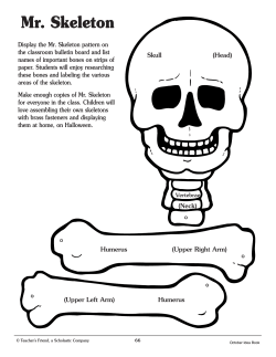


© Copyright 2026