
Topical anti-inflammatory activity of Sal6ia officinalis L. leaves: the
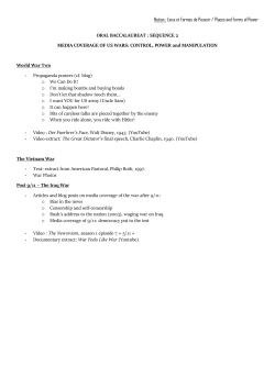
Journal of Ethnopharmacology 75 (2001) 125– 132
www.elsevier.com/locate/jethpharm
Topical anti-inflammatory activity of Sal6ia officinalis L. leaves:
the relevance of ursolic acid
D. Baricevic a,*, S. Sosa b, R. Della Loggia b, A. Tubaro b, B. Simonovska c,
A. Krasna c, A. Zupancic a
a
Agronomy Department, Biotechnical Faculty, Uni6ersity of Ljubljana, 1111 Ljubljana, Slo6enia
b
DEMREP, Uni6ersity of Trieste, Via A. Valerio 6, 34100 Trieste, Italy
c
National Institute of Chemistry Ljubljana, Hajdriho6a 19, 1111 Ljubljana, Slo6enia
Received 15 September 2000; received in revised form 6 November 2000; accepted 5 December 2000
Abstract
Sal6ia officinalis L. leaves, obtained from four plant populations of different origin, were investigated for their topical
anti-inflammatory properties. The n-hexane and the chloroform extracts dose-dependently inhibited the Croton oil-induced ear
oedema in mice, the chloroform extracts being the most active. By contrast, the methanol extracts showed a very low effect and
the essential oil was inactive. Chemical and pharmacological investigation of the most potent chloroform extract, issued from an
autochthonous sage population grown in the submediterranean climatic region of Slovenia, revealed ursolic acid as the main
component involved in its anti-inflammatory activity. The anti-inflammatory effect of ursolic acid (ID50 = 0.14 mMoles/cm2) was
two fold more potent than that of indomethacin (ID50 = 0.26 mMoles/cm2), which was used as a reference non-steroidal
anti-inflammatory drug (NSAID). The content of ursolic acid in sage and sage-based remedies for the topical treatment of
inflammatory diseases is proposed as a parameter for quality control purposes. © 2001 Elsevier Science Ireland Ltd. All rights
reserved.
Keywords: Sal6ia officinalis; Lamiaceae; Anti-inflammatory activity; Plant extracts; Ursolic acid
1. Introduction
The leaves of sage (Sal6ia officinalis L., Lamiaceae)
are well known for their anti-oxidative properties
(Chipault et al., 1956; Farag et al., 1989; Lamaison et
al., 1990; Schwarz and Ternes, 1992; Cuvelier et al.,
1994; Hohmann et al., 1999; Baricevic and Bartol,
2000), used in the food processing industry but applicable also to the area of human health (Pearson et al.,
1997). The plant is reported to have a wide range of
biological activities, such as anti-bacterial, fungistatic,
virustatic, astringent, eupeptic and anti-hydrotic effects
(Dobrynin et al., 1976; Cherevatyıˆ et al., 1980; Anonymus, 1985; Farag et al., 1986). The anti-microbial properties as well as the tannins’ based astringent activities
of sage (active ingredient of dental-care herbal medicinal preparations) benefit the reduction in plaque
* Corresponding author. Fax: +386-61-4231161.
E-mail address: [email protected] (D. Baricevic).
growth, the inhibition of gingival inflammation and
have positive effects on caries prophylaxis (Willershausen et al., 1991).
Furthermore, due to the anti-viral activity of its
water and alcohol extracts, sage is included as an active
ingredient also in combined plant preparations for the
treatment of acute and chronic bronchitis, officially
approved for clinical use in Bulgaria (Manolova et al.,
1995). Other experimental studies on sage extracts or
sage essential oil showed hypotensive properties, central
nervous system-depressant actions and anti-spasmodic
activity (Newall et al., 1996), while the antimutagenic
potential of sage extracts was demonstrated on Escherichia coli repair proficient strains (Baricevic et al.,
1996; Filipic and Baricevic, 1997, 1998). Some constituents of the plant, such as the triterpenes oleanolic
and ursolic acids or the diterpene carnosol, were shown
to present anti-inflammatory properties or related biological activities (Tokuda et al., 1986; Huang et al.,
1994; Liu, 1995). Nevertheless, the anti-inflammatory
0378-8741/01/$ - see front matter © 2001 Elsevier Science Ireland Ltd. All rights reserved.
PII: S 0 3 7 8 - 8 7 4 1 ( 0 0 ) 0 0 3 9 6 - 2
126
D. Barice6ic et al. / Journal of Ethnopharmacology 75 (2001) 125–132
activity of sage and the role of these components in the
anti-phlogistic action of the plant are not yet clearly
defined.
Therefore, S. officinalis leaves were investigated for
their anti-inflammatory property in order to identify
the main compounds involved in this pharmacological
activity. Four different samples of sage were used also
to verify possible variations in the activity due to the
genotype impact. One sample was obtained from autochthonous plants of the submediterranean area of
Slovenia, while the other three samples were obtained
from sage populations of known origin, introduced in
the Slovenian prealpine area. Each sample was extracted by different solvents and the obtained extracts
were evaluated for their ability to inhibit the Croton
oil-induced ear oedema in mice, after topical application (Tubaro et al., 1985). The most active extract was
fractionated in order to find the active principles. Suspected active ingredients were identified by HPLC-UVMS/MS method (Andrensˇek et al., 1999), and
fragmentation spectra of parent ion of external standard were compared to those of the most pharmacologically active fraction to assure the identity of the active
compound.
2. Materials and methods
2.1. Plant material
Samples of Sal6ia officinalis L. leaves were obtained
from spontaneous plants grown in their natural habitat
(sample 1): ‘Petrinje’, autochthonous population of the
submediterranean area of Slovenia, Voucher Nr.DB
13/6-1) or from introduced plant populations grown in
the prealpine region of Slovenia (Genebank Collection
of Medicinal and Aromatic Plants MEDPLANT; sample 2: ‘Petrinje’-population original from the submediterranean area, Voucher Nr.DB 13/6; sample 3:
‘Extracta’-population original from Wies, Austria,
Voucher Nr. Pelz 13/1; sample 4: ‘SALV 25/89’-population original from Gatersleben Genebank, Germany,
Voucher Nr. Ham 13). The plants, harvested at their
full bloom period, were dried in hot-air dryer at 40°C
until constant weight was recorded. The dried leaves,
separated from the stems, were pulverised (sieve No.
0.75) in a mortar grinder and preserved in dark glass
containers until their extraction.
and extracts were dried under a nitrogen gas flow. The
dry plant samples (20 g) were submitted also to steam
distillation in a Clevenger-type apparatus with 200 ml
of water for 2 h to obtain the relevant essential oil.
2.3. Fractionation of the chloroform extract
The dry chloroform extract obtained from sample 1
was fractionated following the scheme presented in Fig.
1. The extract, dissolved in acetone, was bleached with
active carbon and, after filtration, fraction I and a
residue adsorbed on the carbon were obtained. Fraction I was then separated into fractions II and III,
following the method of Wu et al. (1982), slightly
modified: 20 ml water were added to 20 ml of fraction
I — acetone solution obtaining a precipitate (fraction
II) and a supernatant (fraction III).
2.4. High performance thin-layer chromatography
(HPTLC)
Silica gel 60 HPTLC plates (10× 20 cm) with or
without fluorescent indicator (Merck, Darmstadt, Germany) were used. The mobile phase consisted of benzene, ethyl acetate and formic acid (36:12:5, v/v). The
samples and reference substances under analysis were
dissolved in methanol or in acetone (carnosol and
n-hexane extracts) and 20 ml solutions (0.1 mg/ml) were
applied as 8 mm bands 15 mm from the lower edge of
the plate by means of the Linomat IV spotter (Camag,
Muttenz, Switzerland). The plates were developed in an
ascending one-dimensional mode in a saturated glass
chamber. The migration distance of the eluent was 6 cm
(separation time: about 15 min). After separation, the
plates were dried and the chromatographic bands were
2.2. Extraction of plant material
Dried and pulverised leaves (about 30 g) were submitted to three successive extractions with 400 ml of
n-hexane, chloroform or methanol in a Soxhlet apparatus for 24 h. After each extraction, the solvents were
evaporated by Rotavapor (Bu¨chi, Flawil, Switzerland)
Fig. 1. Fractionation scheme of the chloroform extract of sage sample
1.
D. Barice6ic et al. / Journal of Ethnopharmacology 75 (2001) 125–132
detected after spraying the plates with anisaldehyde-sulphuric acid reagent or molybdophosphoric acid
reagent, and observing the plates at 254, 366 or 560 nm
(Jork et al., 1989).
The following reference substances were used:
oleanolic acid, ursolic acid (Sigma-Aldrich Chemie
GmbH, Germany), b-sitosterol, kaempferol, p-coumaric acid, rosmarinic acid, luteolin, luteolin-7-glucoside, apigenin, apigenin-7-glucoside (K. Roth,
Karlsruhe, Germany; Fluka, Buchs, Switzerland;
Aldrich, Milwaukee, USA) and carnosol (a kind gift
from our colleague M. Pukl).
Densitometric analysis of the developed plate was
carried out to quantify the carnosol and ursolic acid.
The carnosol was quantified by densitometric scanning
of the developed plate at 300 nm without derivatization, while the ursolic and oleanolic acids were evaluated after derivatization with anisaldehyde or
molybdophosphoric reagent at 560 nm (Camag TLC
scanner under software control, Muttenz, Switzerland).
Both derivatization reagents gave comparable results.
However, anisaldehyde was more convenient for separation purposes due to the broader colour range for
different compounds.
2.5. High performance liquid chromatography
(HPLC) -UV-Mass spectrometry (MS)
The detection and identification of ursolic acid in
fraction II of chloroform extract was performed on
HPLC-UV-MS system consisting of a pump LDC ConstaMetric 4100 (Thermo Separation Products-TSP, Riviera Beach, CA, USA), a Rheodyne 8125 injector (20
ml) (contact closure; Rheodyne, USA), a LDC Spectromonitor 3200 (TSP) and ion trap LCQ mass spectrometer (Finnigan, MAT, San Jose, CA, USA).
Chromatographic conditions: a stainless-steel column
(150×4.6 mm I.D.; Merck, Darmstadt, Germany)
packed with Hypersil ODS (5 m) was used for in line
separation of ursolic acid with mobile phase consisted
of acetonitril/bi-destilled water mixture in volume ratio
4/1. Flow rate was 2 ml/min. Retention time of ursolic
acid standard was 3.71 min. Injection volume was 20 ml.
Mass detection conditions: capillary temperature was
set at 139.1°C, capillary voltage at −17.25 V, vaporiser
temperature at 403°C, sheat gas flow (N2) rate at 0.46
MPa; auxiliary gas flow (N2) rate at 0.16 MPa, tube
lens offset at − 15.0 V, discharge voltage at 2.3 kV,
discharge current was 5.0 mA and multiplier voltage
was − 950 V. An atmospheric pressure chemical ionisation (APCI) interface was used for direct in-line sample
introduction into mass detector after HPLC separation
and UV detection at 225 nm and negative ions scan
mode was used. For identification of ursolic acid
HPLC/UV/MS measurement of fraction II was made
and compared with external standard of ursolic acid
127
(Sigma-Aldrich Chemie GmbH, Germany). Parent ion
mass of ursolic acid (standard) was 455.5 and isolation
peak width was 1.0 mass unit (Fig. 2A).
2.6. Topical anti-inflammatory acti6ity
Topical anti-inflammatory activity was evaluated as
an inhibition of the Croton oil-induced ear oedema in
mice (Tubaro et al., 1985). Male Albino Swiss mice
(28 –32 g; Harlan Italy, S. Pietro al Natisone, Italy)
were anaesthetised with Ketalar® (145 mg/kg, intraperitoneally; Parke Davis, Milano, Italy). Cutaneous inflammation was induced by application of 15 ml of an
acetone solution containing the irritant (75 mg of Croton oil; Sigma, St. Louis, USA) and the appropriate
amount of the substances under testing to the inner
surface of the right ear of mice (surface: about 1 cm2).
The left ear remained untreated. Control animals received only the irritant. Six hours later, the mice were
sacrificed and a plug (6 mm ¥) was removed from both
the treated and the untreated ears. The oedematous
response was measured as the weight difference between the two plugs. The anti-inflammatory activity
was expressed as a percentage of the oedema reduction
in treated mice compared to the control mice. At least
two experimental groups of five animals were tested for
each dose level. As a reference, the non-steroidal antiinflammatory drug (NSAID) indomethacin was used.
The experimental design was approved by the ethical
committee of the University of Trieste.
2.7. Statistical analysis
The pharmacological data were analysed by Student’s t-test, and a probability level lower than 0.05
was considered to be significant. The doses inhibiting
the oedematous response by 50% (ID50) were calculated
by graphic interpolation of the dose-effect curves.
3. Results
3.1. Extraction yields of plant material
The essential oil contents (%) and extraction yields
(w/w) of the four sage samples are reported in Table 1,
while the yields of fractionation of the chloroform
extract from sample 1 (the most active extract; see the
pharmacological activity) are reported in Fig. 1.
3.2. High performance thin-layer chromatography
(HPTLC)
HPTLC analysis of n-hexane, chloroform and
methanol extracts revealed different chromatographic
profiles. In particular, the n-hexane (with carnosol as
128
D. Barice6ic et al. / Journal of Ethnopharmacology 75 (2001) 125–132
Fig. 2. Chromatograms and spectra — A, shows the standard solution of ursolic acid and B shows the chromatrograms and related spectra of
the fraction II of the chloroform extract of sage (Sal6ia officialis L.) sample 1. The fitting of Rt and fragmentation spectra of standard and
extracted specie clearly confirms the presence of ursolic acid in the fraction II.
the main band at Rf =0.62) and the methanol extracts
(with prevalent rosmarinic acid at Rf =0.15 and caffeic
acid at Rf = 0.41) were characterised by chromatographic lanes in the upper and in the lower part of the
plate, respectively, whereas the chloroform extracts
showed the presence of several bands along the entire
developing path. The chloroform extracts revealed a
pronounced band (Rf =0.65), corresponding to ursolic
and oleanolic acid. This band was observed also in
fractions I and II obtained from the chloroform extract
of sample 1, and, less pronounced, in the n-hexane
extracts. Amongst the reference substances tested, bsitosterol (Rf = 0.66) was well separated from ursolic
and oleanolic acid. The band of carnosol (Rf =0.62)
coloured green with fresh anisaldehyde reagent and
turned to red in a few hours. Kaempferol (orange with
anisaldehyde) and p-coumaric acid (pink) were not
separated (Rf = 0.45). Apigenin and luteolin gave yellow bands at Rf = 0.35 and Rf =0.23, respectively,
while luteolin-7-glucoside and apigenin-7-glucoside remained at start as expected.
No differences in qualitative composition of four
plant samples were identified.
Qualitative HPTLC analyses of the chloroform extract of sample 1 differentiated the ursolic and oleanolic
acid through their different fluorescence after deriva-
tization with anisaldehyde and observation at 366 nm,
i.e. the yellow fluorescence of ursolic acid and the blue
one of oleanolic acid. Moreover, a subsequent densitometric evaluation of the developed plate identified ursolic acid as the main constituent of the band. The
densitometric method was then adopted to quantify
ursolic acid in the chloroform extract of sample 1, in
fractions I, II and III as well as in the relevant n-hexane
extract. The sage constituent carnosol, known for its
anti-inflammatory activity (Huang et al., 1994), was
quantified too. Table 2 shows the results of densitometric quantification of ursolic acid and of carnosol in
pharmacologically active sage extracts of sample 1 and
their fractions.
3.3. High performance liquid chromatography
(HPLC) -UV-Mass spectrometry (MS)
The identity of HPLC/UV peak (Rt= 3.73) recorded
on fraction II sample was subjected to MS/MS spectral
analysis. Comparison of the MS/MS spectra of ursolic
acid standard (Fig. 2A) and that of fraction II (Fig. 2B)
after fragmentation of parent ion at m/z range 454.5 –
456.5, together with retention time (UV-MS detection)
clearly confirms ursolic acid as the main component of
the fraction II.
D. Barice6ic et al. / Journal of Ethnopharmacology 75 (2001) 125–132
129
Table 1
Essential oil contents and extraction yields of samples of Sal6ia officinalis L. leaves
Sample
1
2
3
4
Essential oil content
Dried plant
n-hexane extract
Chloroform extract
Methanol extract
(%)
(g)
(g)
(%)
(g)
(%)
(g)
(%)
1.91
1.74
1.99
1.76
30.44
27.21
29.10
27.93
4.10
2.58
2.04
2.05
13.47
9.48
7.01
7.34
1.51
1.41
1.07
1.24
4.96
5.18
3.68
4.44
6.83
2.71
2.77
3.29
22.44
9.96
9.52
11.78
3.4. Topical anti-inflammatory acti6ity
The essential oil of S. officinalis L. samples did not
show any anti-inflammatory effect after topical application (data not shown). By contrast, the n-hexane and
chloroform extracts obtained from all the four sage
samples induced a dose-dependent oedema-inhibition,
whereas the methanol extracts were almost inactive
(Table 3).
Moreover, considering the extraction yields for each
extract, a drug equivalent (D.E.) value can be derived
from the ID50 values. It represents the amount of crude
drug yielding the ID50 of that extract. The D.E. value
of an extract represents therefore the contribution of
this extract to the activity of the crude drug, in the
sense that a low D.E. value indicates a high contribution to the activity. From the D.E. data reported in
Table 3 it can be seen that for all samples the chloroform extracts gave a higher contribution to the activity
of the S. officinalis L. leaves than the n-hexane ones.
Furthermore, sample 1 proved to be the most active
since both the D.E. of its chloroform and n-hexane
extracts were lower than those of the other three
samples.
Therefore, the chloroform extract from sample 1 was
further investigated, evaluating the activity of its fractions I, II and III, at doses corresponding to 300
mg/cm2 of the extract (Table 4). Fraction I revealed an
activity similar to that of the chloroform extract (85
and 91% inhibition, respectively), showing that the
bleaching procedure of the extract did not remove
significant amounts of its active principles. The activity
passed entirely from fraction I into fraction II, which
induced 91% oedema reduction, whereas fraction III
inhibited the oedematous response only by 18%. The
same doses of fractions II and III, given together,
provoked an almost complete inhibition of the oedematous response (Table 4).
4. Discussion
The results of the study showed, that chloroform
extracts of all four sage samples tested were more active
that the n-hexane extracts since their ID50 values (dose
inducing 50% oedema inhibition) ranged between 105.9
and 139.7 mg/cm2, being lower than those of the n-hexane extracts (ID50 = 438.5 –683.2 mg/cm2). When considering chloroform extracts derived from different
sample sources, the extract of sample 1 proved to be
anti-inflammatory the most potent (ID50 = 105.9 mg/
cm2). Fractionation of sample 1 (spontaneous sage
population from the submediterranean area) chloroform extract yielded about 60% of fraction I, 45% of
fraction II, 15% of fraction III and about 40% of
fraction IV, fraction II being anti-inflammatory the
most active. Since the chemical analyses of fraction II
revealed ursolic acid as its main component, the antiinflammatory activity of this triterpenoid was evaluated, in comparison to that of oleanolic acid and of the
NSAID indomethacin. Both triterpenoid acids induced
a dose-dependent oedema inhibition. In particular,
ursolic acid reduced the oedematous response by 35%
at the dose of 0.1 mMoles/cm2, reaching 84% inhibition
at 0.4 mMoles/cm2, whereas the oedema inhibition induced by oleanolic acid was lower and ranged from 11
to 74%, at the dose range of 0.1 –1 mMole/cm2. The
reference drug indomethacin, administered at 0.12 –0.50
mMoles/cm2, provoked an oedema inhibition ranging
from 29 to 77% (Table 5). The ID50 values of ursolic
acid, oleanolic acid and indomethacin were 0.14, 0.36
and 0.26 mMoles/cm2, the first compound being about
three and two-fold more potent than oleanolic acid and
the NSAID, respectively.
In the chloroform extract carnosol was detected, but
quantification was not possible, because of interference
with a high amount of ursolic acid present in this
Table 2
Ursolic acid and carnosol contents in the active fractions of sage
sample 1
Sample
Ursolic acid (mg/g)
Carnosol (mg/g)
n-hexane extract
Chloroform extract
Fraction I
Fraction II
Fraction III
66
480
756
980
88
82.5
15.0a
25.5a
n.d.
102.5
a
Calculated from the yield and content of carnosol in fraction III,
where interference with ursolic acid was eliminated.
D. Barice6ic et al. / Journal of Ethnopharmacology 75 (2001) 125–132
130
Table 3
Percent oedema inhibition and ID50 values of Sal6ia officinalis L. extracts
Substance
Dose (mg)
Sample 1
Sample 2
Sample 3
Sample 4
n-hexane extr.
100
300
1000
18.6
28.6
60.0
20.6
26.5
67.6
4.2
47.9
69.0
26.8
46.5
60.6
ID50 (mg)
D.E. (mg)a
683.2
5.07
549.6
5.80
438.5
6.26
448.4
6.11
50
75
100
150
200
300
1000
14.3
–
52.9
–
78.6
91.4
98.6
–
–
35.3
55.9
–
86.8
92.6
–
14.1
–
63.4
–
83.1
–
26.8
39.4
60.6
–
88.7
95.8
ID50 (mg)
D.E. (mg)a
105.9
2.13
135.0
2.61
139.7
3.80
124.3
2.80
100
300
1000
2.9
22.9
28.6
1.5
22.1
22.1
–
–
9.9
–
–
26.8
ID50 (mg)
D.E. (mg)a
\1000
\4.46
\1000
\10.04
\1000
\10.51
\1000
\8.49
Chloroform extr.
Methanol extr.
a
D.E.=ID50 expressed equivalents of crude drug.
extract. So, carnosol was quantitatively determined after separation of ursolic acid (in fractionation procedure), i.e. in fraction III, where ursolic acid was found
in a much lower concentration.
The obtained results, reported in Table 2 show, that
ursolic acid was more concentrated in the chloroform
extract than in the n-hexane one. The triterpenic acid
passed from the chloroform extract into fraction I and
then into fraction II, which was constituted almost
completely of ursolic acid. By contrast, the carnosol
was more concentrated in the n-hexane extract than in
the chloroform one; the little amount present in the
chloroform extract passed almost completely into the
fraction III.
Since ursolic acid represents about 50% of the
chloroform extract, its potency (ID50 =0.14 mMoles/
cm2, corresponding to 63.9 mg/cm2) was sufficient to
account for the activity of the extract (ID50 =
105.9 mg/cm2). By contrast, the potency of oleanolic
acid (ID50 =0.36 mMoles/cm2, corresponding to 164.4
mg/cm2) was too low to justify the chloroform
extract activity. Therefore, in accordance with the
chemical results which showed ursolic acid as the main
component of the chloroform extract and of its
fractions I and II, these findings demonstrate the crucial role of this triterpenoid in their anti-inflammatory
activity.
5. Conclusions
The chloroform extracts of Sal6ia officinalis L. leaves
showed strong anti-inflammatory properties after topical application. This activity was influenced by the
plant source. Sample 1, which originated from a spontaneous plant population of the Slovenian submediterranean area, proved to be the most active among the
four samples. Furthermore, ursolic acid, as a component of sage, exhibited strong anti-inflammatory properties, and could explain anti-phlogistic activity of the
official plant drug Sal6iae folium. The ursolic acid
content could thus be considered as a convenient quality control measure in those sage preparations that are
used for their topical anti-inflammatory activity.
Table 4
Anti-inflammatory activity of fractions I–III
Substance
Dose (mg)
N°. animals
Oedema
(mg)
m 9S.E.
% Red.
Controls
Fraction I
Fraction II
Fraction III
Fractions
II+III
–
10
10
10
10
10
7.1 90.3
1.1 90.3*
0.6 90.1*
5.8 90.7*
0.2 90.1*
–
84.5
90.7
18.3
97.2
180
135
45
135+45
* PB0.05 at the Student’s t-test.
D. Barice6ic et al. / Journal of Ethnopharmacology 75 (2001) 125–132
131
Table 5
Anti-inflammatory activity of ursolic acid, oleanolic acid and indomethacin
Substance
Dose (mMoles)
N°. animals
Oedema (mg) m 9 S.E.
% Red.
Ursolic acid
0.00
0.10
0.20
0.40
10
10
10
10
6.5 9 0.3
4.2 90.7a
1.8 90.5a
0.9 90.3a
–
35.4
72.3
86.2
0.00
0.10
0.20
0.40
1.00
10
14
14
12
11
7.2 90.3
6.4 90.3
4.1 9 0.4a
3.4 90.5a
1.9 90.4a
–
11.1
43.0
52.8
73.6
0.00
0.12
0.25
0.50
10
10
20
10
6.5 90.3
4.6 90.4a
3.5 90.4a
1.5 90.2a
–
29.2
46.1
76.9
Oleanolic acid
Indomethacin
a
ID50 (mMoles)
0.14
0.36
0.26
PB0.05 at the Student’s t-test.
Acknowledgements
Part of this work has been supported by a grant from
the Slovenian Scientific Foundation and by the University of Trieste (MURST 60%). We thank Samo Andrensˇek, B. Sc. Chem., for providing us the
HPLC-MS/MS spectra.
References
Andrensˇek, S., S& midovnik, A., Pee`avar, A., Prosˇek, M.., 1999. Routine and sensitive method for determination of nicorandil in
human plasma developed for liquid chromatography with ultraviolet and mass spectrometric detection. Journal of Chromatography, Biomedical Sciences and Applications B735, 103–109.
Anonymus, 1985. Kommission E Monographien, Bundesanzeiger Nr.
90/1985.
Baricevic, D., Bartol, T., 2000. The biological/pharmacological activity of the Salvia genus V., Pharmacology. In: Kintzios, S.E. (Ed.),
Sage: The Genus Salvia. Harwood Academic Publishers, Abingdon, Marston.
Baricevic, D., Filipic, M., Tomazin, E., Cinc, M., Zupancic, A., 1996.
Evaluation of autochthonous and/or introduced genotypes of
medicinal and aromatic plants in Slovenia (Sal6ia officinalis L.).
In: Franz, Ch., Schulz, H., Schumann, G., Sharma, J.R., Tatlioglu, T. (Eds.), Proceedings, Federal Centre for Breeding Research on Cultivated Plants, Quedlinburg, pp. 15–20.
Cherevatyıˆ, V.S., Vashhenko, T.N., Shishkov, G.Z., 1980. Comparative evaluation of the antibacterial action of different extracts
from Sal6ia officinalis. Rastitel’nye Resursy 16, 137–139.
Chipault, J., Mizumo, G., Lundberg, W., 1956. The antioxidant
properties of spices in foods. Food Technology 10, 209–211.
Cuvelier, M.E., Berset, C., Richard, H., 1994. Antioxidant consituents in sage (Sal6ia officinalis). Journal of Agricultural and
Food Chemistry 42, 665–669.
Dobrynin, V.N., Kolosov, M.N., Chernov, B.K., Derbentseva, N.A.,
1976. Antimicrobial substances of Sal6ia officinalis. Khimiya
Prirodnykh Soedineii 5, 686–687.
Farag, R.S., Badei, A.Z.M.A., Hewedi, F.M., El-Baroty, G.S.A.,
1989. Antioxidant activity of some spice essential oils on linoleic
acid oxidation in aqueous media. J. Am. Oil Chem. Soc. 66,
792– 799.
Farag, R.S., Salem, H., Badei, A.Z.M.A., Hassanein, D.E., 1986.
Biochemical studies on the essential oil of some medicinal plants.
Fette Seifen Anstrichmittel 88, 69 – 72.
Filipic, M., Baricevic, D., 1997. Antimutagenic activity of Sal6ia
officinalis extracts against UV-induced mutations in E. coli
strains. Mutation Research 379 (Suppl. 1), 182.
Filipic, M., Baricevic, D., 1998. Inhibitory effect of Sal6ia officinalis
extracts on SOS functions induced by UV-irradiation. Abstract
Book of the 28th Annual Meeting of the Eurropean Environmental Mutagen Society (EEMS), Salzburg, p. 169.
Hohmann, J., Zupko, I., Redei, D., Csanyi, M., Falkay, G., Mathe,
I., Janicsak, G., 1999. Protective effects of the aerial parts of
Salvia officinalis, Melissa officinalis, and Lavandula angustifolia
and their constituents against enzyme-dependent and enzyme-independent lipid peroxidation. Planta Medica 65 (6), 576–578.
Huang, M.T., Ho, C.T., Wuang, Z.Y., Ferraro, T., Luo, Y.R.,
Stauber, K., Ma, W., Georgiadis, C., Laskin, J.D., Conney, A.H.,
1994. Inhibition of skin tumorigenesis by rosemary and ots constituents carnosol and ursolic acid. Cancer Research 54, 701–708.
Jork, H., Funk, W., Fischer, W., Wimmer, H., 1989. Dunnschicht —
Chromatographie, Reagenzien und Nachweismethoden, Band 1 a,
Verlagsgesellschaft mbH, Weinheim, p. 468.
Lamaison, J.L., Petitjean-Freytet, C., Carnat, A., 1990. Teneurs en
acide rosmarinique, en de´rive´s hydroxycinnamiques totaux et
activite´ antioxydante chez les Apiace´es, les Boraginace´e et les
Lamiace´es me´dicinales. Ann. Pharmaceutiques Franc¸aises 48,
103– 108.
Liu, J., 1995. Pharmacology of oleanolic and ursolic acid. Journal of
Ethnopharmacology 49, 57 – 68.
Manolova, N., Serkedjieva, J., Ivanova, V., 1995. Anti-influenza
activity of the plant preparation ‘Broncho Pam’. Fitoterapia LVI,
223– 226.
Newall, C., Anderson, L.A., Phillipson, J.D., 1996. Sage. In: Newall,
C.A., Anderson, L.A., Phillipson, J.D. (Eds.), Herbal Medicines
— A Guide for Health-care Professionals. The Pharmaceutical
Press, London.
Pearson, D.A., Frankel, E.N., Edwin, N., 1997. Inhibition of endothelial cell-mediated oxidation of low-density lipoprotein by
rosemary and plant phenolics. Journal of Agricultural and Food
Chemistry 45, 578– 582.
Schwarz, K., Ternes, W., 1992. Antioxidative constituents of Rosmarinus officinalis and Sal6ia officinalis. II. Isolation of carnosic acid
and formation of other phenolic diterpenes. Z. Lebensm Unters
Forsch 195, 99 – 103.
132
D. Barice6ic et al. / Journal of Ethnopharmacology 75 (2001) 125–132
Tokuda, H., Onigashi, H., Koshimizu, K., 1986. Inhibitory effects of
ursolic and oleanolic acid on skin tumor promotion by 12-O-tetradecanoilphorbol-13-acetate. Cancer Letters 33, 279–285.
Tubaro, A., Dri, P., Delbello, G., Zilli, C., Della Loggia, R., 1985.
The Croton oil ear test revisited. Agents Actions 17, 347–
349.
.
Willershausen, B., Gruber, I., Hamm, G., 1991. The influence of
herbal ingredients on the plaque index and bleeding tendency of
the Gingiva. J. Clin. Dent. 2, 75 – 78.
Wu, J.W., Lee, M.H., Ho, C.T., Chang, S.S., 1982. Elucidation of the
chemical structures of natural antioxidants isolated from rosemary. J. Am. Oil Chem. Soc. 59, 339– 345.


© Copyright 2026