
Introduction Prevalence of Molar-Incisor Hypomineralization in
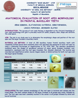
P revalenc e of Molar-Inc is or Hypomineralization in S outh T exas
®
C ervantes Mendez MJ 1 , Abudawood S 1 , Hatc h J P 1 , C hun Y P 2,3
T he Univers ity of T exas Health S c ienc e C enter at S an Antonio, S an Antonio, T X, US A.
1 Department of Developmental Dentis try, 2 Department of P eriodontic s , 3 Department of C ellular and S truc tural B iology
Results
Introduction
Molar-Incisor Hypomineralization (MIH) is defined as lack of mineralization of systemic origin of 1-4
permanent first molars, frequently associated with affected incisors (3). Clinically, it appears as a
hypomineralized lesion of enamel that has a distinct border. Teeth can be affected at variable severity
and can vary in color ranging from white, yellow or brown discoloration (4,5). The reported prevalence
of MIH vary considerably worldwide, ranging from 2.8% in China to 40.2% in Brazil, no data is
available from of the United States (1,2,3).
The aim of this study was to determine the prevalence of MIH in the permanent dentition of children
aged 6 to 14 years old in South Texas. To our knowledge, this is the first report on the prevalence of
MIH in the United States. The second objective of this study was to analyze the distribution pattern of
hypomineralized teeth between different genders, ethnicity and locations, and to specify the more
commonly affected permanent teeth.
Results
Prevalence by Tooth Type
Data was analyzed by examining the
frequencies, percentages at 95% confidence
intervals. Out of 346 investigated children,
119 children were diagnosed with MIH. The
overall prevalence of MIH on permanent
molars and incisors was 34.4% in the study
population.
Overall MIH Prevalence
Affected
None affected
Affected
34%
Score 2: Enamel disintegration
Conclusion
Score 3: Atypical restoration
MIH was the highest at clinics at
Laredo (n=22; 48.9%), followed
by STOHN (n=28; 48.3%),
UTHSCSA (n=22; 32.4%) and
lowest at Ricardo Salinas dental
clinic (n=47; 26.9%).
Materials and Methods
Preliminary data from a larger research project is reported. The research protocol was
approved by Institutional Review Board (IRB). In a cross-sectional design, patients receiving
their initial or semiannual comprehensive dental examination were invited to the study. All
participating dentists were trained and calibrated for diagnosing MIH.
Pediatric patients were recruited from dental practices through the South Texas Oral Health
Network (STOHN) and three clinics affiliated with the Pediatric Dentistry Postgraduate
program at University of Texas Health Science Center at San Antonio (UTHSCSA). Inclusion
criteria for the study were 6-14 years old children with minimum of 1-12 permanent molars
and/or incisors erupted. Exclusion criteria were absence of erupted and/or partially erupted
permanent first molar or incisor at the time of examination and enamel defects caused by
dental fluorosis, amelogenesis imperfecta, dentinogenesis imperfecta, tetracycline, local
infection or trauma.
Demographic information and possible risk factors for hypomineralized enamel were obtained
using a survey. Permanent first molars and incisors were evaluated and tooth surfaces were
scored according to EAPD criteria. The criteria categorizes defects according to it’s severity; a
clearly demarcated opacity at the occlusal or buccal part of the crown of more than 1 mm will
be classified as a score 1. The score will increase depending on the severity of the affected
enamel to a score 4 which is defined as missing teeth that were extracted because of severe
breakdown of MIH as documented in the patient’s record (6).
Max Incisors
1st Max Molar
1st Mand Molar
Mand Incisors
19%
C linic al appearanc e of enamel hypomineralization ac c ording to the E uropean Ac ademy of
P ediatric Dentis try (E AP D) c riteria (6).
Score 1: Demarcated opacity
The tooth type distribution patterns
of MIH were maxillary permanent
incisors (n=59;
19.2%)
most
affected teeth, followed by first
maxillary permanent molars (n=63;
18.6%), mandibular molars (n=55;
16%,) and lower incisors were least
affected at (n=33; 9.8%).
The study sample consisted of 179
(51.7%) females and 167 (48.3%)
males. The frequency of MIH
affected females and males was 69
and 50 respectively. Therefore,
38.5% females and 29.9% males
were affected respectively. The
incidence in females was not
significantly higher than in males at
p<0.05.
18%
16%
10%
Tooth Type
•
The prevalence of MIH in South Texas children is 34.4%. and it is comparable but at the
higher end of the range reported in similar studies in other countries.
•
The most frequently MIH affected teeth were upper permanent incisors (19.2%) vs. lower
incisors (9.8%). Upper first permanent molars were more affected (18.6%) than lower
permanent first molars (16.0%).
•
More research and epidemiological studies are needed to determine the prevalence from
other regions of the US.
Prevalence by Gender
Boys
Girls
39%
30%
Gender
Clinical appearance of MIH in 8 years old male patient: (A)-Moderate yellow opacities on right maxillary FPM (MB cusp) and on the left
maxillary FPM (lingual surface). (B)- Moderate yellow opacities on the lower four permanent incisors and atypical resin restoration on
lower PFMs. (C)-Moderate yellow opacities on maxillary and mandibular permanent incisors (incisal edges), and atypical restorations
on lower PFMs.
References
Prevalence by Ethnicity
Hispanic
Non-Hispanic
44%
32%
Hispanics were represented at 84%
(n=251) and Non-Hispanics at
15.6% (n=39). Demineralization in
Non-Hispanics (43.6%) is not
significantly
higher
than
in
Hispanics (32.0%)
1-Cho SY, Ki Y, Chu V. Molar incisor hypomineralization in Hong Kong Chinese children. Int J Paediatr Dent. 2008;18(5):348-52.
2-Soviero V, Haubek D, Trindade C, Da Matta T, Poulsen S. Prevalence and distribution of demarcated opacities and their
sequelae in permanent 1st molars and incisors in 7 to 13-year-old Brazilian children. Acta Odontol Scand. 2009;67(3):170-5.
3- Weerheijm KL, Jälevik B, Alaluusua S. Molar-incisor hypomineralisation. Caries Res 2001;35: 390–1.
4-Elfrink ME, Schuller AA, Weerheijm KL, Veerkamp JS. Hypomineralized Second Primary Molars: Prevalence Data in Dutch 5Year-Olds. Caries Res 2008;42(4):282–5.
5-Kühnisch J, Heitmüller D, Thiering E, Brockow I, Hoffmann U, Neumann C, Heinrich-Weltzien R, Bauer CP, Berg A, Koletzko S,
Garcia-Godoy F, Hickel R, Heinrich J. Proportion and extent of manifestation of molar-incisor hypomineralizations according to
different phenotypes. J Public Health Dent 2014:74(1)42–9.
6-Lygidakis NA, Wong F, Jälevik B. Best clinical practice guidance for clinicians dealing with children presenting with molar-incisor
hypomineralization (MIH). Eur Arch Paediatr Dent. 2010;11:75-81.
Acknowledgement
Ethnicity
This research was supported by UL1TR001120 from the National Center for Advancing Translational Sciences (MJCM, YPC) and
NIDCR K08 DE 022800 (YPC). The office of the South Texas Oral Health Network (STOHN) directed by Dr. Rahma Mungia and Dr.
Thomas Oates, and by the graduate program in Pediatric Dentistry at UTHSCSA supported this study.


© Copyright 2026