
Clinical Outcomes After Cervical Transcorporeal
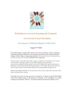
doi: 10.14444/2010
Clinical Outcomes After Cervical Transcorporeal
Microdecompression and Vertebral Body Access Channel
Repair
David W. Lowry, MD, 1 Scott M. Tuinstra, PA-C, 1 Kevin Liang Ph.D, 2 Joseph A. Sclafani MD 2
1 The
Brain and Spine Center, Holland, MI, USA 2 Milestone Research Organization, San Diego, CA
Abstract
Background
Although anterior cervical decompression and fusion (ACDF) can be performed using minimally invasive techniques, the extensive removal of anatomical keystones during decompression requires a segmental fusion to restore
biomechanical stability. Treatment with arthrodesis techniques may result in a prolonged recovery time, loss of
motion, and the need for further treatment if a pseudarthosis or adjacent segment disease occur. Transcorporeal
micro decompression (TCMD) is a newly developed motion sparing, minimally invasive anterior cervical spine decompression procedure that utilizes a small channel through the cervical vertebral body to decompress areas of
central or foraminal stenosis while preserving the native disc. Cervical decompression with TCMD can be performed as a stand-alone or hybrid procedure with ACDF at the adjacent levels. This study retrospectively assesses
patient based clinical outcome measures in patients treated with TCMD.
Methods
A retrospective, non-randomized, single-center chart review of single surgeon experience with patients undergoing
TCMD both with and without adjacent level ACDF using both a trajectory control guide and access channel repair. Statistical analyses were performed on pre and post-operative data collected using visual analog scale (VAS)
and neck disability index (NDI) outcome measures.
Results
Among 62 patients, there were no cases of neurovascular injury, CSF leak, transfusion, or migration of repair implement. Revision surgery was required in 6.4% (n=4) patients. A subanalysis of outcome metrics was performed
for patients that underwent standalone TCMD (TCMD group, n=42) and TCMD with concurrent ACDF at one
or more levels (TCMD+ACDF group, n=20). TCMD group NDI improved from 20.0 to 2.7 at 1 year (p =
0.0001); Axial VAS improved from 5.5 to 0.6 (p = 0.0001); and Radiating VAS improved from 7.0 to 0.7 (p =
0.0001).TCMD+ACDF group NDI improved from 22.0 to 4.0 at 1 year (p = 0.004); Axial VAS improved from 7.1
to 1.2 (p = 0.01); and Radiating VAS trended towards significant improvement from 6.4 to 2.3 (p = 0.09). Mean return to work was 10 days in the TCMD group and 57 days in the TCMD+ACDF group.
Conclusions
Within the limits of a retrospective, single-surgeon study, patients did experience both functional improvement
and pain relief as measured by NDI and VAS respectively from standalone TCMD or combined ACDF / TCMD
procedures. Definitive statements on long-term efficacy, disc space preservation, and motion preservation await
further study.
keywords: minimally invasive spine surgery, motion preservation, clinical outcome, Anterior Cervical Decompression
Volume 9 Article 10 doi: 10.14444/2010
Introduction
Stenosis secondary to cervical spondylosis or an
acute focal cervical disc herniation can manifest clinically as a combination of axial neck pain, cervical
radiculopathy and myelopathy.1 There are two pur-
International Journal of Spine Surgery
1/6
doi: 10.14444/2010
posed mechanisms to explain these clinical sequele.
First, mechanical compression of the exiting nerve
root, either by a soft disc herniation or a disc-osteophyte complex, acts to increase the permeability of
intrinsic nerve root blood vessels. This leads to nerve
root edema, an increased neurologic response
threshold, and ultimately an amplified sensitivity to
pain signals.2,3 Secondly, chemical mediators released
from the pathologic disc fragment (bradykinin, serotonin, histamine, prostaglandins, leukotrienes) as
well as from the compressed nerve root (substance P,
somatostatin, VIP) provoke and perpetuate a dysregulated inflammatory response.4 Surgical decompression of neurovascular structures is indicated if there
is progressive neurologic decompensation or if nonoperative modalities fail to adequately improve the
patient’s quality of life.
Anterior cervical decompression and fusion (ACDF)
is a well-established procedure to treat severe degenerative conditions and focal disc herniation of the
cervical spine.5-8 Although ACDF can be performed
using minimally invasive techniques, the extensive
removal of anatomical keystones during decompression requires a segmental fusion to restore biomechanical stability. Treatment with arthrodesis techniques may result in a prolonged recovery time, loss
of motion, and the need for further treatment if adjacent level disease or a pseudarthosis develop.9 Posterior decompression only techniques such as laminectomy or laminoplasty preserve the adjacent level and
eliminate the risk of pseudarthrosis but increase the
risk of post-operative instability or kyphotic deformity secondary to extensive dissection of the posterior
cervical paraspinal musculature and often times does
not adequately decompress anterior pathologies.10,11
Transcorporeal micro decompression (TCMD) is a
newly developed motion sparing, minimally invasive
cervical spine decompression procedure. TCMD utilizes an anterior approach to channel through the
cervical vertebral body and access the disc pathology.
This technique allows the surgeon to decompress areas of central or foraminal stenosis while preserving
the native disc and posterior stabilizing musculature
of the cervical spine. Cervical decompression with
TCMD can be performed as a stand-alone or hybrid
procedure with an ACDF at the adjacent levels. Pa-
tients presenting with cervical radiculopathy secondary to an isolated soft disc herniation are ideal
candidates for standalone TCMD. Simultaneous
TCMD with adjacent level ACDF is used to address
symptomatic pathology such as a soft disc herniation
with spondylotic myelopathy at other levels. This
study retrospectively assesses perioperative outcomes as well as patient based clinical outcome measures after either a standalone TCMD, or TCMD
with concomitant ACDF performed at the adjacent
level.
Methods
A retrospective, non-randomized, single-center chart
review was conducted for a series of patients that underwent a standalone TCMD procedure using both a
trajectory control guide and access channel repair
(Beta-tricalcium phosphate + local autograft;
TransCorp Spine, Byron Center, Michigan, USA), or
TCMD with concomitant adjacent level ACDF. All
procedures were performed by a single surgeon who
had already overcome the initial procedural learning
curve (DL). Following Institutional Review Board
approval, pre-operative patient demographics (age,
gender, BMI, diagnosis, previous spine surgeries, Visual Analog Scale (VAS) for axial neck and arm pain,
Neck Disability Index (NDI) score, worker’s compensation claim status, co-morbidities) were collected through the chart review. Operative and peri-operative data were obtained from available operative
reports and hospital discharge summaries by an onsite research coordinator. Post-operative complication rate, VAS neck and arm pain score, NDI, time
until return to employment and reoperation rate
were also obtained through chart review. All data
were electronically collected and patient information
was de-identified prior to analysis by an independent
clinical research organization.
Data were segmented for analysis based on age (<50
years versus ≥50 years), procedure (TCMD versus
TCMD with adjacent level ACDF), gender, primary
indication, number of operative levels, previous cervical surgery, pre-operative VAS and NDI, and comorbidity. Data are reported as means ± SD. Statistical analyses were performed via either a one-way
ANOVA or paired t-tests. Statistical analysis of com-
International Journal of Spine Surgery
2/6
doi: 10.14444/2010
plication rate was performed using the chi-squared
method with Yate’s correlation. Significance is defined as p<0.05.
TCMD Procedure
After administration of a general anesthetic, the patient is positioned supine with a rolled towel under
the interscapular region. The incision is made at a
level which permits a direct line of site through the
intended oblique trajectory of the guide, which is
generally similar to where an incision would be
placed if doing an ACDF one segment farther cranially. Once the spine is visualized and levels are confirmed fluoroscopically, the midline is marked at the
disc space above and below the target vertebral body,
and both midline in the sagittal plane and midplane
in the axial plane are identified fluoroscopically,
when possible given patient body habitus. The trajectory control device is then oriented with respect to
midline and midplane, tamped into position on the
anterior surface of the vertebral body, and then secured in position using a single anchoring pin. Positioning is confirmed fluoroscopically when possible.
A hand operated drill with variable depth settings is
used to penetrate the vertebral body in the desired
trajectory to a depth of no more than 3 mm less than
the AP diameter of the vertebral body at midplane as
measured on pre-op studies. (The autograft from the
hand drill is preserved for subsequent channel repair.) The trajectory is obliquely oriented caudally
and laterally to permit access to the anterior aspect of
the foramen while also preserving the uncovertebral
joint. Hemostasis of the cancellous bone is achieved
by passing a hemostatic agent through the trajectory
control guide. After removal of the control guide, the
last 3 mm of bone is removed using a high speed bur.
The epidural plane is then entered in the superomedial quadrant of the access channel by first using a
disc-shaped microdissector. The area of exposed dura is enlarged using Kerrison rongeurs. Osteophytes
are removed using the burr and Kerrisons while herniated disc material is removed using standard microsurgial dissecting instruments. The adequacy of
the decompression is verified both visually and by
feel using a 90-degree ball tip dissector. The access
channel is repaired by using a beta-tricalcium phosphate implant filled with the locally harvested auto-
graft preserved from the hand drill (Figure 1, Figure
2). Prior to placing the implant, a reamer is used to
prepare the channel. After fluoroscopically verifying
implant positioning and achieving hemostasis, the incision is closed.
Results
Sixty-two patients (22 female, 40 male) with a mean
age of 50.6 years (range: 30-71 years) at the time of
surgery were included in the study. The primary surgical indications were degenerative disc disease with
stenosis (DDD, n=37/62, 60%), or acute disc herniation (HNP, n=25/62, 40%). Five patients had undergone previous cervical spine surgery (2 anterior cervical fusion, 3 posterior decompression). Zero patients were treated under pending litigation or a
Worker’s Compensation claim. A standalone TCMD
Fig. 1. Illustration of the stepwise process of a channel repair implant that is
filled with beta-tricalcium phosphate and local autograft.
Fig. 2. Access channel demonstrates ossification at 12 weeks postoperation.
International Journal of Spine Surgery
3/6
doi: 10.14444/2010
was performed in 42/62 patients and 20/62 patients
underwent a combined TCMD with adjacent level
anterior cervical discectomy and fusion procedure
(TCMD + ACDF) at one or more levels (Table 1).
Median hospital admission time was 1 day (range 0-3
days) in the TCMD + ACDF group and 1 day (range
0-2 days) in the standalone TCMD group. Fortythree percent (18/42) of standalone TCMD patients
were discharged home on the day of surgery. Zero
patients required a peri-operative blood transfusion
and there were no cases of neurovascular injury, CSF
leak, or migration of repair implement reported within 90 days of operation. However, revision surgery
was required in 6.4% (n=4/62) patients during the
follow up period. Revision rate was not significantly
higher in the standalone TCMD group (n=2/42)
than in the TCMD + ACDF group (n=2/18, χ2
p>0.05). Mean return to work duration was shorter
in the TCMD group (10 days, n=42, p=0.0001) than
in the TCMD + ACDF group (59 days, n=12) (Figure 3).
TCMD group NDI improved from 20.0 ± 10.4 to 6.5
± 8.1 at 1 month (n=41, p = 0.0001); Axial VAS improved from 5.5 ± 3.1 to 1.6 ± 2.1 (n=42, p = 0.0001);
and Radiating VAS improved from 7.0 ± 2.4 to 1.2 ±
2.0 (n=41, p = 0.0001). At 1 year post-op, the TCMD
group NDI improved from 20.0 ± 10.4 to 2.7 ± 3.9
(n=22, p = 0.0001); Axial VAS improved from 5.5 ±
3.1 to 0.6 ± 0.8 (n=22, p = 0.0001); and Radiating
VAS improved from 7.0 ± 2.4 to 0.7 ± 1.4 (n=22, p =
0.0001). The TCMD + ACDF group NDI improved
from 22.0 ± 11 to 15.3 ± 9.3 at 1 month (n=18, p =
Table 1. Patient demographics.
0.05); Axial VAS improved from 7.1 ± 2.1 to 2.4 ± 2.1
(n=18, p = 0.0001); and Radiating VAS improved
from 6.4 ± 2.9 to 2.3 ± 2.2 (n=18, p = 0.0001). At one
year post-op, the TCMD + ACDF group NDI improved from 22.0 ± 11 to 4.0 ± 3.5 (n=7, p = 0.004);
Axial VAS improved from 7.1 ± 2.1 to 1.2 ± 1.0 (n=7,
p = 0.01); and Radiating VAS trended towards significant improvement from 6.4 ± 2.9 to 2.3 ± 3.2 (n=7, p
= 0.09) (Table 2). There was not a significant difference in outcome measures between groups when the
data was stratified based on patient age, sex, number
of levels treated and specific level treated (p>0.05).
Fig. 3. Mean return to work duration was shorter in the TCMD than in the
TCMD + ACDF group. All patients in the TCMD group returned to work by
post-op day 60 and all patients in the TCMD + ACDF group returned to
work by post-op day 120.
Table 2. Patient based outcome measures for the standalone TCMD group
and TCMD + ACDF group.
TCMD
Preoperation
1 year postoperation
Paired t-test
(p)
NDI
20 ± 10.4 (n =
41)
2.7 ± 2.9 (n = 22)
0.0001
VAS Neck
5.5 ± 3.1 (n =
42)
0.6 ± 0.8 (n = 22)
0.0001
VAS Arm
7.0 ± 2.4 (n =
41)
0.7 ± 1.4 (n = 22)
0.0001
Preoperation
1 year postoperation
Paired t-test
(p)
22 ± 11 (n = 18)
4.0 ± 3.5 (n = 7)
0.004
VAS Neck
7.1 ± 2.1 (n =
18)
1.2 ± 1.0 (n = 7)
0.01
VAS Arm
6.4 ± 2.9 (n =
18)
2.3 ± 3.2 (n = 7)
0.09
n
Total patients
Mean age at surgery (years)
62
50.6
Diagnosis
Degenerative Disc Disease with Stenosis
37
TMCD +
ACDF
Acute Disc Herniation
25
NDI
Procedure
Standalone TCMD
42
TCMD + ACDF procedure
20
International Journal of Spine Surgery
4/6
doi: 10.14444/2010
Discussion
Within the limits of a single center retrospective review, the results of this study demonstrate that
TCMD is a safe and effective minimally invasive surgical option to treat cervical spondylosis with radiculopathy or acute cervical disc herniation. There were
no major complications reported and no cases that
required a peri-operative blood transfusion. Overall
length of hospital stay compares favorably to previous studies of cervical decompression procedures.12,13
Patients in the standalone TCMD group were able to
achieve a level of function necessary to return to
work significantly faster than those treated with
TCMD + ACDF. However, both standalone TCMD
as well as TCMD with concomitant adjacent level
ACDF groups reported significant improvement in
pain scores and functional outcome as early as one
month post-op that continued until the 1 year postoperative clinical follow up visit.
There are several advantages to implementing a
transcorporeal microdecompression with concomitant adjacent level ACDF over a multilevel cervical
fusion in cases of severe multilevel deformity.
TCMD eliminates the risks of postoperative instrumentation failure or pseudoarthosis at the TCMD
level and requires a less extensive dissection than
ACDF to reach the surgical target. Additionally, limiting the extension of arthrodesis preserves an additional native motion segment which can protect
against adjacent level degenerative changes. Prasarn
et al. conducted one of the few studies in the literature on adjacent level biomechanics after single versus multilevel cervical spine fusion.14 These authors
demonstrated that extension of a single level fusion
to a two-level fusion in a cadaveric model system increased sagittal range of motion and stress forces at
the motion segment adjacent to the arthrodesis. In
light of these findings, performing a motion preserving TCMD procedure together with ACDF in cases
of multilevel pathology will likely reduce the rate of
adjacent level hypermobility and accelerated degeneration.
While motion was preserved at the one year post-op
mark for all patients who have reached that point, a
limitation of our study is that long-term efficacy, disc
space maintenance and motion preservation were not
studied. Several previous studies have confirmed that
arthrodesis of a cervical motion segment leads to increased stress, load, and intradiscal pressures at the
level adjacent to the fusion construct.15-19 Such forces
likely accelerate degenerative changes in levels adjacent to arthrodesis which could lead to clinical deterioration necessitating further intervention. Hilibrand and Robbins followed 374 patients over a period of 10 years after ACDF and found that symptomatic adjacent level disease developed at a rate of
2.9% per year.20 Anterior transcorporeal decompression techniques are designed to remove anterior
pathologic compressive fragments while limiting disruption of the native disc and bony stabilizers of the
cervical spine.21 The clinical impact of preserving the
native anatomical architecture during TCMD procedures should be investigated in future dedicated
studies of long term functional and radiographic outcomes. Future studies will also compare the clinical
outcome of standalone TCMD versus standalone
ACDF procedures to better appreciate the best indications for each respective procedure.
References
1. Rao R. Neck Pain, Cervical Radiculopathy, and
Cervical Myelopathy Pathophysiology, Natural History, and Clinical Evaluation. J Bone Joint Surg Am.
2002;84(10):1872-1881.
2. Cooper RG, Freemont AJ, Hoyland JA, Jenkins
JP, West CG, Illingworth KJ, Jayson MI. Herniated
intervertebral disc-associated periradicular fibrosis
and vascular abnormalities occur without inflammatory cell infiltration. Spine. 1995;20:591-8.
3. Humphreys SC, Hodges SD, Patwardhan A, Eck
JC, Covington LA, Sartori M. The natural history of
the cervical foramen in symptomatic and asymptomatic individuals aged 20–60 years as measured by
magnetic resonance imaging. A descriptive approach. Spine. 1998; 23:2180–2184.
4. Chabot MC, Montgomery DM. The pathophysiology of axial and radicular neck pain. Semin Spine
Surg.1995;7:2-8.
5. Cloward RB. The anterior approach for removal
of ruptured cervical disks. J Neurosurg. 1958;
International Journal of Spine Surgery
5/6
doi: 10.14444/2010
15:602–617.
6. Kaiser MG, Haid RWJ, Subach BR, Barnes B,
Rodts GEJ. Anterior cervical plating enhances
arthrodesis after discectomy and fusion with cortical
allograft. Neurosurgery. 2002;50:229–236.
7. Robinson RA, Smith GW. Antero-lateral cervical
disc removal and interbody fusion for cervical disc
syndrome. Bull Johns Hopkins Hosp. 1955;
96:223–224.
8. Wang JC, McDonough PW, Endow K, Delamarter,RB. Increased fusion rates with cervical plating for two-level anterior cervical discectomy and fusion. Spine. 2000;25:41–45.
9. Yonenobu Y, Cervical radiculopathy and
myelopathy: when and what can surgery contribute
to treatment? Eur Spine J. 2000; 9: 1–7.
10. Longstein J. Post-Laminectomy kyphosis spinal
deformities and neurologic dysfunction. New York,
Raven Press. 1978; 53-63.
11. Sim FH, Suien HJ, Bickel WH, Janes JM. Swan
neck deformity following extensive cervical laminectomy. J Bone Joint Surgery. 1974; 56A:564-580.
12. Stieber JR, Brown K, Donald GD, Cohen JD.
Anterior cervical decompression and fusion with
plate fixation as an outpatient procedure. The Spine
Journal. 2005; 5(5), 503-507.
13. Fessler RG, Khoo LT. Minimally invasive cervical microendoscopic foraminotomy: an initial clinical
experience. Neurosurgery. 2002; 51(5), S2-37.
14. Prasarn ML, Baria D, Milne E, Latta L,
Sukovich W. Adjacent-level biomechanics after single versus multilevel cervical spine fusion. Laboratory investigation. J Neurosurg Spine. 2012;
16:172–177.
15. Ren C, Song Y, Xue Y, Yang X. Mid- to longterm outcomes after cervical disc arthroplasty compared with anterior discectomy and fusion: a systematic review and meta-analysis of randomized controlled trials. European Spine Journal. 2014; 23:5,
1115-1123.
16. Kramer J, Javidan Y, Smith J. The value of disk
replacement?. Seminars in Spine Surgery. 2014; 26:1,
45-51.
17. Sundseth J, Jacobsen EA, Kolstad F, Nygaard
OP, Zwart JA, Hol PK. Magnetic resonance imaging
evaluation after implantation of a titanium cervical
disc prosthesis: a comparison of 1.5 and 3 Tesla magnet strength. European Spine Journal. 2013; 22:10,
2296-2302.
18. Zigler JE, Delamarter R, Murrey D, Spivak J,
Janssen M. ProDisc-C and Anterior Cervical Discectomy and Fusion as Surgical Treatment for SingleLevel Cervical Symptomatic Degenerative Disc Disease. Spine. 2013; 38:3, 203-209.
19. Coric D, Kim PK, Clemente JD, Boltes MO,
Nussbaum M, James S. Prospective randomized
study of cervical arthroplasty and anterior cervical
discectomy and fusion with long-term follow-up: results in 74 patients from a single site. Journal of Neurosurgery: Spine. 2013;18:1, 36-42.
20. Hilibrand AS, Robbins M: Adjacent segment
degeneration and adjacent segment disease: the consequence of spinal fusion?. Spine J 6 Suppl. 2004;
4:190S–1944S.
21. Choi G, Lee SH, Bhanot A, Chae YS, Jung B,
Lee S.Modified transcorporeal anterior cervical microforaminotomy for cervical radiculopathy: a technical note and early results. European Spine Journal.
2007; 16(9), 1387-1393.
Corresponding Author
Joseph Sclafani, Milestone Research Organization,
San Diego, CA. [email protected]
Disclosures
Conflicts of Interest and Source of Funding: Milestone Research Organization was hired as the clinical
research organization to conduct the study. This entity was compensated to conduct the study, analyze
results and author the manuscript.
Published 30 March 2015.
This manuscript is generously published free of
charge by ISASS, the International Society for the
Advancement of Spine Surgery. Copyright © 2015
ISASS. To see more or order reprints or permissions,
see http://ijssurgery.com.
International Journal of Spine Surgery
6/6


© Copyright 2026