
Angioedema In The Emergency Department: An Evidence-Based Review November 2012
Angioedema In The Emergency Department: An Evidence-Based Review R. Gentry Wilkerson, MD, FACEP, FAAEM Assistant Professor, Coordinator for Research, Department of Emergency Medicine, University of Maryland School of Medicine, Baltimore, MD Alex Manini, MD, MS Angioedema is the end result of a variety of pathophysiological processes resulting in transient, localized, nonpitting swelling of the subcutaneous layer of the skin or submucosal layer of the respiratory or gastrointestinal tracts. It is now generally accepted that the swelling is mediated by either histamine or bradykinin. Angioedema may result in severe upper airway compromise or—less commonly recognized—compromise in the gastrointestinal tract often associated with severe abdominal pain. A variety of new therapeutic options are becoming available for use in the United States that have the potential to greatly impact the management and outcomes for those with severe clinical manifestations. This review assesses the evidence on the causes and treatments of angioedema in the emergency department and reviews the new therapeutic options available for treatment of angioedema based on their effectiveness, price, and availability. Editor-in-Chief Medical Center, University of North Carolina School of Medicine, Chapel Andy Jagoda, MD, FACEP Hill, NC Professor and Chair, Department of Emergency Medicine, Mount Sinai Steven A. Godwin, MD, FACEP School of Medicine; Medical Director, Professor and Chair, Department Mount Sinai Hospital, New York, NY of Emergency Medicine, Assistant Dean, Simulation Education, Editorial Board University of Florida COMWilliam J. Brady, MD Jacksonville, Jacksonville, FL Professor of Emergency Medicine, Gregory L. Henry, MD, FACEP Chair, Resuscitation Committee, CEO, Medical Practice Risk University of Virginia Health System, Assessment, Inc.; Clinical Professor Charlottesville, VA of Emergency Medicine, University of Peter DeBlieux, MD Michigan, Ann Arbor, MI Louisiana State University Health Science Center Professor of Clinical John M. Howell, MD, FACEP Clinical Professor of Emergency Medicine, LSUHSC Interim Public Medicine, George Washington Hospital Director of Emergency University, Washington, DC; Director Medicine Services, LSUHSC of Academic Affairs, Best Practices, Emergency Medicine Director of Inc, Inova Fairfax Hospital, Falls Faculty and Resident Development Church, VA Francis M. Fesmire, MD, FACEP Shkelzen Hoxhaj, MD, MPH, MBA Professor and Director of Clinical Research, Department of Emergency Chief of Emergency Medicine, Baylor College of Medicine, Houston, TX Medicine, UT College of Medicine, Chattanooga; Director of Chest Pain Eric Legome, MD Center, Erlanger Medical Center, Chief of Emergency Medicine, Chattanooga, TN King’s County Hospital; Professor of Michael A. Gibbs, MD, FACEP Professor and Chair, Department of Emergency Medicine, Carolinas Author Peer Reviewers Abstract Nicholas Genes, MD, PhD Assistant Professor, Department of Emergency Medicine, Mount Sinai School of Medicine, New York, NY November 2012 Volume 14, Number 11 Clinical Emergency Medicine, SUNY Downstate College of Medicine, Brooklyn, NY Keith A. Marill, MD Assistant Professor, Harvard Medical School; Emergency Department Attending Physician, Massachusetts General Hospital, Boston, MA Assistant Professor of Emergency Medicine, Mount Sinai School of Medicine, New York, NY James Scott, MD Professor of Emergency Medicine and Health Policy, The George Washington University of Medicine and Health Sciences, Washington, DC CME Objectives Upon completion of this article, you should be able to: 1. 2. 3. 4. Discuss the various types of angioedema and the mechanism by which the swelling occurs. Describe an appropriate workup of a patient with undifferentiated angioedema. Determine appropriate therapy for treatment of angioedema and understand the rationale behind each therapeutic option. Discuss appropriate disposition for these patients with an understanding of which patients are at higher risk of needing a higher level of care. Date of original release: November 1, 2012 Date of most recent review: October 10, 2012 Termination date: November 1, 2015 Medium: Print and Online Method of participation: Print or online answer form and evaluation Prior to beginning this activity, see “Physician CME Information” on the back page. Charles V. Pollack, Jr., MA, MD, Stephen H. Thomas, MD, MPH George Kaiser Family Foundation FACEP Professor & Chair, Department of Chairman, Department of Emergency Emergency Medicine, University of Medicine, Pennsylvania Hospital, Oklahoma School of Community University of Pennsylvania Health Medicine, Tulsa, OK System, Philadelphia, PA Jenny Walker, MD, MPH, MSW Michael S. Radeos, MD, MPH Assistant Professor, Departments of Assistant Professor of Emergency Preventive Medicine, Pediatrics, and Medicine, Weill Medical College Medicine Course Director, Mount of Cornell University, New York; Sinai Medical Center, New York, NY Research Director, Department of Emergency Medicine, New York Ron M. Walls, MD Hospital Queens, Flushing, New York Professor and Chair, Department of Emergency Medicine, Brigham and Robert L. Rogers, MD, FACEP, Women’s Hospital, Harvard Medical FAAEM, FACP School, Boston, MA Assistant Professor of Emergency Medicine, The University of Scott Weingart, MD, FACEP Maryland School of Medicine, Associate Professor of Emergency Baltimore, MD Medicine, Mount Sinai School of Alfred Sacchetti, MD, FACEP Assistant Clinical Professor, Department of Emergency Medicine, Thomas Jefferson University, Philadelphia, PA Medicine; Director of Emergency Critical Care, Elmhurst Hospital Center, New York, NY Scott Silvers, MD, FACEP Chair, Department of Emergency Medicine, Mayo Clinic, Jacksonville, FL Joseph D. Toscano, MD Emergency Physician, Department of Emergency Medicine, San Ramon Regional Medical Center, San Ramon, CA Senior Research Editor Corey M. Slovis, MD, FACP, FACEP Professor and Chair, Department Research Editor of Emergency Medicine, Vanderbilt University Medical Center; Medical Matt Friedman, MD Director, Nashville Fire Department and Emergency Medical Services Fellow, International Airport, Nashville, TN Fire Department of New York, New York, NY International Editors Peter Cameron, MD Academic Director, The Alfred Emergency and Trauma Centre, Monash University, Melbourne, Australia Giorgio Carbone, MD Chief, Department of Emergency Medicine Ospedale Gradenigo, Torino, Italy Amin Antoine Kazzi, MD, FAAEM Associate Professor and Vice Chair, Department of Emergency Medicine, University of California, Irvine; American University, Beirut, Lebanon Hugo Peralta, MD Chair of Emergency Services, Hospital Italiano, Buenos Aires, Argentina Dhanadol Rojanasarntikul, MD Attending Physician, Emergency Medicine, King Chulalongkorn Memorial Hospital, Thai Red Cross, Thailand; Faculty of Medicine, Chulalongkorn University, Thailand Suzanne Peeters, MD Emergency Medicine Residency Director, Haga Hospital, The Hague, The Netherlands Accreditation: EB Medicine is accredited by the ACCME to provide continuing medical education for physicians. Faculty Disclosure: Dr. Manini, Dr. Scott, Dr. Jagoda, and their related parties report no significant financial interest or other relationship with the manufacturer(s) of any commercial product(s) discussed in this educational presentation. The following disclosure of relevant financial interest with a potentially financially interested entity was made: Dr. Wilkerson reported that he had received research support from Dyax, Corp. Commercial Support: This issue of Emergency Medicine Practice did not receive any commercial support. Case Presentations In the middle of an unusually slow evening shift, a 52-year-old black male presents to the ED from walk-in triage with a complaint of lip swelling. He states that he noticed a tingling in his lips shortly after waking that morning, but it wasn’t until he brushed his teeth that he noticed how large his lips had become. He decided to come to the hospital almost 12 hours later only after family members insisted that he get “checked out.” He denies any recent trauma, infection, or known exposures to possible allergens. He denies any pain or itching. His past medical history is significant for hypertension and borderline diabetes. He is unable to remember the name of the medication that he takes for his blood pressure, but he says he has been taking it for years. His vital signs are: heart rate, 74 beats per minute; blood pressure, 156/82 mm Hg; respiratory rate, 16 breaths per minute; temperature, 36.8°C; and oxygen saturation, 98% on room air. He is comfortable and in no apparent distress. It would be impossible to miss the rather impressive size of his lips. The upper lip looks to be about 10 times the normal size and the lower lip is only somewhat less enlarged. You are able to examine his oropharynx and find no further swelling of the uvula or posterior pharynx. The rest of his examination is unremarkable. Your nurse checks the airway cart out of concern that the patient will need to be immediately intubated. Your medical student asks the following logical questions: • What is the cause of his lip swelling? • Is there a diagnostic test to determine the cause? • What is the appropriate treatment? • Should the patient be intubated immediately to protect his airway? The next week, you see a 19-year-old white female who is brought in by EMS for severe diffuse abdominal pain. She states that the pain began 8 hours earlier. She has had nausea with multiple episodes of vomiting. She denies fever, recent antibiotics, foreign travel, or sick contacts. She does state that she has had similar presentations multiple times in the past but never this pronounced. She denies any past medical history other than recurrent abdominal pain similar to how she is currently presenting. She was recently started on oral contraceptives, but she denies being sexually active. Her vital signs are: heart rate, 112 beats per minute; blood pressure, 92/64 mm Hg; respiratory rate, 22 breaths per minute; temperature, 36.9°C; and oxygen saturation, 100% on room air. She is obviously uncomfortable and actively retching. Her physical exam is remarkable for a soft but markedly distended abdomen with evidence of shifting dullness. She is diffusely tender to palpation without guarding or rebound. A pelvic exam is unremarkable. After reviewing her vital signs and performing her physical exam, you are much more concerned about this patient. You pull over the portable ultrasound to take a look and are immediately impressed by a large amount of free fluid in the abdomen. Emergency Medicine Practice © 20122 You ask yourself several questions: • What is the cause of her recurrent abdominal pain? • Does she need an abdominal CT scan, and, if yes, does it need to be with contrast? • Does she need emergent surgical consultation? • Other than treating the pain, is there any medication that is indicated? Introduction Angioedema is the clinical manifestation of transient, localized, nonpitting swelling of the subcutaneous layer of the skin or submucosal layer of the respiratory or gastrointestinal tracts. The first widely recognized description of angioedema was by Heinrich Quincke in 1882. In honor of his contribution, it is sometimes referred to as Quincke edema. Angioedema is not a disease; rather, it is a physical manifestation of a variety of pathophysiological processes. These processes have the end result of either mast cell degranulation or formation of bradykinin. The incidence of angioedema, overall, is not known. For patients taking angiotensin-converting enzyme (ACE) inhibitors, the incidence is between 0.1% and 0.7%. The prevalence of the various forms of hereditary angioedema is estimated to be between 1 in 10,000 and 1 in 50,000.1 The emergency clinician must understand the various causes of angioedema and tailor the treatment of the patient based on the likely etiology. In this issue of Emergency Medicine Practice, the various causes of angioedema are discussed as well as different diagnostic and treatment strategies. Critical Appraisal Of The Literature A literature search was performed using PubMed from 1964 to present, using the search term angioedema and limited to English-language articles that were systematic reviews, meta-analyses, multicenter studies, clinical trials, or randomized clinical trials. Using this approach, 269 articles were found and assessed for review. In addition, 1502 case reports were identified in the search; however, it was determined that they would add little to the evidence provided by the better-quality resources, and, therefore, these case reports were not systematically reviewed. A search of the National Guidelines Clearinghouse (www.guideline.gov) produced no additional practice guidelines focused on angioedema. There was 1 guideline for urticaria that resulted when a search for angioedema was performed.2 A review of the Cochrane Database of Systemic Reviews failed to find any completed reviews. There is a protocol in place for a review of “angiotensin-converting enzyme inhibitor-induced angioedema in patients with primary hypertension,” but it has not been completed. www.ebmedicine.net • November 2012 aldosterone system. Bradykinin is thought to be the primary mediator of most nonallergic forms of angioedema. In this system, activated factor XII from the coagulation cascade converts prekallikrein to kallikrein in a positive feedback loop. Activated factor XII also has a role in activation of the classical pathway of the complement system. This results in depletion of downstream components such as C4. Kallikrein, in turn, cleaves high-molecular-weight kininogen to form bradykinin. The half-life of bradykinin is approximately 15 to 30 seconds, and the concentration is typically very low.3 There are 2 known types of bradykinin receptors, B1 and B2, that are both encoded on chromosome 14. Bradykinin activates endothelial cells through the B2 receptor, leading to increased vascular permeability and subsequent formation of edema. There are specific zinc metalloproteases that are responsible for the metabolism of the kinins: angiotensin-I-converting enzyme (ACE), aminopeptidase P, neutral endopeptidase, and carboxypeptidases M and N. Etiology And Pathophysiology There are 6 broad categories of angioedema, based on pathophysiology: • Hereditary • Acquired • Immunologic/allergic • ACE inhibitor induced • Physically induced • Idiopathic A brief overview of the mediator pathways is warranted to help understand the various types of angioedema, the treatment options, and the expected clinical course. (See Figure 1.) Mediators Of Angioedema Bradykinin Bradykinin is a vasoactive nonapeptide that is formed as an end product of the kallikrein-kinin system. This system helps to counterbalance the vasoconstrictive properties of the renin-angiotensin- Figure 1. Interaction Of Kallikrein-Kinin System, Coagulation Pathway, And Common Complement Pathway Antigens Antibody Classic complement pathway C4b C1s Kallikrein-kinin system C4a C1s C1q C1r C1r C1-INH C4 Factor XIIf HMWK Factor XII Intrinsic pathway Factor XI Factor XIIa C1-INH ecallantide Bradykinin C1-INH Factor XIa B1-rec Kallikrein Prekallikrein Icatibant Factor IX Factor IXa B2-rec Factor X Factor Xa Common pathway Image courtesy of R. Gentry Wilkerson, MD. Abbreviations: B1-rec, bradykinin 1 receptor; B2-rec, bradykinin 2 receptor; C1-INH, C1 esterase inhibitor; HMWK, high-molecular-weight kininogen. November 2012 • www.ebmedicine.net 3 Emergency Medicine Practice © 2012 C1 esterase inhibitor (C1-INH) is an alpha2-globulin glycoprotein primarily formed within hepatocytes and loosely bound to C1. It is a member of the serine protease-inhibitor (serpin) family, which includes alpha-1-antitrypsin and antithrombin and is encoded by the SERPING1 gene located on the long arm of chromosome 11. C1-INH inhibits the function of C1s and C1r of the classic complement pathway. It also plays a major role in inhibition of other components such as kallikrein, factor XIa, and factor XIIa. Histamine Histamine is an amino acid derivative that is formed by the decarboxylation of histidine. Most of the conversion occurs within granules of mast cells in tissues or basophils circulating in the bloodstream. Histamine has many roles in the body, but its primary role is within the immune system. Mast cells release histamine through a process known as degranulation when they have been sensitized with IgE antibodies and then come in contact with an appropriate antigen. Certain drugs are also capable of causing mast cell degranulation through a nonimmunologic mechanism. Examples of such drugs include morphine and tubocurarine. Additionally, activation of the complement system may also result in mast cell degranulation. There are 4 known receptors for histamine, designated H1 through H4; however, only the receptors H1 and H2 are involved in the formation of edema that can lead to angioedema. H1 receptors are located on endothelial and smooth muscle cells as well as in the central nervous system. Activation of H1 receptors results in vasodilation, bronchoconstriction, and pruritus. H2 receptors are found in vascular smooth muscle cells and parietal cells of the stomach. Activation of H2 receptors results in vasodilation and stimulation of gastric acid secretion. Histamine is metabolized by the enzymes histamine N-methyltransferase and diamine oxidase. Causes Of Angioedema Hereditary Angioedema Hereditary angioedema is a genetic form of angioedema that is most often due to a deficiency in either the quantitative or functional levels of C1INH. William Osler was the first to demonstrate the heritable pattern of hereditary angioedema in 1888. In the original report, he described clinical findings of 5 generations in a single family. Hereditary angioedema is most often inherited in an autosomaldominant fashion, with most cases being heterozygous (although homozygous individuals have been described).4 C1-INH serves as the main regulator of the kallikrein-kinin system. As a result of decreased amounts of functional C1-INH, when the kallikreinkinin system is activated, it is not kept in check. This Emergency Medicine Practice © 20124 leads to increased formation of bradykinin and the resultant increased vascular permeability and edema formation. Nussberger et al showed increased bradykinin levels in patients with hereditary angioedema during acute attacks that returned to normal or near-normal during periods of remission.5 More than 200 mutations of the SERPING1 gene on chromosome 11 have been described.6 It is thought that up to 25% of these mutations are de novo, and, therefore, there will be no prior family history of angioedema. Patients with hereditary angioedema have varying frequency of angioedema episodes. Some present with frequent episodes occurring every couple of weeks, whereas others go years without having an episode. There are at least 3 forms of hereditary angioedema described. Hereditary angioedema type I, which comprises approximately 85% of cases of hereditary angioedema, is the result of mutations in the genetic code that cause decreased transcription of the gene coding for C1-INH. This leads to a deficiency in the quantitative level of C1-INH. Patients with hereditary angioedema type I typically have C1-INH levels < 40%, below the 50% level one would expect for an individual heterozygous for this condition. This is likely due to hypercatbolism of the gene product from the 1 functional gene found in those who are heterozygous. The decreased inhibition of the complement component C1 leads to decreased levels of the downstream components of the complement system, including C4 and C2. Hereditary angioedema type II, which comprises approximately 15% of cases of hereditary angioedema, is the result of a genetic defect that allows for the transcription of a functionally defective C1-INH protein. This results in normal or slightly elevated quantitative (but decreased functional) levels of C1-INH. A third form of hereditary angioedema (type III), which has normal C1-INH levels, was originally discovered in 1986 by Warin et al7 but failed to receive significant interest until 2000 when it was independently described by Bork et al8 and Binkley and Davis.9 Bork initially postulated an X-linked inheritance pattern, whereas Binkley and Davis suggested that the inheritance was most likely autosomal dominant. It is now accepted that the most likely inheritance pattern is autosomal dominant. Initially, only female patients were described; later, males were identified, but with much less frequency.10,11 The gender difference is possibly due to males lacking a female-specific risk factor or possessing a male-specific protective factor. Clinical expression often occurs later, at an average age of 26.8 years.12,13 Clinical symptoms appear to worsen during periods of increased estrogen levels, such as in pregnancy and hormone replacement. Multiple missense mutations have been identified in the gene encoding Hageman www.ebmedicine.net • November 2012 anaphylaxis. The swelling caused by an allergic reaction usually subsides within 24 hours, but relapses are common and unpredictable. Inciting agents are numerous and varied. Most medications that cause angioedema do so through this process. There is frequent confusion among clinicians about what constitutes anaphylaxis. According to a 2006 position paper from the National Institute of Allergy and Infectious Disease,21 anaphylaxis is highly likely when any 1 of 3 possible constellations of clinical findings is present: 1. Acute onset of a reaction (minutes to hours) with involvement of skin and/or mucosal tissue and at least 1 of the following: • Respiratory compromise • Reduced blood pressure or symptoms of end-organ dysfunction 2. Two or more of the following that occur rapidly after exposure to a likely allergen: • Involvement of skin/mucosal tissue • Respiratory compromise • Reduced blood pressure or associated symptoms • Persistent gastrointestinal symptoms 3. Reduced blood pressure after exposure to a known allergen factor (factor XII).14-16 Estrogen stimulation has been previously shown to increase transcription of factor XII. The mutations found in patients with hereditary angioedema type III likely result in upregulation of Hageman factor in response to estrogen and resulting increase in bradykinin generation through the kallikrein-kinin system. Acquired Angioedema The term acquired angioedema refers to angioedema that is the result of C1-INH deficiency that is not due to a genetic defect. It does not refer to angioedema that is acquired due to other mechanisms, such as drug-induced angioedema. Caldwell et al first described this form of angioedema in 1972.17 The incidence of acquired angioedema is exceedingly rare, with only a few hundred cases reported. It usually presents later in life, rarely before the fourth decade of life. The pathophysiology has not been fully determined. Although it is possibly an oversimplification, acquired angioedema is typically described as having 2 distinct forms. In both forms, there is normal initial production of C1-INH. Acquired angioedema type I results from increased catabolism of C1INH. It is often associated with lymphoproliferative or autoimmune disease. As a part of the disease process, there is increased activation of the classical pathway of complement, leading to consumption of available C1-INH. Acquired angioedema type II, first described by Jackson et al in 1986,18 is defined by the presence of an autoantibody to C1-INH that results in increased proteolysis of C1-INH to an inactive molecule.19 There is a blurring of these 2 types in the case of a monoclonal gammopathy in which the antibody overproduced is an antibody to C1-INH. The validity of this typing system has been questioned, due to the lack of rigorous testing in patients with acquired angioedema for lymphoproliferative diseases and autoantibodies to C1-INH. Multiple investigations have found patients with what is classified as acquired angioedema type I also have antibodies to C1-INH.20 ACE Inhibitor-Induced Angioedema Angioedema due to ACE inhibitors is an adverse reaction to a medication that is not mediated through an allergic mechanism. Wilkin et al reported the first cases of ACE inhibitor-induced angioedema in 1980 shortly before the approval of captopril by the United States Food and Drug Administration (FDA).22 The inhibition of ACE prevents its 2 main functions: the conversion of angiotensin I to angiotensin II and the metabolism of bradykinin. In 1998, Nussberger et al demonstrated a 10-fold increase in plasma bradykinin levels in a patient with ACE inhibitor-induced angioedema that returned to normal range after withdrawal of medication and resolution of symptoms.23 The metabolism of bradykinin is also a function of the enzymes neutral endopeptidase (NEP), carboxypeptidase-M and carboxypeptidase-N, and aminopeptidase-P, which are not affected by the use of ACE inhibitors. It is postulated that angioedema may occur in only some users of ACE inhibitors due to a defect in these other pathways of bradykinin breakdown.24 Adam et al showed significantly decreased aminopeptidase-P function in patients with a history of ACE inhibitor-induced angioedema as compared to age-matched and sex-matched individuals taking ACE inhibitors but without a history of angioedema.25 No significant difference in carboxypeptidase-N function was found. The enzyme dipeptidyl peptidase-IV (DPP-IV) and 1 of its substrates, substance P (sP) may also Immunologic/Allergic Angioedema Angioedema that results from immunologic or allergic mechanisms is often accompanied by urticaria. Urticaria is similar to angioedema, but it occurs in the more superficial cutaneous layer of the skin and is often accompanied by pruritus. A true allergic condition requires prior sensitization to the inciting agent. The target cell is the dermal mast cell. When an IgE immunoglobulin binds to the FcR1 receptor, the mast cell releases its preformed mediators, such as histamine, through a process referred to as degranulation. This is a type I hypersensitivity reaction, and the patient may also demonstrate signs of November 2012 • www.ebmedicine.net 5 Emergency Medicine Practice © 2012 have some involvement in formation of angioedema. Substance P has vasodilatory actions similar to that of bradykinin. Normally, DPP-IV is involved in a number of different processes, such as metabolism of incretin. It has increased activity in the metabolism of bradykinin when the normal mechanism of breakdown by ACE is diminished. A new class of diabetes medications, DPP-IV inhibitors, was developed based on DPP-IV’s ability to metabolize incretin. Multiple researchers have suggested that there is an increased risk of angioedema with concomitant treatment with ACE inhibitors and DPP-IV inhibitors.26,27 Angioedema is estimated to occur in 0.1% to 0.7% of patients on ACE inhibitor therapy. The majority of cases occur in the weeks following initiation of therapy; however, cases have been reported after a prolonged course of several years or more. There is variation in the incidence of angioedema according to the specific medication used. The OCTAVE (Omapatrilat Cardiovascular Treatment Assessment Versus Enalapril) trial compared enalapril (Vasotec®) to omapatrilat, a novel antihypertensive agent that inhibits both neutral endopeptidase and ACE. In this study, the incidence of angioedema was 0.7% in the enalapril arm and 2.2% in the omapatrilat arm.28 There is also significant variation of ACE inhibitor-induced angioedema among different races. There is a 3- to 5-fold increased risk among black Americans compared to white Americans.29,30 Individuals of Asian descent have an increased risk of ACE inhibitor-induced cough,31 but not angioedema,32 although both appear to be due to the same final common pathway of increased bradykinin.33 The safety of angiotensin receptor blocker (ARB) usage in patients who have previously experienced ACE inhibitor-induced angioedema is not established. ARBs bypass the function of the ACE and directly inhibit the angiotensin I receptor. There is evidence that blockade of the angiotensin I receptor induces increased numbers of angiotensin II receptors by alteration of normal feedback inhibition. Animal models have shown that stimulation of angiotensin II receptors may actually result in increased synthesis of bradykinin.34 Physically Induced Angioedema There are a number of different physically induced causes of angioedema. These include extremes of temperature, intense physical activity, vibration,35 and ultraviolet radiation.36 The pathophysiology is unclear, but it may be the result of a number of processes, including mast cell degranulation with release of histamine.37 Idiopathic Angioedema The diagnosis of idiopathic angioedema is made after a comprehensive investigation of other known Emergency Medicine Practice © 20126 causes of angioedema has failed to determine the etiology of angioedema in a patient who has had 3 or more attacks within a 6- to 12-month period.38 Differential Diagnosis The differential diagnosis for a patient presenting with swelling includes the types of angioedema presented previously as well as a number of other causes that include the following: • Urticaria • Anaphylaxis • Hydrostatic edema • Oncotic edema • Muckle-Wells syndrome • Gleich syndrome (episodic angioedema associated with eosinophilia) • Ascher syndrome (episodic swelling of eyelids and lips and associated with a euthyroid goiter) • Melkersson-Rosenthal syndrome (intermittent but often persistent swelling of the lips or cheek, fissured or plicated tongue, and Bell palsy) • Dermatitis • Cellulitis • Venous obstructive diseases (superior vena cava syndrome, deep vein thrombosis) • Filariasis (especially infection with Loa loa, the “eye worm”) Angioedema can present with swelling in the submucosa of the gastrointestinal tract. This can lead to abdominal pain, distension, ascites, diarrhea, and signs of bowel obstruction. Gastrointestinal edema can lead to fluid shifts that result in hypotension, lightheadedness, and syncope. The differential diagnosis for isolated abdominal symptoms is broad, and it often presents difficulty for the treating physician. This is especially so if the patient has never had symptoms of angioedema previously. Prehospital Care The focus of prehospital management of angioedema is on maintenance of airway patency. If there is concern for airway compromise, appropriate maneuvers are indicated, based on local protocols. Patients with angioedema symptoms located on the head or neck require oxygenation and cardiac monitoring with oxygen supplementation provided, as indicated. Angioedema that is accompanied by signs of anaphylaxis is treated with epinephrine and intravenous (IV) fluids. Signs of immune-mediated or allergic angioedema may be treated with IV steroids and antihistamines. If bronchospasm is present, the patient may benefit from a nebulized beta agonist such as albuterol. www.ebmedicine.net • November 2012 somewhat differing presentations. Clinically, angioedema has a predilection for the face, extremities, and gastrointestinal tract. Reports of cerebral edema have been described.40 Pruritus is uncommon unless due to immune-mediated or allergic causes. Gastrointestinal tract involvement occurs in up to 93% of patients with hereditary angioedema and can lead to development of severe abdominal pain, obstruction, diarrhea, and ascites.41 The clinical picture can mimic an acute abdomen and lead to unnecessary surgical exploration. The most feared complication is involvement of the airway. The lifetime mortality of hereditary angioedema was, historically, as high as 30%, mostly due to laryngeal edema and obstruction of the airway. This rate of mortality has decreased with increased awareness of the disease and the development of newer treatments.42 Almost all patients with hereditary angioedema report having prodromal symptoms prior to onset of swelling.43 The most frequently cited symptoms include fatigue and rash. The rash is often characterized as erythema marginatum, sometimes called chicken-wire erythema.44 This is a serpiginous rash, which may have raised borders due to vasodilation of surface capillaries. Emergency Department Evaluation Initial Stabilization Evaluation in the emergency department (ED) begins with rapid triage, a full set of vital signs, and an assessment of the airway, breathing, and cardiovascular status. Continuous pulse oximetry and electrocardiogram (ECG) monitoring is indicated. Head, neck, or lung involvement or evidence of hypotension or hypoxia are indications for triage to the resuscitation area of the ED. Patients with symptoms only involving an extremity should be placed in an area where they can be frequently assessed, as there is always potential for rapid progression of symptoms. History The purpose of the history is to help the emergency clinician determine the underlying cause of the patient’s angioedema. Time of onset, activities at onset, recent exposures, and initial symptoms are important facts to obtain. Other important areas of questioning should focus on: • Prior history of similar attacks • Family history of angioedema • Other medical history • Current medications • Associated symptoms such as pruritus, shortness of breath, or lightheadedness Diagnostic Studies Determination of the exact cause of angioedema is usually beyond the means of the emergency clinician. Many of the laboratory tests of interest for diagnosis of angioedema require several days for results. Diagnosis of both hereditary and acquired forms of angioedema requires knowledge of the disease process and a high index of suspicion. There is often a considerable lag between the onset of symptoms and making a formal diagnosis of hereditary angioedema. In 1977, the average delay was 22 years. Recently, the delay was estimated to have decreased to 10 years.1 C1-INH deficiency is suggested by a history of recurrent attacks of angioedema and/or unexplained abdominal pain. The hereditary form would be strongly considered if there is a positive family history of similar attacks. Serum C4 level is often recommended as a screening test for C1-INH deficiency; usually, the level is < 30% of normal. If the C4 level is low, then C1-INH level and function should be measured. A low C1-INH level and function suggests hereditary angioedema type I. If the level is normal but the function is low, then hereditary angioedema type II is likely. Normal levels of these markers are not definitively established in very young children, making it difficult to ascertain the diagnosis in this age group. Use of only C4 as a screening laboratory test has been questioned due to cases of patients with verified hereditary angioedema having normal C4 levels at times.45 Investigation into possible recent trauma should be undertaken. Even minor trauma has been associated with onset of angioedema in patients with hereditary angioedema. There are numerous reports of dental procedures (such as tooth extraction) that have precipitated laryngeal edema and have sometimes resulted in significant morbidity or mortality.39 Physical Examination The physical examination for a patient with suspected angioedema includes a careful examination of the airway with special attention paid to any evidence of swelling. Characteristics that have been associated with need for definitive airway include: voice change, hoarseness, stridor, and dyspnea. The patient should be asked if the tongue or lips feel larger than normal or if there is any change in their voice. Asking the patient to phonate a high-pitched “E” is one way of assessing for laryngeal edema. If the patient is able to phonate a high-pitched “E,” then the presence of laryngeal edema is unlikely. Breath sounds are carefully auscultated to determine whether there is sufficient movement of air and for any adventitious sounds such as stridor, wheezing, or rales. Adequacy of circulation is then assessed. The entire body should be examined for signs of swelling, urticaria, or rashes. Different causes of angioedema tend to have November 2012 • www.ebmedicine.net 7 Emergency Medicine Practice © 2012 Further testing of various complement factors can be undertaken to help differentiate between the causes of angioedema. (See Table 1.) To differentiate hereditary from acquired angioedema, C1q levels are obtained. C1q levels would be normal in hereditary angioedema but markedly decreased in acquired angioedema type I, due to the increased rate of catabolism of the C1 complex.46 Presence of anti-C1-INH autoantibodies defines acquired angioedema type II.47 Activation of the complement system in hereditary angioedema and acquired angioedema results in cleavage of C4 and C2; however, a functional C3 convertase (C4b2a) is not formed, and, therefore, levels of C3 and C5 are typically normal.48 Hereditary angioedema type III is diagnosed when there is a family history of angioedema but normal C1-INH levels and function are found. DNA analysis of factor XII can be undertaken but has had varied results. There is no definitive diagnostic test for ACE inhibitor-induced angioedema. The diagnosis remains one based on clinical presentation, history of concurrent ACE inhibitor use, and physical examination. In a single retrospective cohort study by Bas et al, all (n = 25) of the patients with ACE inhibitorinduced angioedema had elevated C-reactive protein (CRP) levels. The average level was increased over 7-fold the upper limit of normal.49 A smaller retrospective review of patients with angioedema found that none of the patients on ACE inhibitors had an elevation of CRP.50 In allergic angioedema, measurements of markers of mast cell degranulation (such as serum tryptase) may be increased during an acute attack.51 If a distinct offending agent is suspected, IgE-specific assays can be performed. The ice cube test is used to diagnose cold-induced angioedema. In this test, an ice cube is placed on the patient’s volar forearm for 4 minutes. The skin is then observed as it passively rewarms. Appearance of isolated swelling confirms the diagnosis.37 Patients with angioedema of the small intestine present a significant diagnostic dilemma for even the most astute clinician. This is particularly true if an- gioedema has not been previously diagnosed. Small intestine involvement has been reported in cases of hereditary angioedema, acquired angioedema, and ACE inhibitor-induced angioedema. Computed tomography (CT) findings include segmental bowel wall edema with or without skip segments, straightening of bowel segments, and ascites.52 Use of the white blood cell (WBC) count to differentiate between an infectious process and angioedema is not definitive. Episodic swelling of the gastrointestinal system has been associated with a marked leukocytosis as high as 31,000/mm3.53 Ultrasonography has been used to demonstrate bowel wall thickening and the presence of ascites.54 One benefit to the use of ultrasound is the ability to perform serial examinations without increased exposure to radiation. Also, the anatomy of the larynx is visualized well on ultrasound evaluation despite being an air-filled structure. Ultrasonography has been used in the evaluation of laryngeal edema for other conditions. A potential clinical application of this modality would be for assessing resolution of known laryngeal edema seen on direct fiber optic evaluation. Treatment Treatment of angioedema due to any cause prioritizes airway management, with a low threshold for establishment of a definitive airway. If there is concern that the airway is compromised or that compromise is imminent, the airway should be secured early in the ED evaluation. It should be cautioned that physical manipulation of the airway during evaluation can potentially increase the amount of edema present and result in further compromise to the airway. Nasotracheal intubation with fiber-optic visualization may be useful in cases of massive tongue swelling in the absence of significant laryngeal edema. Standard rescue devices such as the laryngeal mask airway or the esophageal-tracheal double-lumen airway will not be effective in cases of laryngeal edema. It is suggested that a “double setup” with equipment for an emer- Table 1. Complement Profile Testing For Different Causes Of Angioedema Quantitative C1-INH Functional C1-INH C4 Level C1q Level C3 Level HAE type I Decreased Decreased Decreased Normal Normal HAE type II Normal Decreased Decreased Normal Normal HAE type III Normal Normal Normal Normal Normal AAE type I Decreased Decreased Decreased Decreased Normal or decreased AAE type II Decreased Decreased Decreased Decreased Normal or decreased ACE inhibitor-induced Normal Normal Normal Normal Normal Allergic/immunologic Normal Normal Normal Normal Normal Abbreviations: AAE, acquired angioedema; ACE, angiotensin-converting enzyme; C1-INH, C1 esterase inhibitor; HAE, hereditary angioedema. Emergency Medicine Practice © 20128 www.ebmedicine.net • November 2012 gent surgical airway be at the bedside. Rapid transfer to the operating room should be considered, if available, for patients with airway compromise who are not crashing but may need a surgical airway due to severe oropharyngeal edema. Studies assessing the prevalence of airway obstruction in patients with angioedema have produced numbers ranging from 5% to 15%. Of patients requiring airway intervention, the need for cricothyrotomy or tracheotomy has ranged from 0% to more than 50%.55,56 There is large heterogeneity of the populations, the causes of angioedema, and the additional treatments provided that limit conclusions from this data. Supplemental oxygen should be given to all patients with angioedema of the head and neck, especially if the patient is hypoxic or if there is any possibility that intubation will be required. A nasal trumpet may be used to increase airflow past an enlarged tongue. Vascular access should be secured. IV crystalloid infusion is essential in patients with large fluid shifts that result in hypotension or if the angioedema is associated with anaphylaxis. is repeated every 5 to 15 minutes as needed. The dose for children is 0.01 mg/kg (maximum 0.3 mg) of the 1:1000 dilution. It is important to choose an appropriate-length needle to ensure administration into the muscle rather than the subcutaneous tissue. In a study of 220 healthy adult volunteers using high-frequency ultrasound, a standard 5/8-inch-long (16-mm-long) needle would have failed to penetrate the deltoid muscle in 17% of men and 48% of women.59 In obese patients, consideration should be given to use of a longer needle or an alternative site of injection. IV administration is usually reserved for patients requiring multiple doses of IM epinephrine or if the patient is in cardiac arrest.60 The recommended dose of epinephrine for IV infusion is 1 to 4 mcg/min.61 Nebulized epinephrine is not the first-line agent for angioedema presenting with anaphylaxis. It may be considered as an adjunct therapy in these cases or in cases of mild angioedema of the oropharynx. In cases of bradykinin-mediated angioedema, epinephrine given by any route is unlikely to have a significant response. There are no prospective studies assessing the efficacy of epinephrine in any type of bradykinin-mediated angioedema. There are multiple case reports that demonstrate a failure of bradykinin-mediated angioedema to respond to epinephrine.62,63 Pharmacologic Treatment The management of angioedema has 3 phases: • Management of acute episodes • Short-term prophylaxis • Long-term prophylaxis ED management focuses on the first 2 phases. For any type of allergic or drug-induced angioedema, removal of the offending agent is the first step. Angioedema, regardless of cause, has historically been treated as a histaminergic reaction with antihistamines, steroids, and epinephrine. In cases of bradykinin-mediated angioedema, these medications have limited, if any, utility. For allergic or immunologic-induced angioedema, these would be considered the mainstay of treatment. Glucagon Patients on beta-blocker therapy may not have the expected response to epinephrine therapy. In these cases, the clinician should consider giving glucagon. Glucagon and epinephrine both exert their clinical effects through the elevation of cyclic adenosine monophosphate (cAMP) levels. The effect of glucagon on cAMP levels is mediated through activation of adenylate cyclase independent of the beta receptor.21 The adult dose is 1 to 5 mg given intravenously over 5 minutes. If necessary, an infusion can be initiated at a rate of 5 to 15 mcg/min. In children, the dose is 20 to 30 mcg/kg (maximum 1 mg) given intravenously over 5 minutes.21 Epinephrine Epinephrine is a potent nonselective agonist of all types of adrenergic receptors. Its action on alpha-1 receptors results in vasoconstriction. It is the drug of choice for angioedema associated with anaphylaxis. The preferred route of administration of epinephrine is intramuscular (IM), into the anterolateral aspect of the middle third of the thigh. There is some limited evidence that administration in the lateral thigh results in better absorption than administration in the deltoid. Subcutaneous administration of epinephrine has been shown to result in erratic and delayed absorption, as it is dependent on cutaneous blood flow, which may be compromised during these episodes.57,58 The initial dose in adults is 0.2 mL to 0.5 mL (0.2-0.5 mg) of a 1:1000 dilution (1 mg/mL). This November 2012 • www.ebmedicine.net Terbutaline Terbutaline is a selective beta-2-receptor agonist that results in bronchodilation. It can be administered as a 0.25 mg subcutaneous injection every 20 minutes to a maximum of 3 doses. Terbutaline is not FDA approved for patients younger than 12 years of age. Antihistamines Antihistamines are considered a second-line agent in the treatment of angioedema with signs of anaphylaxis due to the relatively slow onset of action as compared to epinephrine. They should never be used as the sole agent in anaphylaxis treatment. 9 Emergency Medicine Practice © 2012 Clinical Pathway For The Management Of Patients With Angioedema Patient presents with angioedema Airway obstruction present or imminent? YES NO YES Evidence of anaphylaxis? NO Consider underlying cause Allergic/immunologic (histaminergic): • Remove offending agent • Treat with corticosteroid (Class I) • Treat with H1 antagonist (Class I) • Consider addition of H2 antagonist (Class III) ACE inhibitor-induced: • Stop treatment with ACE inhibitors • Consider H1 antagonist (Class Indeterminate) • Consider H2 antagonist (Class Indeterminate) • Consider corticosteroids (Class Indeterminate) • Consider treatment with FFP 10-15 mL/kg IV (Class II) • Consider treatment with icatibant, if available (off-label use) (Class II) • Determine disposition based on severity of illness and response to treatment. • Consider using Ishoo classification in determining appropriate disposition (see page 15). Secure the airway: • Direct laryngoscopy • Fiberoptic intubation • Cricothyrotomy • Tracheotomy • Administer epinephrine 0.2-0.5 mL of 1:1000 dilution IM in anterolateral thigh • Repeat every 5 min as needed (Class I) • In patients on beta-blocker therapy not responsive to epinephrine, consider glucagon 1-5 mg IV (Class III) HAE or AAE known or likely: • Consider treatment with FFP 10-15 mL/kg IV (Class II) • Consider treatment with Berinert® 20 U/kg IV (Class II) • Consider treatment with ecallantide 10 mg SC x 3 (Class II) • Consider treatment with icatibant 30 mg SC (Class II) • Consider H1 antagonist (Class Indeterminate) • Consider H2 antagonist (Class Indeterminate) • Consider corticosteroids (Class Indeterminate) Abbreviations: AAE, acquired angioedema; ACE, angiotensin-converting enzyme; FFP, fresh frozen plasma; HAE, hereditary angioedema; IM, intramuscular; IV, intravenous; SC, subcutaneous. Class Of Evidence Definitions Each action in the clinical pathways section of Emergency Medicine Practice receives a score based on the following definitions. Class I • Always acceptable, safe • Definitely useful • Proven in both efficacy and effectiveness Level of Evidence: • One or more large prospective studies are present (with rare exceptions) • High-quality meta-analyses • Study results consistently positive and compelling Class II • Safe, acceptable • Probably useful Level of Evidence: • Generally higher levels of evidence • Nonrandomized or retrospective studies: historic, cohort, or case control studies • Less robust randomized controlled trials • Results consistently positive Class III • May be acceptable • Possibly useful • Considered optional or alternative treatments Level of Evidence: • Generally lower or intermediate levels of evidence • Case series, animal studies, consensus panels • Occasionally positive results Indeterminate • Continuing area of research • No recommendations until further research Level of Evidence: • Evidence not available • Higher studies in progress • Results inconsistent, contradictory • Results not compelling Significantly modified from: The Emergency Cardiovascular Care Committees of the American Heart Association and represen- tatives from the resuscitation councils of ILCOR: How to Develop Evidence-Based Guidelines for Emergency Cardiac Care: Quality of Evidence and Classes of Recommendations; also: Anonymous. Guidelines for cardiopulmonary resuscitation and emergency cardiac care. Emergency Cardiac Care Committee and Subcommittees, American Heart Association. Part IX. Ensuring effectiveness of communitywide emergency cardiac care. JAMA. 1992;268(16):2289-2295. This clinical pathway is intended to supplement, rather than substitute for, professional judgment and may be changed depending upon a patient’s individual needs. Failure to comply with this pathway does not represent a breach of the standard of care. Copyright © 2012 EB Medicine. 1-800-249-5770. No part of this publication may be reproduced in any format without written consent of EB Medicine. Emergency Medicine Practice © 201210 www.ebmedicine.net • November 2012 Older H1 antagonists such as diphenhydramine have a long history of use in allergic reactions. Adults receive a dose of 25 to 50 mg IV. Pediatric dosing is 1 mg/kg IV (up to 50 mg). If given for mild allergic symptoms associated with angioedema, the oral route may be considered. Diphenhydramine undergoes extensive first-pass liver metabolism that results in only approximately 50% bioavailability.64 Second-generation H1 antagonists (cetirizine, loratadine, fexofenadine) may also be given, although this would be limited to the oral route as there are no parenteral versions available in the United States. The addition of an H2 blocker may be beneficial, as 15% of cutaneous histamine receptors are of the H2 subtype.65 A small, underpowered study of 39 patients by Runge et al did not show improvement in pruritus with the addition of cimetidine to diphenhydramine; however, the researchers were able to show that cimetidine alone or cimetidine in combination with diphenhydramine was superior to diphenhydramine for urticaria. This study was not designed to assess improvement in angioedema symptoms.66 Lin et al performed a placebo-controlled double-blind trial assessing the addition of ranitidine to standard therapy with diphenhydramine.67 At 2 hours, there was significant relief in urticaria but not in angioedema symptoms. Patients that present to the ED with angioedema with obvious allergic or immunologic cause should receive an H1 blocker with strong consideration to also receiving an H2 blocker. In cases where the etiology of angioedema is not clear, it is reasonable to give these medications, as they have a favorable side-effect profile. The choice of H2 blocker should be based on availability, as there is no evidence to support the use of one type over another. specific recommendation can be made as to the type and dose of steroid used. Fresh Frozen Plasma Fresh frozen plasma is widely available and rapidly accessible for administration. There are no studies evaluating its effectiveness, but multiple case reports appear to support its use in acute attacks of hereditary68-70 and ACE inhibitor-induced71,72 angioedema. In 1975, Jaffe et al demonstrated the safe use of fresh frozen plasma as prophylactic therapy for patients with hereditary angioedema who were undergoing dental procedures.73 Since that case series, there have been no systematic studies of this therapy, but other reports have shown fresh frozen plasma to have varying degrees of success. No reports were found showing the successful use of fresh frozen plasma in the treatment of acute attacks of acquired angioedema. The underlying mechanism of angioedema (reduced C1-INH) is similar in acquired angioedema and hereditary angioedema. Presumably, replacement of this substrate should be as effective. Caution should be given that, in cases of acquired angioedema type II, there is an autoantibody that is responsible for the decreased levels of C1-INH, and providing additional substrate for these autoantibodies presumably increases the potential for worsening clinical outcome. The possibility of infectious transmission, hypersensitivity reactions, and fluid overload precludes fresh frozen plasma’s widespread use as therapy. It requires 50 times the volume of fresh frozen plasma compared to C1-INH concentrate to achieve the same level of enzyme.74 While fresh frozen plasma contains the desired C1 inhibitor, it also contains the substrates (such as high-molecular-weight kininogen) that could worsen the clinical expression of angioedema. This has further limited its use; however, no reports substantiate this argument.75 In acute attacks, the usual dose is 2 units (10 to 15 mL/kg). For prophylaxis prior to planned surgical procedures in patients with hereditary angioedema, the usual dose is 2 units given 1 hour prior to the procedure.76 Steroids Glucocorticoids are another second-line agent and are often given as part of the “shotgun” approach. Their use in type I immediate hypersensitivity reactions are based on their inhibition of T helper cells and production of inflammatory mediators such as leukotrienes, prostaglandins, histamine, and bradykinin. Any effect from steroid administration would be expected to be significantly delayed; however, it may help prevent a rebound reaction. The use of glucocorticoids in all types of angioedema is an extension of their use in angioedema with anaphylaxis, which itself is an extension of their use in asthma. There are no high-quality investigations assessing the use of glucocorticoids for angioedema due to any pathological process. Based on the low likelihood of harm from a short course of steroids, it is reasonable for the emergency clinician to include this treatment unless contraindications exist and all other appropriate therapies are utilized. Due to lack of evidence, no November 2012 • www.ebmedicine.net C1 Inhibitor Concentrate Replacement therapy is the mainstay of treatment for a number of inherited protein deficiencies such as hemophilia and hypogammaglobulinemia. Use of C1-INH concentrate for the treatment of acute attacks of hereditary angioedema was first demonstrated in 1973.77 There are currently 2 different formulations of C1-INH concentrate available in the United States, Berinert® and Cinryze®. A recombinant form, Rhucin®, is in development stages. (See Table 2, page 12.) 11 Emergency Medicine Practice © 2012 Berinert® (CSL Behring; Marburg, Germany) Berinert® C1-INH is produced from pooled plasma obtained from United States donors. It is administered at room temperature by slow IV infusion. It has been used for many years in Europe. The median half-life is > 30 hours in both adult and pediatric patients.78 It has also been used effectively in acute attacks of ACE inhibitor-induced angioedema.79 In 2001, Bork and Barnstedt published a study in which 18 patients with known hereditary angioedema experiencing 193 episodes of hereditary angioedema were treated with Berinert® at an initial dose of 500 units intravenously. An additional dose of 500 units was given if symptoms had not resolved by 30 to 60 minutes. The mean time to reversal of development of symptoms was 42.2 (+/- 19.9) minutes.80 CSL Behring funded the IMPACT trials (International Multi-centre Prospective Angioedema C1-Inhibitor Trials) to evaluate the effectiveness of Berinert® in acute attacks of hereditary angioedema. The IMPACT-1 trial was a randomized double-blind placebo-controlled study that assessed Berinert® given at IV doses of 10 or 20 U/kg, as compared to placebo. The 20 U/kg dose showed a significant decrease in time to onset of symptom relief (0.5 h vs 1.5 h, P = 0.0025). The 10 U/kg dose did not show significant improvement in the primary end point.81 The validity of this study is questionable, given the rather rapid time to onset of relief of symptoms in the placebo group. The IMPACT-2 study was an open-label, uncontrolled extension of the IMPACT-1 trial in which Berinert® was used to treat 1085 episodes of acute exacerbations of hereditary angioedema in 57 patients. The median time to onset of symptom relief was 0.46 h.82 Berinert® was given full FDA approval in 2009 for the treatment of acute abdominal, facial, or laryngeal attacks of hereditary angioedema in adult and adolescent patients at a dose of no less than 20 U/kg. Cinryze® (ViroPharma, Inc.; Exton, PA) Cinryze® C1-INH was developed by Lev Pharmaceuticals in partnership with Sanquin Blood Supply Foundation. This pooled plasma product undergoes an additional nanofiltration step, which is particularly effective against enveloped viruses, to ensure purity. In 1 small study of 6 patients, Cinryze® was administered at a dose of 1000 units once or twice weekly as prophylaxis against hereditary angioedema attacks during pregnancy. The number of attacks and ED visits was decreased by more than 85%.83 In a double-blind placebo-controlled trial, 68 hereditary angioedema patients with acute episodes of angioedema involving the extremities, groin, or abdomen were randomized to receive either Cinryze® 1000 units intravenously over 10 minutes or placebo. The primary end point was the time to relief of symptoms. In the study group, 60% had onset of relief by 4 hours as compared to 42% in the placebo group. This failed to achieve significance with a P value of 0.06; however, the median time to complete resolution of symptoms of 12.3 hours in the study group as compared to 25.0 hours in the control reached significance, with a P value of 0.006. The same study team performed a double-blind crossover trial of 21 patients from the initial trial looking at the use of Cinryze® as a prophylactic agent. The results showed a decrease in the number, duration, and severity of attacks.84 An extension of the previous study was performed as a multicenter prospective open-label study that was not placebo controlled.85 In this study, 609 attacks in 101 patients were treated with 1000 units of Cinryze® intravenously. Patients without relief at 60 minutes could receive a second dose of the study medication; 68% of these patients had unequivocal relief at 1 hour and 87% within 4 hours. Cinryze® was approved by the FDA in October 2008 for use in prophylactic treatment of hereditary Table 2. Comparison Of New Medications For Treatment Of Angioedema97 Drug Mechanism FDA-Approved Indication Dose Adverse Effects Cost Per Dose C1-INH concentrate (Berinert®) Replacement of C1INH; inhibition of bradykinin formation Acute abdominal, facial, or laryngeal attacks of HAE in adult and adolescent patients 20 U/kg IV Headache, nausea, vomiting, diarrhea, anaphylaxis, transmission of infection $5796 C1-INH concentrate (Cinryze®) Replacement of C1INH; inhibition of bradykinin formation Prophylactic treatment of HAE in adult and adolescent patients 1000 U IV q3d or q4d Headache, nausea, rash, vomiting, development of antibodies, transmission of infection $4680 Ecallantide (Kalbitor®) Inhibition of kallikreinmediated formation of bradykinin Acute attacks of HAE in any location in patients > 16 y 30 mg SC Headache, nausea, injection-site irritation, anaphylaxis, development of antibodies, diarrhea $9540 Icatibant (Firazyr®) Bradykinin B2 receptor antagonist Acute attacks of HAE in any location in adults 30 mg SC Headache, nausea, dizziness, injection-site irritation, elevated liver enzymes, fever $6800 Abbreviations: HAE, hereditary angioedema; FDA, United States Food and Drug Administration; INH, inhibitor; IV, intravenous; q, every; SC, subcutaneous; U, unit. Emergency Medicine Practice © 201212 www.ebmedicine.net • November 2012 angioedema in adult and adolescent patients. The dose is 1000 units intravenously every 3 or 4 days. With an average wholesale price of $2340 per 500 units,86 the yearly cost of this medication would be about $500,000. were treated with ecallantide at doses of 5, 10, or 20 mg/m2 as a 10-minute IV infusion or as a 30-mg subcutaneous injection. The subcutaneous dosing had the greatest proportion of patients with successful outcome and the lowest rate of patients having only a partial response. Based on the results of this study, further studies were based on treatment with ecallantide with a subcutaneous dose of 30 mg.90 The EDEMA3 trial was a 2-stage (open label and double blind) phase 3 trial evaluating ecallantide 30 mg by subcutaneous injection versus placebo. The primary endpoint was a patient-reported treatment outcome score at 4 hours from -100 to 100. Patients treated with ecallantide had statistically significant improvement in the treatment outcome score as compared to placebo (53.8 vs 18.5, P = 0.02).91 In the EDEMA4 trial, patients with moderate to severe attacks of hereditary angioedema were assigned 1:1 to receive either ecallantide 30 mg subcutaneously or placebo. The primary efficacy endpoint was change in the patient-reported assessment, mean symptom complex severity score, at 4 hours. In this scoring system, a decrease in score indicates improvement in symptom severity. A total of 96 patients were enrolled. The results showed a significant difference between the ecallantide-treated group as compared to the control group (-0.8 vs -0.4, P = 0.01).92 In December 2009, ecallantide was given FDA approval for use in adults over the age of 16 with acute attacks of hereditary angioedema. The recommended dose is 3 subcutaneous injections of 10 mg each for a total of 30 mg. Due to concerns regarding potential for anaphylaxis and anaphylactoid reactions with administration of ecallantide, the FDA requires that administration of the drug be performed by clinicians capable of treating these reactions. Rhucin® (Pharming; Leiden, The Netherlands) Rhucin® (conestat alfa) is a recombinant human C1INH (rHuC1INH) made from the milk of transgenic rabbits. An open-label study without placebo control was performed on 9 patients with 13 severe angioedema attacks. Patients were treated with 100 U/kg of Rhucin® intravenously over 15 minutes. Median time to symptom relief was 30 minutes.87 A placebocontrolled, double-blind study assessing the efficacy of Rhucin® at doses of 50 U/kg and 100 U/kg versus saline was stopped early, on the advice of data monitoring committees, due to significant positive findings and lack of adverse events. The median time to relief of symptoms was 66 minutes in the 100 U/kg group, 122 minutes in the 50 U/kg group, and 495 minutes in the control group.88 The protein undergoes extensive glycosylation that reduces its half-life to about 3 hours, but it does not appear to reduce its clinical efficacy nor result in rebound of angioedema symptoms. Conestat alfa was approved by the Committee for Medicinal Products for Human Use (CHMP) of the European Medicines Agency (EMA) for use in Europe in 2010 under the trade name Ruconest®. In 2011, the FDA sent Pharming a Refusal to File letter for its application for Rhucin®, stating that there was insufficient scientific evidence to do a proper analysis. Ecallantide (Kalbitor®/DX-88; Dyax; Burlington, MA) Ecallantide is a 60-amino-acid recombinant protein that functions as a plasma kallikrein inhibitor and is produced in Pichia pastoris yeast. Inhibition of kallikrein results in decreased formation of kinins, including bradykinin. Dyax has sponsored a series of phase 2 and 3 trials with the use of the name EDEMA (Evaluating DX-88’s Effects on Mitigating Angioedema). In the EDEMA1 trial, patients were assigned to receive ecallantide intravenously at a dose of 5, 10, 20, or 40 mg/m2 versus placebo. The primary efficacy endpoint was the percentage of patients that achieved significant symptom improvement at the primary site by 4 hours after drug administration. Comparing the pooled data of patients treated with ecallantide versus placebo, there was a significant result for the primary efficacy endpoint (72.5% vs 25%, P = 0.0169). Comparing the individual doses, only the 40 mg/m2 dose achieved statistical significance, although the study was likely underpowered to fully assess the lower doses.89 The EDEMA2 trial was a phase 2 dose ranging trial in which 77 patients with 240 separate episodes November 2012 • www.ebmedicine.net Icatibant (Firazyr®/HOE-140; Shire Human Genetic Therapies, Inc.; Lexington, MA) Icatibant is a synthetic decapeptide structurally similar to bradykinin but containing 5 nonproteinogenic amino acids. It functions as a specific inhibitor to the bradykinin B2 receptor. It has 96% bioavailability after subcutaneous administration and is not degraded by the 2 main bradykinin metabolizing enzymes, ACE and carboxypeptidase. A series of clinical trials sponsored by the original manufacturer, Jerini, were called FAST (For Angioedema Subcutaneous Treatment). The results of the FAST-1 and FAST-2 trials were published together but had significant differences in their study designs.93 The FAST-1 study was a double-blind placebo-controlled study comparing subcutaneous administration of 30 mg of icatibant to placebo. FAST-2 was also a double-blind study, but it was of double-dummy design, without placebo, in which icatibant 30 mg subcutaneous was compared to oral tranexamic acid at a dose of 3 g daily for 3 13 Emergency Medicine Practice © 2012 days. The results of FAST-1 showed a significant decrease in onset of symptom relief (0.8 vs 16.9 h; P < 0.001); however, it failed to show a statistical difference in the primary end point of time to significant symptom relief (2.5 vs 4.6 h; P = .142). The FAST-2 study showed superiority of icatibant as compared to tranexamic acid. The time to onset of symptom relief was 0.8 hours versus 7.9 hours (P < 0.001), and median time to significant symptom improvement was 2.0 versus 12.0 hours (P < 0.001). FAST-3, a phase 3 randomized double-blind placebo-controlled trial of 88 patients with hereditary angioedema showed significantly improved results for icatibant 30 mg subcutaneous versus placebo using a visual analog score. The primary efficacy endpoint was time to onset of symptom relief, which took 2.0 hours for icatibant versus 19.8 hours for placebo (P < 0.001). There was also a decrease in the time to onset of primary symptom relief (1.5 vs 18.5 h; P < 0.001) and median time to almost complete symptom relief (8.0 vs 36.0 h; P = 0.012).94 Based on the results of these studies, the FDA approved icatibant for patient-administered treatment of acute attacks of hereditary angioedema in patients 18 years of age and older. Use of icatibant in ACE inhibitor-induced angioedema has been limited to case reports and series. Schmidt et al reported a case of a man with ACE inhibitor-induced angioedema of the neck, tongue, and larynx. He was treated unsuccessfully with steroids, diphenhydramine, inhaled epinephrine, and C1-INH concentrate. After administration of icatibant, his symptoms began to improve within 10 to 15 minutes.95 The most significant study, to date, was a case series of 8 patients with ACE inhibitorinduced angioedema who received icatibant in a single subcutaneous dose of 30 mg.96 The clinical results of these patients were then compared to a historical group of 47 patients with similar presentations. In the study group, the mean time of first symptom improvement was 50.6 minutes, and there was complete relief of symptoms in 4.4 hours. The historical group had a mean time to complete relief of symptoms of 33 hours. A prospective doubleblind placebo-controlled study assessing the use of icatibant for ACE inhibitor-induced angioedema is currently enrolling patients. Prophylactic Medications Androgens Danazol, stanozolol, and oxandrolone are synthetic 17-alpha-alkylated androgens that are used for the long-term prophylaxis of hereditary angioedema. Androgens have also been used as prophylaxis in acquired angioedema type I, but they are not effective for type II.98 Androgens are thought to exert their effects by increasing hepatic synthesis of C1-INH.99 There are likely other mechanisms involved, since patients with hereditary angioedema type III (who have Emergency Medicine Practice © 201214 normal levels of C1-INH) have shown improvement in symptoms with their use. Recent literature has shown that these androgens also cause an increased synthesis of aminopeptidase P, another major enzyme responsible for the breakdown of bradykinin.100 These medications take a minimum of a few days to exert any effect and are, therefore, not indicated for the treatment of acute attacks. Attenuated androgens have been used as short-term prophylaxis prior to planned surgical procedures. High-dose therapy is initiated 1 week prior to the operation. The side-effect profile of these medications greatly limits their use. These adverse effects include hirsutism, weight gain, menstrual irregularities, liver function abnormalities, and hepatic neoplasms. They are contraindicated in pregnancy and lactation, in childhood, and in patients with prostate cancer undergoing androgen therapy. A small pilot study of 8 patients with hereditary angioedema treated with the synthetic steroid, tibolone, showed similar efficacy to danazol but a reduced side-effect profile.101 Antifibrinolytic Agents Antifibrinolytic agents, including tranexamic acid (Cyklokapron®) and sigma-aminocaproic acid (Amicar®) have been used for prophylaxis in hereditary angioedema. The mechanism of action is not completely understood. Use of antifibrinolytic agents does not result in increased levels of C1-INH. Antifibrinolytics are not as efficacious as androgens for hereditary angioedema, but they are considered first-line agents for prophylaxis in children due to the reduced side-effect profile in comparison to androgen therapy. Tranexamic acid is ineffective in hereditary angioedema type III. Some researchers consider antifibrinolytic agents to be superior to attenuated androgens for the treatment of acquired angioedema.102 There is an increased risk of thromboembolic events with the use of these medications. Controversies And Cutting Edge Controversies In Treatment Angioedema is very well described in the literature, but its treatment is relatively poorly studied. It was not until recent years that well-designed clinical trials assessing the efficacy of various newly developed treatments were undertaken. The most standard of treatments (steroids, antihistamines, and epinephrine) have very little evidence to support their use in most types of angioedema. Epinephrine, however, is the medication of choice when angioedema is accompanied with anaphylaxis. The lack of options in the emergency clinician’s armamentarium has begun to change. With the approval of the C1-INH concentrates ecallantide and icatibant, there is finally an option to treat nonhistaminergic angioedema. These medications have been www.ebmedicine.net • November 2012 rigorously studied only in hereditary angioedema, but they are now being tested in cases of ACE inhibitor-induced angioedema. Case reports and series demonstrate the possibility that these medications will prove beneficial. The next hurdles will be lowering the cost of treatment and increasing the availability of these medications Due to the lack of evidence regarding the use of steroids in angioedema, no recommendation can be made as to dosing and duration of therapy. The emergency clinician should follow local standards for this treatment. For patients with angioedema associated with anaphylaxis, there is some concern that patients may have a rebound episode of anaphylaxis. Rates of recurrent anaphylaxis as high as 20% have been reported, but this was a study with a small sample size.103 A larger study of 282 patients demonstrated a rate of recurrent anaphylaxis of 5.3%. Four of the 15 patients had onset of the second phase more than 8 hours later.104 This highlights the importance of patient education and the need for discharge with an epinephrine autoinjector device if the patient is to be discharged from the ED. Disposition Several authors have attempted to predict the risk of airway compromise in angioedema attacks. Ishoo et al performed a single-center retrospective review of patients admitted over an 11-year period with angioedema due to all causes.107 They found that the following factors were associated with an increased risk of need for definitive airway: voice change, hoarseness, stridor, and dyspnea. Patients in the study by Ishoo et al were categorized according to the anatomic location of the edema.107 (See Table 3.) These same factors were found in another review performed by Bentsianov et al 1 year later.108 Based on the assigned stage, the percentage of patients who required intensive care unit (ICU) admission and airway intervention was determined. All patients with stage I and II angioedema were managed as outpatients or on the regular medical floor. Stage III angioedema required ICU admission in 67% of cases and airway management in 7%. Stage IV angioedema was managed in the ICU in all cases, and 24% required airway management. This study had several limitations to its applicability in clinical practice. First, it was retrospective and has not been validated in a prospective manner. Second, the study was based in a single center, where practice patterns and physician comfort with angioedema may vary greatly from that of other locations. Third, the study period was from 1985 to 1995, and, since that time, there have been multiple advances in the care of angioedema. Patients should be observed in the ED for a minimum of 4 to 6 hours after the peak clinical expression of angioedema before consideration for discharge. If the patient had angioedema associated with anaphylaxis and required epinephrine, admission to the hospital or observation for at least 24 hours should be considered. This is based on the concern for recurrent anaphylaxis. Patients with only mild reactions who show no progression of symptoms during a period of observation in the ED may be discharged. If the angioedema was due to an allergic or immunologic response, then a prescription for a short course of steroids and antihistamines should be provided. Use Of Angiotensin Receptor Blockers The use of ARBs after an episode of ACE inhibitorinduced angioedema is controversial because the use of ARBs is also associated with angioedema. The safety of ARB use after an episode of ACE inhibitorinduced angioedema is not established. A metaanalysis published in 2008 found the risk of developing angioedema while on an ARB after having confirmed ACE inhibitor-induced angioedema was 3.5% (95% confidence interval [CI], 0.0%-9.2%) and for possible cases, was 9.4% (95% CI, 1.6%-17%).105 A recent meta-analysis found that the incidence of angioedema in all patients taking ARBs was 0.11% (95% CI, 0.09%-0.13%) and that this was not statistically different than the rate in patients taking placebo, 0.07% (95% CI, 0.05%-0.09%).106 Despite this, in this author’s opinion, because there are other treatments available that are not associated with angioedema, ARB therapy should be avoided in patients who have previously had an episode of angioedema while on an ACE inhibitor. Table 3. Ishoo Staging Of Angioedema107 Stage Site Episodes (%) Outpatient Treatment (%) Floor Treatment (%) ICU Treatment (%) Intervention (%) I Face, lip 31 48 52 0 0 II Soft palate 5 60 40 0 0 III Tongue 32 26 7 67 7 IV Larynx 31 0 0 100 24 Ishoo E, Shah UK, Grillone GA. Otolaryngology - Head and Neck Surgery. (Volume 121, Issue 3), page 265, copyright © 1999 by SAGE Publications. Reprinted by permission of SAGE Publications. November 2012 • www.ebmedicine.net 15 Emergency Medicine Practice © 2012 Patients treated for anaphylactic reactions should be prescribed an epinephrine autoinjector. There are currently 2 formulations available. The EpiPen® contains 0.3 mL of a 1:1000 dilution (0.3 mg) of epinephrine and is used for patients weighing 30 kg or more. The EpiPen Jr® contains 0.3 mL of a 1:2000 dilution (0.15 mg) of epinephrine. It is approved for patients weighing 15 to 30 kg. It is important that patients receive sufficient training regarding storage and administration of these devices. Patients who present with ACE inhibitorinduced angioedema should be carefully instructed to never take this class of medication again. There are case reports of patients who remained on ACE inhibitors after an episode of angioedema who had subsequent episodes of angioedema. Summary Angioedema, the swelling of subcutaneous or submucosal tissue, is a clinical manifestation of various pathological processes. Most cases are due to allergic or immunologic reactions, ACE inhibitor use, or a genetic or functional deficiency of C1-INH. Severe upper airway compromise is one of the most devastating presentations. Consideration must be taken early to secure the airway in a patient with signs of airway compromise. By understanding the physiology of these processes, the emergency clinician will be able to more effectively treat these patients. For allergic and immunologic angioedema, treatment focuses on steroids, antihistamines, and epinephrine. Other bradykinin-mediated forms of angioedema have new forms of treatment available. These are either replacement of C1-INH, inhibition of kallikrein to decrease formation of bradykinin, or blockage of the bradykinin B2 receptor. Risk Management Pitfalls For Angioedema (Continued on page 17) 1. “The patient had been on an ACE inhibitor for years. I assumed the angioedema was due to another cause, so I did not tell the patient to stop taking it.” ACE inhibitor-induced angioedema can develop at any time, although most cases are reported to occur in the weeks following initiation of treatment. There are multiple cases of angioedema developing after years of being on the ACE inhibitor. If a patient is on an ACE inhibitor and develops angioedema, the patient should be told to stop taking that medication immediately. 2. “The patient had no family history of hereditary angioedema, so I thought it must be due to some other cause.” Not all patients with hereditary angioedema have a family history of the disease. While the disease is due to a genetic defect and is therefore considered hereditary, up to 25% of these mutations occur de novo. An investigation for hereditary angioedema should be undertaken in patients with recurrent angioedema even if there is no family history. 3. “I didn’t give this patient epinephrine because I thought this was angioedema and not anaphylaxis.” It is important to remember that angioedema is a physical manifestation of a disease process. This may include allergic reactions and anaphylaxis. If the patient is having an anaphylactic reaction Emergency Medicine Practice © 201216 and epinephrine is indicated, then the presence of angioedema does not change the need for epinephrine. 4. “The patient’s swelling only involved the lips, so I sent him home after an hour of observation.” Patients with angioedema should be observed in the ED for a minimum of 4 to 6 hours before there is any consideration for discharge. The swelling of angioedema may present with a benign initial evaluation but then significantly worsen in a short period of time. 5. “The patient wasn’t on an ACE inhibitor or an ARB, there was no family history, and there was no sign of an allergic reaction. I didn’t think she needed follow-up.” It is important to remember that there are multiple causes of angioedema. One possible cause, although rare, is acquired angioedema. This is usually associated with a lymphoproliferative or autoimmune disease. Angioedema may be the presenting complaint for some of these patients. For recurrent angioedema without an obvious cause, an investigation into possible malignancy or autoimmune disease should be initiated. 6. “I knew the patient had a history of hereditary angioedema, but she presented with a rigid, tender abdomen and elevated WBC, so I thought emergency surgery was indicated.” www.ebmedicine.net • November 2012 might present with intermittent, severe abdominal pain. You were not swayed by the fact that she had no family history of hereditary angioedema because you read that 25% of cases are the result of new mutations. You did get a CT scan with IV and oral contrast that showed marked small bowel edema and large amount of ascites. There was no definitive source of infection or sign of active bleeding. You got a surgical consult but discussed with the surgeon that you thought that this may be angioedema. You treated her pain and gave IV fluids, which improved her situation. You discussed with her the possibility of treating with a new medication called C1-INH concentrate. After discussion of risks and potential benefits, she agreed. One hour after infusion, she had improved significantly. You decided to admit her overnight for observation. The next day, you reviewed her labs and saw that the complement panel you sent had returned with a low C4 level and a low C1-INH level. The hospitalist called you later and thanked you for your astute diagnosis and informed you that she was referred to an immunologist for management of newly diagnosed hereditary angioedema. Case Conclusions The patient with the lip swelling was able to tell you later that the medication he takes is lisinopril. You realized that the diphenhydramine, cimetidine, and prednisone that you already gave him were unlikely to change his clinical course; however, you were reassured that despite how impressive his lip swelling may have been, this would be considered Ishoo stage I and thus unlikely to need airway intervention. You decided to observe him in the ED. After 6 hours, he had marked improvement. You decided to discharge the patient after contacting his primary care provider who would be able to see him the next afternoon. You instructed the patient that the lisinopril is most likely the cause of his swelling and that he should never take this medication or any medication of the same class again. Luckily for the young girl with severe abdominal pain, you had reviewed the literature on angioedema after your recent encounter with ACE inhibitor-induced angioedema, and you remembered that hereditary angioedema Risk Management Pitfalls For Angioedema (Continued from page 16) Angioedema affects not only the subcutaneous tissue but also the submucosal layer of the gastrointestinal tract. Patients with hereditary angioedema often have recurrent abdominal pain. The presentation may be so profound that unnecessary surgical exploration may be undertaken. An elevated WBC count is unlikely to be helpful in differentiating an acute infectious abdominal process from abdominal angioedema. These patients present a diagnostic challenge to the treating physician. available medications and their indications and to establish a protocol on the use of these new agents, proactively, with other services. 9. “This patient with hereditary angioedema was complaining of shortness of breath. I didn’t see any sign of swelling after observing her for 4 hours, so I thought she was OK to be discharged.” Some patients with hereditary angioedema are placed on antifibrinolytic agents such as sigma-aminocaproic acid and tranexamic acid as prophylaxis against angioedema episodes. There is an increased risk of thromboembolic events with the use of these medications. Alternative diagnoses unrelated to angioedema should be considered in these patients. 7. “The tongue swelling was getting worse, but I thought I could wait to secure the airway.” The airway is of paramount importance in any patient presenting with swelling that involves the head or neck. It is better to err on the side of caution and secure the airway before the edema prevents the use of normal airway devices. At times, the progression of swelling can be rapid. A case of death from asphyxiation within 20 minutes of onset of laryngeal edema has been reported. Once there is significant swelling, the airway may only be secured with either fiberoptic means or by using a surgical technique. 10. “I didn’t think that the patient needed any further medications because her angioedema and urticaria resolved in the ED.” Patients with allergic/immunologic angioedema who respond to initial therapy and are able to be discharged should be sent home with a prescription for at least 3 days of steroids and instructions to continue antihistamines. Despite initial improvement, there is always a concern for a second episode. This is due to a biphasic nature of up to 20% of allergic reactions. The second phase may be delayed as much as 72 hours after the initial presentation. 8. “I had given steroids, diphenhydramine, cimetidine, and epinephrine. The patient didn’t get better, so I didn’t think that there was any other therapeutic option.” The FDA has approved many new therapeutic options over the past few years. It is important for the emergency clinician to stay current on November 2012 • www.ebmedicine.net 17 Emergency Medicine Practice © 2012 References Evidence-based medicine requires a critical appraisal of the literature based upon study methodology and number of subjects. Not all references are equally robust. The findings of a large, prospective, randomized, and blinded trial should carry more weight than a case report. To help the reader judge the strength of each reference, pertinent information about the study Time- And Cost-Effective Strategies • Perform a detailed history, including family history, to help determine the possible cause of angioedema. • For patients with obvious allergic/immunologic angioedema, consider that the patient may have anaphylaxis and treat as such. • Because many cases of angioedema are undifferentiated on presentation in the ED, it is appropriate to consider the “shotgun” approach to treatment with steroids and antihistamines. There is likely to be little, if any, benefit in angioedema mediated by bradykinin; however, there is little risk involved in giving these medications. • For patients who have undifferentiated angioedema, consider sending C4 levels and C1-INH functional and quantitative levels. It is important to remember that many hospitals do not perform these tests routinely, and, therefore, results may be delayed. If the results are not available at the time of patient disposition, the treating clinician must ensure that there is timely follow-up of the results. • Inform the patient early on in the course of evaluation that they will need a period of observation in the ED even if their symptoms have significantly improved. • In patients with angioedema of the head and neck, pay careful attention to the airway. Establishment of a definitive airway early in the course of disease can help prevent airway catastrophe. • Be aware that there are a number of new therapeutic options available. Learn what is available at your hospital and the indications and possible adverse reactions of those agents. As most of these are very costly, they should not be given without a clear indication. • Consider using the Ishoo staging system as a guideline for patient disposition. Nonetheless, remember the limitations of this system and that the patient’s overall clinical status is the guiding force in the decision as to disposition. Emergency Medicine Practice © 201218 will be included in bold type following the ref erence, where available. In addition, the most informative references cited in this paper, as determined by the authors, are noted by an asterisk (*) next to the number of the reference. 1. 2. 3. 4. 5. 6. 7. 8. 9. 10. 11. 12. 13. 14. 15. 16. 17. 18. Zuraw BL. Clinical practice. Hereditary angioedema. N Engl J Med. 2008;359(10):1027-1036. (Review) Grattan CE, Humphreys F. Guidelines for evaluation and management of urticaria in adults and children. Br J Dermatol. 2007;157(6):1116-1123. (Consensus statement) Maurer M, Bader M, Bas M, et al. New topics in bradykinin research. Allergy. 2011;66(11):1397-1406. (Review) Blanch A, Roche O, Urrutia I, et al. First case of homozygous C1 inhibitor deficiency. J Allergy Clin Immunol. 2006;118(6):1330-1335. (Review) Nussberger J, Cugno M, Cicardi M. Bradykinin-mediated angioedema. N Engl J Med. 2002;347(8):621-622. (Prospective cohort study; 14 patients) Cicardi M, Johnston DT. Hereditary and acquired complement component 1 esterase inhibitor deficiency: a review for the hematologist. Acta Haematol. 2012;127(4):208-220. (Review) Warin RP, Cunliffe WJ, Greaves MW, et al. Recurrent angioedema: familial and oestrogen-induced. Br J Dermatol. 1986;115(6):731-734. (Case series; 2 patients) Bork K, Barnstedt SE, Koch P, et al. Hereditary angioedema with normal C1-inhibitor activity in women. Lancet. 2000;356(9225):213-217. (Case series; 24 patients) Binkley KE, Davis A III. Clinical, biochemical, and genetic characterization of a novel estrogen-dependent inherited form of angioedema. J Allergy Clin Immunol. 2000;106(3):546550. (Case series; 7 patients) Gupta S, Yu F, Klaustermeyer WB. New-variant hereditary angioedema in three brothers with normal C1 esterase inhibitor level and function. Allergy. 2004;59(5):557-558. (Case series; 3 patients) Bork K, Gul D, Dewald G. Hereditary angio-oedema with normal C1 inhibitor in a family with affected women and men. Br J Dermatol. 2006;154(3):542-545. (Case series; 8 patients) Bork K, Gul D, Hardt J, et al. Hereditary angioedema with normal C1 inhibitor: clinical symptoms and course. Am J Med. 2007;120(11):987-992. (Retrospective analysis of case reports; 138 patients) Bork K, Frank J, Grundt B, et al. Treatment of acute edema attacks in hereditary angioedema with a bradykinin receptor-2 antagonist (icatibant). J Allergy Clin Immunol. 2007;119(6):1497-1503. (Prospective uncontrolled intervention; 15 patients) Dewald G, Bork K. Missense mutations in the coagulation factor XII (Hageman factor) gene in hereditary angioedema with normal C1 inhibitor. Biochem Biophys Res Commun. 2006;343(4):1286-1289. (Case-control DNA analysis; 20 patients) Cichon S, Martin L, Hennies HC, et al. Increased activity of coagulation factor XII (Hageman factor) causes hereditary angioedema type III. Am J Hum Genet. 2006;79(6):1098-1104. (Case control DNA analysis; 2 families and case-controlled enzymatic activity analysis; unknown number of patients) Prieto A, Tornero P, Rubio M, et al. Missense mutation Thr309Lys in the coagulation factor XII gene in a Spanish family with hereditary angioedema type III. Allergy. 2009;64(2):284-286. (Case report with DNA analysis; 4 patients) Caldwell J, Ruddy S, Schur P, et al. Acquired C1 inhibitor deficiency In lymphosarcoma. Clin Immunol Immunopathol. 1972;1:39-52. (Case series; 2 patients) Jackson J, Sim RB, Whelan A, et al. An IgG autoantibody www.ebmedicine.net • November 2012 19. 20. 21. 22. 23. 24. 25. 26. 27. 28. 29. 30. 31. 32. 33. 34. 35. which inactivates C1-inhibitor. Nature. 1986;323(6090):722724. (Case report) Chevailler A, Arlaud G, Ponard D, et al. C-1-inhibitor binding monoclonal immunoglobins in three patients with acquired angioneurotic edema. J Allergy Clin Immunol. 1996;97(4):998-1008. (Case series; 3 patients) D’Incan M, Tridon A, Ponard D, et al. Acquired angioedema with C1 inhibitor deficiency: is the distinction between type I and type II still relevant? Dermatology. 1999;199(3):227-230. (Case series; 2 patients) Sampson HA, Munoz-Furlong A, Campbell RL, et al. Second symposium on the definition and management of anaphylaxis: summary report--second National Institute of Allergy and Infectious Disease/Food Allergy and Anaphylaxis Network Symposium. Ann Emerg Med. 2006;47(4):373-380. (Consensus statement) Wilkin JK, Hammond JJ, Kirkendall WM. The captoprilinduced eruption. A possible mechanism: cutaneous kinin potentiation. Arch Dermatol. 1980;116(8):902-905. (Case series; 7 patients) Nussberger J, Cugno M, Amstutz C, et al. Plasma bradykinin in angio-oedema. Lancet. 1998;351(9117):1693-1697. (Case control; 27 patients) Blais C Jr, Rouleau JL, Brown NJ, et al. Serum metabolism of bradykinin and des-Arg9-bradykinin in patients with angiotensin-converting enzyme inhibitor-associated angioedema. Immunopharmacology. 1999;43(2-3):293-302. (Case control biochemical analysis; 20 patients) Adam A, Cugno M, Molinaro G, et al. Aminopeptidase P in individuals with a history of angio-oedema on ACE inhibitors. Lancet. 2002;359(9323):2088-2089. (Case control enzyme kinetic analysis; 39 patients) Brown NJ, Byiers S, Carr D, et al. Dipeptidyl peptidase-IV inhibitor use associated with increased risk of ACE inhibitorassociated angioedema. Hypertension. 2009;54(3):516-523. (Retrospective cohort analysis of pooled data from premarketing phase 3 studies of vildagliptin; 27 patients) Byrd JB, Touzin K, Sile S, et al. Dipeptidyl peptidase IV in angiotensin-converting enzyme inhibitor associated angioedema. Hypertension. 2008;51(1):141-147. (Case control; 50 patients) Kostis JB, Packer M, Black HR et al. Omapatrilat and enalapril in patients with hypertension: the Omapatrilat Cardiovascular Treatment vs. Enalapril (OCTAVE) trial. Am J Hypertens. 2004;17(2):103-111. (Multicenter randomized double-blind active-controlled; 25,302 patients) Kostis JB, Kim HJ, Rusnak J, et al. Incidence and characteristics of angioedema associated with enalapril. Arch Intern Med. 2005;165(14):1637-1642. (Retrospective cohort study; 86 patients) Miller DR, Oliveria SA, Berlowitz DR et al. Angioedema incidence in US veterans initiating angiotensin-converting enzyme inhibitors. Hypertension. 2008;51(6):1624-1630. (Retrospective cohort study; 869 patients) Woo KS, Norris RM, Nicholls G. Racial difference in incidence of cough with angiotensin-converting enzyme inhibitors (a tale of two cities). Am J Cardiol. 1995;75(14):967-968. (Case control; 160 patients) Morimoto T, Gandhi TK, Fiskio JM, et al. An evaluation of risk factors for adverse drug events associated with angiotensin-converting enzyme inhibitors. J Eval Clin Pract. 2004;10(4):499-509. (Retrospective cohort study; 422 patients) Byrd JB, Adam A, Brown NJ. Angiotensin-converting enzyme inhibitor-associated angioedema. Immunol Allergy Clin North Am. 2006;26(4):725-737. (Review) Bas M, Adams V, Suvorava T, et al. Nonallergic angioedema: role of bradykinin. Allergy. 2007;62(8):842-856. (Review) Lawlor F, Black AK, Breathnach AS, et al. Vibratory angioedema: lesion induction, clinical features, laboratory and ul- November 2012 • www.ebmedicine.net 36. 37. 38. 39. 40. 41. 42. 43. 44. 45. 46. 47. 48. 49. 50. 51. 52. 53. 54. 55. 19 trastructural findings and response to therapy. Br J Dermatol. 1989;120(1):93-99. (Case report) Rose RF, Bhushan M, King CM, et al. Solar angioedema: an uncommonly recognized condition? Photodermatol Photoimmunol Photomed. 2005;21(5):226-228. (Case series; 2 patients) Bentley B II. Cold-induced urticaria and angioedema: diagnosis and management. Am J Emerg Med. 1993;11(1):43-46. (Case report) Frigas E, Nzeako UC. Angioedema. Pathogenesis, differential diagnosis, and treatment. Clin Rev Allergy Immunol. 2002;23(2):217-231. (Review) Bork K, Barnstedt SE. Laryngeal edema and death from asphyxiation after tooth extraction in four patients with hereditary angioedema. J Am Dent Assoc. 2003;134(8):1088-1094. (Case series; 4 patients) Decloedt E, Freercks R, Maartens G. Cerebral angioedema associated with enlapril. Br J Clin Pharmacol. 2009;68(2):271273. (Case report) Bork K, Meng G, Staubach P, et al. Hereditary angioedema: new findings concerning symptoms, affected organs, and course. Am J Med. 2006;119(3):267-274. (Retrospective cohort study; 221 patients) Agostoni A, Aygoren-Pursun E, Binkley KE, et al. Hereditary and acquired angioedema: problems and progress: proceedings of the third C1 esterase inhibitor deficiency workshop and beyond. J Allergy Clin Immunol. 2004;114(3 Suppl):S51S131. (Consensus statement) Khan DA. Hereditary angioedema: historical aspects, classification, pathophysiology, clinical presentation, and laboratory diagnosis. Allergy Asthma Proc. 2011;32(1):1-10. (Review) Yucelten D, Kus S. Chicken-wire erythema, but not urticaria, as the presenting sign of hereditary angioedema. Eur J Dermatol. 2006;16(2):197-198. (Case report) Tarzi MD, Hickey A, Forster T, et al. An evaluation of tests used for the diagnosis and monitoring of C1 inhibitor deficiency: normal serum C4 does not exclude hereditary angio-oedema. Clin Exp Immunol. 2007;149(3):513-516. (Case series; 49 patients) Markovic SN, Inwards DJ, Phyliky RP. Acquired C1 esterase inhibitor deficiency. Ann Intern Med. 2000;133(10):839. (Case series; 22 patients) Alsenz, J, Bork K, et al. Autoantibody-mediated acquired deficiency of C1 inhibitor. N Engl J Med. 1987;316(22):13601366. (Case series; 2 patients) Geha RS, Quinti I, Austen KF, et al. Acquired C1-inhibitor deficiency associated with antiidiotypic antibody to monoclonal immunoglobulins. N Engl J Med. 1985;312(9):534-540. (Case series; 4 patients) Bas M, Hoffmann TK, Bier H, et al. Increased C-reactive protein in ACE inhibitor-induced angioedema. Br J Clin Pharmacol. 2005;59(2):233-238. (Retrospective cohort study; 25 patients) Taki M, Watanabe H, Hasegawa T, et al. Angioedema: 6 years experience with 14 cases. Auris Nasus Larynx. 2010;37(2):199204. (Case series; 14 patients) Temino VM, Peebles RS Jr. The spectrum and treatment of angioedema. Am J Med. 2008;121(4):282-286. (Review) Scheirey CD, Scholz FJ, Shortsleeve MJ, et al. Angiotensinconverting enzyme inhibitor-induced small-bowel angioedema: clinical and imaging findings in 20 patients. AJR Am J Roentgenol. 2011;197(2):393-398. (Case series; 20 patients) Calbo L, Quattrocchi P, Ferlazzo B. Abdominal attack of hereditary angioedema associated with marked leucocytosis. A case report. Ital J Gastroenterol. 1992;24(8):464-465. (Case report) Sofia S, Casali A, Bolondi L. Sonographic findings in abdominal hereditary angioedema. J Clin Ultrasound. 1999;27(9):537540. (Case series; 2 patients) Al-Khudari S, Loochtan MJ, Peterson E, et al. Management of angiotensin-converting enzyme inhibitor-induced angio- Emergency Medicine Practice © 2012 56. 57. 58. 59. 60. 61. 62. 63. 64. 65. 66. 67.* 68. 69. 70. 71. 72. 73. edema. Laryngoscope. 2011;121(11):2327-2334. (Prospective cohort observational study; 40 patients) Bork K, Hardt J, Schicketanz KH, et al. Clinical studies of sudden upper airway obstruction in patients with hereditary angioedema due to C1 esterase inhibitor deficiency. Arch Intern Med. 2003;163(10):1229-1235. (Retrospective cohort study; 123 patients) Simons FE, Roberts JR, Gu X, Simons KJ. Epinephrine absorption in children with a history of anaphylaxis. J Allergy Clin Immunol. 1998;101(1 Pt 1):33-37. (Prospective randomized blinded parallel-group study; 17 patients) Simons FE, Gu X, Simons KJ. Epinephrine absorption in adults: intramuscular versus subcutaneous injection. J Allergy Clin Immunol. 2001;108(5):871-873. (Prospective randomized blinded placebo-controlled 6-way crossover study; 13 patients) Poland GA, Borrud A, Jacobson RM, et al. Determination of deltoid fat pad thickness. Implications for needle length in adult immunization. JAMA. 1997;277(21):1709-1711. (Prospective cohort study; 220 patients) Johnston SL, Unsworth J, Gompels MM. Adrenaline given outside the context of life threatening allergic reactions. BMJ. 2003;326(7389):589-590. (Case series; 2 patients) ECC Committee, Subcommittee, and Task Forces of the American Heart Association. 2005 American Heart Association guidelines for cardiopulmonary resuscitation and emergency cardiovascular care. Part 10.6: anaphylaxis. Circulation. 2005;112(24 Suppl):IV1-IV203. (Guideline) Bramante RM, Rand M. Images in clinical medicine. Angioedema. N Engl J Med. 2011;365(2):e4. (Case report) Richman MJ, Talan DA, Lumry WR. Treatment of laryngeal hereditary angioedema. J Emerg Med. 2012;42(1):44-47. (Case report) Albert KS, Hallmark MR, Sakmar E, et al. Pharmacokinetics of diphenhydramine in man. J Pharmacokinet Biopharm. 1975;3(3):159-170. (Pharmacokinetic study; 2 patients) Kaplan AP. Clinical practice. Chronic urticaria and angioedema. N Engl J Med. 2002;346(3):175-179. (Review) Runge JW, Martinez JC, Caravati EM, et al. Histamine antagonists in the treatment of acute allergic reactions. Ann Emerg Med. 1992;21(3):237-242. (Prospective randomized double-blind placebo-controlled trial; 39 patients) Lin RY, Curry A, Pesola GR, et al. Improved outcomes in patients with acute allergic syndromes who are treated with combined H1 and H2 antagonists. Ann Emerg Med. 2000;36(5):462-468. (Prospective randomized double-blind placebo-controlled trial; 91 patients) Pickering RJ, Good RA, Kelly JR, et al. Replacement therapy in hereditary angioedema. Successful treatment of two patients with fresh frozen plasma. Lancet. 1969;1(7590):326--330. (Case series; 2 patients) Hill BJ, Thomas SH, McCabe C. Fresh frozen plasma for acute exacerbations of hereditary angioedema. Am J Emerg Med. 2004;22(7):633. (Case report) Pekdemir M, Ersel M, Aksay E, et al. Effective treatment of hereditary angioedema with fresh frozen plasma in an emergency department. J Emerg Med. 2007;33(2):137-139. (Case series; 3 patients) Warrier MR, Copilevitz CA, Dykewicz MS, et al. Fresh frozen plasma in the treatment of resistant angiotensin-converting enzyme inhibitor angioedema. Ann Allergy Asthma Immunol. 2004;92(5):573-575. (Case report) Bolton MR, Dooley-Hash SL. Angiotensin-converting Enzyme Inhibitor Angioedema. J Emerg Med. 2011. (Case report) Jaffe CJ, Atkinson JP, Gelfand JA, et al. Hereditary angioedema: the use of fresh frozen plasma for prophylaxis in patients undergoing oral surgery. J Allergy Clin Immunol. 1975;55(6):386-393. (Prospective uncontrolled intervention; 7 patients) Emergency Medicine Practice © 201220 74. Longhurst HJ. Emergency treatment of acute attacks in hereditary angioedema due to C1 inhibitor deficiency: what is the evidence? Int J Clin Pract. 2005;59(5):594-599. (Review) 75.* Prematta M, Gibbs JG, Pratt EL, et al. Fresh frozen plasma for the treatment of hereditary angioedema. Ann Allergy Asthma Immunol. 2007;98(4):383-388. (Abstract: chart review and literature search) 76. Epstein TG, Bernstein JA. Current and emerging management options for hereditary angioedema in the US. Drugs. 2008;68(18):2561-2573. (Review) 77. Brackertz D, Kueppers F. Hereditary angioneurotic oedema. Lancet. 1973;2(7830):680. 78. Martinez-Saguer I, Rusicke E, Aygoren-Pursun E et al. Pharmacokinetic analysis of human plasma-derived pasteurized C1-inhibitor concentrate in adults and children with hereditary angioedema: a prospective study. Transfusion. 2010;50(2):354-360. (Prospective single-center pharmacokinetic study; 6 children, 34 adults) 79. Nielsen EW, Gramstad S. Angioedema from angiotensinconverting enzyme (ACE) inhibitor treated with complement 1 (C1) inhibitor concentrate. Acta Anaesthesiol Scand. 2006;50(1):120-122. (Case report) 80.* Bork K, Barnstedt SE. Treatment of 193 episodes of laryngeal edema with C1 inhibitor concentrate in patients with hereditary angioedema. Arch Intern Med. 2001;161(5):714-718. (Prospective uncontrolled intervention; 42 patients) 81. Craig TJ, Levy RJ, Wasserman RL, et al. Efficacy of human C1 esterase inhibitor concentrate compared with placebo in acute hereditary angioedema attacks. J Allergy Clin Immunol. 2009;124(4):801-808. (Prospective randomized double-blind placebo controlled intervention trial; 125 patients) 82. Craig TJ, Bewtra AK, Bahna SL, et al. C1 esterase inhibitor concentrate in 1085 Hereditary Angioedema attacks--final results of the I.M.P.A.C.T.2 study. Allergy. 2011;66(12):16041611. (Prospective open-label intervention trial; 57 patients) 83. Baker J, Sheffer A, Christensen J, et al. Cinryze Replacement Therapy In Hereditary Angioedema and Pregnancy. J Allergy Clin Immunol. 2009;123(2):S106. (Abstract: Prospective uncontrolled intervention; 7 patients) 84. Zuraw BL, Busse PJ, White M, et al. Nanofiltered C1 inhibitor concentrate for treatment of hereditary angioedema. N Engl J Med. 2010;363(6):513-522. (Part 1, double-blind placebo-controlled intervention trial; 68 patients; Part 2, double-blind placebo-controlled crossover intervention trial; 22 patients) 85. Riedl MA, Hurewitz DS, Levy R, et al. Nanofiltered C1 esterase inhibitor (human) for the treatment of acute attacks of hereditary angioedema: an open-label trial. Ann Allergy Asthma Immunol. 2012;108(1):49-53. (Prospective open-label intervention trial; 113 patients) 86. Cocchio C, Marzella N. Cinryze, a human plasma-derived C1 esterase inhibitor for prophylaxis of hereditary angioedema. P T. 2009;34(6):293-328. (Review) 87. Choi G, Soeters MR, Farkas H, et al. Recombinant human C1-inhibitor in the treatment of acute angioedema attacks. Transfusion. 2007;47(6):1028-1032. (Prospective uncontrolled intervention; 9 patients) 88. Zuraw B, Cicardi M, Levy RJ, et al. Recombinant human C1-inhibitor for the treatment of acute angioedema attacks in patients with hereditary angioedema. J Allergy Clin Immunol. 2010;126(4):821-827. (2 independent prospective randomized double-blind placebo-controlled intervention trials; 70 patients) 89. Schneider L, Lumry W, Vegh A, et al. Critical role of kallikrein in hereditary angioedema pathogenesis: a clinical trial of ecallantide, a novel kallikrein inhibitor. J Allergy Clin Immunol. 2007;120(2):416-422. (Prospective randomized double-blind placebo-controlled ascending-dose intervention trial; 48 patients) 90. Lumry W, Li H, Schneider L, et al. Results of a repeat-dosing www.ebmedicine.net • November 2012 study of intravenous and subcutaneous administration of ecallantide (DX-88), a recombinant plasma kallikrein inhibitor, in patients with hereditary angioedema. Ann Allergy Asthma Immunol. 2007;98(Supp 1):A29. (Abstract: Prospective uncontrolled tiered-dosing intervention trial; 77 patients) 91. Levy R, McNeil D, Li H, et al. Results of a 2-stage, phase 3 pivotal trial, EDEMA3: a study of subcutaneous DX-88 (ecallantide), a plasma kallikrein inhibitor, in patients with hereditary angioedema (HAE). J Allergy Clin Immunol. 2008;121(2):S103. (Abstract: 2-stage prospective intervention trial; Stage 1, placebo-controlled, 72 patients; Stage 2, openlabel, 67 patients) 92.* Levy RJ, Lumry WR, McNeil DL, et al. EDEMA4: a phase 3, double-blind study of subcutaneous ecallantide treatment for acute attacks of hereditary angioedema. Ann Allergy Asthma Immunol. 2010;104(6):523-529. (Prospective randomized double-blind placebo-controlled intervention trial; 96 patients) 93. Cicardi M, Banerji A, Bracho F, et al. Icatibant, a new bradykinin-receptor antagonist, in hereditary angioedema. N Engl J Med. 2010;363(6):532-541. (2 independent prospective randomized double-blind controlled intervention trials; 56 and 74 patients) 94.* Lumry WR, Li HH, Levy RJ, et al. Results from FAST-3: a phase 3 randomized, double-blind, placebo-controlled, multicenter study of subcutaneous icatibant in patients with acute hereditary angioedema (HAE) attacks. J Allergy Clin Immunol. 2011;127(2):AB1. (Abstract: Prospective randomized double-blind placebo-controlled intervention trial; 88 patients) 95. Schmidt PW, Hirschl MM, Trautinger F. Treatment of angiotensin-converting enzyme inhibitor-related angioedema with the bradykinin B2 receptor antagonist icatibant. J Am Acad Dermatol. 2010;63(5):913-914. (Case report) 96.* Bas M, Greve J, Stelter K, et al. Therapeutic efficacy of icatibant in angioedema induced by angiotensin-converting enzyme inhibitors: a case series. Ann Emerg Med. 2010;56(3):278-282. (Prospective; intervention with historical patients as control trial; 8 patients) 97. Martello JL, Woytowish MR, Chambers H. Ecallantide for treatment of acute attacks of hereditary angioedema. Am J Health Syst Pharm. 2012;69(8):651-657. (Review) 98. Heymann WR. Acquired angioedema. J Am Acad Dermatol. 1997;36(4):611-615. (Review) 99. Greaves M, Lawlor F. Angioedema: manifestations and management. J Am Acad Dermatol. 1991;25(1 Pt 2):155-161. (Review) 100. Drouet C, Desormeaux A, Robillard J, et al. Metallopeptidase activities in hereditary angioedema: effect of androgen prophylaxis on plasma aminopeptidase P. J Allergy Clin Immunol. 2008;121(2):429-433. (Prospective cohort enzymatic assay trial; 216 patients) 101. Ott HW, Mattle V, Hadziomerovic D, et al. Treatment of hereditary angioneurotic oedema (HANE) with tibolone. Clin Endocrinol (Oxf). 2007;66(2):180-184. (Prospective, uncontrolled, open-label intervention trial; 8 patients) 102. Zingale LC, Castelli R, Zanichelli A, et al. Acquired deficiency of the inhibitor of the first complement component: presentation, diagnosis, course, and conventional management. Immunol Allergy Clin North Am. 2006;26(4):669-690. (Review) 103. Smit DV, Cameron PA, Rainer TH. Anaphylaxis presentations to an emergency department in Hong Kong: incidence and predictors of biphasic reactions. J Emerg Med. 2005;28(4):381-388. (Retrospective chart review; 282 patients) 104. Stark BJ, Sullivan TJ. Biphasic and protracted anaphylaxis. J Allergy Clin Immunol. 1986;78(1 Pt 1):76-83. (Prospective observation study) November 2012 • www.ebmedicine.net 105.*Haymore BR, Yoon J, Mikita CP, et al. Risk of angioedema with angiotensin receptor blockers in patients with prior angioedema associated with angiotensin-converting enzyme inhibitors: a meta-analysis. Ann Allergy Asthma Immunol. 2008;101(5):495-499. (Meta-analysis) 106. Makani H, Messerli FH, Romero J, et al. Meta-analysis of randomized trials of angioedema as an adverse event of renin-angiotensin system inhibitors. Am J Cardiol. 2012;110(3):383-391. (Meta-analysis) 107.*Ishoo E, Shah UK, Grillone GA, et al. Predicting airway risk in angioedema: staging system based on presentation. Otolaryngol Head Neck Surg. 1999;121(3):263-268. (Retrospective chart review; 80 patients) 108.*Bentsianov BL, Parhiscar A, Azer M, et al. The role of fiberoptic nasopharyngoscopy in the management of the acute airway in angioneurotic edema. w 2000;110(12):2016-2019. (Retrospective chart review; 70 patients) CME Questions Take This Test Online! Current subscribers receive CME credit absolutely free by completing the following test. Each issue includes 4 AMA PRA Category 1 CreditsTM, 4 ACEP Category I credits, 4 AAFP Prescribed Take This Test Online! credits, and 4 AOA category 2A or 2B credits. Monthly online testing is now available for current and archived issues. To receive your free CME credits for this issue, scan the QR code below or visit www.ebmedicine.net/E1112. 1. The mediator of angioedema in cases of ACE inhibitor-induced angioedema is: a.Cortisol b.Histamine c.Bradykinin d.Renin 2. Acquired angioedema that is due to __________ does not fit neatly into the proposed classification scheme. a. ACE inhibitor-induced angioedema b. B cell lymphoma c.Sarcoid d. Monoclonal gammopathy with autoantibody to C1-INH 21 Emergency Medicine Practice © 2012 3. Signs and symptoms associated with need for definitive airway include all of the following EXCEPT: a.Stridor b. Voice change c.Hoarseness d. Lip swelling 7. Which of the following airway techniques is inappropriate for securing the airway of a patient with angioedema? a. Nasotracheal intubation b. Endotracheal intubation c. Placement of a laryngeal mask airway d.Cricothyrotomy 4. The most common cause of mortality in patients with angioedema is: a. Shock due to fluid shifts b.Infection d. Asphyxiation due to laryngeal edema c. There is no mortality associated with angioedema 8. Therapeutic options for acute management of ACE inhibitor-induced angioedema may include all of the following except: a.Steroids b. Tranexamic acid c.Diphenhydramine d.Epinephrine 5. The best laboratory screen for hereditary angioedema is: a. Quantitative C4 level b. Quantitative C1-INH level c. Functional C1-INH level d. Quantitative bradykinin levels 9. Patients with mild angioedema associated with an allergic reaction without anaphylaxis should be observed in the ED for how long? a. 1 to 2 hours b. 2 to 4 hours c. 4 to 6 hours d. 24 hours 6. Which type of hereditary angioedema is correctly matched with its genetic abnormality? a. Hereditary angioedema type I: normal quantitative level of C1-INH but increased function b. Hereditary angioedema type II: normal quantitative level of C1-INH but decreased function c. Hereditary angioedema type III: decreased quantitative level of C1-INH and decreased function d. Hereditary angioedema type IV: increased level of factor XII (Hageman Factor) Emergency Medicine Practice © 201222 10. According to Ishoo et al, patients with angioedema affecting what structure required ICU admission 100% of the time? a.Larynx b.Tongue c.Lips d. Gastrointestinal tract www.ebmedicine.net • November 2012 Board-Certified Emergency Physicians: Are You Prepared For The ABEM LLSA Exam? Locating, reading, and studying the required readings for the ABEM exam is both time- and cost-intensive. But it doesn’t have to be. With EB Medicine’s LLSA Study Guide, you receive full article reprints of the required readings PLUS easy-to-read summaries with key points. YOU ALSO RECEIVE: • An in-depth discussion of each article to clarify and elaborate on the key points • Sample questions to help you quiz yourself on your knowledge of the material • Answers and explanations to the sample questions that drive home the main points • 35 AMA PRA Category 1 CreditsTM and 35 ACEP Category 1 CME Credits • A critique of the article that answers the question, “what does this article really tell us?” • NEW with the 2013 LLSA Study Guide: audio summaries, by Scott Weingart, MD, of each article Free Article Reprints READER REVIEWS: “Easy to use; informative and useful.” – James Schultz, DO “The EB Medicine LLSA review product is lucid, efficient and practice-oriented. The content is clearly presented. The layout supports learning. The questions relate to key content. I am impressed with the product, year after year.” – James Espinosa, MD “The EB Medicine LLSA study guides have proven an invaluable resource for taking the LLSA tests. The editorial staff has gathered the needed articles in one convenient collection, and added commentary to help the reader gain the most from each article without wasting time of the “fluff.” I have taken more CME courses in my 30 years of practice than I can count. I certified by ABEM, and recertified twice. The EB Medicine Study Guides represent one of the best resources and best values for emergency medicine continuing education. The guides emphasize the information the clinician needs for treating patients or for taking tests, making the learning process as painless and interesting as possible.” – Dean Dobkin, MD Save time and money preparing for the 2010-2013 ABEM exams with EB Medicine’s LLSA Study Guides. Order today by visiting www.ebmedicine.net/LLSA or by calling 1-800-249-5770. November 2012 • www.ebmedicine.net 23 Emergency Medicine Practice © 2012 Emergency Medicine Practice Has Gone Mobile! You can now view all Emergency Medicine Practice content on your iPhone or Android smartphone. Simply visit www.ebmedicine.net from your mobile device, and you’ll automatically be directed to our mobile site. On our mobile site, you can: • View all issues of Emergency Medicine Practice since inception • Take CME tests for all Emergency Medicine Practice issues published within the last 3 years – that’s over 100 AMA Category 1 CreditsTM! • View your CME records, including scores, dates of completion, and certificates • And more! Check out our mobile site, and give us your feedback! Simply click the link at the bottom of the mobile site to complete a short survey to tell us what features you’d like us to add or change. Physician CME Information Date of Original Release: November 1, 2012. Date of most recent review: October 10, 2012. Termination date: November 1, 2015. Accreditation: EB Medicine is accredited by the ACCME to provide continuing medical education for physicians. Credit Designation: EB Medicine designates this enduring material for a maximum of 4 AMA PRA Category I Credits™. Physicians should claim only the credit commensurate with the extent of their participation in the activity. ACEP Accreditation: Emergency Medicine Practice is approved by the American College of Emergency Physicians for 48 hours of ACEP Category I credit per annual subscription. AAFP Accreditation: This Medical Journal activity, Emergency Medicine Practice, has been reviewed and is acceptable for up to 48 Prescribed credits by the American Academy of Family Physicians. AAFP accreditation begins July 31, 2012. Term of approval is for one year from this date. Each issue is approved for 4 Prescribed credits. Credit may be claimed for one year from the date of each issue. Physicians should claim only the credit commensurate with the extent of their participation in the activity. AOA Accreditation: Emergency Medicine Practice is eligible for up to 48 American Osteopathic Association Category 2A or 2B credit hours per year. Needs Assessment: The need for this educational activity was determined by a survey of medical staff, including the editorial board of this publication; review of morbidity and mortality data from the CDC, AHA, NCHS, and ACEP; and evaluation of prior activities for emergency physicians. Target Audience: This enduring material is designed for emergency medicine physicians, physician assistants, nurse practitioners, and residents. Goals: Upon completion of this article, you should be able to: (1) demonstrate medical decision-making based on the strongest clinical evidence; (2) cost-effectively diagnose and treat the most critical ED presentations; and (3) describe the most common medicolegal pitfalls for each topic covered. Discussion of Investigational Information: As part of the newsletter, faculty may be presenting investigational information about pharmaceutical products that is outside Food and Drug Administration-approved labeling. Information presented as part of this activity is intended solely as continuing medical education and is not intended to promote off-label use of any pharmaceutical product. Faculty Disclosure: It is the policy of EB Medicine to ensure objectivity, balance, independence, transparency, and scientific rigor in all CME-sponsored educational activities. All faculty participating in the planning or implementation of a sponsored activity are expected to disclose to the audience any relevant financial relationships and to assist in resolving any conflict of interest that may arise from the relationship. In compliance with all ACCME Essentials, Standards, and Guidelines, all faculty for this CME activity were asked to complete a full disclosure statement. The information received is as follows: Dr. Manini, Dr. Scott, Dr. Jagoda, and their related parties report no significant financial interest or other relationship with the manufacturer(s) of any commercial product(s) discussed in this educational presentation. The following disclosure of relevant financial interest with a potentially financially interested entity was made: Dr. Wilkerson reported that he had received research support from Dyax, Corp. Method of Participation: •Print Semester Program: Paid subscribers who read all CME articles during each Emergency Medicine Practice 6-month testing period, complete the post-test and the CME Evaluation Form distributed with the June and December issues, and return it according to the published instructions are eligible for up to 4 hours of CME credit for each issue. •Online Single-Issue Program: Current, paid subscribers who read this Emergency Medicine Practice CME article and complete the online post-test and CME Evaluation Form at www.ebmedicine.net/CME are eligible for up to 4 hours of Category 1 credit toward the AMA Physician’s Recognition Award (PRA). Hints will be provided for each missed question, and participants must score 100% to receive credit. Hardware/Software Requirements: You will need a Macintosh or PC to access the online archived articles and CME testing. Additional Policies: For additional policies, including our statement of conflict of interest, source of funding, statement of informed consent, and statement of human and animal rights, visit http://www.ebmedicine.net/policies. CEO & Publisher: Stephanie Williford Managing Editor: Dorothy Whisenhunt Managing Editor & CME Director: Jennifer Pai Director of Member Services: Liz Alvarez Director of Marketing: Robin Williford Direct all questions to: EB Medicine 1-800-249-5770 or 1-678-366-7933 Fax: 1-770-500-1316 5550 Triangle Parkway, Suite 150 Norcross, GA 30092 E-mail: [email protected] Website: www.ebmedicine.net To write a letter to the editor, please email: [email protected] Subscription Information: 12 monthly evidence-based print issues; 48 AMA PRA Category 1 CreditsTM, 48 ACEP Category 1 credits, 48 AAFP Prescribed credits, and 48 AOA Category 2A or 2B CME credits; and full online access to searchable archives and additional CME: $329 Individual issues, including 4 CME credits: $30 (Call 1-800-249-5770 or go to http://www.ebmedicine.net/EMP issues to order) Emergency Medicine Practice (ISSN Print: 1524-1971, ISSN Online: 1559-3908, ACID-FREE) is published monthly (12 times per year) by EB Medicine (5550 Triangle Parkway, Suite 150, Norcross, GA 30092). Opinions expressed are not necessarily those of this publication. Mention of products or services does not constitute endorsement. This publication is intended as a general guide and is intended to supplement, rather than substitute, professional judgment. It covers a highly technical and complex subject and should not be used for making specific medical decisions. The materials contained herein are not intended to establish policy, procedure, or standard of care. Emergency Medicine Practice is a trademark of EB Medicine. Copyright © 2012 EB Medicine. All rights reserved. No part of this publication may be reproduced in any format without written consent of EB Medicine. This publication is intended for the use of the individual subscriber only and may not be copied in whole or part or redistributed in any way without the publisher’s prior written permission — including reproduction for educational purposes or for internal distribution within a hospital, library, group practice, or other entity. Emergency Medicine Practice © 201224 www.ebmedicine.net • November 2012


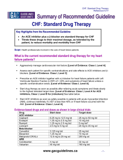

© Copyright 2026