
Read PDF
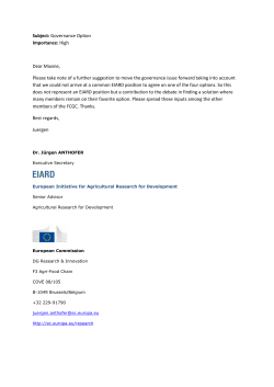
637
ORIGINAL ARTICLE
MPV and other inflammatory markers in diagnosing acute appendicitis
Seyran Bozkurt,1 Ataman Köse,2 Semra Erdogan,3 Gülten Iraöz Bozali,4 Cüneyt Ayrik,5 Rabia Bozdogan Arpaci,6 Anil Özgür,7
Güllü Akbaydogan Dündar,8 Özgür Türkmenoglu9
Abstract
Objective: To investigate whether mean platelet volume can be used as an inflammatory marker for the diagnosis
of acute appendicitis, and to determine the role, if any, of white blood cell count, C-reactive protein and neutrophil
count in this regard.
Methods: The retrospective study was conducted at Mersin University (MEU) Health Research and Application
Center, Emergency Department, Mersin, Turkey, and included medical record of patients having gone
appendectomy between April 2012 to July 2013. Based on pathology examination, the cases were grouped as
uncomplicated, complicated, and non-appendicitis cases. Preoperative white blood cell, neutrophil, C-reactive
protein and mean platelet volume were noted. SPSS 16 was used for statistical analysis.
Results: Records of 275 patients were studied. Overall, 90(32.7%) patients were uncomplicated, 120(43.7%)
complicated, and 65(23.6%) were non-appendicitis cases. The first two groups had a significantly higher white blood
cell (p=0.001) and neutrophil (p<0.001) counts than the third one. Mean platelet volume levels were not statistically
different (p=0.478).The neutrophil count had a sensitivity of 76.19%, specificity of 56.92%, positive predictive value
of 85.11%, and negative predictive value of 42.53%; white blod cell count had sensitivity 68.10%, specificity 61.54%,
positive predictive value 85.12%, and negative predictive value 37.38%; mean platelet volume level had sensitivity
74.76%, specificity 35.38%, positive predictive value 78.89%, and negative predictive value 30.26%; and C-reactive
protein level had sensitivity 84.29%, specificity 30.77%, positive predictive value 79.73%, and negative predictive
value 37.74%.
Conclusion: Elevated white blood cell and neutrophil counts may be used as diagnostic tests in cases of acute
appendicitis, while C-reactive protein and mean platelet volume levels were not useful as diagnostic markers.
Keywords: Acute appendicitis, Mean platelet volume, C-reactive protein, Leucocyte count. (JPMA 65: 637; 2015)
Introduction
Acute appendicitis (AA) is the most common cause of
urgent abdominal operations and it is still difficult to
diagnose despite its classical signs and symptoms being
well known.1,2 A delay in diagnosis is associated with
perforation and increased complication rate.3 In AA the
negative laparotomy rate is generally around 20%.2
Therefore, distinguishing nonspecific abdominal pain
from AA is of paramount importance. The value of
various parameters — C-reactive protein (CRP), white
blood cell (WBC) count, lymphocyte/leucocyte ratio,
interleukin-6 (IL-6), IL-10, IL-4, IL-5, IL-12, tumour necrosis
factor
alpha
(TNF),
endotoxin,
erythrocyte
sedimentation rate (ESR), procalcitonin, fibrinogen,
alpha 2-macroglobulin, alpha 1- antitrypsin, D-lactate —
for diagnosis of AA have been studied.4 However, they
have proved non-superior to clinical history, physical
1,2,4,5,8Emergency Medicine Department, 3Biostatistics and Medical Informatics
Department, 6Pathology Department, 7Radiology Department, 9General
Surgery Department, Mersin University Medical Faculty, Mersin, Turkey.
Correspondence: Seyran Bozkurt. Email: [email protected]
examination, and usual laboratory studies in diagnosing
AA at an early stage.5
Mean platelet volume (MPV) is a measure of
thrombocyte size provided by complete blood count
(CBC) analysers as a routine part of CBC and it is usually
overlooked by clinicians.6 Elevated MPV levels have been
reported in chronic obstructive lung disease (COPD),
myocardial infarction (MI), diabetes, and high altitude.3
On the other hand, some studies have reported that
MPV levels decrease in rheumatoid disorders such as
rheumatoid arthritis and ankylosing spondylitis,
ulcerative colitis, and acute pancreatitis.6-9 Furthermore,
there are studies that have showed that MPV is affected
by inflammation. 3,6-9 There are only a few studies
examining the relationship between MPV and AA and
their results are variable.1,3,4
The current study was planned to examine whether MPV
level can be used as an inflammatory marker in nonappendicitis,
uncomplicated
appendicitis
and
complicated appendicitis cases. In addition, we planned
to determine if there was any difference between the
groups with respect to WBC, CRP and neutrophil count.
J Pak Med Assoc
638
MPV and other inflammatory markers in diagnosing acute appendicitis
Patients and Methods
The retrospective study was conducted at Mersin
University (MEU) Health Research and Application Center,
Emergency Department, Mersin, Turkey, and included
medical record of patients having gone appendectomy
between April 2012 and July 2013. After approval from
the ethics committee of Mersin University Faculty of
Medicine, Turkey, data pertaining to patients aged at least
17 years who had presented to the emergency
department (ED) with abdominal pain and had
undergone appendectomy after a diagnosis of acute
appendicitis on pathological basis was studied. Those
excluded were patients with acute cerebrovascular
disease, MI, pregnancy, heart failure, peripheral vascular
disease, COPD, haematological disorder, diabetes
mellitus, acute or chronic disorders, cancer, or liver
disease; those using anticoagulants or non-steroidal antiinflammatory drugs (NSAIDs); or those who had no
leucocyte, neutrophil, MPV, CRP levels measured or
ultrasonography performed the same day prior to the
operation. Based on the pathology results, data was
divided into 3 groups: uncomplicated AA (uncomplicated;
AA without peritonitis or phlegmonous appendicitis);
complicated appendicitis (perforated, plastrone,
necrotising appendicitis and appendicitis with
peritonitis); and non-appendicitis (normal appendix, and
reactive lymph node hyperplasia) cases. Preoperative
WBC, neutrophil, MPV, and CRP levels were recorded. CBC
analysis had been performed with Sysmex XT-2000I
(Roche, Istanbul, Turkey) and CRP analysis with Cobas
integra 800 (Roche, Istanbul, Turkey) device. The reference
intervals for MPV and CRP levels and WBC and neutrophil
counts were set by the local hospital's laboratory (WBC:
4.5-11 x103/µL; neutrophil: 1.5-6.7 x10³/ µL; MPV: 7.4-10.4
fL; CRP: <5 mg/L). Sensitivity, specificity, negative
predictive value (NPV), positive predictive value (PPV) and
cut-off value of WBC, neutrophil counts and MPV and CRP
levels were calculated. The likelihood ratios were
calculated as positive (LR+) and negative (LR-).
SPSS 16.0 was used for statistical analysis. Normality of
data for continuous variables was analysed using Shapiro
Wilk test. Age and CRP levels did not have normal
distribution. Inter-group differences were analysed with
one-way analysis of variance (ANOVA) for normally
distributed parameters and with Kruskal-Wallis test for
non-normally distributed parameters. Paired comparisons
were done with Bonferroni test. Descriptive statistics
included mean and standard deviation (mean±SD) for
normally distributed parameters and minimum,
maximum, median, and 25-75% percentiles for nonnormally distributed parameters. Pearson's Chi-Square
Vol. 65, No. 6, June 2015
test was used for categorical variables. Frequencies and
percentages were presented as descriptive statistics. A
receiver operating curve (ROC) analysis was performed for
sensitivity, specificity, LR+ and LR- values for the cut-off
points belonging to continuous variables. Uncomplicated
and complicated AA cases were merged into a single
group and designated as the AA group when performing
ROC analysis. Hence, ROC analysis was performed
between the cases of AA and non-appendicitis. A p value
less than 0.05 was considered statistically significant.
Results
Records of 363 patients were studied, but 88(24.24%)
were left out on the basis of exclusion criterion or missing
data. The records of the remaining 275(75.75%) patients
represented the final sample. Of them, 90(32.7%) patients
were uncomplicated, 120(43.7%) complicated, and
65(23.6%) were non-appendicitis cases.
Male gender had a higher rate of acute appendicitis
(p<0.001). While 69(76.7%) uncomplicated cases and
79(65.8%) complicated appendicitis cases were male,
41(63.1%) non-appendicitis cases were female (Table-1).
Of the male patients, 69(40.1%) were diagnosed to have
uncomplicated appendicitis and 79(45.9%) were
diagnosed with complicated appendicitis. Besides,
Table-1: Pathological diagnostic groups by gender.
Gender
Uncomplicated
Appendicitis
Complicated
Appendicitis
NonAppendicitis
p
Male
Female
69(76.7)
21(23.3)
79 (65.8)
41 (34.2)
24 (36.9)
41 (63.1)
<0.001
MPV: Mean platelet volume. WBC: White blood cell.
Figure: Receiver Operating Curve (ROC) of MPV, WBC, Neutrophil and CRP.
639
S. Bozkurt, A. Köse, S. Erdogan, et al
Table-2: Comparison of CRP and Age parameters.
Uncomplicated Appendicitis (n=90)
Min-Max
Median [25-75% percentiles]
Age
CRP
17-78
0-341.66
31 [23-40]
13.05 [3.21-39.65]
Complicated Appendicitis (n=120)
Min-Max
Median [25-75% percentiles]
17-76
0.10-431.80
33 [25-42]
17.76 [3.62-52.35]
Min-Max
Non-Appendicitis (n=65)
Median [25-75% percentiles]
17-69
0.16-443.00
p
34 [25.5-42.5]
17.65 [2.24-70.78]
0.270
0.555
Note: Test values were expressed as median (25th/75th percentile) range.
CRP: C-reactive protein.
Table-3: Comparison of MPV, neutrofil and WBC parameters between pathological diagnosis groups.
Uncomplicated Appendicitis
(n=90)
Complicated Appendicitis
(n=120)
Non-Appendicitis
(n=65)
p
14.048 ± 3.735
10.945 ± 3.73
10.402 ± 0.934
14.333 ± 3.727
11.227 ± 3.399
10.272 ± 0.935
12.112 ± 4.467*,†
8.891 ± 4.328*,†
10.423 ± 1.000
0.001
<0.001
0.478
WBC
Neutrofil
MPV
MPV: Mean platelet volume
WBC: White blood cell.
*Differences with uncomplicated appendicitis, †: Differences with complicated appendicitis.
Table-4: ROC analysis.
MPV
CRP
Neutrophil
WBC
Cut-off
AUC (p)
10.8
59.41
8.23 x10³/ µL'
12.51 x10³/ µL'
0.532 (0.4455)
0.514 (0.7627)
0.677 (<0.0001)
0.654 (0.0002)
Sensitivity (%)
Specificity (%)
74.76 [68.32 - 80.49] 35.38 [23.92 - 48.23]
84.29 [78.65-88.93] 30.77 [19.91-43.45]
76.19 [69.84 - 81.78] 56.92 [44.04 - 69.15]
68.10 [61.33 - 74.34] 61.54 [48.64 - 73.35]
PPV (%)
NPV (%)
Accuracy
LR+
LR-
78.89 [72.56-84.35]
79.73 [73.83-84.81]
85.11 [79.20-89.87]
85.12 [78.82-90.13]
30.26 [20.25-41.87]
37.74 [24.67-52.25]
42.53 [31.99-53.59]
37.38 [28.18-47.31]
65.45
71.64
71.64
66.54
1.16
1.22
1.77
1.77
0.71
0.51
0.42
0.52
Note: Test values were expressed 95% Confindence interval.
MPV: Mean platelet volume. CRP: C-reactive protein. WBC: White blood cell. PPV: Postive predictive value. NPV: Negative predictive value. ROC: Receiver operating curve. AUC: Area under the curve.
24(14%) males and 41(39.8%) females were ultimately
found to have no appendicitis.
There were no significant differences among the groups
with respect to age and CRP level (Table 2).
WBC (p=0.001) and neutrophil (p<0.001) counts were
higher in uncomplicated and complicated appendicitis
cases compared to non-appendicitis cases. MPV levels
were not significantly different across the groups
(p=0.478) (Table 3).
ROC analysis found that neutrophil and WBC
measurements were significant, and, as such, a neutrophil
value above 8.23 x10³/µL was allowed to diagnose AA
(p<0.0001). The sensitivity, specificity, PPV, and NPV of
neutrophil count for diagnosing AA were found to be
76.19%, 56.92%, 85.11%, and 42.53%, respectively. A WBC
level above 12.51 x10³/µL was allowed to diagnose AA
(p=0.0002). WBC level had sensitivity 68.10%, specificity
61.54%, PPV 85.12%, and NPV 37.38%. Values above 10.8
fL for MPV and above 59.41 mg/L for CRP were able to
diagnose AA, albeit statistically non-significant (p>0.05).
MPV had a sensitivity, specificity, PPV, and NPV of 74.76%,
35.38%, 78.89%, and 30.26%, respectively. MPV level was
lower in the complicated appendicitis patients, although
it did not reach statistical significance (p>0.05). CRP level
had sensitivity 84.29%, specificity 30.77%, PPV 79.73%,
and NPV 37.74%. The likelihood of neutrophil and WBC
counts for differentiation of true patients was high (Table4, Figure).
Discussion
Debate surrounding the benefit of laboratory tests in
diagnosing AA still continues.10 In this study we planned
to investigate the results of MPV, leucocyte, neutrophil
and CRP values based on the pathology results in patients
undergoing appendectomy with an early diagnosis of AA.
It is believed that there is a link between thrombocyte
activation, thrombosis and the pathophysiology of the
diseases with a tendency for inflammation. Many
thrombocyte markers, including MPV, have been related
to thrombosis and inflammation.11 In a study, cases with
acute pancreatitis had lower MPV levels.6 Another study
J Pak Med Assoc
640
MPV and other inflammatory markers in diagnosing acute appendicitis
reported that cases with ankylosing spondylitis and
rheumatoid arthritis had a lower MPV level compared to
control group.7 One study found that during an ulcerative
colitis attack, MPV level decreased and MPV could be used
for diagnosis of that disease.8 Similarly, one study showed
that MPV level was unchanged compared to the control
group outside the attacks, but was significantly reduced
during attacks, which was associated with lower
morbidity.12 There are a few studies regarding the role of
MPV in AA diagnosis in children and adults, albeit with
variable results.1,3,4,13 A study comparing a healthy adult
control group and an adult patients group with AA found
significantly lower MPV level. It reported a cut-off MPV
value of 7.6 for AA, a sensitivity of 73%, specificity 84%,
PPV 84%, and specificity 74%. It also stated that MPV level
should not be overlooked in cases with suspected AA.3
One study reported a significant reduction in MPV level in
patients with AA. It found a cut-off level of 7.55, sensitivity
87%, and specificity 60% for MPV.13 One study did not find
any relationship between paediatric appendicitis and
MPV levels.1 Another found a significantly higher MPV
level in cases with AA compared to the control group. It
reported a cut-off value of 7.87, sensitivity 66%, and
specificity 51% for MPV.4 In our study, the cut-off level of
MPV was 10.8ft, sensitivity 74.76%, specificity 35.38%, PPV
78.89%, and NPV 30.26%. There was no significant
difference between the appendicitis cases (complicated
or uncomplicated) and non-appendicitis cases with
respect to MPV levels. However, the complicated
appendicitis group had a lower MPV measurement value
compared to other groups. The reduction in MPV levels
was explained as the sequestration of larger
thrombocytes in the vascular segments of the inflamed
intestines.9 However, as there are not many studies on this
subject, it is clear that further studies should be
conducted in this field.
Leucocyte count is a commonly employed test in AA
diagnosis. Many studies have reported use of increased
leucocyte level in early diagnosis of appendicial
inflammation since AA cases continue commonly with
leucocytosis. Neutrophil is usually associated with
bacterial infections. Earlier studies reported WBC
sensitivities 67-97.8%, specifities 31.9-90.8%, NPV 77.982%, and PPV 42-91.8%.2,10,13-17 An increased neutrophil
count has been reported to have sensitivity 68.6-98.9%,
specificity 33.1-91%, PPV 84.7-89.21%, and NPV 5371.5%.2,3,10,13,16 In agreement with the literature, we found
higher WBC and neutrophil counts in complicated and
uncomplicated appendicitis compared to the nonappendicitis cases. The WBC count had a higher specificity
and PPV value compared to other parameters. The WBC
parameter had a cut-off level of 12.51 x10³/µL, sensitivity
Vol. 65, No. 6, June 2015
68.10%, specificity 61.54%, PPV 85.12%, and NPV 37.38%.
The neutrophil parameter had a cut-off value of 8.23
x103/µL, sensitivity 76.19%, specificity 56.92%, PPV
85.11%, and NPV 42.53%.
CRP is an acute phase protein and can be used as a marker
and diagnostic tool in some inflammations and attacks of
some disorders.18 A meta-analysis exploring the
diagnostic accuracy of CRP revealed a very wide range of
sensitivity and specificity (47-74%, and 55-89%,
respectively).19 Other studies investigating the role of CRP
in diagnosing AA have reported sensitivities ranging
between 76.5% and 95.6%, specifities ranging between
26.1% and 77.7%, and PPV 95.6.2,10 The cut-off level of CRP
was 59.41mg/dl, sensitivity 84.29%, specificity 30.77%,
PPV 79.73%, and NPV 37.74% in our study. Furthermore,
CRP values had a higher sensitivity but lower specificity
compared to other parameters. A large body of studies
has shown that CRP levels increase in parallel with the
severity of the inflammatory response in AA.2,10,16,18,20 A
study found that there was not any definite level of CRP
that will diagnose AA, but its elevated level may suggest
abscess formation in AA cases.16 One study reported that
the mean CRP level was higher in perforated appendix
compared to normal appendix, but there were no
significant differences between uncomplicated
appendicitis and non-appendicitis cases.10 This study
found no difference between the groups with respect to
CRP level. The sensitivity of CRP was higher, but its
specificity was lower compared to those of other
parameters. CRP level was within normal limits in some
AA cases in this study. This condition may be explained by
the fact that CRP level starts to increase 12-24 hours after
the symptom onset.18 We additionally observed that CRP
concentration was also higher in cases having no
appendicitis. A study made the same observation that the
cases with normal appendix had an elevated CRP level.16
In our study, the patients who had an elevated CRP level
but had no appendicitis had no signs of acute infection,
an additional pathology in ultrasonography, or any other
intraabdominal pathology after appendectomy either.
CRP elevation in patients with no appendicitis may be
associated with the presence of other acute inflammatory
disorders remaining undiagnosed.
Conclusion
There was no statistically significant difference between
patients
with
appendicitis
(complicated
or
uncomplicated) and those having no appendicitis with
respect to MPV level. However, the complicated
appendicitis group had a lower MPV value compared to
other groups. WBC and neutrophil counts were observed
to be increased in AA cases, and more so in complicated
641
S. Bozkurt, A. Köse, S. Erdogan, et al
appendicitis cases. CRP levels did not significantly differ
across the groups. Considering the largely varied results
reported in literature, it is essential to conduct large-scale
prospective studies in future.
10.
11.
References
1.
2.
3.
4.
5.
6.
7.
8.
9.
Uyanik B, Kavalci C, Arslan ED, Yilmaz F, Aslan O, Dede S, et al. Role
of Mean Platelet Volume in Diagnosis of Childhood Acute
Appendicitis. Emerg Med Int. 2012;2012:823095.
Shafi SM, Afsheen M, Reshi FA. Total Leucocyte Count, C-reactive
Protein and Neutrophil Count: Diagnostic Aid in Acute
Appendicitis. Saudi J Gastroenterol. 2009;15:117-20.
Albayrak Y, Albayrak A, Albayrak F, Yildirim R, Aylu B, Uyanik A, et
al. Mean Platelet Volume: A New Predictor in Confirming Acute
Appendicitis Diagnosis. Clin Applied Thrombosis/Hemostasis
2011;17: 362-6.
Narci H, Turk E, Karagulle E, Togan T, Karabulut K. The Role of Mean
Platelet Volume in the Diagnosis of Acute Appendicitis: A
Retrospective Case-Controlled Study. Iran Red Crescent Med J
2013;15:e11934.
Sack U, Biereder B, Elouahidi T Bauer K, Keller T, Tröbs RB.
Diagnostic value of blood inflammatory markers for detection of
acute appendicitis in children. BMC Surg 2006: 6;15.
Beyazit Y, Sayilir A, Torun S, Suvak B, Yesil Y, Purnak T, et al. Mean
platelet volume as an indicator of disease severity in patients with
acute pancreatitis. Clin Res Hepatol Gastroenterol 2012:36;162-8.
Kisacik B, Tufan A, Kalyoncu U, Karadag O, Akdogan A, Ozturk MA,
et al. Mean platelet volume (MPV) as an inflammatory marker in
ankylosing spondylitis and rheumatoid arthritis. Joint Bone Spine
2008;75:291-4.
Yuksel O, Helvaci K, Ba?ar O, Köklü S, Caner S, Helvaci N, et al. An
overlooked indicator of disease activity in ulcerative colitis: mean
platelet volume. Platelets. 2009;20:277-81.
Danese S, Motte C, Fiocchi C. Platelets in inflammatory bowel
disease: clinical, pathogenic, and therapeutic implications. Am J
12.
13.
14.
15.
16.
17.
18.
19.
20.
Gastroenterol. 2004;99:938-45.
Yang HR, Wang YC, Chung PK, Chen WK, Jeng LB, Chen RJ.
Laboratory tests in patients with acute appendicitis. ANZ J Surg
2006; 76: 71-4.
Gasparyan AY, Ayvazyan L, Mikhailidis DP, Kitas GD. Mean platelet
volume: a link between thrombosis and inflammation? Curr
Pharm Des. 2011;17:47-58.
Makay B, Türkyilmaz Z, Unsal E. Mean platelet volume in children
with familial Mediterranean fever. Clin Rheumatol 2009;28:975-8.
Bilici S, Sekmenli T, Göksu M, Melek M, Avci V. Mean Platelet
volume in diagnosis of acute appendicitis in children. Afr Health
Sci 2011; 11: 427-32.
Wang LT, Prentiss KA, Simon JZ, Doody DP, Ryan DP. The use of
white blood cell count and left shift in the diagnosis of
appendicitis in children. Pediatr Emerg Care. 2007;23:69-76.
Cardall T, Glasser J, Guss DA. Clinical value of the total white blood
cell count and temperature in the evaluation of patients with
suspected appendicitis. Acad Emerg Med. 2004;11:1021-7.
Birchley D. Patients with clinical acute appendicitis should have
pre-operative full blood count and C reactive protein assays. Ann
R Coll Surg Engl 2006; 88: 27-32
Keskek M, Tez M, Yoldas O, Acar A, Akgül O, Goçmen E, et al.
Receiver operating characteristic analysis of leukocyte counts in
operations for suspected appendicitis. Am J Emerg Med 2008; 26:
769-72.
Wu HP, Lin CY, Chang CF, Chang YC, Huang CY. Predictive value of
C-reactive protein at different cutoff levels in acute appendicitis.
Am J Emerg Med 2005; 23: 449-53.
Yu Cw, Juan LI, Wu MH, Shen CJ, Wu JY, Lee CC. Systematic review
and meta-analysis of the diagnostic accuracy of procalcitonin, Creactive protein and white blood cell count for suspected acute
appendicitis. Br J Surg 2013;100: 322-9.
Yokoyama S, Takifuji K, Hotta T, Matsuda K, Nasu T, Nakamori M, et
al. .C-Reactive protein is an independent surgical indication
marker for appendicitis: a retrospective study. World J Emerg Surg
2009, 4:36.
J Pak Med Assoc


© Copyright 2026