
Effect of Treatment of Temporomandibular Disorders Single-Blind, Randomized Controlled Study
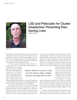
n PHYSICAL THERAPY
Effect of Treatment of Temporomandibular Disorders
(TMD) in Patients with Cervicogenic Headache: A
Single-Blind, Randomized Controlled Study
Harry von Piekartz, Ph.D., M.Sc., P.T., M.T.; Kerstein Lüdke, M.Sc., P.T.
0886-9634/2901000$05.00/0, THE
JOURNAL OF
CRANIOMANDIBULAR
PRACTICE,
Copyright © 2011
by CHROMA, Inc.
Manuscript received
November 13, 2009; revised
manuscript received
April 13, 2010; accepted
May 24, 2010
Address for correspondence:
Dr. Harry von Piekartz
Physiotherapy Clinic for
Manual Therapy and Applied
Neurobiomechanic Science
Stobbenkamp 10,
7631 CP Ootmarsum
The Netherlands
E-mail: H.von-Piekartz@
fh-osnabrueck.de
ABSTRACT: The present study was comprised of 43 patients (16 men) with cervicogenic headaches for
over three months, diagnosed according to the International Classification of Diagnostic Criteria of
Headaches (ICDH-ll). The patients were randomly assigned to receive either manual therapy for the
cervical region (usual care group) or additional manual therapy techniques to the temporomandibular
region to additionally influence temporomandibular disorders (TMD). All patients were assessed prior to
treatment, after six sessions of treatment, and at a six-month follow-up. The outcome criteria were: intensity of headaches measured on a colored analog scale, the Neck Disability Index (Dutch version), the
Conti Anamnestic Questionnaire, noise registration at the mandibular joint using a stethoscope, the
Graded Chronic Pain Status (Dutch version), mandibular deviation, range of mouth opening, and pressure/pain threshold of the masticatory muscles. The results indicate in the studied sample of cervicogenic headache patients, 44.1% had TMD. The group that received additional temporomandibular
manual therapy techniques showed significantly decreased headache intensities and increased neck
function after the treatment period. These improvements persisted during the treatment-free period
(follow-up) and were not observed in the usual care group. This trend was also reflected on the questionnaires and the clinical temporomandibular signs. Based on these observations, we strongly believe
that treatment of the temporomandibular region has beneficial effects for patients with cervicogenic
headaches, even in the long-term.
T
here is evidence that long-term upper cervical
dysfunction may influence the function of the
temporomandibular region and vice versa. 1-4
However, there are no well-designed studies that demonstrate that temporomandibular treatment provided by
physical therapists has an influence on craniocervical
dysfunction and the resulting complaints. 4 An effect
study by Hoving, et al.,5 demonstrated that directly referring patients with neck complaints to a manipulative
physical therapist is more effective and cost-saving than
guidance from a first-line medical professional. It has
also been stated that in randomized effect studies, regarding neuromusculoskeletal treatment of long-term neck
complaints, temporomandibular disorders (TMD) are
rarely incorporated in the inclusion and exclusion criteria.6-13 Furthermore, a literature review revealed that the
temporomandibular region can influence the cervical
region, although most studies included had a low level of
evidence.4 An investigation by the Netherlands Institute
for Health Services Research (NIVEL) showed that in
Dr. Harry von Piekartz is a professor in
physical therapy and rehabilitation
science on the Faculty of Business,
Management and Social Science,
University of Applied Science Osnabruck
Germany. He received his Ph.D. Degree
in 2005 from the Staffordshire University
(UK). His primary research interest is
orofacial dysfunction and pain related to
posture and movement. He is an international lecturer in neuromusculoskeletal
dysfunction and pain and works in a
specialized practice for craniofacial pain
and applied neurobiomechanical science
in Ootmarsum in The Netherlands.
1
EFFECT OF ORTHOPEDIC MANUAL THERAPY ON TMD HEADACHE
daily practice, TMD is rarely considered during neuromusculoskeletal treatment of long-term, nontraumatic
neck and headache complaints. In 2006, 11,12 patients
undergoing physical therapy were classified as nonspecific neck complaints (n=1,220) and specific neck syndromes (n=714), of which 63.5% and 66.9% received
more than six treatments (average, 8.1 and 8.9: SD, 14.3
and 17.6), respectively. After 13 weeks, 24.2% (n=297)
of the nonspecific neck complaints group and 26.5%
(n=189) of the neck syndrome group were still being
treated. This group of nonspecific neck complaints occupies the second place after the nonspecific shoulder complaints14 in the list of the longest and highest number of
treatments.
Therefore, patients with chronic neck complaints,
including headaches related to neck dysfunction, need
more treatment in relation to other neuromusculoskeletal
syndromes, and it is unknown to what extent TMD plays
a causal and/or a contributing role in this patient population. A subgroup of chronic neck complaints forms the
group of cervicogenic headache patients,15 which is recognized by the International Headache Society (IHS) as a
separate entity (Headache Committee of the International
Headache Society, 2004).
The diagnostic criteria for cervicogenic headaches
described by the IHS contain subjectively described complaint patterns and dysfunction (impairments) that might
be found during a cervical functional examination (IHS,
2004) (Table 1).
Although the hypothesis is supported that the craniocervical region can be a contributing factor in different
types of headache, such as migraine and tension headache,
neuromusculoskeletal dysfunctions of the cervical spine
appear to play a role in only 14% to 18% of chronic
headaches.16,17 Some authors suggest that in patients who
are diagnosed with cervicogenic headaches, the cervical
spine as the potential source of the symptoms is often
overvalued and other contributing factors, such as TMD,
do not receive sufficient attention.18,19 Headaches may
therefore be a symptom of TMD. It is known that significantly more (p<0.001) headache complaints occur in a
TMD group diagnosed according to the RDC/TMD criteria compared to a usual care group without headaches.20
Some authors advocate that the same pathophysiologic
mechanisms form the basis for different types of headaches, such as myofascial TMD, tension-type headaches,
and cervicogenic headaches.21,22 Also, in children, significantly more (p<0.05) TMD was detected in different
headache typologies,23,24 which suggests that in children,
the temporomandibular region plays an important (associated) role in the etiology of different types of headache.
When TMD is associated with maintaining cervicogenic
2
THE JOURNAL OF CRANIOMANDIBULAR PRACTICE
VON PIEKARTZ AND LüDKE
headaches, treatment of TMD may be successful, if the
clinical signs and symptoms are relevant to the patient’s
complaints. Hence this study aimed to:
1. Identify the prevalence of TMD in a sample of
patients diagnosed with cervicogenic headaches;
2. Determine the tests that are clinically relevant to
detect TMD in cervicogenic headache patients; and
3. Evaluate the effect of additional orofacial physical
therapy (the experimental group) after three and six
months in comparison with the usual care group
(control group).
Material and Methods
Patients
Forty-three (43) patients (27 women and 16 men, age
range 18-65 years: average age 36±7.7 years) and diagnosed with cervicogenic headache by a neurologist met
the following criteria: (i) diagnosed with cervicogenic
headache according to the International Classification of
Diagnostic Criteria of Headache (ICDH, 2004), with
headaches for >3 months, and no prior TMD treatment,
and (ii) Neck Disability Index (NDI) >15 points. Anesthetic
blockades were not used as a criterion for cervicogenic
headache, as the procedure was considered too invasive
and costly for this study and is not readily accessible to
most clinicians. To be certain that the recruited patients
could be treated with orofacial physical therapy, a minimum of one of the four signs of TMD: joint sounds, deviation during mouth opening, extraoral muscle pain at a
minimum of two tender points in the masseter or temporalis muscles and pain during passive mouth opening, had
to be present.25,26
In addition, the patients had not received any orthodontic treatment and had not experienced any neuropathic pain in the head region during the previous three
years. The subjects were recruited from different physical
therapy practices in The Netherlands. The project was
conducted in accordance with the Helsinki guidelines and
approved by the Ethics Committee of the Rehabilitation
Center ‘Het Roessingh’ in Enschede, the Netherlands
(Table 1, Figure 1).
Study Design and Procedure
The study is a randomized clinical trial (RCT). To calculate the sample size, a pilot study was performed to
determine the changes (before and after six treatments) in
patients with standard physical therapy, including manual
techniques and treatment of the temporomandibular
region. The measuring tool used as the primary outcome
parameter was the colored analog scale (CAS). This
analog scale was initially designed to record the pain
JANUARY 2011, VOL. 29, NO. 1
VON PIEKARTZ AND LüDKE
EFFECT OF ORTHOPEDIC MANUAL THERAPY ON TMD HEADACHE
intensity in children, but also showed a good concurrent
and construct validity in adult headache patients and
others.27 These results were applied to calculate the group
sizes: An alpha value of 0.05 and a power value of 0.8
were used. To obtain a difference of three points on the
CAS with a SD of three points, 17 patients were required
in each group. According to this required minimum
number of patients, 43 patients were divided into two
groups by a third researcher, using a computerized
random number generator. Subsequent measurements
were obtained after 4-6 weeks of treatment and at sixmonths follow-up (see flow diagram of Figure 1). The
patients in the experimental group (n=22) received treatment from a manual therapist who specialized in orofacial pain. Both therapy groups were discharged after six
treatments. A blinded investigator performed three
assessments, as follows: before the first treatment, after
six treatments within a time period of 4-6 weeks, and
after six months.
Investigators
All investigators participating in the study were first
contact practitioners, had more than five years of work
experience, and had completed a training program for
manual therapy recognized by the International Federation
of Orthopedic Manual Therapy (IFOMT). The group of
therapists who treated the experimental group had moreover received an additional regular training consisting of
200 hours, focusing on the assessment and management
of the craniomandibular and craniofacial pain.
Measurements
During the study, the following standardized measuring instruments were used:
• Colored analog scale (CAS): The CAS is a pain
intensity scale similar to the visual analogue scale
•
•
•
•
(VAS) that was designed especially for patients with
headache of different age categories.27 The patient
indicates the intensity of the craniofacial complaints
by marking the point on an increasingly colored line
that best represents his or her symptoms. On the
back, the marking line gives a score corresponding to
the VAS.
Neck Disability Index (NDI) Dutch version: the NDI
is a questionnaire commonly used in clinical trials
to measure the functional status of patients with neck
pain and is a dimension-specific index that reflects
the domain “functional limitation” (disability) of the
International Classification of Function (ICF). The
questionnaire includes 10 items (activities) with six
different response options, ranging from “no disability” (0) to “complete disability.”5 The total score
is 50. A higher score indicates more pain and
disability.28
Anamnestic Questionnaire CMD (Conti): The Conti
questionnaire contains 10 questions that are related
to problems originating from the craniomandibular
region. Each question has three ranking options
(0=none: 1=present: and 3=strong or bilateral). The
likelihood of a CMD is divided into 4 subgroups: 49, none: 9-14, minimal: 15-21, moderate: 21-23,
strong.29 The questionnaire has shown a strong statistical association to the modified Helkimo’s Clinical
Dysfunction index at a 95% level of confidence.30
Noise Registration at the Mandibular Joint: By
using a stethoscope, the sounds (cracking and crepitation) in the left and right mandibular joints were
determined. Assessment: cracking or crepitation present or absent, both left and right.
Graded Chronic Pain Status (GCPS-NL) Dutch version: The reliability coefficient of the GCPS on the
Gutmann scale indicates that it is a highly reliable
Table 1
The Diagnostic Criteria of Cervicogenic Headache of the
International Classification of Headache (ICHD-ll 2004)
A. Pain referred from the neck and perceived in one or more regions of the head and/or face,
fullfilling criteria C and D.
B. Clinical laboratory and/or imaging evidence of a disorder within the cervical spine.
C. Evidence that the pain can attribute to the neck disorder or lesion based on at least
one of the following:
- Clinical signs that implicate a source of pain in the neck
- Diagnostic blockade or cervical structures
D. Pain resolves within 3 months after successful treatment.
JANUARY 2011, VOL. 29, NO. 1
THE JOURNAL OF CRANIOMANDIBULAR PRACTICE
3
EFFECT OF ORTHOPEDIC MANUAL THERAPY ON TMD HEADACHE
VON PIEKARTZ AND LüDKE
Figure 1
Recruitment
Private practice
Physical therapy
Flowchart
Informed consent
! $! , )*%
#" $! , (+%
•
•
•
•
IHS criteria cervicogenic
headache
NDI > 15 points
> 3 months headache
No TMD treatment
In (n = 43)
Out (n = 23)
Stratification/Randomization
+ first evaluation responsive variables
Usual care group
Continuing physical treatment
Experimental
Orofacial treatment
In ( n= 22 )
In ( n= 21 )
Second evaluation responsive
variables
6-week follow-up
) weeks follow-up
Orofacial treatment
In (n = 22)
In (n = 21)
Half year follow-up (6
months)
Out (n = 2)
20
4
Out (n = 3)
Complete case analysis (n = 38)
THE JOURNAL OF CRANIOMANDIBULAR PRACTICE
18
JANUARY 2011, VOL. 29, NO. 1
VON PIEKARTZ AND LüDKE
EFFECT OF ORTHOPEDIC MANUAL THERAPY ON TMD HEADACHE
measuring tool for the classification of TMD pain.30,31
The questionnaire results are based on the responses
to seven questions: four are pain-related limitations
and three items refer to pain intensity. The outcomes
are classified into four subgroups, with grades I and
II seen as a slight limitation (functional chronic pain)
and grades III and IV as strong limitations (dysfunctional chronic pain).31
• Mandibular Deviation: Deviation of the mandible is
another important clinical sign of TMD.32 During
maximum mouth opening and closing, the deviation
is assessed visually by the researcher who stands in
front of the patient at mouth level. A deviation is present if there is a difference of more than two mm to
the midline.33
• Mouth Opening Measurement: (range and pain):
Using a ruler of 15 cm, starting at 0, the mouth opening is measured. Measuring the mouth opening is
seen as a nonstable biological variable and is strongly
influenced by repeated measurements.34-36 Repeated
measuring reduces the standard error of measurement,37,38 hence repeated measurements were also
included in our study (three times) with the largest
recorded range taken. During maximum mouth opening, the pain intensity is measured using the CAS.
• Pain Threshold Measurement of the Masticatory
Muscles by Means of Algometry (Pain Threshold
Meter [PTM]): The measurement is carried out at 12
points of the masseter and the temporalis muscles. A
digital algometer (Wagner Instruments, 2004) was
used (Figure 2). The algometer is a reliable instrument for measuring the sensitivity of the masticatory
muscles. An increased pressure rate is expressed by
an increased PTM value (mean regression coefficient b=0.70). The pressure is measured in kilogram
force (Kgf).39
Treatment
Prior to the treatment, all patients in the experimental
group received an orofacial examination by the treating
physical therapist to guide the treatment schedule. The
treatment techniques consisted of accessory (translatory)
movements of the temporomandibular region and/or
masticatory muscle techniques, such as tender-trigger
point treatment and muscle stretching. Active and passive
movements facilitating optimal function of cranial nerve
tissue, coordination exercises, and home exercises were
also included. The techniques used depended on the therapists’ clinical decisions. The therapist could also, when
necessary, opt for additional neuromusculoskeletal treatment of the cervical region (see “study design”). The
usual care group continued their treatment of the cranio-
JANUARY 2011, VOL. 29, NO. 1
Figure 2
Pain threshold measurement (PTT) measured at 12 points of the
masseter and temporalis muscles, left and right, using a digital algometer (Wagner Instruments, Model, FDI, 2004)
cervical region and the therapist selected the technique
and treatment or exercise type he or she considered to be
beneficial for the patient. The duration of the treatment
was maximally 30 minutes, and the treatment interval
depended on the therapists’ decision. The six treatments
had to be concluded after a minimum of 21 and a maximum of 42 days.
Statistical Analysis
The outcomes of the first, second, and third measurements were analyzed using ANOVA’s (with TukeyKramer post hoc analysis when significant) or KruskalWallis (with Dunn’s multiple comparison when significant) or chi-square test. The level of significance was set
at 0.05.
Results
The patient sample consisted of 43 patients. Of this
group, 71.8% had unchanged headache complaints for
>12 months. There was no significant difference among
age, gender, and duration of the complaints between the
usual care (control) and experimental groups (Table 2a ).
All underwent the first measurement, participated in
all six treatments, and also underwent the second measurement. Between the second and the third evaluation,
three patients in the usual care group dropped out for
the following reasons: an increase in complaints (n=2)
and a household accident (fall down the stairs [n=1]). In
the experimental group, two patients dropped out because
of an increase in complaints (n=1) and a sudden death in
the family (n=1). As a result, 38 patients (25 women
THE JOURNAL OF CRANIOMANDIBULAR PRACTICE
5
EFFECT OF ORTHOPEDIC MANUAL THERAPY ON TMD HEADACHE
Table 2(a)
Average Age, Gender Distribution, and Total
Duration of Cervicogenic Headache in the
Control (usual care) and the Experimental Groups
During the First Measurement
Age
Gender % female
Control
Group
(n=21)
36.1 (6.5)
66.7
Duration %
0-6 months
6-9 months
9-12 months
>12 months
0
9.1
18.2
72.7
Experimental
Group
(n=22)
34.7 (7.1)
63.6
0
14.3
14.3
71.4
------------------------------------------------------------------------
Table 2(b)
Average Age, Gender Distribution, and Total
Duration of Cervico Headache in the Control
(Usual Care) and the Experimental Groups After
the Final Measurement (Six Months)
Age
Gender % female
Control
Group
(n=18)
35.1 (6.4)
66.7
Duration %
0-6 months
6-9 months
9-12 months
>12 months
0
11.1
16.7
72.2
Experimental
Group
(n=20)
35.5 (6.9)
65.0
0
20.0
20.0
60.0
and 13 men) with an average age of 32 years (SD 6.5),
and a range between 18 and 62 years remained, of which
18 were in the usual care group and 20 in the experimental group (Table 2b).
The primary outcome measure was the intensity of
headaches measured using the CAS (Figure 3a). The
change in TMD complaints (AQ, GCPS, and TMD tests)
and neck complaints (NDI) were the secondary and third
measurements and are displayed in Figure 3 (b,c) and
Figures 4, 5, and Table 3.
Based on the results of the primary outcome (CAS), it
was observed that there was no significant difference
between the usual care group and the experimental group
(p<0.05) at baseline, whereas the ANOVA results of the
6
THE JOURNAL OF CRANIOMANDIBULAR PRACTICE
VON PIEKARTZ AND LüDKE
CAS and the two questionnaires at the times of the second
and the third measurements displayed a significant difference between the groups (p<0.001). After six treatments,
the average values (mean range) of CAS and AQ had
decreased by more than 50%. The decreasing trend of the
CAS, the AQ, and the NDI was also observed when comparing the second and the third measurement. In the usual
care group, there was a significant increase of the average
value, indicating a possible deterioration of the headache
complaints (Figure 3).
The outcomes of the GCRS at the first measurement
showed an average score in the control and the experimental group. After the third measurement, the scores in
percentages in the grade II and III groups were clearly
reduced in the experimental group and slightly increased
in the usual care group (Figure 4).
The results of mouth opening in mm and the presence
of mandibular deviation and mandibular joint sounds are
presented in Table 3.
The distribution of the data on the pain threshold
measurement for the masticatory muscles displays a less
even distribution: there was no significant difference
(p<0.05) for 10 of the 12 examined tender point regions.
Only tender points, both left and right in the anterior
temporal muscle, did display a significant difference
(p>0.001) at the second and third measurement. These
results and those for the anterior masseter muscle are
shown in Table 4.
Effect of the Intervention
Effects after the Treatment Series (Second Measurement)
and at Follow-up (Third Measurement)
In the usual care group, the results for CAS , NDI , AQ
and TMD signs, (mouth opening: pain and range, deviation, sounds and PTM of the anterior temporal muscles)
were not significantly different, observed at the second
and third measurements compared with the first measurement (Figure 3, Tables 3 and 4). The results of the
second measurement for CAS, NDI, AQ, the TMD signs
(mouth opening, range and pain) and PTM from the anterior temporal muscles) of the experimental group differed
significantly from the results of the usual care group
(Figures 3, 5 and Table 3). During the treatment-free
period (comparing the second with the third measurement), the experimental group showed a trend towards
decreased headache symptoms (CAS) and a reduction of
TMD signs that was not observed in the usual care group.
Only the NDI and the range of the mouth opening did not
change significantly. Comparing the results of the initial
and the third measurement in Chronic Pain Status (CPGS),
the experimental group shows a clear shift towards grade
JANUARY 2011, VOL. 29, NO. 1
VON PIEKARTZ AND LüDKE
EFFECT OF ORTHOPEDIC MANUAL THERAPY ON TMD HEADACHE
a
Figure 3 (a, b, c)
Results of the response variables before
(a) after three months (b) and six months
post-intervention (c) in two treatment
groups (usual care group, n=21: experimental group, n=22) and after six months
(usual care group, n=18: experimental
group, n=20).
b
c
JANUARY 2011, VOL. 29, NO. 1
THE JOURNAL OF CRANIOMANDIBULAR PRACTICE
7
EFFECT OF ORTHOPEDIC MANUAL THERAPY ON TMD HEADACHE
VON PIEKARTZ AND LüDKE
Figure 4
Chronic Pain Grade Status: Prevalence of Grade I-IV on the Chronic Pain Grade Classification of the control and experimental group during
the first measurement (before intervention) and after the third measurement (six weeks after the last intervention) scored according to the
research criteria/temporomandibular disorder (RCD/TMD) grade I, low intensity; grade II, high intensity; grade III, moderately limiting; grade IV,
severely limiting.
I that was not present in the usual care group (Figure 4).
Smallest Detected Difference (SDD) and Clustering of
Tests
The CAS is seen as a sensitive instrument for measuring neck pain intensity and headache,27-39 and was therefore chosen as the primary outcome parameter. Research
has revealed that clustering of the CAS with other reliable
instruments, such as questionnaires and manual test combinations (known as manual examination procedures
[MEP]) in patients with neck pain, results in an increase
of the diagnostic value.40,41 Cut-off points are often used
that correspond to the smallest statistically significant
change measured with the particular measuring instrument, and also called the smallest detectable change
(SDC) or difference (SDD).42,43 The SDD thus measures
the minimal statistical and clinical relevant change that
may result in improved clinical decision-making.44
For cervicogenic headaches, it has been shown that the
8
THE JOURNAL OF CRANIOMANDIBULAR PRACTICE
SDD on a VAS45 was determined as >20 mm,46 as 3.5
points47 for the NDI, as five mm for the restriction of
mouth opening, and as 22 mm 48 for the minimal and
maximal pain intensity during the mouth opening on
a VAS.
In daily practice, the question occurs to what degree
the amount of improvement of mouth opening and pain
are prognostic indicators for the treatment course. In this
study, all subjects with an improvement of >5 mm and a
decrease in pain intensity of >22 mm between the first,
second, and third measurements were rated. Subsequently,
it was determined how many patients had a SDD of
mouth opening range (>5 mm) and pain (>22 mm), the
NDI >3.5 and a VAS for the headache >20 mm improvement. In this case, 17 of the 20 patients (85%) of the
experimental group met these criteria, while there were
none in the usual care group (0%). Table 5 lists the average values of the measurements of the 17 subjects from
the initial and the third measurement.
JANUARY 2011, VOL. 29, NO. 1
VON PIEKARTZ AND LüDKE
EFFECT OF ORTHOPEDIC MANUAL THERAPY ON TMD HEADACHE
Mouth Opening(A) and Pain (B)
Figure 5
Results of the range of mouth opening in mm (A) and the pain intensity measured by the CAS in mm (B) before, after three months, and after six
months post-intervention, in two treatment groups (usual care group, n=21: experimental group, n=22) and after six months (usual care group, n=18:
experimental group, n=20).
Discussion
The Prevalence of TMD in Cervicogenic Headache
Patients
It is known that TMD signs do not need to be relevant
for the presence of complaints. Population-based studies
report that the prevalence of TMD ranges from 8% to
15% in women and from 3% to 10 % in men.49 In headache patients, the prevalence of TMD is estimated to be
much higher 51.6%.50 In this study, a preselected sample
of cervicogenic headache patients, who did not respond
to neuromusculoskeletal therapy, were randomized without prescreening for TMD. Based on the reference values
for TMD in other studies, participants were classified
as TMD patients when all outcomes agreed with the
reference values described in Table 6. From this point
of view, it can be concluded that in the present sample
of patients with cervicogenic headache, 44.1% (n=19)
have a TMD, a result that is significantly higher than
the described prevalences in studies on healthy populations but lower than the described prevalence in head-
JANUARY 2011, VOL. 29, NO. 1
ache studies.
The average score for the NDI at the initial measurement is 15.4 and 15.6 points for the usual care and the
experimental group respectively, indicating a moderate
neck disability (maximal score is 50 points). Similar
results were reported by other authors in studies on neck
pain.5 The experimental group shows a tendency to a further reduction of neck disability even after the orofacial
physical treatment was stopped (Figure 3c).
According to the GCPS, 42.8% and 45% of the usual
care and experimental groups, respectively, in this sample of cervicogenic headache patients, had a slight disability (low intensity) and a high disability (moderately
limiting) of 33.4% and 32%, respectively (Figure 4).
Also, the percentage of women in the four groups (I-IV)
was >50% greater, which corresponds with studies on
TMD-related headache.50 The authors are not aware of
the existence of any comparable studies of GCPS with
cervicogenic headache. During the third measurement
(after six months), a clear shift towards the Grade 0-1-11
classification was observed in the experimental group,
THE JOURNAL OF CRANIOMANDIBULAR PRACTICE
9
EFFECT OF ORTHOPEDIC MANUAL THERAPY ON TMD HEADACHE
VON PIEKARTZ AND LüDKE
Table 3
Results of the Response Variables from Mouth Opening in mm: Pain During Mouth Opening (VAS) in mm:
Presence of Mandibular Deviation and Sounds Before, After Three Months, and After Six Months PostIntervention in Two Treatment Groups (After Three Months Control Group, n=21: Experimental Group,
n=22), and After Six Months (Control Group, n=18: Experimental Group, n=20)
Response
variables
Mouth opening (MO)
Mean
SD
Range
Before intervention
Control
Experimental
group
group
Follow-up (6 mos.)
Control
Experimental
group
group
43.2
4.3
34-50
42.5
3.5
35-49
42.4
3.7
35-49
51.8
4.9
42-62
41.6
4.3
34-49
53.5
3.2
49-62
VAS during MO (mm)
Mean
SD
Range
52.0
14.0
29-80
57.0
18.0
33-85
51.0
13.0
29-74
31.0
11.0
10-50
53.0
7.0
40-65
0.9
8.0
0-33
Deviation
Present (%)
38.9
45.0
33.3
20.0
33.9
10.0
Sound (click)
Present (%)
44.4
55.0
44.0
35.0
42.0
25.0
indicating that according to von Korff, et al.,31 the severity of chronic pain is clearly reduced. This may fit into the
current concept that different entities like chronic tension
headaches and myofascial temporomandibular disorders
share the same pathobiological mechanisms. Similarities
regarding sensitization of the nociceptive pathways, dysfunction of the endogenous pain modulatory systems, as
well as contributing genetic factors play a role in these
chronic pain state of headache.22 Cervical headache may
be one of these entities where neuromusculoskeletal
treatment of TMD may alter pathobiological mechanisms, and thereby reduce chronification in a prospective
setting such as present in this study.
The average mouth opening values of the usual care
and experimental groups are 43.2 mm (±4.3) and 42.5
mm (±3.5), respectively, and are clearly below the average of most reference value studies among healthy volunteers who indicate an average value score >53 mm.34-36
Pain intensity during maximal mouth opening in nonTMD patients has been reported, with an average of 32
mm on a VAS (±24.2).51 In our sample, it ranged around
57 mm (±18) and 52 mm (±14) in the experimental and
usual care group, respectively, and is therefore clearly
higher than the published reference values (Figure 5).
Joint sounds are distinguished into clicking or crepitation. Clicking is often associated with internal derange-
10
After intervention (3 mos.)
Control
Experimental
group
group
THE JOURNAL OF CRANIOMANDIBULAR PRACTICE
ments of the mandibular joint, and crepitation is associated with degenerative changes of the mandibular joint.52
Sounds of the mandibular joints may also indicate ligament or muscular morphologic changes.25 The timing of
cracking during mouth opening can also be classified and
is related to the nature of the internal derangement.25
Epidemiologic studies indicate that in a general population, the prevalence of temporomandibular joint sounds
ranges from 15% to 40% and in adult TMD patients
groups, it ranges from 56%25 to 79%.53 In the studied
sample, the percentage in the usual care group is 38.9%
and 45% in the experimental group, thereby corresponding more with the studies on populations without TMD. It
should be noted that no distinction was made between the
type of sound and the timing of the cracking as described
previously.
Deviation of the mandible in the frontal plane can also
be seen as a dysfunction caused by intra-articular or muscular imbalance.54,55
In a normal population, a deviation of >2 mm from the
midline occurs in 0% to 14%,56,57 whereas in TMD populations with or without headache, it ranges between 5%
and 43%.58,59 In the studied cervicogenic headache group,
the percentage distribution of the control and experimental groups was 44.4% and 45%, respectively, indicating
that the prevalence of mandibular deviation was clearly
JANUARY 2011, VOL. 29, NO. 1
VON PIEKARTZ AND LüDKE
EFFECT OF ORTHOPEDIC MANUAL THERAPY ON TMD HEADACHE
Table 4
Results of the Response Variables from the Pain Threshold of the Tender Point of the Anterior Temporalis
Muscle (Left and Right), Measured with a Pressure Threshold Meter (PTM) in kgf/cm2 After Three Months
and Six Months Post-Intervention in Two Treatment Groups (After Three Months, Control Group, n=21:
Experimental Group, n=22) and After Six Months (Control Group, n=18: Experimental Group, n=20)
Response
variables
Anterior temporalis
muscle (left)
Mean
SD
Range
Anterior temporalis
muscle (right)
Mean
SD
Range
First measurement
Control
Experimental
group
group
Control
group
3 months
Experimental
group
Control
group
6 months
Experimental
group
0.98
1.0
0.3-2.3
0.97
0.81
0.8-3.2
0.10
1.02
0.3-3.3
1.14
0.75
0.3-3.2
1.01
1.0
0.6-3.3
1.31
0.78
0.2-3.2
0.35
0.47
0.2-1.4
0.36
0.47
0.2-1.4
0.38
0.49
0.1-1.4
0.62
0.50
0.3-1.5
0.36
0.45
0.2-1.3
0.91
0.52
0.3-1.9
greater than in a population without TMD, but corresponded reasonably with previously studied TMD populations.
Muscle sensitivity (muscle tenderness) is an important
clinical sign of TMD and can be observed in 90% of the
patients with TMD.60 The pain pressure threshold (PTT)
is therefore a parameter that can easily be measured in
daily practice with a reliable digital or pressure-measuring device, such as an algometer.61 Due to the high standard deviations at all three measurements, no statement
can be made regarding significant change to the most
commonly selected muscle regions. Only the anterior
masseter muscle (left and right) showed a highly reliable
(p>0.001) sensitivity to pressure. These observations
match the findings of a similar study by Silva, et al.,61 that
also detected the highest sensitivity (77%) and likelihood
ratio in the anterior masseter muscle, while using a similar study set-up and an algometer as a measuring tool. A
similar conclusion was drawn from a sample of female
TMD patients (n=49) with approximately the same
Table 5
Mean, Standard Deviation, and Minimal and Maximal Ranges of Subjects in the Experimental
Patient Group (n=20) Who Fulfilled the SDD (Smallest Detectable Difference) of VAS,
Range of Mouth Opening, the Headache VAS, and the Neck Disability Index (NDI)
VAS mouth opening
(mm)
Mouth opening
(mm)
VAS
headache
NDI
(points)
Measure 1
Mean
SD
Range
59
12
33-85
42
34
35-48
74
8
59-89
16.2
5.4
10-28
Measure 3
Mean
SD
Range
10
9
0-33
54
32
49-62
23
14
0-50
6.2
3.0
0-12
JANUARY 2011, VOL. 29, NO. 1
THE JOURNAL OF CRANIOMANDIBULAR PRACTICE
11
EFFECT OF ORTHOPEDIC MANUAL THERAPY ON TMD HEADACHE
age distribution (mean, 28.8 years: range, 17-52 years)
in which sensitivity, specificity, and the positive predictive values of the PTT of the anterior masseter muscle
(48%) and the temporalis muscle (55%) were assessed.
Reference values of the PTT from the usual care groups
were 3.46 kgf/cm2 (±1.08) on the right and 3.67 (±1.23)
on the left (Silva, et al.61). The outcomes in this study
were 0.35 kgf/cm2 (±0.47) on the right and 0.98 kgf/cm2
(±1.0) on the left, which is significantly lower than in the
study by Silva, et al.61
According to the TMD reference values that include
limited mouth opening, presence of deviation, cracking,
and minimally lowered pain thresholds of the anterior
masseter muscle, 42.1% of the studied patient sample had
TMD. This is significantly more than the prevalence in
studies on healthy populations that range between 5%
and 10%, 48 but is also lower than the prevalence in
headache studies58,61,62 (Table 6).
Effects After the Treatment Series (Second Measurement)
and At Follow-up (Third Measurement)
The CAS , NDI , AQ, mouth opening (range and pain),
and the PTM from the anterior masseter muscle of the
usual care group did not show any significant differences
in the second and third measurements (p>0.05) in comparison with the first measurement. In the experimental
group, there was a significant difference (p<0.001) at
both the second and third measurements in relation to the
first measurement (Table 4). There was a significant difference in the CAS, AQ and mouth opening (pain) and
the PTM from the anterior temporal muscles, but not
VON PIEKARTZ AND LüDKE
from the NDI and the mouth opening range (Table 7).
This suggests that the decrease of the sensitivity of the
orofacial region on stress (mouth opening) and pressure
(PTM anterior masseter muscles) did not relate directly to
the amount of mouth opening. This supports the actual
pathophysiological mechanisms derived from head-neck
and facial pain50 in the studied sample.
In general, it can be postulated that in the experimental
group, a trend towards a decreased primary measure
(headache intensity) and increased neck function was
found after the treatment period (second measurement).
This also applied to the treatment-free period (third measurement) in contrast to the usual care group (Table 7).
Based on these observations, we strongly believe that
treatment of the TMD region has beneficial effects in this
patient population with cervicogenic headache, even in
the longer term and can be explained by the existing
(anatomical, biomechanical, and neurophysiologic
models4,26).
Conclusions
From this study, it can be concluded that:
1. Cervicogenic headache patients in the investigated
group showed a higher prevalence of TMD than the
healthy population but a lower prevalence than in
previously described headache samples.
2. Clustering of tests such as mouth opening (range and
pain), NDI, and VAS of headaches, contribute to an
improved diagnosis of TMD in chronic cervicogenic
headache patients.
Table 6
Mean, Standard Deviation of the Anamnestic Questionnaire (AQ), Mouth Opening (MO-Range), Pain
Pressure Algometry and Percentages of Joint Sounds of the Sample, and Reference Values
12
Measure
AQ
Sample (n=43)
13.4 (SD 4.6)
Reference values
9-14 points (minimal TMD)
Authors
Conti, et al.29
MO (range)
42.6 (SD 3.5)
53-55 mm
Wood, et al. 34
Achterberg35
Mezitis, et al.36
MO (pain) VAS
58 mm (SD 14)
32 mm (SD 12.4)
Van der Kloot, et al.51
Joint sounds
42.3%
15-40%
Elving et al.53
PTM ant. mass. L
Kgf/cm2
0.35 (SD 0.47)
3.67 (SD 1.23)
Silva, et al.61
PTM ant. mass. R
Kgf/cm2
0.98 (SD 1.0)
3.46 (SD 1.08)
Silva, et al.61
THE JOURNAL OF CRANIOMANDIBULAR PRACTICE
JANUARY 2011, VOL. 29, NO. 1
VON PIEKARTZ AND LüDKE
EFFECT OF ORTHOPEDIC MANUAL THERAPY ON TMD HEADACHE
Table 7
Significance of Changes (Effects) After Treatment Series (Second Measurement) and
At Follow-Up (Third Measurement) for Colored Analog Scale; Anamnestic Questionnaire;
Neck Disability Index; Mouth Opening; Pain Threshold Measurement (After Three Months, Control Group,
n=21; Experimental Group, n=22) and After 6 Months (Control Group, n=18; Experimental Group, n=20)
Parameter
2nd measurement
after 3 mos.
Control
group
Experimental
group
CAS
NS
NDI
NS
AQ
NS
S
p<0.001
S
p<0.001
S
p<0.001
3rd measurement
after 6 mos.
MO
range
NS
S
p<0.001
Control
NS
NS
NS
NS
group
Experimental
S
NS
S
NS
group
p<0.05
p<0.05
CAS: colored analog scale; AQ: anamnestic questionnaire; NDI: neck disability index;
MO: mouth opening; PTM: pain threshold measurement
3. The beneficial treatment effect in the experimental
group remains or is improved at six months followup and a decreased chronification occurs in contrast
to the usual care group that remains status quo or
worsens after discharge from the treatment and indicates that TMD may be a contributing or etiological
factor in chronic cervicogenic headache patients.
4. Further research is required for other treatment interventions, such as splint therapy, and/or the combination with specialized physical therapy, such as
applied here with a follow-up over a longer period
(12 months).
Acknowledgement
We would thank the Cranial Facial Therapy Academy
(CRAFTA) Researchgroup and the University of Applied
Science Osnabruck (Germany) for their financial support.
References
1.
2.
3.
4.
5.
De Wijer A, Steenks MH, de Leeuw JR, Bosman F, Helders PJ: Symptoms of
the cervical spine in temporomandibular and cervical spine disorders.
J Oral Rehabil 1996; 23:742-750.
Nicolakis P, Nicolakis M, Piehslinger E, Ebenbichler G, Vachuda M, Kirtley
C, Fialka-Moser V: Relationship between craniomandibular disorders and
poor posture. J Craniomandib Pract 2000; 18:106-112.
Rocabado M: Biomechanical relationship of the cranial, cervical, and hyoid
regions. J Craniomandib Pract 1983; 1(3)61-66.
Olivo SA, Bravo J, Magee DJ, Thie NM, Major PW, Flores-Mir C: The association between head and cervical posture and temporomandibular disorders: a systematic review. J Orofac Pain 2006; 20:9-23.
Hoving JL, Pool JJ, van Mameren H, Devillé WJ, Assendelft WJ, de Vet HC,
de Winter AF, Koes BW, Bouter LM: Reproducibility of cervical range of
motion in patients with neck pain. BMC Musculoskelet Disord 2005;
JANUARY 2011, VOL. 29, NO. 1
6.
7.
8.
9.
10.
11.
12.
13.
14.
15.
16.
17.
18.
19.
20.
21.
MO
pain
NS
PTM
NS
S
p<0.001
S
p<0.05
NS
NS
S
p<0.001
S
<0.05
13:6:59-65.
Jordan A, Bendix T, Nielsen H, Hansen F, Host D, Winkel A: Intensive training, physiotherapy, or manipulations for patients with chronic neck pain.
Spine 1998; 23:311-319.
Jull G, Amiri M, Bullock-Saxton J, Darnell R, Lander C: Cervical musculoskeletal impairment in frequent intermittent headache. Part 1: Subjects
with single headaches. Cephal 2007; 27:793-802.
Jull G, Trott P, Potter H, Zito G, Niere K, Shirley D, Emberson J, Marschner
I, Richardson I: A randomized controlled trial of exercise and manipulative
therapy for cervicogenic headache. Spine 2002; 27:1835-1843.
Jull G, Kristjansson E, Dall’Alba P: Impairment in the cervical flexors: a
comparison of whiplash and insidious onset neck pain patients. Man Ther
2004; 8:89-94.
Jull G, Falla D, Treleaven J, Sterling M,‘O Leary S: A therapeutic exercise
approach for cervical disorders. In: Grieve’s modern manual therapy: the
vertebral column. Edinburg: Churchill Livingstone, 2004:451-471.
Falla D, Jull G, Dall’Alba P, Rainoldi A, Merletti R: An electromyographic
analysis of the deep cervical flexor muscles in performance of craniocervical flexion. Phys Ther 2003; 8:899-906.
Leaver AM, Refshauge KM, Maher CG, Latimer J, Herbert RD, Jull G,
McAuley JH: Efficacy of manipulation for nonspecific neck pain of recent
onset: design of a randomised controlled trial. BMC Musculoskelet Disord
2007; 26:8-18.
Gross AR, Hoving JL, Haines TA, Goldsmith CH, Kay T, Aker P, Bronfort
G: Cervical Overview Group. A Cochrane review of manipulation and
mobilization for mechanical neck disorders. Spine 2004; 29:1541-1548.
NIVEL: Nederlands Instituut voor onderzoek van de gezondheidszorg.
www.nivel.nl 2008.
Headache Classification Subcommittee of the International Headache
Society: The international classification of headache disorders. Cephal
2004; (Suppl 1):9-160.
Pfaffenrath V, Kaube H: Diagnostics of cervicogenic headache. Funct Neurol
1990; 5:159–164.
Nilsson N: The prevalence of cervicogenic headache in a random population
sample of 20–59 year olds. Spine 1995; 20:1884–1888.
Leone M, D’Amico D, Grazzi L, Attanasio A, Bussone G: Cervicogenic
headache: a critical review of the current diagnostic criteria. Pain 1998;
78:1-5.
Antonaci F, Ghirmai S, Bono B, Sandrini G, Nappi G: Cervicogenic
headache: evaluation of the original diagnostic criteria. Cephal 2001;
21(5):573–583.
Glaros AG, Urban D, Locke J: Headache and temporomandibular disorders:
evidence for diagnostic and behavioural overlap. Cephal 2007; 27:542549.
Mongini F: Temporomandibular disorders and tension-type headache. Curr
THE JOURNAL OF CRANIOMANDIBULAR PRACTICE
13
EFFECT OF ORTHOPEDIC MANUAL THERAPY ON TMD HEADACHE
22.
23.
24.
25
26.
27.
28.
29.
30.
31.
32.
33.
34.
35.
36.
37.
38.
39.
40.
41.
42.
43.
44.
45.
46.
14
Pain Headache Rep 2007; 11:465-470.
Svensson P: What can human experimental pain models teach us about clinical TMD? Arch Oral Biol 2007; 52:391-394.
Liljeström M, Le Bell Y, Anttila P, Aromaa M, Jämsä T, Metsähonkala L,
Helenius H, Viander S, Jäppilä E, Alanen P & Sillanpää M: Headache children with temporomandibular disorders have several types of pain and
other symptoms. Cephal 2005; 25:1054–1060.
Bertoli FM, Antoniuk SA, Bruck I, Xavier GR, Rodrigues DC, Losso EM:
Evaluation of the signs and symptoms of temporomandibular disorders in
children with headaches. Arq Neuropsiquiatr 2007; 65:251-255.
Palla S: Grundsätzliches zur diagnose der myoarthropathien. In:
Myoarhropathien des kausystems und orofaziale schmerzen. 1998:61-67.
Von Piekartz H: Physical examination of dysfunctions. In: The craniomandibular region. Craniofacial pain, assessment, treatment and management. von Piekartz H, ed. Edinburgh: Butterworth-Heinemann
2007:159-214.
McGrath PA ,Seifert CE, Speechley KN, Booth JC, Stitt L, Gibson MC: A
new analogue scale for assessing children’s pain: an initial validation study
Pain 1996; 64:435-443.
Vernon H, Mior S: The neck disability index. A study of reliability and validity. J Manip Physiol Ther 1998; 8:271-273.
Conti PC, Ferreira PM, Pegoraro LF, Conti JV, Salvador MC: A cross-sectional study of prevalence and etiology of signs and symptoms of temporomandibular disorders in high school and university students. J Orofac Pain
1996; 10(3):254-262.
Bevilaqua-Grossi D, Chaves TC, de Oliveira AS, Monteiro-Pedro V:
Anamnestic index severity and signs and symptoms of TMD. J Craniomandib Pract 2006; 24:112-118.
Von Korff M, Ormel J, Keefe FJ, Dworkin SF: Grading the severity of
chronic pain. Pain 1992; 50(2):133-149.
Grosfeld O, Jackowska J, Czarnecka B: Results of epidemiological examinations of the temporomandibular joint in adolescents and young adults.
J Oral Rehabil 1985; 12:95-105.
Pahkala R, Qvarnström M: Can temporomandibular dysfunction signs be predicted by early morphological or functional variables? Eur J Orthod 2004;
26:367-373.
Wood GD, Branco JA: A comparison of three methods of measuring maximal opening of the mouth. Int Oral Surg 1979; 37:175-217.
Agteberg G: Longitudinal variation of maximal mandibular mobility: an
intra-individual study. J Prosthet Dent 1987; 58:370-373.
Mezitis M, Rallis G, Zachariades N: The normal range of mouth opening.
J Oral Maxillofac Surg 1989; 47:1028-1029.
Cronbach JL, Gleser GC, Nanda H, Rajaratman N: The dependability of
behavioral measurements: theory of generalizability for scores and profiles. New York: John Wiley and Sons, 1972.
Komiyama O, Arai M, Kawara M, Kobayashi K, De Laat A: Pain patterns
and mandibular dysfunction following experimental trapezius muscle pain.
J Orofac Pain 2005; 19:119-126.
List T, Helkimo M, Karlsson R: Influence of pressure rates on the reliability
of a pressure threshold meter. J Craniomandib Disord 1991; 5:173-178.
De Hertogh W, Vaes P, Duquet W: The validity of the manual examination in
the assessment of patients with neck pain. Spine 2007; 7:628-629.
Lobbezoo-Scholte AM, de Wijer A, Steenks MH, Bosman F: Inter-examiner
reliability of six orthopaedic tests in diagnostic subgroups of craniomandibular disorders. J Oral Rehabil 1994; 21:273-285.
Brennan R: Elements of generalizability theory. Rev. ed.1992, Iowa City, IA:
ACT Publications, P.O. Box 168, Iowa City, IA 52243.
Roebroeck M, Harlaar J, Lankhorst GJ: The application of generalizability
theory to reliability assessment: an illustration using isometric force measurements. Phys Ther 1993; 73:386-401.
Ostelo RW, Deyo RA, Stratford P, Waddell G, Croft P, Von Korff M, Bouter
LM, de Vet HC: Interpreting change scores for pain and functional status in
low back pain: towards international consensus regarding minimal important change. Spine 2008; 33:90-94.
McCormack HM, Horne DJ, Sheather S: Clinical applications of visual analogue scales: a critical review. Psychol Med 1988; 18:1007-1019.
De Hertogh WJ, Vaes PH, Vijverman V, De Cordt A, Duquet W: The clinical examination of neck pain patients: the validity of a group of tests. Man
Ther 2007; 12:50-55
THE JOURNAL OF CRANIOMANDIBULAR PRACTICE
47
48.
49.
50.
51.
52.
53.
54.
55.
56.
57.
58.
59.
60.
61.
62.
VON PIEKARTZ AND LüDKE
Pool JJ, Ostelo RW, Hoving JL, Bouter LM, de Vet HC: Minimal clinically
important change of the Neck Disability Index and the Numerical Rating
Scale for patients with neck pain. Spine 2007; 32:3047-3051.
Kropmans TJ, Dijkstra PU, van Veen A, Stegenga B, de Bont LG: The smallest detectable difference of mandibular function impairment in patients
with a painfully restricted temporomandibular joint. J Dent Res 1999;
78:1445-1449.
LeResche L: Epidemiology of temporomandibular disorders: implications for
the investigation of etiologic factors. Crit Rev Oral Biol Med 1997; 8:291305.
Ballegaard V, Thede-Schmidt-Hansen P, Svensson P, Jensen R: Are headache
and temporomandibular disorders related? A blinded study. Cephal 2008;
May 21 Epub preprint.
Van der Kloot WA, Oostendorp RA, van der Meij J, van den Heuvel J. The
Dutch version of the McGill pain questionnaire: a reliable pain questionnaire. Ned Tijdschr Geneeskd 1995:139:669-673.
Huddleston Slater JJ, Lobbezoo F, Naeije M: Mandibular movement characteristics of an anterior disk displacement with reduction. J Orofac Pain
2002; 16:135-142.
Elfving L, Helkimo M, Magnusson T: Prevalence of different temporomandibular joint sounds, with emphasis on disk-displacement, in patients
with temporomandibular disorders and controls. Swed Dent J 2002; 26:919.
Agteberg G, Carlsson GE: Symptoms of functional disturbances of the masticatory system: a comparison of frequencies in a population sample and in
a group of patients. Acta Odontol Scand 1975; 33:183-190.
Feteih RM: Signs and symptoms of temporomandibular disorders and oral
parafunctions in urban Saudi Arabian adolescents: a research report. Head
Face Med 2006; 6(2):25.
Bianchini EM, Paiva G, de Andrade CR: Mandibular movement patterns
during speech in subjects with temporomandibular disorders and in asymptomatic individuals. J Craniomandib Pract 2008; 26:50-58.
Farsi NM: Symptoms and signs of temporomandibular disorders and oral
parafunctions among Saudi children. J Oral Rehabil 2003; 12:1200-1208.
Antoniuk SA, Bruck I, Xavier GR, Rodrigues DC, Losso EM: Evaluation of
the signs and symptoms of temporomandibular disorders in children with
headaches. Arq Neuropsiquiatr 2007; 65:251-255.
Truelove EL, Sommers EE, LeResche L, Dworkin SF, Von Korff M: Clinical
diagnostic criteria for TMD. New classification permits multiple diagnoses.
J Am Dent Assoc 1992; 123:47-54.
Davenport JC: Pressure-pain thresholds in the oral cavity in man. Arch Oral
Biol 1969; 14:1267-1274.
Silva R S, Conti PC, Lauris JR, da Silva RO, Pegoraro LF: Pressure pain
threshold in the detection of masticatory myofascial pain: an algometerbased study. J Orofac Pain 2005; 19:318-324.
De Kanter RJ, Truin GJ, Burgersdijk RC, Van ‘t Hof MA, Battistuzzi PG,
Kalsbeek H, Käyser AF: Prevalence in the Dutch adult population and a
meta-analysis of signs and symptoms of temporomandibular disorder.
J Dent Res 1993; 72:1509-1518.
Ms. Kerstin Lüdtke is a physiotherapist specializing in musculoskeletal
rehabilitation. Her clinical work focuses on the management of chronic
low back pain and headache patients at the Rueckenzentrum Am Michel
in Hamburg, Germany. She is currently working as a research assistant
at the Institute of Systems Neurosciences in Hamburg. Her M.Sc. degree
was awarded in 2002 by the Sheffield Hallam University, UK. She is
now a Ph.D. candidate at the University of Birmingham, UK.
JANUARY 2011, VOL. 29, NO. 1


© Copyright 2026