
Document 138097
Q J Med 1997; 90:61-73
Cerebral vasculitis—recognition, diagnosis and management
N.J. S C O L D I N G 1 , D.R.W. JAYNE 2 , J.P. ZAJICEK\ P.A.R. MEYER 3 , E.P. W R A I G H T
and C M . L O C K W O O D 2
Departments of ^ Neurology,2Medicine,3Ophthalmology,
Addenbrooke's Hospital, Hills Road, Cambridge, UK
and4Nuclear Medicine,
Received 8 November 7 996
Summary
previously studied in cerebral vasculitis was
assessed: ophthalmological examination using lowdose fluorescein angiography with slit-lamp video
microscopy of the anterior segment (abnormal in
4/5 patients); spinal fluid oligoclonal band analysis
(abnormal in 3/6 patients); anti-neutrophil cytoplasm ic antibody assay (abnormal in 3/8 patients);
and indium-labelled white-cell cerebral imaging
(positive in only one patient). Treatment was with
steroid alone (n = 2) or steroid with cyclophosphamide ( n = 6 ) . Seven patients responded clinically.
Introduction
The vasculitides are a heterogenous group of disorders which share certain pathological features, in
particular intramural inflammation and leukocytoclastic changes within the walls of blood vessels. They
may be classified according to additional histological
characteristics, including the size of vessel involved
and the presence of granulomata, or according to
the clinical context. Primary vasculitic disorders
include Wegener's granulomatosis, microscopic
polyangiitis, and temporal arteritis; alternatively, vasculitis can be secondary to collagen vascular or
rheumatological disorders, malignancy (particularly
myeloproliferative disease), drugs, or infections such
as hepatitis B.
Involvement of the central or peripheral nervous
system can occur in any of the systemic vasculitides.1'2 Additionally, primary or isolated vasculitis of
the CNS or of the PNS is recognized, where little or
no inflammation is apparent outside the nervous
system.3"5 In both isolated and secondary CNS vascu-
litis, the neurological features arising from inflammation and necrosis of the vasculature are similar, and
represent principally the consequences of infarction.
Cerebral vasculitis might therefore be considered a
syndrome, of diverse aetiologies but a (largely)
common cerebral pathological process. The manifestations may be devastating and permanent, but
CNS vasculitis remains under-recognized and notoriously difficult to diagnose, further accentuating the
clinical problem.
In recent years, there have been a number of
significant advances in the diagnostic approach to
multisystem vasculitis. Specifically, testing for antinuclear cytoplasmic antibodies, indium-labelled
white-cell scanning, and ophthalmological examination of ocular vessels have all been shown to
offer substantial diagnostic contributions.6""8 Novel
immunological therapies have also been described,
and there has been a significant improvement in the
prognosis of systemic vasculitic disorders.7'9"12 It is
Address correspondence to Dr N.J. Scolding, Department of Neurology, Addenbrooke 's Hospital, Hills Road, Cambridge
CB2 2QQ
© Oxford University Press 1997
Downloaded from http://qjmed.oxfordjournals.org/ by guest on September 9, 2014
Cerebral vasculitis is a serious but uncommon condition which presents considerable difficulties in
recognition, diagnosis and treatment. We studied
eight consecutive patients in whom this diagnosis
was made. Despite the great diversity of symptoms
and signs, we noted three clinical patterns: (i) acute
or sub-acute encephalopathy, (ii) a picture with
some similarities to multiple sclerosis ('MS-plus'),
and (iii) features of a rapidly progressive spaceoccupying lesion. The identification of these patterns
may help recognition of cerebral vasculitis. The
diagnostic value of four investigative procedures not
62
N.J. Scolding et al.
Methods
Patients
Eight patients in whom a diagnosis of cerebral
vasculitis had been made were studied. Six were
studied prospectively from the time the diagnosis
was first considered, and their previous courses
retrospectively examined; the admission records of
the remaining two were reviewed restrospectively.
Anti-neutrophil cytoplasmic antibody
(ANCA) assay
All ANCA assays were performed in the same laboratory as previously described.13 Briefly, sera were
tested by indirect immunofluorescence on alcoholfixed normal human neutrophils, by acid-extract
ELISA, and by proteinase 3 and myeloperoxidase
ELISAs.
Radioisotope-labelled white-blood-cell scan
Autologous leucocytes were separated by differential
centrifugation and labelled using 16MBq 111 indium
tropolonate as previously described.8'14 Whole-body
scans were performed at 3 h and 20-24 h after
intravenous injection of labelled leucocytes, to obtain
early and late images; local images of the brain were
also obtained.
Cerebral perfusion scans by emission
tomography (SPECT)
These were performed after injection of 500 mBq
"Tc-HMPAO in a quiet room with subdued lighting.
Medical ophthalmological assessment
Patients were examined using conventional slit-lamp
microscopy and fundus fluorescein angiography to
study both anterior and posterior ocular circulations,
supplemented by video slit-lamp microscopy recording of erythrocyte flow 15 and low-dose anterior
segment fluorescein angiography, both as previously
described.16
Results
Illustrative case histories
Patient 7, FT
This previously-well 28-year-old female became
depressed one week after the uncomplicated delivery
of her second child. She developed headaches, early
morning vomiting, and became drowsy. A CT brain
scan in her local hospital showed a non-enhancing
left thalamic lesion with some space occupation
(Figure 1a), felt most likely to be a glioma. She was
transferred to the regional neurosurgery unit where
no focal neurological signs were apparent on admission. A CT-guided stereotactic biopsy was performed.
She recovered well from surgery, but two days later
suffered a cardio-respiratory arrest; prompt resuscitation successfully re-established a normal cardiac
output, but she did not recover consciousness and
required continued mechanical ventilation. She
remained deeply comatose, unresponsive to pain,
with fixed gaze deviation to the right and bilateral
decorticate rigidity.
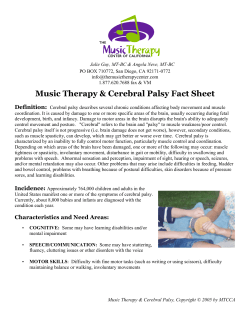
The biopsy showed clear necrotizing leukocytoclastic vasculitis, with an infiltrate predominantly of
neutrophils and no granuloma formation (Figure 2).
Her ESR was 60 (5 days after surgery), C-reactive
protein (CRP) 49; anti-nuclear factor, rheumatoid
factor, hepatitis B, cryoglobulins, serum angiotensinconverting enzyme and ANCA assay were normal or
negative. Her CSF contained no cells, protein 0.3
g/dl, normal glucose, and no evidence of oligoclonal
immunoglobulin bands. MRI (Figure 1b-e) showed
multiple T2-weighted high-signal-intensity lesions in
the periventricular areas, with larger areas in the left
Downloaded from http://qjmed.oxfordjournals.org/ by guest on September 9, 2014
therefore timely to re-examine vasculitic disease of
the nervous system in the light of these recent
advances, and to investigate whether they present
new opportunities for improving the diagnosis, treatment and outlook of these difficult and challenging
disorders.
As part of a structured clinical collaboration
investigating vasculitis and the nervous system, we
present an analysis of eight patients with the syndrome of cerebral vasculitis. We have included a
spectrum of aetiologies, ranging from primary CNS
vasculitis to intracranial vasculitis secondary to rheumatoid disease or lymphoma. This represents an attempt
to address the practical diagnostic difficulties posed
by the patient with suspected CNS vasculitis—where
a history of rheumatoid arthritis (for example) might
increase suspicion of cerebral vasculitis, but also (with
an associated history of immune suppressant treatment) raise other diagnostic possibilities, increasing
the importance of diagnostic accuracy. We have
attempted to analyse clinical patterns of disease, to
facilitate recognition of the disorder, and have assessed
the diagnostic value of four clinical or investigational
procedures not previously assessed in cerebral vasculitis: ophthalmological examination using low-dose
fluorescein angiography with slit-lamp video microscopy of the anterior segment; spinal fluid oligoclonal
band analysis; anti-neutrophil cytoplasmic antibody
assay; and indium-labelled white-cell cerebral
imaging. We have also assessed the therapeutic
implications of the response of our patients to immune
suppressive treatments.
Cerebral vasculitis
63
thalamus, the heads of both caudate nuclei, and the
left occipital cortex. Some lesions showed high signal
on T1-weighted images, suggesting a haemorrhagic
element to focal ischaemic areas.
She was treated with intravenous cyclophosphamide, 2 mg/kg/day, commencing 6 days after biopsy,
and intravenous methylprednisolone, 500 mg daily
for 5 days, then 60 mg prednisolone orally per day.
She remained comatose for 9 days, but thereafter
steadily recovered, breathing independently 16 days
after starting treatment, at which stage she had a
right hemiparesis with bilateral extensor plantar
responses. Her improvement continued and she was
discharged home 4 weeks later. When examined at
6 months, she was asymptomatic, with no residual
neurological signs on a tapering dose of cyclophosphamide and prednisolone.
Patient 2, JM
This 61-year-old retired, previously-well female presented with 2 weeks of increasing confusion and
forgetful ness, and suffered two generalized tonicclonic convulsions. No abnormalities were apparent
on general physical examination. She was disorientated, with poor and fluctuating registration, immediate recall, and episodic memory. She exhibited
choreiform movements of the limbs, predominantly
distal and symmetrical, together with facial grimacing, but no other focal neurological signs.
Psychiatric assessment suggested periodical hallucinations without delusions, paranoid features, occasional Ganserian responses, and crocydidmos.
Investigations included normal blood count and
film, ESR, CRP, liver function, urea and electrolytes
and thyroid function, and negative rheumatoid factor,
anti-nuclear factor and thyroid antibody. A CT head
scan showed only minimal atrophic changes, while
MR scanning was marred by gross movement
artefact. CSF: normal protein and glucose, no organisms seen or cultured, 4 neutrophils and 2 lymphocytes were present (per fil). ANCA serology was
positive at 22% on RIA, with competitive inhibition
Downloaded from http://qjmed.oxfordjournals.org/ by guest on September 9, 2014
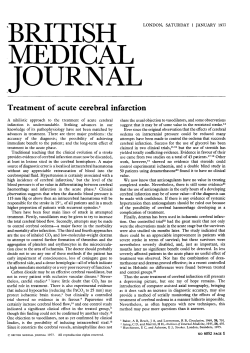
Figure 1. Cerebral imaging, patient 1. a CT brain scan. A non-enhancing lesion is shown in the left thalamus, exhibiting
slight mass effect. MRI (b-e) showed multiple T2-weighted high-signal-intensity lesions in the periventricular areas, with
larger areas in the left thalamus, the heads of both caudate nuclei, and the left occipital cortex. Some lesions showed high
signal on T1 -weighted images (d,e), suggesting a haemorrhagic element to focal ischaemic areas.
64
N.J. Scolding etal.
revealing specific antibody. Indium-labelled whitecell scan showed patchy uptake in both cerebral
hemispheres, more marked in the left (Figure 3), with
additional more marked uptake in the lungs. Cerebral
perfusion SPECT scanning showed an isolated area
of reduced perfusion in the left parietal region.
Bilateral carotid contrast arteriography showed variation in arterial calibre on both sides, particularly
affecting the distal vessels.
A diagnosis of isolated cerebral vasculitis was
made and she was treated with oral prednisolone
(60 mg daily). She made a good if slow recovery,
ANCA serology becoming negative within 6 weeks,
but was re-admitted 2 months later (on 30 mg
prednisolone daily) with recurring confusion, albeit
less severe. She was treated with 150mg daily of
oral cyclophosphamide, and again recovered well.
When seen 3 years later (6 months after changing
cyclophosphamide to azathioprine) she was well,
with no chorea, neurological signs or psychiatric
abnormalities,
although
cognitive
assessment
revealed mild residual deficits of frontal and memory
functions.
Patient 3, MF
This previously-well female had episodes of rightsided twitching starting in 1988 when aged 44. From
1990, she felt her memory steadily deteriorated. In
early 1992 she developed nocturnal convulsions,
and in March 1993, she had a short episode of status
epilepticus. CT head scan (normal in 1988) showed
a low-density area in the left temporal area. In July
1993, she developed acute visual loss in her left eye
accompanied by headache. Visual acuity was
reduced to 6/60, with an afferent pupillary defect
and a pale optic disc. She had bilateral Babinski
responses but no other neurological signs. ESR and
temporal artery biopsy were normal. Other serological tests, CRP, coagulation screen, cardiolipin antibody, echocardiography, ACE, and ANCA tests were
normal. Her headache, but not her vision, improved
with 80 mg prednisolone daily.
Four months later she was re-admitted in coma.
Her consciousness level fluctuated over the next 3
days but then improved and stabilized; at this stage,
she was amnesic, with reduced registration. She had
a pout reflex, an exaggerated jaw jerk, a right spastic
paretic arm (MRC grade 4) with hyper-reflexia,
bilateral flexor plantar responses and a nonspecifically unsteady gait. Re-investigation revealed
an ESR of 29 (repeated at 30), with a CRP of 68.
CSF was acellular, but contained oligoclonal
immunoglobulin bands: identical serum bands were
present. CT head scan now showed non-enhancing
low attenuation areas in the left frontal and both
occipital lobes, and perfusion HMPAO SPECT scanning showed multiple focal ischaemic defects,
affecting the left posterior parietal region, the left
fronto-parietal, and the right temporal areas
(Figure 4). Bilateral carotid angiography was normal.
An open cerebral biopsy of the left frontal lobe
(abnormal on CT), which included meninges, white
matter and cortex, showed multiple small ischaemic
areas, with nerve cell loss, myelin pallor, some
Downloaded from http://qjmed.oxfordjournals.org/ by guest on September 9, 2014
Figure 2. Cerebral biopsy, patient 1. Clear changes of necrotising leukocytoclastic vasculitis are present, with an intense
cellular infiltrate composed predominantly of neutrophils.
Cerebral vasculitis
65
The remaining five patients presented with acute
or subacute neurological encephalopathic illnesses;
two had raised intracranial pressure. Three experienced seizures, two had accompanying focal neurological signs, and one had chorea.
Three patients had an associated systemic disease:
two had previously-established rheumatoid disease
(without active arthritis at the time of neurological
presentation), and one had cerebral and systemic
vasculitis in the context of lymphoma. The remaining
five patients had isolated CNS vasculitis (though one
had livedo reticulares).
Investigation and diagnosis
macrophages but no vasculitic changes. Fluorescein
angiography, however, showed clear changes of
retinal vasculitis.
She was treated with intravenous cyclophosphamide (four 750 mg doses over 6 weeks), followed by
oral steroids. While symptomatically well thereafter,
she had a bilateral inferior altitudinal hemianopia
when examined 4 months later.
Clinical features
Details of all patients are summarized in Table 1. As
expected, a wide variety of clinical features were
encountered. Three patients (3, 5 and 7) had relapsing and spontaneously remitting disease characterized by optic neuropathy (3 episodes), brain stem
events (2), acute or sub-acute encephalopathic episodes (3), generalized or partial seizures (2 patients),
and hemispheric stroke-like episodes (one patient,
three events). Two such patients had additional
progressive cognitive or neuropsychological disturbance.
Discussion
We have described eight patients with vasculitis of
the central nervous system—in five confined to the
Downloaded from http://qjmed.oxfordjournals.org/ by guest on September 9, 2014
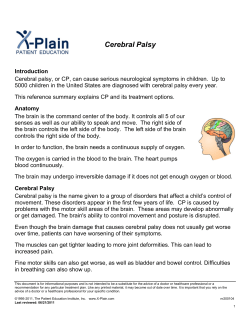
Figure 3. Indium-labelled white-cell scan, patient 2.
Patchy uptake is apparent in both cerebral hemispheres,
more marked in the left. Additional pulmonary uptake
was also apparent and was found also in patient 7, who
had normal brain images.
An elevated ESR or CRP accompanied neurological
symptoms in 5/8 patients; during two episodes (in
one patient), dissociation was found, with a raised
CRP in the presence of a normal ESR. Conventional
autoimmune serology was abnormal in three, while
ANCA serology was positive in two (patients 2 and
8), with an additional false-positive result in one
patient with rheumatoid arthritis. Routine spinal-fluid
examination was abnormal in only 2/8, in one of
whom (patient 2) the abnormality was very marginal
(6 white cells). CSF oligoclonal bands were present
in 3/6 patients, in one (patient 7) showing a shifting
pattern during the protracted course.
Ophthalmological examination made an important
contribution to the diagnosis of cerebral vasculitis
(Table 2). Vasculitic changes were found in 4/5
patients studied (patient 8 illustrated, Figure 5).
Abnormalities revealed by dynamic video recording
variably included marked slowing of flow, multifocal
attenuation of arterioles, and erythrocyte aggregates.
Fluorescein studies confirmed the variation in vessel
calibre, slowing of flow and red-cell aggregation,
and also demonstrated areas of small-vessel infarction, together with multifocal segments of intense
leakage from post-capillary and collecting venules.
CT scanning was abnormal in 7/8 patients, MRI
in all four patients imaged. Cerebral perfusion SPECT
was abnormal in 2/3 patients, while labelled whitecell scanning indicated cerebral inflammation in 1/2
patients—although in the patient with normal brain
appearances, increased uptake in other sites was
found and contributed usefully to diagnosis.
Cerebral biopsy was carried out in three patients;
all were abnormal though only two showed unequivocal vasculitis, the third (patient 3), showed multiple
small infarcts but no active vasculitis.
66
N.J. Scolding et al.
B
Figure 4. Perfusion HMPAO SPECT scanning, patient 3. Multiple focal ischaemic defects are visible; these involved the left
posterior parietal region, the left fronto-parietal, and the right temporal areas.
brain, the remainder proving to have CNS vasculitis
with laboratory evidence or past history of systemic
disease. Of the latter three, two (patients 5 and 8)
had established seropositive rheumatoid disease, of
which cerebral vasculitis is a rare but well-reported
complication. 2 ' 17 The third (patient 7) exhibited a
raised lymphocyte count at the onset of his illness
which eventually proved a consequence of a
low-grade B-cell lymphoma; his illness clinically
resembled lymphomatoid granulomatosis which not
uncommonly transforms to lymphoma 18 , a recognized cause of systemic and cerebral vasculitis.
We elected to describe all eight patients together
for three reasons. Firstly, in all patients inflammatory
disease of the brain was overwhelmingly the principal cause of morbidity at the time of presentation,
regardless of the underlying cause. Secondly, it seems
likely that all share similar cerebral processes.
Thirdly, the investigation and management of all
patients was largely independent of any systemic
process—which was either clinically quiescent at
the time of neurological presentation, or diagnosed
only in the course of investigation for suspected
cerebral vasculitis. As mentioned above, the clinical
diagnostic problems presented by all eight patients
were similar, regardless of their underlying medical
background.
Cerebral vasculitis is unusual—in our unit
accounting for a maximum of 0.5% admissions, no
more than 3-4 patients per year (with a neurological
catchment area of approximately 2.4 million). It is
difficult to recognize, to diagnose, and to treat, but
reports of successful therapies for other inflammatory
neurological diseases (j5-interferon and monoclonal
antibodies for multiple sclerosis, plasmapheresis and
intravenous immunoglobulin for inflammatory neuropathies and myopathies19"21 and for multi-system
vasculitis9'12) provide new hope for the treatment of
cerebral vasculitis, placing new emphasis on recognition and treatment.
Our patients reflect the previously emphasized
wide variation in manifestations, course and severity,
and the absence of a pathognomic or even typical
clinical picture.1'2'5'22 Focal and generalized seizures,
stroke-like episodes, acute and sub-acute encephalopathies, brain-stem events, progressive cognitive
changes, chorea, optic and other cranial neuropathies
all were seen. Despite this variability, it is possible
(and may be useful) to divide our patients into three
broad clinical groups.
Downloaded from http://qjmed.oxfordjournals.org/ by guest on September 9, 2014
B
Subacute post-partum ND
headache,
vomiting,
drowsiness;
cardiorespiratory
arrest and
persistent coma
after biopsy; no
systemic features.
Sub-acute confusion ND
with choreiform
movements and
generalized fits
Patient 1, PT (F)
dob 7.4.66
Acute psychosis,
N
followed by
confusional
state + cognitive
failure. Extensor /.
plantar. P/H
rheumatoid disease
Patient 4, VS (F)
dob 25.4.35
Acute CNS disease
in the context of
rheumatoid
arthritis
Chronic isolated
relapsing CNS
disease
5 year evolution,
FFA: retinal
starting with
vasculitis
unilateral twitching
nocturnal fits.
Cognitive
symptoms. Optic
neuropathy. Acute
encephalopathy.
Patient 3, MF (F)
dob 4.12.44
Subacute isolated
CNS disease
Patient 2, JM (F)
dob 27.4.40
Subacute isolated
CNS disease
Ocular
findings
Clinical features
Patient
78
30
N
60
90
68
N
49
ESR CRP
+ ve
N
ANCA
testing
RhF + ve
False +ve
1230lu/ml
ACA + ve
(lgM-68)
N
N
N
Inflammatory Routine
indices
serology
Summary of the clinical features and investigation results in all eight patients
Table 1
N, ogc-ve
No cells
ogc + v e
serum -l-ve
Past history,
current
rheumatoid &
cardiolipin
serology, ESR
& CRP, CT
scan
ESR/CRP; CSF
electrophoresis;
SPECT;
cerebral
biopsy and
FFA
ANCA, whitecell scan and
carotid
angiography
ND
CT—N (slight
prominence of
ventricles)
wcs—patchy
cerebral
uptake
SPECT— single
ischaemic area
angiography—
variation of
vessel calibre
CT—low
temporal artery
bx.—N
density
temporal lobe cerebral bx
SPECT—multiple
(meninges
cortex & white
perfusion
matter):
defects
angiography—
multiple small
normal
infarcts; no
visible
vasculitis
CT—
None
periventricular
low densities
SPECT—normal
Diagnosis
based on
4n,2l ogc -ve
Histopathology
CT—thalamic
Thalamic bx:
Thalamic biopsy
lesion
necrotizing
MRI—multiple
leukocytoclastic
lesions
vasculitis
caudate,
thalami,
periventricular
area
Imaging
N, ogc -ve
Spinal
fluid
Downloaded from http://qjmed.oxfordjournals.org/ by guest on September 9, 2014
o
Si"
8-si
i"
</>
c
(continued)
Clinical features
Patient 5, KR (M)
dob 3.12.46
Acute jaw, tooth &
scalp pain, then /.
visual loss,
Subacute relapsing
followed by r.:
CNS disease,
then transient /.
hemisphere
livedo reticulares.
episodes. Livedo
reticulares.
Cognitive changes
with impaired
frontal function.
Patient 6, SP (M)
Subacute headache,
vomiting,
dob 29.8.58
papilloedema,
ataxia
Subacute isolated
CNS disease
Patient
Table 1
14
1
ND
<6
<6
ESR CRP
N
ACA + ve,
(lgM-69)
Inflammatory Routine
indices
serology
Suggests
vasculitis
Ocular
findings
N
N
ANCA
testing
38L, 1N
protein 0.3
pressure—
80 cm water
N
Spinal
fluid
TA—normal
Liver—mild
cirrhotic
change
Histopathology
Stereotactic bx:CT—cerebellar
granulomatous
mass lesion
vasculitis with
with
epithelioid and
hydrocephalus
giant cells,
MRI—focal high
lymphocytic
signal r.
cuffing, reactive
cerebellar
changes,
hemisphere +
swollen
upper pons,
mid-brain,
astrocytes
external
Kveim -ve
capsule and r.
putamen,
periventricular
areas and
centrum
semiovale
CT—
periventricular
low densities,
probable
infarcts,
generalised
atrophy
Imaging
Downloaded from http://qjmed.oxfordjournals.org/ by guest on September 9, 2014
CT, MRI and
biopsy
High dose
steroids and
cyclosporin A.
Relapsing
course, died
with
hydrocephalus
and localized
haemorrhagic
changes (on
CT)
ACA serology,
CT scan and
ocular findings
Diagnosis
based on
r-
2-
<§'
5;
8
o
^.
•>-
ND
27
39
191
239
61
6
4
Subacute fluctuating Clear vasculitis 94
encephalopathy,
changes
70
headache, painful
numb extremities,
fever, night sweats;
dysarthria,
peripheral sensory
loss, transient
punctate macular
rash lower legs.
Past treatment for RA
included
Campath-1H, gold,
azathioprine,
cyclosporine
N
N
N
RhF +239, True +ve,
269 iU/ml
ag not
defined
N
N
N
CT—small
ND
ogc +ve, 1
infarct, r.
corona
band serum
irradiata
ogc +ve, serum
CT—N
+ ve (diff
bands)
MRI—multiple
high signal
ogc +ve, serum
lesions
-ve
WCS—increased
uptake lungs
and iliac fossa,
normal brain
Angiography—
normal
MRI—high
signal
periventricular
area, internal
capsule, subcortical area,
pons, /. globus
pallidus
N ocg +ve
CT—extensive
low
serum +ve
attenuation
cerebral white
matter
MRI—diffuse
high signal
intensity
centrum semiovale,
posterior
frontal lobe,
external
capsules,
periventricular
areas
Downloaded from http://qjmed.oxfordjournals.org/ by guest on September 9, 2014
Systemic
symptoms,
active
rheumatoid
serology, ESR,
CRP, ANCA,
ocular
findings.
Good response
Renal Bx—N
(transient
impaired renal
function)
cyclophosphamide
to
Rectal biopsy,
MRI, ocular
findings
lung bx.—
inflammatory
cells
rectal bx.—
arteritis with
fibrinoid
necrosis
Kveim—normal
bone marrow—
low grade
B-cell
lymphoma
dob, date of birth; ND, not done; N, normal; ogc, oligoclonal band analysis; /, left; r, right; wcs, white-cell scan; bx, biopsy; FFA, fundus fluorescein angiography;
n, neutrophil; I, lymphocyte; RhF, rheumatoid factor; ACA, anticardiolipin antibody.
Subacute CNS
disease in the
context of
rheumatoid
disease
Patient 8, LH (M)
dob 4.9.29
1986: wt loss, cough,
headache;
pulmonary
Relapsing CNS and
infiltrate, raised
Suggests
lymphocyte count
arteritis
systemic disease
1992: diarrhoea,
night sweats, wt
loss; followed by
acute febrile
encephalopathy
1993: fever, night
sweats, abdo. pain
headaches, brain
stem episode
1994: recurrent brain
stem episode with
/. VI palsy
Patient 7, JW (M)
dob 11.10.34
in"
O
C;
In
2
?8.
>ral
N.J. Scolding etal.
70
Table 2
Details of ocular findings in the five cases fully examined
Patient
Visual activity
Examination
Results
Ocular diagnosis
3
r1/60
/ 1/60
Anterior segment
IOP 31/30
Posterior segment
Pale discs; arterial attenuation,
venous beading, old venous
sheathing r
No active vasculitis
No rbc aggregation
/-focal arteriolar narrowing &
dry macular degeneration
r-homonymous inferior
quadrantanopia
Multifocal venular dilation
and stasis
Normal
Steroid-induced ocular
hypertension
Large vessel ischaemia in
ophthalmic artery territories
4
r6/9
/ 6/18
5
r6/5
/6/5
FFA
Conjunctival vessels
Posterior segments
Fields
Conjunctival vessels
Anterior and posterior
Previous clear retinal vasculitis
No vasculitis
/-posterior parietal infarct
Consistent with vasculitis
segments
r6/6
/6/6
8
Not reliable
Conjunctival vessels
Anterior segment
Fundus
FFA
Ocular examination
Conjunctival fluorescein
videoangiogram
(i) Atypical multiple sclerosis (more accurately
'MS-plus'). Three patients (patients 3, 5 and 7)
exhibited a spontaneously relapsing and remitting
course, with neurological features which included
optic neuropathy and brain stem episodes. Two had
CSF oligoclonal bands, one (of two scanned) had
multifocal white-matter lesions on MRI; multiple
sclerosis had been considered a possible diagnosis
in all at some time. Each, however, had additional
features less common in multiple sclerosis: seizures
(1/3), severe headaches (2/3), encephalopathic episodes (2/3), or hemispheric stroke-like events (1/3).
Systemic features were found in 2/3, including livedo
reticulares, fever, or oligoarthropathy.
(ii) Intracranial mass lesion. This was found in
two patients on 'first pass' investigation (CT).
(Hi) Acute or sub-acute encephalopathy the presentation in the remaining three patients.
The great majority of cases detailed in the literature
also conform to these patterns, which we do not
suggest carry pathological or therapeutic implications. Their occurrence might, however, lead to a
suspicion of cerebral vasculitis, so they may improve
recognition of this condition.
ANCA assays are now routinely used in the
serological diagnosis of systemic vasculitis; these
antibodies may also have a pathogenic role.23 A
number of case reports indicate the expected positive
ANCA testing in patients with Wegener's granulomatosis complicated by direct cerebral spread,24'25 but
Normal
Normal
r-cotton wool spot (nerve fibre
layer infarct)
No other vascular abnormality
Normal
Multifocal microvascular nonperfusion and leakage
Compatible with systemic
vasculitis
Characteristic of systemic
vasculitis
we have been unable to identify any previous
investigation of the role of ANCA serology in the
diagnosis of cerebral vasculitis. We tested all eight
patients. Two had true-positive ANCA testing; in one
(patient 2), the result was particularly useful since
she had isolated CNS disease and (otherwise) normal
blood tests. We also performed ANCA-testing on one
CSF sample (patient 8, with a positive serum test).
No antibody was detectable, concurring with our
experience of CSF ANCA testing in cerebral disease
from ANCA-positive Wegener's granulomatosis
(unpublished observations).
Spinal fluid was examined in all eight patients.
Only one had a substantially elevated cell count
(patient 6; 38 lymphocytes); Patient 2 had a very
marginally abnormal count (6 white cells). None had
a raised total protein level. Previous published series
report abnormal results in 50-80% of cases,2'3'26"28
but comparisons are difficult, since the definition of
an abnormal result varies considerably (from > 1 , to
> 4 cells per high-power field 3 ).
There have been no published systematic studies
of CSF oligoclonal immunoglobulin band analysis in
cerebral vasculitis, although 2/4 cases of herpeszoster-related cerebral vasculitis were positive,29 as
were '25-66%' of cases with various manifestations
of cerebral lupus.30 3/6 of our patients showed
abnormalities. Patient 3 had different immunoglobulin bands in CSF and in serum, patient 8 had
identical bands. In patient 7, a fluctuating pattern
Downloaded from http://qjmed.oxfordjournals.org/ by guest on September 9, 2014
7
Cerebral vasculitis
was found on serial testing over a 2-year period (see
Table 1). We suggest that oligoclonal band analysis
is therefore worthwhile in suspected cerebral vasculitis, an abnormal result (and perhaps particularly of
variable pattern, or indicating intrathecal and systemic immunoglobulin synthesis) providing some
diagnostic support, albeit without specificity.
MRI abnormalities were present in 4/5 of our
patients. In the fifth, the images were not interpretable
due to movement. MRI has been suggested as a
sensitive but not specific screening test in cerebral
vasculitis: Harris ef a/.31 reported significant abnormalities in 9/9 cases of proven disease, and no falsenegative scans. However, in an interesting correlative
study, Greenan ef a/.32 found on careful regional
analysis that 12/33 vascular territories with angiographic vasculitis exhibited no lesions on MR. Up to
25% of cases may in fact have normal MRI scans,28'33
some additionally with normal angiography.34
The value of angiography is difficult to assess,
many published series relying on this investigation
for diagnostic confirmation. 26 ' 28 Studies depending
on pathological examination indicate a false-negative
rate for angiography of 30-45%, 3 ' 27 although other
series suggest angiography is diagnostically useful in
only 20-27% of cases.35'36 A 10% risk of transient
neurological deficit is reported,37 with permanent
deficit in 1 % . Only 1/4 of our patients had abnormal
angiography, and although the value of this investigation may have been over-emphasized in the past,
it clearly remains important to exclude atheromatous
and other disease, and when positive, affords more
specificity than MRI.
We have found no reference to labelled-whitecell nuclear scanning in cerebral vasculitis, notwithstanding its increasingly recognized value in systemic
vasculitides.8 We found abnormal cerebral accumulation of indium-labelled leucocytes in 1/2 patients
tested, although the (clinically silent) increased pulmonary uptake in both was also diagnostically useful.
Vasculitis in other organs is demonstrated indirectly
by leucocyte uptake in necrotic tissue, or ischaemic
areas supplied by vasculitic vessels,14 whereas leucocyte infiltration of intracerebral vessels is unlikely to
be visualized directly. Nevertheless, further studies
may be worthwhile. Functional cerebral imaging,
including SPECT, was abnormal in 8/12 patients in
a recent series,36 and in 1/2 patients we examined.
Cerebral perfusion SPECT may well therefore be a
useful if non-specific test in cerebral vasculitis,
demonstrating focal ischaemia secondary to the
vasculitic process.
Most helpfully, we found ophthalmological examination with video microscopy and low-dose fluoresecin angiography of the anterior chamber to be
extremely valuable in the assessment of our patients.
Four of five patients examined had abnormal findings
suggestive of vasculitis. Studies of the ocular vasculature have been shown to contribute to the
diagnosis of multi-system vasculitis,6 and retinal
vasculitis has been much studied in multiple sclerosis
(where vasculitis generally refers to vascular leakage
and occlusion without implying a histopathology of
leukocytoclastic vascular damage). We have, however, been unable to find any reference in the
literature to the diagnostic contribution of these
ophthalmological techniques to cerebral vasculitis.
Previous studies have confirmed the clinicopathological correlation between fluorescein angiographic
changes and (localized ocular) vasculitis,38'39 and
our findings suggest that ophthalmological assessment is a useful and important addition to the
investigation of patients with suspected cerebral
vasculitis.
Our study might be criticized for lacking pathological data. This issue is, however, far from straightforward. In two of the largest series surveying previously
published cases,3'27 biopsies had been performed in
only 29% (14/48) and 52% (37/71) of cases, and
were diagnostic only in 10 and 26 patients, respect-
Downloaded from http://qjmed.oxfordjournals.org/ by guest on September 9, 2014
Figure 5. Anterior ocular vascular study, patient 8. Still
frames from slit-lamp video recording are shown.
Abnormalities included multifocal attenuation of arterioles,
and erythrocyte aggregates (a) Low-dose fluorescein studies (b) confirmed the variation in vessel calibre, and also
demonstrated areas of small-vessel infarction, together
with multifocal segments of intense leakage from postcapillary and collecting venules.
71
72
N.J. Scolding et al.
with cerebral vasculitis who have more benign
disease which may, in fact, not require any treatment,41 a suggestion supported by others.26 In many
cases of cerebral vasculitis, however, relapses may
occur when steroids alone are used35 and we would
advocate early recourse to cyclophosphamide in
patients not responding rapidly to high dose intravenous steroids—as previously recommended in both
cerebral and systemic vasculitis.5'22'43 We have not
as yet treated patients with cerebral vasculitis with
any of the more experimental approaches, although
the promise of Campath-1 H humanized monoclonal
antibody treatment in inflammatory demyelination 20
and in systemic vasculitis12 may indicate significant
potential, cerebral vasculitis unresponsive to cyclophosphamide being well-described. 26
References
1. Moore PM, Calabrese LH. Neurologic manifestations of
systemic vasculitides. Semin Neurol 1994; 14:300-6.
2. Sigal LH. The neurologic presentation of vasculitic and
rheumatologic syndromes. A review. Medicine 1987;
66:157-80.
3. Calabrese LH, Mallek JA. Primary angiitis of the central
nervous system. Report of 8 new cases, review of the
literature, and proposal for diagnostic criteria. Medicine
1988; 67:20-39.
4. Dyck PJ, Benstead TJ, Conn DL, et al. Nonsystemic
vasculitic neuropathy. Brain 1987; 110:843-54.
5. Moore PM. Vasculitis of the central nervous system. Semin
Neurol 1994; 14:307-12.
6. Charles SJ, Meyer PAR, Watson PC. Diagnosis and
management of systemic Wegener's granulomatosis
presenting with anterior ocular inflammatory disease. Br
J Ophthalmol 1991; 75:201 -7.
7. Jennette JC, Falk RJ, Andrassy K, ef al. Nomenclature of
systemic vasculitides: Proposal of an international
consensus conference. Arthritis Rheum 1994; 37:187-92.
8. Reuter H, Wraight EP, Qasim FJ, Lockwood CM.
Management of systemic vasculitis: Contribution of
scintigraphic imaging to evaluation of disease activity and
classification. QJM 1995; 88:509-16.
9. Jayne DRW, Davies MJ, Fox CJV, et al. Treatment of
systemic vasculitis with pooled intravenous
immunoglobulin. Lancet 1991; 337:1137-9.
10. Jennette JC, Falk RJ. Diagnostic classification of
antineutrophil cytoplasmic autoantibody- associated
vasculitides. Am J Kidney Dis 1991; 18:184-7.
11. Lai KN, Jayne DRW, Brownlee A, Lockwood CM. The
specificity of anti-neutrophil cytoplasm autoantibodies in
systemic vasculitis. Clin Exp Immunol 1990; 82:233-7.
12. Mathieson PW, Cobbold SP, Hale G, et al. Monoclonalantibody therapy in systemic vasculitis. N EnglJ Med 1990;
323:250-4.
13. Hagen EC, Andrassy K, Chernok E, ef al. The value of
indirect immunofluorescence and solid phase techniques for
ANCA detection. J Immunological Methods 1993;
159:1-16.
Downloaded from http://qjmed.oxfordjournals.org/ by guest on September 9, 2014
ively. Three of our eight patients underwent cerebral
biopsy: two in the course of urgent assessment for
acute space-occupying lesions, biopsy revealing
unsuspected vasculitis, the third undergoing 'blind'
biopsy of cortex, white matter and meninges for
suspected vasculitis; in this patient, the findings were
non-specific and not diagnostic. Our results are
therefore numerically typical and representative of
published reports and series.
Cerebral biopsy is not a trivial procedure, carrying
a significant risk of serious morbidity estimated at
0.5-2% 4 0 , and in the majority of our patients we
found it impossible to justify. Patient 8 is illustrative:
findings included strongly positive rheumatoid serology, positive ANCA serology, marked elevations of
both ESR and CRP, and unequivocal evidence of
small-vessel vasculitis in the anterior ocular circulation, with no other explanation for his subacute
encephalopathy after extensive investigation. We felt
unable to build a compelling case for biopsy, particularly since, with our own experience of one nondiagnostic biopsy and published data consistently
showing the diagnostic yield of biopsy to be relatively
low at 70%, 3 ' 27 the decision to treat with cyclophosphamide would not have been influenced by a nondiagnostic biopsy. A pronounced response to treatment in this patient provided further retrospective
support for the diagnosis.
The low incidence of vasculitic cerebral disease
renders formal
prospective therapeutic
trials
extremely difficult: none has so far been reported.
An informed approach to treatment therefore depends
on the cumulative experience described in published
retrospective and pooled series, and our patients
may offer some useful lessons. Patients 1 and 6 had
very similar features: biopsy-proven isolated cerebral
vasculitis presenting with single cerebral mass lesions
on CT but MRI evidence of multifocal disease, both
with negative routine and ANCA serology. One was
treated with cyclophosphamide and steroids and
made a complete recovery with no further symptoms
some 6 months later; the other received high-dose
steroids with cyclosporin and suffered a chronic
relapsing and ultimately fatal illness. Four other
patients received cyclophosphamide. One had a
single further episode of optic neuropathy (whilst on
oral steroid maintenance treatment alone, 3 months
after four pulses of intravenous cyclophosphamide)
and remained well thereafter, and three others experienced a striking resolution of their illness. The fourth
patient, who had B-cell lymphoma, also died (of
pseudomonas pneumonia).
Two patients received steroids alone: one (patient
4) with past rheumatoid disease, and one (patient
5) whose initial presentation had suggested giant
cell arteritis. Both responded well. Interestingly,
Calabrese ef al. have defined a group of patients
Cerebral vasculitis
73
14. Fink AM, Miles KA, Wraight EP. lndium-111 labelled
leucocyte uptake in aortitis. Clin Radiol 1994; 49:863-6.
and magnetic resonance imaging. J Rheumatol 1994;
21:1277-82.
15. Meyer PAR. Patterns of blood flow in episcleral vessels
studied by low dose fluorescein videoangiography. Eye
1988; 2:533-46.
29. Hilt DC, Buchholz D, Krumholz A, etal. Herpes zoster
ophthalmicus and delayed contralateral hemiparesis caused
by cerebral angiitis: Diagnosis and management
approaches. Ann Neurol 1983; 14:543-53.
16. Meyer PAR, Watson PC. Low dose fluorescein angiography
of the conjunctiva and episclera. BrJ Ophthalmol 1987;
71:2-10.
17. Ramos M, Mandybur Tl. Cerebral vasculitis in rheumatoid
arthritis. Arch Neurol 1975; 32:271-5.
18. Katzenstein A, Carrington CB, Liebow AA. Lymphomatoid
granulomatosis; A clinicopathological study of 152 cases.
Cancer 1979; 43:360-73.
19. Jacobs LD, Cookfair DL, Rudick RA, etal. Intramuscular
interferon beta-1a for disease progression in relapsing
multiple sclerosis. Ann Neurol 1996; 39:285-94.
21. Thornton CA, Criggs RC. Plasma exchange and intravenous
immunoglobulin treatment of neuromuscular disease. Ann
Neurol 1994; 35:260-8.
22. Calabrese LH, Duna GF. Evaluation and treatment of central
nervous system vasculitis. Curr Opin Rheumatol 1995;
7:37-44.
23. Kallenberg CCM, Brouwer E, Weening JJ, Cohen Tervaert
JW. Anti-neutrophil cytoplasmic antibodies: Current
diagnostic and pathophysiological potential. Kidney Int
1994; 46:1-15.
24. Nishino H, Rubino FA, Parisi JE. The spectrum of neurologic
involvement in Wegener's granulomatosis. Neurology 1993;
43:1334-7.
25. Weinberger LM, Cohen ML, Remler BF, etal. Intracranial
Wegener's granulomatosis. Neurology 1993; 43:1831 - 4 .
26. Abu Shakra M, Khraishi M, Grosman H, etal. Primary
angiitis of the CNS diagnosed by angiography. QJM 1994;
87:351-8.
27. Hankey G. Isolated angiitis/angiopathy of the CNS.
Prospective diagnostic and therapeutic experience.
Cerebrovasc Dis 1991; 1:2-15.
28. StoneJH, Pomper MG, Roubenoff R, etal. Sensitivities of
noninvasive tests for central nervous system vasculitis: A
comparison of lumbar puncture, computed tomography,
31. Harris KG, Tran DD, Sickels WJ, etal. Diagnosing
intracranial vasculitis: The roles of MR and angiography.
Am) Neuroradioh994; 15:317-30.
32. Greenan TJ, Grossman Rl, Goldberg HI. Cerebral vasculitis:
MR imaging and angiographic correlation. Radiology 1992;
182:65-72.
33. Alhalabi M, Moore PM. Serial angiography in isolated
angiitis of the central nervous system. Neurology 1994;
44:1221-6.
34. Vanderzant C, Bromberg M, MacGuire A, McCune
J. Isolated small-vessel angiitis of the central nervous system.
Arch Neurol 1988; 45:683-7.
35. Koo EH, Massey EW. Granulomatous angiitis of the central
nervous system: Protean manifestations and response to
treatment. J Neurol Neurosurg Psychiatry 1988;
51:1126-33.
36. VollmerTL, GuarnacciaJ, Harrington W, etal. Idiopathic
granulomatous angiitis of the central nervous system:
Diagnostic challenges. Arch Neurol 1993; 50:925-30.
37. Hellmann DB, Roubenoff R, Healy RA, Wang H. Central
nervous system angiography: Safety and predictors of a
positive result in 125 consecutive patients evaluated for
possible vasculitis. J Rheumatol 1992; 19:568-72.
38. Ffytche TJ. Retinal vasculitis: a review of the clinical signs.
Trans Ophthal Soc UK 1977; 97:457-61.
39. Stanford MR, Graham EM, Kasp E, etal. Retinal vasculitis:
correlation of animal and human disease. Eye 1987;
1:69-77.
40. Barza M, Pauker SG. The decision to biopsy, treat, or wait
in suspected herpes encephalitis. Ann Intern Med 1980;
92:641-9.
41. Calabrese LH, Gragg LA, Furlan AJ. Benign angiopathy: a
subset of angiographically defined primary angiitis of the
central nervous system. J Rheumatol 1993; 20:2046-50.
42. Lockwood CM. Approaches to specific immunotherapy for
systemic vasculitis. Semin Neurol 1994; 14:387-92.
Downloaded from http://qjmed.oxfordjournals.org/ by guest on September 9, 2014
20. Moreau T, Thorpe J, Miller D, etal. Preliminary evidence
from magnetic resonance imaging for reduction in disease
activity after lymphocyte depletion in multiple sclerosis.
Lancet-\ 994; 344:298-301.
30. Van Dam AP. Diagnosis and pathogenesis of CNS lupus.
Rheumatol Int 1991; 11:1-11.


© Copyright 2026