
Mitral Valve Prolapse: Old Beliefs vs New Knowledge
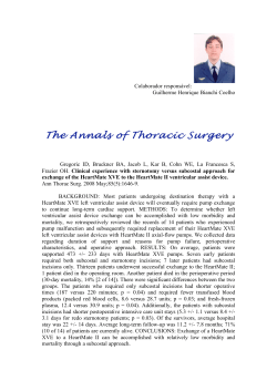
HEART VALVE UPDATE
CME
CREDIT
BRIAN GRIFFIN, MD, EDITOR
EMIL HAYEK, MD
BRIAN GRIFFIN, MD
Department of Cardiovascular Medicine,
The Cleveland Clinic
Co-Director, Valve Management Program,
Director, Cardiovascular Training
Program, Department of Cardiovascular
Medicine, The Cleveland Clinic
Mitral valve prolapse:
Old beliefs yield to new knowledge
■ A B S T R AC T
we once believed about
valve prolapse (MVP) have
proved false, as understanding of this condition has improved.
MVP was first described by Barlow and
Bosman in the 1960s.1 It is now recognized as
the most common cardiac valvular abnormality in the United States, but there has been
much less agreement about other aspects of
its epidemiology, diagnosis, and clinical features.
This paper describes how current knowledge is replacing previous beliefs about
MVP.
M mitral
ANY THINGS
Much of the conventional wisdom about the prevalance,
causes, diagnosis, symptoms, effects, and treament of mitral
valve prolapse (MVP) is changing. MVP has a benign course
and excellent prognosis for most patients, with only a small
minority developing serious complications.
■ KEY POINTS
Mitral valve prolapse (MVP) is not as common as once
believed, particularly in young women.
Two-dimensional echocardiography (parasternal and apical
long-axis views) is the diagnostic test of choice for MVP;
cardiac auscultation has a low sensitivity.
Accumulating evidence suggests that the MVP syndrome is
not directly linked to the valvular abnormality.
Rarely, MVP is associated with serious complications, but
recent data show that, in the absence of mitral
regurgitation, it does not increase the risk of arrhythmias,
stroke, or sudden death.
MVP is an important cause of progressive severe mitral
regurgitation requiring mitral valve surgery, particularly in
older men.
Some patients with MVP need antibiotic prophylaxis
against infective endocarditis; those with a history of stroke
or transient ischemic attack need aspirin or warfarin.
■ HOW COMMON IS MVP?
Previous belief: MVP is very common,
especially in young women.
Current knowledge: MVP is not as common as originally thought, with no gender difference in prevalence.
In the United States, the prevalence of MVP
is estimated to be 2.4%,2 based on
Framingham data and using the current criteria for diagnosis with two-dimensional
echocardiography.
Earlier studies, which used less-specific
echocardiographic criteria, had placed the estimate much higher, particularly in young women,
for whom the figure was as high as 17%.3
Why were the earlier figures so high? Part
of the reason is that the earlier studies used
less-specific echocardiographic criteria for
diagnosis (see Echocardiography, below).
Selection bias may also have accounted for
the much higher prevalence of MVP among
young women with symptoms in earlier, referral-based studies.
CLEVELAND CLINIC JOURNAL OF MEDICINE
VOLUME 69 • NUMBER 11
NOVEMBER 2002
Downloaded from www.ccjm.org on September 9, 2014. For personal use only. All other uses require permission.
889
MITRAL VALVE PROLAPSE
HAYEK AND GRIFFIN
Mitral valve prolapse: Dynamic maneuvers
affect the midsystolic click
Heart sounds at rest
S1
C
Ventricular volume
hastens the click
Valsalva maneuver
Standing
Amyl nitrate
S1 C
S2
S2
Ventricular volume
delays the click
Squatting
Maximal isometric
exercise
Leg raising
S1
C
S2
FIGURE 1. The timing of the midsystolic click (C)
associated with MVP is altered by dynamic maneuvers
that affect ventricular volume.
Listen for
a midsystolic
click, then
a murmur,
at the apex
890
diseases that affect connective tissue, such as
Marfan syndrome and Ehlers-Danlos syndrome. Prolapse of other cardiac valves, most
commonly the tricuspid and occasionally the
aortic, has been described in patients with
MVP.
Secondary MVP refers to prolapse of
leaflets that do not show myxomatous change.
It may occur when the leaflets are disproportionately large compared with the left ventricular cavity, most often in young women with a
relatively small left ventricular cavity in association with a hyperdynamic state. With
advancing age, the incidence of secondary
MVP among women declines as the size of the
ventricle better approximates that of the
mitral leaflets.
MVP has also been found in patients with
ischemic heart disease, hypertrophic cardiomyopathy, and rheumatic heart disease, but
whether these diseases actually cause the
MVP is not always clear.
■ HOW IS MVP BEST DIAGNOSED?
■ WHAT CAUSES MVP?
Previous belief: MVP is best diagnosed by
auscultation and M-mode echocardiography.
Current knowledge: Auscultation sometimes suffices, but 2-D echocardiography
(parasternal and long-axis views) is usually
necessary.
MVP—systolic billowing of one or both mitral
leaflets into the left atrium—may result from
structural or functional abnormalities of any
component of the mitral apparatus, including
the leaflets, chordae, papillary muscle, or adjacent left ventricle and atrium.
Primary MVP is characterized by myxomatous degeneration with an increased
amount of leaflet tissue and proliferation of
the spongiosa layer. We recently found
that, compared with normal valves, myxomatous valves contain different proportions of glycosaminoglycan, their leaflets
and chordae are stretchier, and their chordae are weaker.4
While primary MVP is usually sporadic,
familial clustering of MVP has been well
described, with autosomal-dominant inheritance modified by age and gender.3,5 MVP
may also occur in conjunction with certain
The classic finding of MVP on physical examination is a midsystolic click followed by a
murmur of varying duration, heard best at the
cardiac apex. The click is caused by tensing of
the mitral apparatus as the leaflets prolapse in
midsystole.
Dynamic maneuvers that diminish preload, such as the Valsalva maneuver or
standing, or drugs that decrease afterload,
such as amyl nitrate, tend to make the
sounds occur earlier in systole. In contrast,
the sounds occur later in systole during
maneuvers that increase ventricular filling,
such as squatting, or increase afterload, such
as isometric handgrip (FIGURE 1).
The midsystolic click of MVP should be
differentiated from aortic or pulmonic ejection clicks, which occur earlier in systole and
the timing of which is not altered by dynamic
maneuvers that affect ventricular volume.
A person’s lifetime risk of MVP is more
difficult to estimate, as the natural history of
its development is unknown.
CLEVELAND CLINIC JOURNAL OF MEDICINE
VOLUME 69 • NUMBER 11
NOVEMBER 2002
Downloaded from www.ccjm.org on September 9, 2014. For personal use only. All other uses require permission.
Echocardiography
Although the finding of the combination of
a midsystolic click and a systolic murmur is
relatively specific for MVP, its sensitivity is
limited.6 Furthermore, the finding of either
a systolic click without a murmur or a murmur without a click is not well correlated
with echocardiographic prolapse or with
mitral valve thickening or redundancy.7,8
Therefore, transthoracic echocardiography
is often necessary for patients suspected of
having MVP on the basis of cardiac auscultation.
Even when a patient has the classic auscultatory findings of MVP, echocardiography
may have an important role in risk stratification by assessing the degree of regurgitation
and chamber enlargement and features of
myxomatous mitral valve disease such as
leaflet thickening and redundancy. It is also
useful in assessing left ventricular systolic
function.
Echocardiography can also be used to
exclude MVP in first-degree relatives of
patients with known myxomatous valve disease; however, there is insufficient evidence
to support its general use as a screening
test.9
Criteria for diagnosis. The echocardiographic diagnosis of MVP should be made
only if one or both leaflets are displaced by at
least 2 mm above the mitral annulus during
systole in either the parasternal or apical
long-axis view on two-dimensional imaging
(FIGURE 2).
A caveat: in certain two-dimensional
echocardiographic views, notably the apical four-chamber view, the mitral leaflets
may appear to prolapse while actually
remaining below the level of the entire
mitral valve, possibly leading to an incorrect diagnosis of MVP. Use of the apical
four-chamber view may have contributed
to the relatively high prevalence of MVP
found in earlier echocardiographic studies.
A detailed three-dimensional echocardiographic analysis has revealed that the mitral
annulus is saddle-shaped, not planar,10 and
physicians should look for leaflet displacement above the most superior points of the
mitral annulus.
Mitral valve prolapse:
Echocardiographic appearance
FIGURE 2. Two-dimensional parasternal long-axis systolic
frame showing prolapse of the posterior leaflet (arrow)
well behind the plane of the annulus (dotted line). LV = left
ventricle, LA = left atrium.
■ DOES MVP CAUSE THE MVP SYNDROME?
Previous belief: MVP causes a variety of
diffuse symptoms.
Current knowledge: Little direct evidence links these symptoms with MVP.
Consider an
echo if you
suspect MVP
on auscultation
Although most patients with MVP have no
symptoms whatsoever, a small subset have a
variety of symptoms and signs that are termed
the MVP syndrome.
Symptoms of the MVP syndrome include
atypical chest pain, dyspnea, palpitations,
dizziness, fatigue, and anxiety or panic attacks.
However, several studies have shown no true
increased frequency of chest pain or dyspnea
or other symptoms such as dizziness in patients
with MVP.2,11–13 The original descriptions of
increased prevalence probably reflected selection bias.
MVP is associated with thin body habitus,
pectus excavatum, scoliosis, and low resting
blood pressure.
Electrocardiographic abnormalities described in association with MVP include atri-
CLEVELAND CLINIC JOURNAL OF MEDICINE
VOLUME 69 • NUMBER 11
NOVEMBER 2002
Downloaded from www.ccjm.org on September 9, 2014. For personal use only. All other uses require permission.
891
MITRAL VALVE PROLAPSE
HAYEK AND GRIFFIN
al and ventricular arrhythmias, prolonged
QTc interval, nonspecific ST-T wave changes,
and inferior T-wave inversion.14 However,
other studies have found no increased frequency of electrocardiographic abnormalities
in patients with MVP.2,11,12
Pathophysiology unknown
The pathophysiology of the MVP syndrome is
unknown, and the symptoms cannot be
explained by the valvular abnormality alone.
Several studies have documented abnormal
autonomic function in patients with symptomatic MVP, including elevated circulating
levels of catecholamines, enhanced betaadrenergic receptor affinity, increased vasoconstrictor tone, decreased plasma volume,
and diminished vagal responsiveness.15–19
However, other studies have shown no evidence of abnormal autonomic or neuroendocrine function, either at rest or during tilttesting, in MVP patients with or without
symptoms.20,21
■ NATURAL HISTORY
AND COMPLICATIONS OF MVP
The mitral
annulus is
saddle-shaped,
not planar
MVP has a benign course and excellent prognosis for most patients, with a survival rate
similar to that in an age-matched and sexmatched population without MVP.22
However, a minority of patients develop serious complications, including infective endocarditis, cerebrovascular events, progressive
severe mitral regurgitation, arrhythmias, and
sudden cardiac death.
The risk of complications is highest in
men, patients older than 45 years, patients
with familial MVP, and patients with a
holosystolic murmur or left-sided chamber
enlargement.23,24 Echocardiographic predictors of increased complications have also been
found, including significant mitral regurgitation, leaflet thickening, and leaflet redundancy.22,25–27
Infective endocarditis
MVP is probably the most common cardiovascular abnormality predisposing to infective
bacterial endocarditis. Patients with MVP are
at a fivefold to eightfold greater risk of infective endocarditis compared with controls
892
CLEVELAND CLINIC JOURNAL OF MEDICINE
VOLUME 69 • NUMBER 11
without MVP.24,28,29 However, in prospective
studies, the risk that a patient with MVP will
develop infective endocarditis has been
demonstrated to be very low.2,22,23,27
The risk of developing endocarditis is
highest in older men and patients with a murmur24,30 or those with redundant and thickened valve leaflets.22,25 Endocarditis in these
patients is associated with considerable morbidity and mortality: the 5-year cumulative
incidence of death or mitral valve surgery is
estimated to be 60%.31
Cerebrovascular events
Previous belief: MVP often causes stroke
in young patients.
Current knowledge: Stroke in patients
with MVP, regardless of age, is usually due to
causes other than MVP.
Whether MVP causes strokes is controversial.
Several studies found an association
between MVP and cerebrovascular ischemic
events, particularly among younger patients
without other known risk factors for cerebrovascular disease.32–36 Although these
events have been attributed to fibrin emboli
from the prolapsing mitral leaflet, possibly in
association with an underlying prothrombotic
state, this has rarely been documented in the
literature.37
On the other hand, recent populationbased and case-control studies demonstrated
no increased frequency of MVP among
patients with stroke or transient ischemic
attacks, including young patients.38–40 In addition, patients with MVP who had strokes were
similar in age to the general stroke population,
and many had other risk factors for strokes,
including atrial fibrillation.22,38,39
Several prospective studies have found
that cerebrovascular events occur only rarely
in patients with MVP, with an incidence of
less than 1% per year.2,22,23
Mitral regurgitation
Most patients with MVP do not develop significant mitral regurgitation.2 Nevertheless,
MVP is a common cause of progressive, severe
mitral regurgitation, which often requires
valve repair or replacement.41,42
Several prospective studies have found
NOVEMBER 2002
Downloaded from www.ccjm.org on September 9, 2014. For personal use only. All other uses require permission.
that the risk of developing severe mitral regurgitation requiring mitral valve repair or
replacement is less than 1% per year.22,23,26
The cumulative risk rises with age; an
Australian study found that by age 70 it is 4%
in men and 1% in women.43
Subsets of patients with MVP who are at
higher risk of developing severe mitral regurgitation requiring mitral valve surgery are
older men and those with higher systolic pressure and body weight.44 Other clinical predictors of the need for mitral valve surgery for
mitral regurgitation include a left ventricular
diastolic dimension greater than 60 mm and
cardiomegaly on chest radiography.22 Older
patients and those with prolapse of the posterior mitral leaflet have been found to have
more severe regurgitation.26
Cardiovascular complications, including
atrial fibrillation and congestive heart failure,
are much more likely if mitral regurgitation is
severe or if it progresses over time.26,27
Severe mitral regurgitation can develop
from progressive myxomatous degeneration
with an increasing degree of prolapse, or it can
be due to chordal rupture. Rupture of chordae
tendineae nearly always results in severe
mitral regurgitation and a flail leaflet.24,42,45
Arrhythmias
Previous belief: MVP often causes atrial
and ventricular arrhythmias.
Current knowledge: No excess of
arrhythmias is associated with MVP without
regurgitation.
Early studies indicated that many patients
with MVP had palpitations and various dysrhythmias, including supraventricular tachycardia and atrial and ventricular premature
depolarizations.
However, these studies were likely affected by selection bias, as the subjects typically
were patients referred for evaluation of symptoms such as palpitations.
Furthermore, the Framingham investigators46 found that these dysrhythmias are also
common among people without MVP, and
that patients with MVP had no statistically
significant excess of dysrhythmias on resting
12-lead electrocardiography, treadmill electrocardiography, or 1-hour or 24-hour ambula-
LA
LV
LA
LV
FIGURE 3. Top, transesophageal systolic image of severe
mitral regurgitation (arrow) due to posterior mitral leaflet
prolapse before surgical repair. Bottom, trivial residual
mitral regurgitation (arrow) after mitral valve repair.
LA = left atrium, LV = left ventricle.
tory electrocardiographic monitoring compared with people without MVP.
An exception, however: patients with
MVP and mitral regurgitation have a higher
prevalence of ventricular dysrhythmias,
including ventricular tachycardia.47
Sudden cardiac death
Sudden cardiac death is a rare complication of
MVP, with an estimated incidence of 0.1% to
0.4% per year, similar to the risk in the general adult population of the United
States.22,23,48 The risk appears to be greatest in
MVP patients with complex ventricular
arrhythmias on Holter monitoring, a prolonged QT interval, hemodynamically signifi-
CLEVELAND CLINIC JOURNAL OF MEDICINE
VOLUME 69 • NUMBER 11
NOVEMBER 2002
Downloaded from www.ccjm.org on September 9, 2014. For personal use only. All other uses require permission.
893
MITRAL VALVE PROLAPSE
HAYEK AND GRIFFIN
cant mitral regurgitation, and redundant
mitral leaflets.22,49
ed for those with an isolated systolic click and
equivocal or no evidence of MVP on echocardiography.
■ HOW SHOULD MVP BE TREATED?
Patients with MVP who have no symptoms
(ie, most patients with MVP) require no specific treatment other than antibiotic prophylaxis in some cases (see below), and should be
reassured of their excellent prognosis.
Patients with either syncope or palpitations should be evaluated with Holter ambulatory monitoring to rule out significant
supraventricular or ventricular arrhythmias.
Patients without mitral regurgitation
should be reexamined every 3 years, and
echocardiography should be performed if cardiovascular symptoms develop or a new murmur or mitral regurgitation is found on cardiac
auscultation.
Beta-blockers
Beta-blockers may be useful empirically in
alleviating symptoms of palpitations, anxiety,
and chest pain in some patients.50
Follow up
severe mitral
regurgitation
at least every
6 - 12 months
with stress
echos
Antibiotic prophylaxis
Antibiotic prophylaxis to prevent infective
bacterial endocarditis has been shown to be
cost-effective ($3,000 per year of life saved)
when given to patients with MVP and a systolic murmur.51
Guidelines9,52 call for antibiotic prophylaxis before dental procedures or other invasive procedures for patients with MVP who
have either of the following:
• Both a systolic click and murmur on auscultation
• An isolated systolic click and echocardiographic evidence of MVP and mitral
regurgitation.
Antibiotic prophylaxis may be considered
for those with only a systolic click and no murmur if certain high-risk echocardiographic
characteristics are present, such as leaflet
redundancy, leaflet thickening, or left atrial or
ventricular enlargement.
Antibiotic prophylaxis is not recommend■ REFERENCES
1. Barlow JB, Bosman CK. Aneurysmal protrusion of the
posterior leaflet of the mitral valve: an auscultatory-elec-
894
CLEVELAND CLINIC JOURNAL OF MEDICINE
VOLUME 69 • NUMBER 11
Anticoagulation
Patients with MVP should receive aspirin
(80–325 mg/day) if they have either of the following:
• A history of transient ischemic attacks
• Atrial fibrillation (if they are younger
than 65 and have no mitral regurgitation,
hypertension, or congestive heart failure).
Patients should receive warfarin (target
international normalized ratio 2–3) if they
have any of the following:
• Documented stroke
• Recurrent transient ischemic attacks
• Atrial fibrillation with an increased risk of
thromboembolism (eg, if they are older
than age 65 or have mitral regurgitation,
hypertension, or congestive heart failure).
Management of mitral regurgitation
Serial echocardiography is necessary to document changes in left ventricular size and systolic function, which are important indicators
of the need for surgery.
Patients with MVP who have mitral
regurgitation of mild to moderate severity
should undergo echocardiography every year.
Patients with MVP and severe mitral
regurgitation should undergo stress echocardiography every 6 to 12 months or more frequently, depending on their clinical condition.
There is no evidence that afterload-reducing drugs favorably alter the natural history of
mitral regurgitation if left ventricular function
is normal.
The indications for mitral valve surgery
when severe mitral regurgitation is present are
similar to those for causes of severe mitral regurgitation other than MVP and include the
development of symptoms or even mild impairment of left ventricular systolic function.
When surgery is required, mitral valve repair is
usually feasible (FIGURE 3); the 10-year reoperation-free survival rate is 93% to 96%.53,54
trocardiographic syndrome. Am Heart J 1966; 71:166–178.
2. Freed LA, Levy D, Levine RA, et al. Prevalence and clinical
outcome of mitral-valve prolapse. N Engl J Med 1999;
341:1–7.
NOVEMBER 2002
Downloaded from www.ccjm.org on September 9, 2014. For personal use only. All other uses require permission.
3. Savage DD, Garrison RJ, Devereux RB, et al. Mitral valve
prolapse in the general population, I: epidemiologic features—the Framingham Study. Am Heart J 1983;
106:571–576.
4. Barber JE, Kasper FK, Ratliff NB, Cosgrove DM, Griffin BP,
Vesely I. Mechanical properties of myxomatous mitral
valves. J Thorac Cardiovasc Surg 2001; 122:955–962.
5. Devereux RB, Brown WT, Kramer-Fox R, Sachs I.
Inheritance of mitral valve prolapse: effect of age and sex
on gene expression. Ann Intern Med 1982; 97:826–832.
6. Attenhofer Jost CH, Turina J, Mayer K, et al.
Echocardiography in the evaluation of systolic murmurs
of unknown cause. Am J Med 2000; 108:614–620.
7. Weis AJ, Salcedo EE, Stewart WJ, Lever HM, Klein AL,
Thomas JD. Anatomic explanation of mobile systolic
clicks: implications for the clinical and echocardiographic
diagnosis of mitral valve prolapse. Am Heart J 1995;
129:314–320.
8. Etchells E, Bell C, Robb K. Does this patient have an
abnormal systolic murmur? JAMA 1997; 277:564–571.
9. Bonow RO, Carabello B, de Leon AC Jr, et al. Guidelines
for the management of patients with valvular heart disease: executive summary—a report of the American
College of Cardiology/American Heart Association Task
Force on Practice Guidelines (Committee on Management
of Patients with Valvular Heart Disease). Circulation 1998;
98:672–707.
10. Levine RA, Handschumacher MD, Sanfilippo AJ, et al.
Three-dimensional echocardiographic reconstruction of
the mitral valve, with implications for the diagnosis of
mitral valve prolapse. Circulation 1989; 80:589–598.
11. Devereux RB, Kramer-Fox R, Brown WT, et al. Relation
between clinical features of the mitral prolapse syndrome and echocardiographically documented mitral
valve prolapse. J Am Coll Cardiol 1986; 8:763–772.
12. Savage DD, Devereux RB, Garrison RJ, et al. Mitral valve
prolapse in the general population, II: clinical features—
the Framingham Study. Am Heart J 1983; 106:577–581.
13. Retchin SM, Fletcher RH, Earp J, Lamson N, Waugh RA.
Mitral valve prolapse: disease or illness? Arch Intern Med
1986; 146:1081–1084.
14. Bhutto ZR, Barron JT, Liebson PR, Uretz EF, Parrillo JE.
Electrocardiographic abnormalities in mitral valve prolapse. Am J Cardiol 1992; 70:265–266.
15. Gaffney FA, Karlsson ES, Campbell W, et al. Autonomic
dysfunction in women with mitral valve prolapse syndrome. Circulation 1979; 59:894–901.
16. Puddu PE, Pasternac A, Tubau JF, Krol R, Farley L, de
Champlain J. QT interval prolongation and increased
plasma catecholamine levels in patients with mitral valve
prolapse. Am Heart J 1983; 105:422–428.
17. Pasternac A, Tubau JF, Puddu PE, Krol RB, de Champlain J.
Increased plasma catecholamine levels in patients with
symptomatic mitral valve prolapse. Am J Med 1982;
73:783–790.
18. Gaffney FA, Bastian BC, Lane LB, et al. Abnormal cardiovascular regulation in the mitral valve prolapse syndrome. Am J Cardiol 1983; 52:316–320.
19. Anwar A, Kohn SR, Dunn JF, et al. Altered beta adrenergic receptor function in subjects with symptomatic mitral
valve prolapse. Am J Med Sci 1991; 302:89–97.
20. Chesler E, Weir EK, Braatz GA, Francis GS. Normal catecholamine and hemodynamic responses to orthostatic tilt
in subjects with mitral valve prolapse: correlation with
psychologic testing. Am J Med 1985; 78:754–760.
21. Lenders JW, Fast JH, Blankers J, de Boo T, Lemmens WA,
Thien T. Normal sympathetic neural activity in patients
with mitral valve prolapse. Clin Cardiol 1986; 9:177–182.
22. Nishimura RA, McGoon MD, Shub C, et al.
Echocardiographically documented mitral-valve prolapse:
23.
24.
25.
26.
27.
28.
29.
30.
31.
32.
33.
34.
35.
36.
37.
38.
39.
40.
41.
long-term follow-up of 237 patients. N Engl J Med 1985;
313:1305–1309.
Zuppiroli A, Rinaldi M, Kramer-Fox R, Favilli S, Roman MJ,
Devereux RB. Natural history of mitral valve prolapse.
Am J Cardiol 1995; 75:1028–1032.
Devereux RB, Hawkins I, Kramer-Fox R, et al.
Complications of mitral valve prolapse: disproportionate
occurrence in men and older patients. Am J Med 1986;
81:751–758.
Marks AR, Choong CY, Sanfilippo AJ, Ferre M, Weyman
AE. Identification of high-risk and low-risk subgroups of
patients with mitral-valve prolapse. N Engl J Med 1989;
320:1031–1036.
Kamei F, Nakahara N, Yuda S, Kobayashi N, Tsuchihashi
K, Shimamoto K. Long-term site-related differences in
the progression and regression of the idiopathic mitral
valve prolapse syndrome. Cardiology 1999; 91:161–168.
Kim S, Kuroda T, Nishinaga M, et al. Relationship
between severity of mitral regurgitation and prognosis
of mitral valve prolapse: echocardiographic follow-up
study. Am Heart J 1996; 132:348–355.
MacMahon SW, Roberts JK, Kramer-Fox R, Zucker DM,
Roberts RB, Devereux RB. Mitral valve prolapse and
infective endocarditis. Am Heart J 1987; 113:1291–1298.
Clemens JD, Horwitz RI, Jaffe CC, Feinstein AR, Stanton
BF. A controlled evaluation of the risk of bacterial endocarditis in persons with mitral-valve prolapse. N Engl J
Med 1982; 307:776–781.
MacMahon SW, Hickey AJ, Wilcken DE, Wittes JT, Feneley
MP, Hickie JB. Risk of infective endocarditis in mitral valve
prolapse with and without precordial systolic murmurs.
Am J Cardiol 1987; 59:105–108.
Frary CJ, Devereux RB, Kramer-Fox R, Roberts RB, Ruchlin
HS. Clinical and health care cost consequences of infective endocarditis in mitral valve prolapse. Am J Cardiol
1994; 73:263–267.
Barnett HJ, Boughner DR, Taylor DW, Cooper PE, Kostuk
WJ, Nichol PM. Further evidence relating mitral-valve
prolapse to cerebral ischemic events. N Engl J Med 1980;
302:139–144.
Kelley RE, Pina I, Lee SC. Cerebral ischemia and mitral
valve prolapse: case-control study of associated factors.
Stroke 1988; 19:443–446.
Jones HR Jr, Naggar CZ, Seljan MP, Downing LL. Mitral
valve prolapse and cerebral ischemic events: a comparison between a neurology population with stroke and a
cardiology population with mitral valve prolapse
observed for five years. Stroke 1982; 13:451–453.
Sandok BA, Giuliani ER. Cerebral ischemic events in
patients with mitral valve prolapse. Stroke 1982;
13:448–450.
Giovannoni G, Fritz VU. Transient ischemic attacks in
younger and older patients: a comparative study of 798
patients in South Africa. Stroke 1993; 24:947–953.
Gross CM, Nichols FT, von Dohlen TW, D’Cruz IA. Mitral
valve prolapse and stroke: echocardiographic evidence
for a missing causative link. J Am Soc Echocardiogr 1989;
2:94–97.
Petty GW, Orencia AJ, Khandheria BK, Whisnant JP. A
population-based study of stroke in the setting of mitral
valve prolapse: risk factors and infarct subtype classification. Mayo Clin Proc 1994; 69:632–634.
Orencia AJ, Petty GW, Khandheria BK, et al. Risk of
stroke with mitral valve prolapse in population-based
cohort study. Stroke 1995; 26:7–13.
Gilon D, Buonanno FS, Joffe MM, et al. Lack of evidence
of an association between mitral-valve prolapse and
stroke in young patients. N Engl J Med 1999; 341:8–13.
Waller BF, Morrow AG, Maron BJ, et al. Etiology of clinically isolated, severe, chronic, pure mitral regurgitation:
CLEVELAND CLINIC JOURNAL OF MEDICINE
VOLUME 69 • NUMBER 11
NOVEMBER 2002
Downloaded from www.ccjm.org on September 9, 2014. For personal use only. All other uses require permission.
895
HAYEK AND GRIFFIN
42.
43.
44.
45.
46.
47.
48.
49.
50.
51.
52.
53.
54.
analysis of 97 patients over 30 years of age having mitral
valve replacement. Am Heart J 1982; 104:276–288.
Hickey AJ, Wilcken DE, Wright JS, Warren BA. Primary
(spontaneous) chordal rupture: relation to myxomatous
valve disease and mitral valve prolapse. J Am Coll Cardiol
1985; 5:1341–1346.
Wilcken DE, Hickey AJ. Lifetime risk for patients with
mitral valve prolapse of developing severe valve regurgitation requiring surgery. Circulation 1988; 78:10–14.
Singh RG, Cappucci R, Kramer-Fox R, et al. Severe mitral
regurgitation due to mitral valve prolapse: risk factors for
development, progression, and need for mitral valve
surgery. Am J Cardiol 2000; 85:193–198.
Jeresaty RM, Edwards JE, Chawla SK. Mitral valve prolapse and ruptured chordae tendineae. Am J Cardiol 1985;
55:138–142.
Savage DD, Levy D, Garrison RJ, et al. Mitral valve prolapse in the general population, III: dysrhythmias—the
Framingham Study. Am Heart J 1983; 106:582–586.
Kligfield P, Hochreiter C, Kramer H, et al. Complex arrhythmias in mitral regurgitation with and without mitral valve
prolapse: contrast to arrhythmias in mitral valve prolapse
without mitral regurgitation. Am J Cardiol 1985;
55:1545–1549.
Myerburg RJ, Interian A Jr, Mitrani RM, Kessler KM,
Castellanos A. Frequency of sudden cardiac death and
profiles of risk. Am J Cardiol 1997; 80:10F–19F.
Kligfield P, Levy D, Devereux RB, Savage DD. Arrhythmias
and sudden death in mitral valve prolapse. Am Heart J
1987; 113:1298–1307.
Winkle RA, Lopes MG, Goodman DJ, Fitzgerald JW,
Schroeder JS, Harrison DC. Propranolol for patients with
mitral valve prolapse. Am Heart J 1977; 93:422–427.
Devereux RB, Frary CJ, Kramer-Fox R, Roberts RB, Ruchlin
HS. Cost-effectiveness of infective endocarditis prophylaxis
for mitral valve prolapse with or without a mitral regurgitant murmur. Am J Cardiol 1994; 74:1024–1029.
Dajani AS, Taubert KA, Wilson W, et al. Prevention of bacterial endocarditis: recommendations by the American
Heart Association. JAMA 1997; 277:1794–1801.
Gillinov AM, Cosgrove DM, Blackstone EH, et al.
Durability of mitral valve repair for degenerative disease.
J Thorac Cardiovasc Surg 1998; 116:734–743.
David TE, Omran A, Armstrong S, Sun Z, Ivanov J. Longterm results of mitral valve repair for myxomatous disease
with and without chordal replacement with expanded
polytetrafluoroethylene sutures. J Thorac Cardiovasc Surg
1998; 115:1279–1285.
LET US
HEAR
FROM YOU
■ Let us hear your opinions about the Cleveland
Clinic Journal of Medicine.
■ Do you like current articles and sections?
■ What topics would you
like to see covered and how
can we make the Journal
more useful to you?
PHONE 216.444.2661
FAX 216.444.9385
E-MAIL [email protected]
WWW http://www.ccjm.org
CLEVELAND CLINIC JOURNAL OF MEDICINE
The Cleveland Clinic Foundation
9500 Euclid Avenue, NA32
Cleveland, Ohio 44195
ADDRESS: Brian Griffin, MD, Department of Cardiovascular
Medicine, F15, The Cleveland Clinic Foundation, 9500 Euclid
Avenue, Cleveland, OH 44195; e-mail [email protected].
Visit our web site at
http://www.ccjm.org
Contact us by e-mail at
[email protected]
896
CLEVELAND CLINIC JOURNAL OF MEDICINE
VOLUME 69 • NUMBER 11
NOVEMBER 2002
Downloaded from www.ccjm.org on September 9, 2014. For personal use only. All other uses require permission.


© Copyright 2026