
About Normal Pressure Hydrocephalus (NPH) A Book for Adults and Their Families
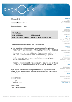
About Normal Pressure
Hydrocephalus (NPH)
A Book for Adults and Their Families
About Normal Pressure Hydrocephalus—A Book for Adults
and Their Families was written for adults with NPH, their families, friends and caregivers with the intention of providing information about the diagnosis and treatment of adult-onset normal
pressure hydrocephalus (NPH). It is a companion piece to our
booklet About Hydrocephalus—A Book for Families, the most widely
distributed resource on infant and childhood hydrocephalus
in the United States.
It is our belief that individuals and families dealing with the
complex issue of adult-onset NPH must educate themselves
about the condition in order to make informed decisions regarding treatment and care. While each case differs, the information
presented in this booklet is intended to give a general overview of
the condition without making judgments or recommendations for
individual care. For making clinical decisions, patients must rely
on the guidance and recommendations of the clinicians providing
their care.
A BOOK FOR ADULTS AND THEIR FAMILIES
1
Foreword
Hydrocephalus is a condition characterized by the expansion of
the cavities or ventricles in the brain, caused by an abnormal accumulation of cerebrospinal fluid (CSF). Hydrocephalus can develop
with high or normal CSF pressure. Normal pressure hydrocephalus (NPH), originally described by Salomon Hakim, MD, PhD, in
1964, is a clinical condition which principally affects the elderly.
It is characterized by a triad of symptoms: motor disturbances
(mostly gait impairment), incontinence and dementia, associated
with ventricular enlargement in the absence of elevated intracranial
pressure. The diagnosis of NPH does not require all three symptoms to be present.
Almost 50 years after the original description of NPH, there
still remain many unknowns, including its diagnostic criteria. It is
not uncommon to find many of the symptoms of NPH in the
elderly. These may occur as an isolated finding or associated with
other diseases. Changes in some of the intellectual functions are
expected to occur during the process of aging in the same way
that other physiologic functions of the body become altered with
advancing age. In spite of all this, NPH is a known and unique
clinical entity justifying its own differential diagnosis with other
brain atrophies. There is great importance in identifying patients
who have NPH and had previously been diagnosed as hopeless
cases of degenerative brain disease, Alzheimer’s or Parkinson’s disease, since there is the opportunity to provide them with a treatment
for NPH that will allow them to have a better quality of life.
During the last years, much has been learned to help provide a
more accurate diagnosis and better treatment for NPH. The families of patients who might have NPH should be well informed
of the symptoms which are characteristic of this clinical entity,
since it is with them that the process of making a diagnosis starts.
The families should be encouraged to take a first step, since many
patients with NPH have experienced “miraculous” recoveries after
treatment and are now living much fuller lives.
2
NORMAL PRESSURE HYDROCEPHALUS (NPH)
My family has been very involved in the field of hydrocephalus
for many years and we have seen many persons recover dramatically after being treated for NPH. The most rewarding experience
has always been to help someone with NPH. Even though sometimes a patient might not recover after treatment as was expected,
the results are many times very rewarding and definitely worth the
effort. As has been previously said, “There is life after NPH.”
—Carlos Hakim, PhD
Because some of the symptoms of NPH can
appear similar to Alzheimer’s or Parkinson’s
or are often associated with the aging process,
NPH can be misdiagnosed or go undiagnosed
for years. Milt Newman went 13 years before
being diagnosed with NPH and successfully
treated with a shunt. “The difference in how
I feel is like night and day. Now I’m living;
I wasn’t living before. I waited for my wife Phyllis to do
everything for me. Now I can do things I couldn’t do before.”
— dr .
milt newman
A BOOK FOR ADULTS AND THEIR FAMILIES
3
What Is Hydrocephalus?
Hydrocephalus is a condition characterized by an abnormal accumulation of cerebrospinal fluid (CSF) or “spinal fluid” within
cavities called ventricles that are inside the brain. CSF surrounds
the brain and spinal cord. The functions of CSF include physical support or cushioning of the brain, excretion of some waste
products and distribution of important substances within the
central nervous system. The average adult produces about one
pint (500 cc) of clear spinal fluid daily. When the circulatory path
of the CSF is blocked, fluid begins to accumulate, causing the
ventricles to enlarge and the pressure inside the head to increase,
resulting in hydrocephalus.
What Is Normal Pressure Hydrocephalus?
Normal pressure hydrocephalus (NPH) is an accumulation of
cerebrospinal fluid that causes the ventricles in the brain to become enlarged, sometimes with little or no increase in intracranial
pressure (ICP). It is most commonly seen in older adults, and is
accompanied by some or all of the following triad of symptoms:
◼◼ Gait disturbances
◼◼ Mild dementia
◼◼ Impaired bladder control
In most cases of NPH, it is not clear what causes the CSF
absorptive pathways to become blocked.
The name “normal pressure hydrocephalus” came out of Dr.
Salomon Hakim’s 1964 paper describing certain cases of hydrocephalus where a triad of neurologic symptoms occurred in the
presence of “normal” CSF pressure. This was before continuous
pressure-recording techniques were available. We now know that
the phrase “normal pressure” is misleading, because many patients have fluctuations in CSF pressure ranging from high to nor4
NORMAL PRESSURE HYDROCEPHALUS (NPH)
Cerebrospinal fluid (CSF) circulatory pathway: The drawing shows a view of the
brain. The black arrows show the major pathway of CSF flow. The gray arrows
show additional pathways.
Arachnoid villi
Subarachnoid space
Lateral ventricle
Sagittal sinus
Choroid plexus
Third ventricle
Fourth ventricle
Aqueduct of Sylvius
mal to low. However, normal pressure hydrocephalus, or NPH,
continues to be the common name for the condition.
What Causes Normal Pressure Hydrocephalus?
The majority of cases of NPH are idiopathic, meaning of unknown cause (also known as primary NPH).
NPH can also develop as the result of a known cause, in which
case it is referred to as secondary NPH. Some of these causes
are head injury, cranial surgery, subarachnoid hemorrhage, tumor
or cysts, as well as subdural hematomas, bleeding during surgery,
A BOOK FOR ADULTS AND THEIR FAMILIES
5
meningitis and other brain infections. All of these predisposing
conditions can cause inflammation that affects the CSF pathways,
impeding CSF flow.
What Are the Symptoms?
Normal pressure hydrocephalus is usually characterized by a
triad of symptoms: gait disturbance (difficulty walking), mild
dementia and impaired bladder control. These symptoms may
not occur all at the same time, and sometimes only one or two
symptoms are present.
◼◼ Gait disturbances range in severity from mild imbalance
to the inability to stand or walk at all. Gait is described as a
“magnetic gait,” often wide-based, short-stepped, slow and
shuffling. People with NPH may have trouble picking up their
feet, making stairs and curbs difficult and frequently resulting
in falls. They may also have difficulty turning around, and turn
very slowly with multiple little steps. Gait disturbance is often the
most pronounced symptom and the first to become apparent.
◼◼ Mild dementia can be described as a loss of interest in daily
activities, forgetfulness, difficulty dealing with routine tasks and
“The placement of my shunt
provides me with new hope,
physical balance and freedom
from falling, allowing me to
live a normal lifestyle including
visiting family, travel and
serving as an arbitrator.”
— dr .
6
james o ’ grady
NORMAL PRESSURE HYDROCEPHALUS (NPH)
short-term memory loss. The cognitive symptoms associated
with NPH are usually less severe than full-blown dementia,
and are often overlooked for years or accepted as an inevitable
consequence of aging. People with NPH do not usually lose
language skills, but they may be less aware of their deficits
than those around them, and may even deny that there are any
problems. Not all individuals have an obvious cognitive impairment. In mildly affected cases, conversational skills may be
preserved and thinking abilities may be relatively unchanged.
In some cases, cognitive changes may only be detectable with
formal neuropsychological testing.
◼◼ Impairment in bladder control is usually characterized by
urinary frequency and urgency in mild cases, whereas a complete loss of bladder control (urinary incontinence) can occur
in more severe cases. Urinary frequency is the need to urinate
more often than usual, sometimes as often as every one to two
hours. Urinary urgency is a strong, immediate sensation of the
need to urinate. This urge is sometimes so strong that it cannot
be held back, resulting in incontinence. In very rare cases, fecal
incontinence may occur. Some people never display signs of
bladder problems.
Because the triad of symptoms are often associated with the
aging process in general, and a majority of the NPH population is
older than 60 years, people often assume that they must live with
the problems and adapt to the changes occurring within their bodies as they age.
Symptoms of NPH can also resemble those of other conditions
affecting the elderly. For example, the cognitive deficits of NPH can
resemble those associated with early Alzheimer’s, and the gait disturbances of NPH can look similar to those of Parkinson’s. (In some
cases, NPH can occur in combination with these diseases.)
There are challenges to distinguishing disease processes that
mimic NPH symptoms. Any type of senile or pre-senile dementia,
A BOOK FOR ADULTS AND THEIR FAMILIES
7
View of enlarged ventricle: CT scan (top); orientation of CT scan in the head (bottom)
Front horn: Lateral ventricle
Temporal horn: Lateral ventricle
Third ventricle
including Alzheimer’s, may be associated with atrophy or shrinking of the brain, resulting in large CSF spaces that are visible on
MRI and CT scans. Because the appearance is quite similar to
NPH, differentiating these diseases can be very difficult. People
with Parkinson’s may have the typical gait disturbance, dementia
and incontinence associated with NPH, but they rarely have the
enlarged CSF spaces. People with spinal stenosis, a condition
in which the nerves supplying the bladder and lower limbs are
compressed from arthritic changes in the lumbar spine, may have
incontinence and gait disturbance. However, they do not necessarily have enlarged CSF spaces.
Symptoms can be present for months or even years before a
8
NORMAL PRESSURE HYDROCEPHALUS (NPH)
person sees a physician. The symptoms of hydrocephalus seem to
progress with time. The rate of progress is variable, and it is often
a critical loss of function, or disability, that brings a person to
seek evaluation and treatment. It seems that the longer and more
severe the symptoms, the less likely it is that treatment will be successful. As a general rule, the earlier the diagnosis, the better the
chance for successful treatment; however, some patients who have
had symptoms for years can improve with treatment.
How Is Normal Pressure Hydrocephalus Diagnosed?
It is often the affected person or close family member who first
brings the symptoms of NPH to the attention of a doctor. Occasionally, enlarged ventricles are discovered on a brain image performed
for another purpose. Once NPH is suspected by a primary physician,
a variety of tests may be recommended to confirm the diagnosis and
assess the person’s candidacy for shunt treatment.
At this point, it is important that a neurosurgeon and/or a neurologist (or neuropsychologist) become part of the medical team.
The involvement of these medical professionals from the diagnostic stage onward is helpful not only in interpreting test results
and selecting likely candidates for shunting but also in discussing
the actual surgery and follow-up care as well as expectations and
risks of surgery.
The decision to order a given test may depend on the specific
clinical situation, as well as the preference and experience of the
medical team. Not all of the tests described here need to be performed in order to make a diagnosis.
Clinical exams to evaluate symptoms consist of an interview
and/or a physical/neurologic examination. In a clinical examination, a doctor might:
◼◼ discuss and observe walking and turning to determine the
extent and type of gait disturbance;
A BOOK FOR ADULTS AND THEIR FAMILIES
9
◼◼ assess cognition by asking a few questions or administering a full
neuropsychological evaluation using pencil-and-paper tests to
probe such qualities as attention, reaction time, memory, reasoning, language and emotional state; and
◼◼ verbally assess urinary urgency and frequency or incontinence.
The presence of all three symptoms is not necessary to the diagnosis of NPH. The medical team will consider the pattern and
severity of impairments, along with results of other tests mentioned below, in differentiating NPH from other conditions.
Brain images to detect enlarged ventricles commonly
include magnetic resonance imaging (MRI) and computerized
tomography (CT). Each of these imaging techniques is described
in more detail below.
◼◼ MRI uses radio signals and a very powerful magnet to create
a picture of the brain. It is safe for most people, reliable and
painless, but takes longer than a CT scan and some may find
the physically confining circumstances of being in an MRI
machine challenging. MRI can detect enlarged ventricles as well
as evaluate the CSF flow and provide information about the
surrounding brain tissues. MRI scans can also assess how fast
CSF moves through a particular part of CSF pathways called
the cerebral (or Sylvian) aqueduct (the CSF flow void sign).
Some physicians believe that high CSF flow through the aqueduct predicts improvement with treatment of NPH. The MRI
provides more information than the CT, and is therefore the
test of choice in most cases, but people with cardiac pacemakers or certain other metallic implants may not be able
to have MRI scans because of potential interference with these
devices.
◼◼ CT (or CAT) scan is a picture of the brain created by using
X-rays and a special scanner. An X-ray beam passes through
10 NORMAL PRESSURE HYDROCEPHALUS (NPH)
the head, allowing a computer to take a picture of the brain. It
is safe, reliable, painless and relatively quick (about 15 minutes).
A CT scan will show if the ventricles are enlarged.
CSF tests to predict shunt responsiveness and/or determine shunt pressure include lumbar puncture, external lumbar
drainage, measurement of CSF outflow resistance, intracranial
pressure (ICP) monitoring and isotopic cisternography. Though
there is no way to accurately predict a particular patient’s responsiveness to a particular shunt, many doctors find these tests
helpful in determining the likelihood of a positive response to
shunting. Each test is described in more detail below. People who
have abnormal bleeding tendencies or take medications that affect
bleeding should talk with their medical team about any special
precautions before invasive procedures are performed.
◼◼ Lumbar puncture, or spinal tap, allows an estimation of
CSF pressure and analysis of the fluid. Under local anesthetic,
a thin needle is passed into the spinal fluid space of the lower
back. Up to 50 cc of CSF is removed to see if symptoms are
temporarily relieved by this CSF volume reduction. If removal
of some CSF dramatically improves symptoms, even temporarily, then surgical treatment is likely to be successful. A limitation of lumbar puncture and removal of a small volume of
CSF as a screening test for NPH is that some people may have
little or no improvement after the test, and yet may still improve with a shunt. When the response to a lumbar puncture is
“negative” or uncertain, further evaluation may be helpful.
◼◼ External lumbar drainage, also called lumbar catheter insertion or continuous lumbar drainage, is a variation of the
lumbar puncture where a thin, flexible tube, called a catheter, is
left in place to drain CSF. The procedure, which is performed
in the hospital, allows for either intermittent or continuous removal of spinal fluid over several days to imitate the effect that
A BOOK FOR ADULTS AND THEIR FAMILIES 11
a shunt would have. It also allows for more accurate recording of
CSF pressure. With an intermittent drainage protocol the person
is free to move around when the fluid is not being drained.
Spinal fluid drainage over time can be thought of as a “test
drive” of a shunt without actually undergoing shunt surgery.
However, because it requires hospitalization and has associated
risks it may not be recommended for all patients. People who
respond dramatically to such spinal fluid drainage are likely to
respond to shunt surgery, which will be necessary for long-term
treatment of NPH. Some physicians advocate using the pressure
results for selecting the type of shunt or initial shunt setting for
programmable and adjustable shunts.
◼◼ The measurement of CSF outflow resistance is a more
involved test that requires a specialized clinical setting. This test
begins with a lumbar tap and assesses the degree of blockage
of CSF absorption back into the bloodstream. It requires the
simultaneous infusion of artificial spinal fluid and measurement of CSF pressure. If the calculated resistance value is
abnormally high, then there is a very good chance that the
patient will improve with shunt surgery, since the shunt mimics
the function of the body’s normal CSF drainage pathways.
◼◼ For intracranial pressure (ICP) monitoring or spinal pressure monitoring, a small pressure monitor is inserted through
the skull into the brain or ventricles or in the lumbar region
to measure the ICP. Pressure monitoring, either by the lumbar
catheter or the intracranial method, requires admission to a
hospital. It can detect an abnormal pattern of pressure waves
as well as low or high pressure. It is possible for NPH to occur
even when CSF pressure is not measurably high. The results
of this test can also be used to select initial shunt pressure if a
shunt is being implanted.
12 NORMAL PRESSURE HYDROCEPHALUS (NPH)
◼◼ Isotopic cisternography is no longer in frequent use because a “positive” cisternogram result does not reliably predict
whether a patient will respond to implantation of a shunt, and
results are often ambiguous. It involves having a radioactive
isotope injected into the lower back through a spinal tap, in order to monitor the absorption of CSF over a period of several
days. This test is done in the hospital.
What Treatment Is Available?
The most common and usually the only available treatment for
NPH is the surgical implantation of a shunt, a device that channels CSF away from the brain to another part of the body where it
can be absorbed. Most shunt systems consist of three components:
(1) a collection catheter situated within the cerebral ventricles or the
lumbar spinal canal; (2) a valve mechanism to control how much CSF
flows; and (3) an exit catheter to drain the CSF to another part of the
body where it can be absorbed (see drawing on page 18). The most
common part of the body for drainage is the peritoneal (abdominal)
cavity, and the most common system is a ventriculoperitoneal (VP)
shunt (from the cerebral ventricle to the peritoneum). The drainage catheter can also be placed in a vein that leads to the heart, a
configuration called a ventriculoatrial (VA) shunt. After surgery is
completed, all components of the shunt system are entirely under the
skin, and nothing is exposed to the outside.
The shunt valve is a critical component of the shunt system.
The design that has been in use the longest is a fixed differentialpressure (DP) valve. This valve opens when the fluid pressure at
the inlet of the valve exceeds the pressure at the outlet by a certain
amount. Traditionally, DP valves have come in low, medium and
high pressure varieties. For the adult with hydrocephalus, sometimes the valve is overwhelmed by the effects of gravity, leading
to the drainage of too much CSF. This is commonly known as
siphoning. Siphoning is sometimes asymptomatic, sometimes associated with headaches or nausea in the upright position, and in
A BOOK FOR ADULTS AND THEIR FAMILIES 13
some instances, can cause so much spinal fluid to drain from the
head that small blood vessels between the brain and skull are disrupted, causing a type of bleeding known as subdural hematoma,
a serious complication of shunting.
In order to counteract this potential problem, valve mechanisms have been designed that incorporate anti-siphon and
gravity-compensating mechanisms or flow-regulated mechanisms. Anti-siphon devices are triggered by pressure change;
gravity-compensating devices are triggered by postural change.
Anti-siphon devices are not effective with lumbar shunts, but
gravity-compensating devices are. Both devices are designed to
counteract overdrainage of CSF when sitting or standing.
The flow-regulated valve is designed to minimize overdrainage
by limiting flow to the approximate rate of CSF production when
conditions of overdrainage occur (when sitting, standing or straining).
This valve acts like a differential-pressure valve when the risk of over-
“Caring for patients with NPH and their families
is a rich and rewarding experience. One of the
greatest joys for me is when an elderly couple comes
back after the shunt operation and tells us, ‘You’ve
given us our lives back’ because they can now
travel, and socialize, and enjoy their families in
ways they couldn’t before the NPH was treated.
And for those patients we evaluate who turn out
not to have NPH, while we’re disappointed that
we can’t help them get better, they and their families are almost
always grateful that we undertook the time and effort to investigate
the possibility.” — michael williams , md
14 NORMAL PRESSURE HYDROCEPHALUS (NPH)
drainage is low (when the body is prone). It is designed specifically
of Shunt
Valves
to minimize excessExamples
CSF drainage
during straining—such
as bowel
movements, coughing, sneezing or sexual exertion.
Adjustable/“programmable” valves are differential-pressure
valves that can be adjusted externally, using magnetic programmers or adjustment tools. These valves allow the opening pressure of ProGAV
the shunt to be
fine-tuned Aesculap, Inc.
without additional
surgery
and may help to
optimize treatment and avoid repeated surgery in some cases.
The valves can be adjusted within a range of differential pressures, from low to medium to high, in multiple steps. In some
cases, adjustable
Hakim Programmable Valve
valves include an
Codman, a Johnson and Johnson Company
anti-siphon or
gravity-compensating device. Because such valves are generally
reset with magnetic devices, they should be checked after an MRI
or after exposure to other strong magnets.
It is important to realize that each of these valve designs has
Diamond Valve
po- Polaris Adjustable Valve tential
Vygon Neuro
ad- Sophysa
vantages
and
disadvantages, and that there is no single design or setting that works
in all patients. The valve a neurosurgeon selects for an individual
depends on a number of factors, including age, size of ventricles,
intra- OSV®
cranial pressure, the availability
SmartValve™
of the Integra
valve and the experience of the
neurosurgeon. People with NPH may
want to ask their neurosurgeon to demonstrate a sample shunt
and discuss the pros and cons of the system being recommended.
As an alternative to a VP or VA shunt, some neurosurgeons
may recommend a lumboperitoneal
Strata
Adjustable
Valve
(LP) shunt.
An LP shunt is inserted
Medtronic Neurosurgery
into the spinal
space in the lower back
A BOOK FOR ADULTS AND THEIR FAMILIES 15
-drainage is low (when the body is prone). It is designed
specifically to minimize excess CSF drainage during straining—such as bowel movements, coughing, sneezing or sexual
exertion.
Adjustable/“programmable” valves are differentialpressure valves that can be adjusted externally, using magnetic
programmers or adjustment tools. These valves allow the
opening pressure of the shunt to be fine-tuned without additional surgery and may help to optimize treatment and avoid
repeated surgery in cases. The valves can be adjusted within a
range of differential pressures, from low to medium to high,
in multiple steps. In some cases, adjustable valves include an
anti-siphon or gravity-compensating device. Because such
valves are generally reset with magnetic devices, they should
be checked after an MRI or after exposure to other strong
magnets.
It is important to realize that each of these valve designs
has potential advantages and disadvantages, and that there is
no single design or setting that works in all patients. The valve
a neurosurgeon selects for an individual depends on a number of factors, including age, size of ventricles, intracranial
pressure, the availability of the valve and the experience of
the neurosurgeon. People with NPH may want to ask their
neurosurgeon to demonstrate a sample shunt and discuss the
pros and cons of the system being recommended.
As an alternative to a VP or VA shunt, some neurosurgeons may recommend a lumboperitoneal (LP) shunt. An
LP shunt is inserted into the spinal space in the lower back
15b NORMAL PRESSURE HYDROCEPHALUS (NPH)
(in the same spinal region where a spinal tap is done). The tubing,
which is narrower than that used in ventricular shunts, is tunneled
under the skin to the abdomen, where it is inserted into the peritoneal cavity, much like a VP shunt. Some people who are particularly apprehensive about the insertion of a ventricular shunt
through the brain may be more comfortable with an LP shunt,
yet it is worth noting that insertion of a ventricular shunt rarely
harms the brain. Although LP shunts may have some potential
advantages, in general they are more prone to obstruction over
the long term and if there is a problem with the shunt, it is more
difficult to assess what the problem is.
For people who have aqueductal stenosis, a surgical procedure
called endoscopic third ventriculostomy (ETV) may be considered as an alternative to a shunt. In this procedure, the neurosurgeon uses a special endoscope to create an alternative CSF
passageway that bypasses the obstruction at the cerebral aqueduct.
The determination of aqueductal stenosis can be made by MRI.
The success of ETV in adults is variable, and some people who
have a third ventriculostomy may later require shunt surgery to
treat their symptoms. Clinical trials are currently exploring ETV
as a treatment for NPH.
Will Shunting Help?
This question cannot be answered definitively. Many tests and
evaluation criteria have been proposed, but, unfortunately, no one
single factor is reliable in predicting success from implantation of
a shunt for NPH. The following findings are generally associated
with a better outcome following shunt placement:
◼◼ The onset of gait disturbance as the first and most prominent
symptom
◼◼ A known cause for NPH, such as trauma or hemorrhage
◼◼ The scan (MRI or CT) shows the ventricular size to be disproportionately larger than the CSF in the subarachnoid space
16 NORMAL PRESSURE HYDROCEPHALUS (NPH)
With the VP shunt in place, cerebrospinal fluid (CSF) flows into the collection
catheter and down the exit catheter, which shunts the fluid into the abdominal
(peritoneal) cavity.
Collection catheter
Valve
Lateral ventricle
Exit catheter
A BOOK FOR ADULTS AND THEIR FAMILIES 17
◼◼ Removal of spinal fluid via lumbar puncture or lumbar catheter
gives dramatic, temporary relief of symptoms
◼◼ ICP or spinal fluid pressure monitoring shows an abnormal
range or pattern of spinal fluid pressure or an elevated CSF
outflow resistance
◼◼ Minimal evidence of disease of the small blood vessels nourishing
the brain
Because some people with NPH have additional medical or
neurological problems, it is important for them, their families and
their neurosurgeons to discuss their expectations of shunt surgery.
Does “success” mean that they will regain the levels of motor skill
or mental ability they had before the symptoms presented themselves? Does it mean that the condition will not worsen? Or does
it mean something else? The definition of success must be individualized, and it is important to know that it is possible for any
or all of the hydrocephalus symptoms to improve with shunting.
One way to evaluate the success of shunt surgery is to consider
whether it has reduced the disabilities that were present before
surgery and increased the individual’s functional abilities.
Although everyone hopes for a complete recovery, it is not
often seen, and many individuals and their families are satisfied
when shunt surgery results in reduced disability or dependence than
they had before surgery, or prevention of further neurological deterioration. People with NPH, their families and their physicians need
to be supportive and hopeful, but they should also know the possible
complications, risks and realities of shunt implantation.
Not all people with enlarged ventricles need treatment. Some
people with enlarged ventricles have no symptoms and no neurologic deficits at all, even when evaluated by neurosurgeons and
neurologists who specialize in hydrocephalus. This is a condition
often called “compensated hydrocephalus.” People who have
compensated hydrocephalus cannot be “made better” by treating
the hydrocephalus, and in such circumstances, there are no ben-
18 NORMAL PRESSURE HYDROCEPHALUS (NPH)
efits that would offset the potential risks of treatment (see below).
People with compensated hydrocephalus may, however, develop
symptoms later in their lives, and may benefit from treatment with
shunting at that time. Therefore, it is important for individuals with
compensated hydrocephalus to see a neurosurgeon or neurologist
periodically to assess whether subtle symptoms are developing.
What Does a Shunt Operation Entail?
VP: The surgical procedure to implant a VP shunt usually requires less than an hour in the operating room. After the patient
is placed under general anesthesia, his or her scalp overlying the
shunt insertion site is shaved and the patient is scrubbed with
an antiseptic from the scalp to the abdominal area. These steps
are taken in order to reduce the chances of an infection. Small
incisions are made in the head and in the abdomen to allow the
neurosurgeon to pass the shunt tubing through the fatty tissue
just under the skin. A small hole is made in the skull, opening the
membranes between the skull and brain to allow the ventricular
end of the shunt to be passed through the brain and into the lateral ventricle. The abdominal (peritoneal) end is passed through a
small opening into the abdominal cavity where the excess CSF will
eventually be absorbed. The incisions are then closed and sterile
bandages are applied.
LP: Under general anesthesia, the LP shunt catheter is inserted
using a hollow needle between two vertebrae in the lumbar CSF
region of the spine. The valve is implanted below the skin and the
drainage catheter is directed into the peritoneal cavity. Once in
place the LP shunt drains the excess cerebrospinal fluid removed
from the lumbar region and transports it to the peritoneal cavity,
where it is eventually absorbed by the organs and passed out of
the body during urination. Patients with lumboperitoneal shunts
are left with two scars: a vertical scar down the lower (lumbar)
part of the spine and a horizontal scar across the upper abdomen.
The patient stays under careful neurological observation for the
A BOOK FOR ADULTS AND THEIR FAMILIES 19
“Following a stroke at the age of 59, I was
eventually correctly diagnosed with NPH after
seeing my family doctor, a psychologist “and
taking several meaningless tests and wasting
about a month. Once a neurologist made the
correct diagnosis, a shunt was installed and
my symptoms immediately disappeared.”
— martin
mittleman
first 24 hours following a procedure. The incisions are monitored
for signs of infection. The patient will stay in the hospital from
one to seven days. Follow-up visits will be necessary to check
post-operative status and resolution of symptoms. After surgery,
physical therapy, occupational therapy and other rehabilitation
strategies may be advised to help patients attain as much resolution of symptoms as possible. People should talk with their neurosurgeon about his or her particular protocols following surgery.
How Successful Is Shunting?
The symptoms of gait disturbance, mild dementia and bladder
control problems may improve within days of shunt surgery, or
may take weeks to months to abate. There is currently no way to
predict how fast, or to what extent, this improvement will occur.
For those who do improve, changes are often seen in the first
weeks, although there are late responders and some people take
longer to recover from surgery. In addition, this improvement may
range from mild to dramatic. It is not possible to predict how long
the improvement will last, as the course of clinical improvement
varies for each person. Some people seem to reach a plateau, while
others improve for months but then seem to decline again. Unfortunately, there are no guarantees. A recurrence of symptoms in a
20 NORMAL PRESSURE HYDROCEPHALUS (NPH)
person who had improved should prompt consideration of shunt
malfunction or one of the complications described below.
The rate of success for shunting normal pressure hydrocephalus is quite variable. Neurosurgeons do not agree on the factors
that lead to a successful procedure, nor do they have similar rates
of success. Although the success rate for shunting is higher when
proper diagnostic and treatment procedures are followed, it is not
possible to predict how much a patient will improve after surgery.
It is important to note that if initial success is followed by a recurrence of symptoms, it may be due to a valve or shunt failure, or the
need for a lower pressure valve—rather than failure of the procedure.
The presence of other neurological or medical conditions may
also affect the outcome after shunt surgery. One of the most
common is the long-term effect of high blood pressure on the
brain, which can cause multiple tiny strokes to the same areas of
the brain that are affected by hydrocephalus, causing virtually the
same symptoms. Other factors to consider in recovery are arthritis
involving the back, hips or knees; impaired sensation in the legs
and feet (neuropathy); other causes of urinary dysfunction; or the
presence of Alzheimer’s disease. The issue is often not whether
the symptoms are caused by one diagnosis or another, but how
much is caused by one diagnosis and how much by another. The
more the symptoms are due to NPH, rather than the associated
conditions, the greater the likelihood of successful recovery
after shunting.
What Are the Complications and Risks Involved
with Shunting?
Although the insertion of a shunt is a relatively simple neurosurgical procedure, the decision to undergo surgery should not be
taken lightly. The treatment of normal pressure hydrocephalus
carries greater risks compared to the treatment of hydrocephalus
in children; therefore, this operation should be undertaken only if
the degree of disability or the progression of the disorder warrants
A BOOK FOR ADULTS AND THEIR FAMILIES 21
Hazel’s Story
“My mom is a classic case of misdiagnosis
for NPH. Her symptoms began to appear in 2007. The doctors thought she
had a dropped bladder and suspected a
stroke, but there was no evidence of
either. Later on, they diagnosed her
with Parkinson’s then with Alzheimer’s
disease. I had done some volunteer
work in nursing homes with Alzheimer’s and Parkinson’s patients and I knew my mom didn’t act like a patient with either one.
“During my dad’s illness and after his death, my mom came
to live with me. I set her up with a general practitioner and she
agreed to send my mother to a neurologist to run furthers tests.
Finally the neurologist diagnosed my mother with normal pressure hydrocephalus and they referred her for surgery. It took us
a while to find the right surgeon, but when we finally went to the
Cleveland Clinic and met Dr. Mark Luciano, my mom fell in
love. With her permission, the surgery was performed by Dr.
Luciano over the live video feed as part of the 11th National
Conference on Hydrocephalus.
“Mom still gets frustrated because the change is taking time.
But for me it’s remarkable. It’s as if she went back in time five
years and each day she improves a little more. She hasn’t cooked
in about eight years, but last night she said, ‘I think it is about
time I start cooking again—I think I’ll start with something
easy, shrimp dip.’ I know it doesn’t sound like much to most
people, but to me, it’s amazing.” —richard chard
22 NORMAL PRESSURE HYDROCEPHALUS (NPH)
such intervention. An additional factor to consider is whether the
expected benefits of surgery outweigh the risks. Ways to assess the
expected benefits include diagnostic tests that reveal information
about the abnormal CSF flow that the shunt is intended to correct,
such as the response to CSF removal, abnormal CSF pressure patterns or insufficient CSF absorption. Most people feel more comfortable proceeding with shunt surgery if they have good reason to
expect a favorable outcome from the surgery.
The potential complications of shunt surgery include those related to the actual operation, as well as those that may occur days to
years later. Unlike many other operations, in which the surgical risks
are highest during the operation, most of the problems associated
with shunting can occur weeks or even years after the surgery.
One of the most common problems with shunt systems is
obstruction (the shunt becomes clogged). It is not possible to
predict which patients will have a shunt obstruction, nor when the
obstruction will occur. A shunt obstruction is usually suspected
when the original symptoms of NPH reappear over a period of
days, weeks or months. Fortunately, shunt obstruction in NPH is
usually easily fixed and rarely results in serious problems, although
further surgery may be necessary.
Another common complication of shunt surgery is shunt
malfunction, which can occur when either end of the shunt is
incorrectly positioned, the valve fails to function properly or the
CSF is not efficiently reabsorbed. The typical remedy involves a
further surgical procedure.
It is important for people with NPH and their families to know
the symptoms they experienced prior to shunt implantation and
to alert their physician if the symptoms return after the shunt is
implanted, as this may be a sign of an obstructed or malfunctioning shunt.
Additional complications include an infection involving the
surgical wound, the shunt or the CSF (meningitis); bleeding into
the brain or ventricles; or a seizure. A shunt infection may be inA BOOK FOR ADULTS AND THEIR FAMILIES 23
dicated by fever, redness or swelling along the shunt track. In very
rare instances, a person can have a reaction to the implanted shunt
materials. Fortunately, these complications are uncommon and can
be managed successfully in almost all cases, although treatment
may require additional surgery.
One of the more serious complications that can occur following
insertion of a shunt is a subdural hematoma (blood clot). The risk
of a subdural hematoma in people with NPH and a shunt is approximately 5 to 10 percent. Because most shunts drain CSF from
the center of the brain (the ventricles), this may cause the surface
of the brain to pull away from the skull, thus stretching and tearing
blood vessels that go from the scalp to the surface of the brain. This
can sometimes be seen on a CT scan as a fluid space between the
brain and the skull called a hygroma. Although a hygroma may not
have any clinical symptoms, it may increase the risk of hematoma.
The symptoms of a subdural hematoma may vary from headache to
paralysis or even coma or death. Shunt-related subdural hematomas
most commonly occur following a fall, even a minor one with no apparent injuries at the time of the fall.
The interval between the fall and the onset of symptoms
can be as long as days or weeks. Subdural hematoma can also
be caused by overdrainage, when a shunt drains too much CSF.
Therefore, people with NPH should not hesitate to seek medical
attention if they develop worrisome symptoms.
In patients who have had multiple abdominal operations, there
is a rare complication of injury to the bowel.
Given these potential complications, individuals and their families need to assess their own situations to determine if the possible benefits of surgery outweigh the possible risks.
NPH Left Untreated
People with NPH usually present with progressive symptoms,
and there is no reason to believe that the progression will stop
on its own. No one is able to predict how fast the symptoms
24 NORMAL PRESSURE HYDROCEPHALUS (NPH)
“My wife and I did quite a bit of research once I
made the decision to have the surgery, which precipitated many questions for my surgeon. Confidence in my surgeon by both my wife and
I was crucial, and I must say that my surgeon
is the person who finally put me at ease with the
surgery along with talking to a person who already had the procedure and could give me some
insight as to what I could expect afterward. The surgery
was a success and my recovery from it was relatively brief
(a few weeks).” — peter morris
will progress, and the seriousness of symptoms may vary day to
day. Evidence suggests that the longer the symptoms have been
present and the more severe the symptoms are, the less likely it is
that the treatment will be successful. As a general rule, the earlier
the diagnosis and treatment, the better the chance of recovery.
However, some patients who have had symptoms for years can
improve with treatment. For patients whose symptoms are very
mild, their doctor may advocate closely monitoring the clinical
condition without proceeding immediately to a shunt operation.
The Importance of Family Support
The process of diagnosing possible normal pressure hydrocephalus can often be frustrating. The symptoms of gait disturbance,
mild dementia and bladder control problems can also occur with a
number of other conditions that affect people over 60. Sometimes
these conditions coexist with hydrocephalus, making the diagnosis
and treatment even more difficult. Families and individuals who stay
informed and ask questions of their physicians will likely feel more
involved in the management of their care. It is important to be opA BOOK FOR ADULTS AND THEIR FAMILIES 25
timistic about surgical treatment. We live in a society where people
are living longer, and more and more adult children are being asked
to care for their aging parents. The frustration and complexity of
being ill, or caring for an ill parent or spouse, can be stressful for
all involved. Adults with hydrocephalus may resent being dependent
on their spouse, children and family. Families often feel better and cope
better by acknowledging and discussing their emotional responses to a
chronic, and possibly disabling, condition such as hydrocephalus.
What Can We Expect for the Future?
There is currently a great deal of interest in the diagnosis and
management of NPH, with the expectation that the reported
incidence of this condition will increase dramatically as the baby
boomers reach retirement age and beyond. Physicians, medicaldevice manufacturers and patient advocates are cooperating on a
variety of levels to improve methods of diagnosis, techniques and
devices for treatment, and public awareness of NPH. It is anticipated that these efforts will result in years of better quality of life
for people affected by this condition.
Conclusions
The Hydrocephalus Association is positioned at the nexus of a
movement to strategically drive hydrocephalus research. We are
aligned with professionals and peer advisors, with Congress and
members of government, with other organizations and with key
agencies. It is our intent to drive and support the research agenda
by directly funding hydrocephalus research.
There are many unknowns surrounding the diagnosis and
treatment of adult-onset normal pressure hydrocephalus. Although the success rate for shunting is higher when proper
diagnostic and treatment procedures are followed, it is not possible to predict the degree of improvement that will follow shunt
surgery. However, NPH is not a hopeless condition. Advances in
shunt technology and surgical techniques are continually being
26 NORMAL PRESSURE HYDROCEPHALUS (NPH)
introduced and developed. Adults diagnosed with normal pressure
hydrocephalus, and their families, should be encouraged to ask
questions, gather information and network with others.
The Hydrocephalus Association was formed to support and
foster these goals by bringing together individuals and families
such as yours. Because normal pressure hydrocephalus can be a
complex medical condition, it is essential that everyone involved
learn as much as possible about the particular case so that informed decisions can be made.
A BOOK FOR ADULTS AND THEIR FAMILIES 27
Glossary
aqueductal stenosis: A narrowing of the aqueduct of Sylvius.
This is one cause of obstructive hydrocephalus; it may be treated
using a CSF shunt or by a surgical procedure called endoscopic
third ventriculostomy (ETV).
arachnoid: The middle layer of the meninges. It covers the brain
and spinal cord smoothly without conforming to the irregularities
of their surfaces; CSF flows within the arachnoid space.
arachnoid villi: Small projections in the dura mater that project
into the dural venous (blood) sinuses. CSF is reabsorbed from the
arachnoid space by passing through the arachnoid villi and entering the venous system. Also known as arachnoid granulations.
catheter: Flexible, hollow tube used to shunt fluid. For CSF
shunting, the proximal catheter of a shunt is the inflow catheter
and the distal catheter is the outflow catheter.
cerebral aqueduct (aqueduct of Sylvius): A narrow channel
for CSF flow in the midbrain that connects the third and fourth
ventricles.
cerebrospinal fluid (CSF): Clear, colorless liquid secreted primarily by the choroid plexus and contained within the ventricles and
the subarachnoid space. CSF functions primarily to float and cushion
the brain and spinal cord.
communicating hydrocephalus: Hydrocephalus in which the
openings between the ventricular spaces and between the fourth
ventricle up to the subarachnoid space are functioning.
choroid plexus: The structures in the lateral, third and fourth
ventricles that produce cerebrospinal fluid.
dura mater (or dura): The outermost and heaviest layer of the
meninges covering the brain and spinal cord; this layer is closest
to the skull.
28 NORMAL PRESSURE HYDROCEPHALUS (NPH)
foramen of Monro (interventricular foramen): An opening between the lateral ventricle and third ventricle through which CSF
flows from the lateral ventricle into the third ventricle.
fourth ventricle: A cavity within the brain that is situated between the brain stem and the cerebellum. The fourth ventricle
receives CSF from the cerebral aqueduct, and CSF exits the fourth
ventricle via the foramina of Luschka and Magendie into the subarachnoid space.
hematoma: A localized collection of blood, usually clotted. hemorrhage: The escape of blood from blood vessels.
hydrocephalus: An abnormal condition that occurs when there is
an imbalance between the rate of CSF production and the rate of
absorption, leading to gradual accumulation of CSF.
hygroma: A sac, cyst or bursa distended with fluid. A subdural
hygroma is a collection of fluid between the brain and the skull.
intraventricular hemorrhage: Bleeding into the ventricles.
lateral ventricle: One of two normal cavities within the cerebral
hemispheres that contain cerebrospinal fluid. CSF flows from
the lateral ventricles into the third ventricle via the foramen of
Monro.
meninges: Membranous coverings of the brain and spinal cord
consisting of the dura mater, arachnoid and pia mater.
meningitis: Inflammation of the meninges. Meningitis can result
from bacterial or viral infection. Scarring of the arachnoid that
results from meningitis can restrict or block CSF flow and absorption.
noncommunicating hydrocephalus: Hydrocephalus in which
there is obstruction of the flow of CSF through the cerebral aqueduct or from the fourth ventricle to the subarachnoid space.
normal pressure hydrocephalus (NPH): A syndrome characterized by enlarged ventricles and a triad of symptoms including
A BOOK FOR ADULTS AND THEIR FAMILIES 29
gait disturbance, dementia and impaired bladder control; this form
of hydrocephalus that occurs most often in middle-age and older
persons.
obstructive hydrocephalus: Hydrocephalus caused by a blockage along the CSF flow pathway.
third ventricle: A midline cavity within the brain that is situated
between the right and left thalamus. It receives CSF from each
lateral ventricle via the foramen of Monro; CSF exits the third
ventricle via the cerebral aqueduct (aqueduct of Sylvius).
third ventriculostomy: A surgical operation to create an opening
through the membranous floor of the third ventricle, permitting
CSF to exit the third ventricle directly into the subarachnoid space
at the base of the brain.
ventricle: A cavity within the brain that contains cerebrospinal fluid.
30 NORMAL PRESSURE HYDROCEPHALUS (NPH)
external lumbar drainage. See lumbar drainage
Index
family support, 25
fever, 24
flow-regulated valve. See valves
abdominal cavity, 13, 17, 19
adjustable valve. See valves
aging, 2, 3, 7, 26
Alzheimer’s disease, 2, 3, 7, 8, 21, 22
anesthesia, 19
anti-siphon devices. See valves
aqueduct of Sylvius, 5, 10, 28, 30
aqueductal stenosis, 16, 28
arachnoid villi, 5, 28
artificial spinal fluid, 12
gait disturbance, 2, 4, 6, 7, 8, 9, 16, 20, 25, 29
gravity-compensating device. See valves
headache, 13, 24
head injury, 5
hematoma, 5, 14, 24, 29
hemorrhage, 5, 16, 29
hydrocephalus
compensated, 18, 19
defined, 4
normal pressure. See NPH
hygroma, 24, 29
bladder control, 4, 6, 7, 20, 25, 29
impairment, 4, 6, 7
loss of, 7
blockage, 12, 30
blood clot, 24
catheter, 11, 12, 13, 17, 18, 19, 28
CAT scan. See CT scan
cerebral aqueduct, 16, 28, 29, 30
cerebrospinal fluid. See CSF
choroid plexus, 5, 28
clinical exam, 9
coma, 24
compensated hydrocephalus, 18, 19
complications, 18, 21, 23, 24
computerized tomography. See CT scan
continuous lumbar drainage. See lumbar drainage
cranial surgery, 5
CSF, 2, 4 - 6, 8, 10 – 14, 16 – 19, 23, 24, 28
flow void sign, 10
outflow resistance, 11, 12, 18
removal, 11, 18, 23
CT scan, 8, 10, 11, 24
cysts, 5
death, 22, 24
dementia, 2, 4, 6, 7, 8, 19, 25, 29
diagnosis, 2, 9 – 13, 20, 21, 25, 26
diagnostic tests, 9 – 13, 23
differential-pressure valve. See valves
endoscopic third ventriculostomy (ETV), 16, 28
enlarged ventricles, 4, 8, 9, 10, 18, 29
estimation of CSF pressure, 11
ICP, 4, 11, 12, 18
ICP pressure monitoring, 12
idiopathic, 5
incisions, 19, 20
incontinence, 2, 7, 8, 10
infections, 6, 19, 20, 23, 24, 29
meningitis, 6, 23, 29
shunt infection, 23
intracranial pressure. See ICP
isotopic cisternography, 11, 13
LP shunt, 16, 19
lumbar catheter, 11, 12, 18
lumbar drainage, 11
lumbar puncture, 11, 18
lumboperitoneal shunt. See LP shunt
magnetic resonance imaging. See MRI
malfunction. See shunt malfunction
meningitis, 6, 23, 29
metallic implants, 10
mild dementia, 4, 6, 20, 25
MRI, 8, 10, 16
nausea, 13
neurologist, 9, 18 – 20, 22
neurosurgeon, 9, 15, 18 – 21, 34
normal pressure hydrocephalus. See NPH
A BOOK FOR ADULTS AND THEIR FAMILIES 31
NPH
causes, 5
defined, 4, 5
diagnosis, 9 – 13
symptoms, 6 – 9
treatment, 13 – 16
untreated, 24, 25
obstruction. See shunt obstruction
overdrainage, 14, 24
pacemaker, 10
paralysis, 24
Parkinson’s disease, 2, 3, 7, 8, 22
peritoneal cavity, 13, 16, 17, 19
pressure monitoring, 12, 18
programmable valves. See valves
redness, 24
removal of spinal fluid. See CSF removal
shunt
infection, 19, 20, 23
malfunction, 21, 23
obstruction, 16, 23, 29
responsiveness, 11
surgery, 12, 13, 16, 18, 20 – 26
types of, 13 – 17
valve. See valves
siphoning, 13
spinal cord, 4, 28, 29
spinal fluid pressure, 13, 18
spinal needle, 11
spinal tap, 11, 13, 16
subdural hematoma, 5, 14, 24
subarachnoid hemorrhage, 5
subarachnoid space, 5, 18, 30, 31
surgery, 7. See also shunt surgery
surgical risks, 9, 12, 18, 21 – 24
swelling, 24
symptoms of NPH, 2, 3, 7, 9, 23, 6 – 9, 10, 11
urinary frequency, 7, 10
urinary urgency, 7, 10
valves
adjustable, 12, 15
anti-siphon device, 14
differential-pressure, 13, 14
flow-regulated, 14
gravity-compensating device, 14
mechanism, 13, 14
programmable, 12, 15
VA shunt, 13
ventricle
fourth ventricle, 5, 28
lateral ventricle, 5, 8, 17, 19
third ventricle, 5, 8
ventriculoatrial shunt. See VA shunt
ventriculoperitoneal shunt. See VP shunt
ventriculostomy. See endoscopic third ventriculostomy
VP shunt, 13, 16, 17, 19
walking, 6, 9. See also gait disturbance
X-rays, 10
third ventriculostomy. See endoscopic third
ventriculostomy
trauma, 16
triad of symptoms, 2, 4, 6, 7, 29
tube, 11, 28
tumor, 5
32 NORMAL PRESSURE HYDROCEPHALUS (NPH)
Resources
The Hydrocephalus Association is a national, 501(c)(3) nonprofit
organization founded in 1983 to provide support, education and
advocacy to individuals, families and professionals. Our mission is
to eliminate the challenges of hydrocephalus by stimulating innovative research and providing support, education and advocacy for
individuals, families and professionals dealing with the complex
issues of the condition.
The Association provides comprehensive services that empower individuals and families to seek out the best medical care, programs and resources that meet their needs now and in the future.
As the nation’s largest and most widely respected organization dedicated solely to hydrocephalus, the Association has been
instrumental in creating a community of individuals, families and
healthcare professionals addressing the complexities of hydrocephalus in all age groups—infants, children, young adults and
adults. We continually update and expand our resources to keep
pace with new technologies in the diagnosis and treatment of
hydrocephalus and stay current with the needs of the individuals
we serve.
Hydrocephalus is a chronic condition. With early detection,
effective treatment and appropriate interventional services, the
future for individuals with hydrocephalus is promising. We invite
your inquiries.
About Normal Pressure Hydrocephalus—A Book for Adults and Their
Families was originally published by the Hydrocephalus Association in 1998, with financial support from The George H. Sandy
Foundation.
Revision of this booklet was made possible through funds
contributed by Codman, a Johnson & Johnson Company, The
Medtronic Foundation and Medtronic Neurological Technologies.
A BOOK FOR ADULTS AND THEIR FAMILIES 33
Editorial review was provided by:
Marvin Bergsneider, MD Harold L. Rekate, MD
Peter M. Black, MD, PhD Norman Relkin, MD, PhD
Michael D. Heafner, MD Marvin L. Sussman, PhD
Sharon Lamb, RN Charles Teo, MD
Edward R. Laws, Jr., MD Marion L. Walker, MD
Anthony Marmarou, PhD Michael Williams, MD
2010 Edition
Graphic Design: Briar Levit, BriarMade
Photography: Our special thanks to the many families who
graciously allowed the use of their photos for this booklet.
Editor: Rachel Fudge
Illustrations: Lynne Larson © 2002
© Copyright 2010 Hydrocephalus Association
Educational Booklets and Fact Sheets
◼◼ About Hydrocephalus—A Book for Families (available in English and
Spanish)
◼◼ Prenatal Hydrocephalus—A Book for Parents
◼◼ Healthcare Transition Guide for Teens and Young Adults with
Hydrocephalus
◼◼ Hydrocephalus Diagnosed in Young and Middle-Aged Adults
◼◼ A Teacher’s Guide to Hydrocephalus
◼◼ More than 20 one- to four-page fact sheets that answer common questions about hydrocephalus and its complications
Directories of Medical Professionals
◼◼ Pediatric Neurosurgeons
◼◼ Congenital Neurosurgeons
◼◼ Neurosurgeons and Neurologists for Older Adults
◼◼ Neuropsychologists
34 NORMAL PRESSURE HYDROCEPHALUS (NPH)
Notes
A BOOK FOR ADULTS AND THEIR FAMILIES 35
Notes
36 NORMAL PRESSURE HYDROCEPHALUS (NPH)
Hydrocephalus Association
870 Market St, Suite 705
San Francisco, CA 94102
Toll-free: (888) 598-3789
E-mail: [email protected]
Website: www.hydroassoc.org


© Copyright 2026