
64 T B U R N S
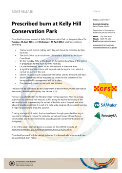
64
B U R N S
MALCOLM LESAVOY
T
hermal injuries are a result of the destruction of the
skin envelope of the body due to traumatic injury from
thermal energy such as heat, chemicals, electricity, radiation, or severe cold. The treatment of these severe and
sometimes catastrophic injuries requires knowledge of
the management of not only the local burn wound, but
also the confounding problems of the hemodynamic,
metabolic, nutritional, psychological, and immunologic
phenomena that occur. Discussion in this chapter is
predominantly on heat thermal injuries. Chemical and
electrical burns are significant problems, but occur less
frequently.
CASE 1
A CAR EXPLOSION
A 45-year-old mechanic was cleaning a carburetor when a
spark from the lamp he was using ignited the gasoline. His
co-workers extinguished his burning clothing rapidly, but
he still had charring of his right upper arm, chest, and abdomen. He was quickly undressed on arrival at the hospital and his burn was estimated at 40% TBSA. No evidence
of burn about the face or singed nasal hair was noted. He
was asked to cough and produced a small amount of clear
sputum that had no carbonaceous particles. He was able
to breathe without difficulty. Oxygen by nasal cannulae at
4 L/min was begun. An arterial blood gas showed a carboxyhemoglobin concentration of less than 5%. Several
intravenous catheters were placed through nonburned
skin. The physicians planned to administer 5,600 ml of
lactated Ringer’s solution to this 70-kg patient over the
next 8 hours (700 ml/hr).
On arrival at the burn unit, nasogastric and bladder
catheters were placed. Tetanus toxoid was given, and intravenous narcotics were administered. Additional blood
samples were sent for CBC, electrolytes, fasting blood
sugar, and cross-matching. A chest x-ray and ECG were
obtained. His wounds were cleansed and superficial debris removed. The burned areas were dressed with silver
sulfadiazine and sterile dressings.
CASE 2
ELECTRICAL WIRING INJURY
A 40-year-old male decided to replace his porch light. The
moment his screwdriver touched the wiring he saw a bright
flash, felt his muscles contract, and fell to the ground. On
arrival at the emergency room he was dazed and sore. He
had obviously broken his left arm because of the pain he
felt and the visible deformity. An IV catheter infused lactated Ringer’s solution at 150 ml/hr. His ECG, head CT,
and chest x-ray were all normal. He had a small burn on the
dorsum of his right forearm, but otherwise seemed unscathed. He was amazed when the nurse told him that he
was being admitted to the ICU for observation.
467
4 6 8
B
P L A S T I C
S U R G E R Y
GENERAL CONSIDERATIONS
urn injuries cause destruction of the skin. The
amount of tissue destruction is related to the intensity of
the heat and to the time of exposure. In general, a burn is
produced by an agent with a temperature of 40°C or
above. Hot baths and showers are common causes of
burns around the house, as parents of young children or
caretakers of the elderly may not realize how hot the
water actually is. Fires and explosions are other common
causes (Case 1) when people become careless around
flammable liquids. House and office fires are particularly
dangerous not only because of the burns they produce,
but also because of the toxic fumes that wood and plastics
emit when they burn. Victims of closed space fires (cars
and unvented rooms) may sustain severe inhalational injuries from these products of combustion. Additionally,
inhalation of carbon monoxide produces carboxyhemoglobin in which hemoglobin is unavailable for transport of
oxygen. Chemical burn severity depends on the nature of
the agent (gasoline, phenol, hydrofluoric acid, white
phosphorous, etc.). Electrical injuries depend on the type
of circuit (AC or DC), the voltage, the resistance offered
by the body, the duration of contact, and the amperage of
current flowing through the tissue (Case 2). When there
is no direct path through the patient to ground, the injury
tends to be less. Use of insulating rather than conductive
materials around electric circuits helps increase the resistance and decrease the flow through the patient. Tissue
resistance to electrical current increases from nerve
(which is the least resistant) to vessel, muscle, skin, tendon, fat, and finally bone (most resistant).
K E Y
The burn size or percentage of total body surface area
(TBSA) that is burned can be estimated by the “rule of
nines.” This divides the body into multiples of nine. The
chest and abdomen are 18% combined, and the back and
buttocks are also 18% combined. The anterior portion of
the one lower extremity is 9%, the posterior aspect is 9%,
each upper extremity is 9%, the head and neck area is 9%,
and the genitalia are 1%. In children, the head and neck
and the chest and abdomen are relatively larger than the
extremities (Fig. 64.1).
The depth of the burn is dependent on the amount of
superficial or deep tissue destruction (Fig. 64.2). Firstdegree burns are limited to the epidermis and are selflimiting and self-healing. Second-degree burns include
destruction of the epidermis and portions of the dermis,
leaving the mid- to deep dermis intact. Superficial blisters
with clear fluid can appear. Because nerve endings are still
intact (Fig. 64.3), second-degree burns are extremely
9%
9%
9%
9%
9%
P O I N T S
• Burn is produced by agent with a temperature of 40°C or
above
• Victims of closed space fires (cars and unvented rooms) may
sustain severe inhalational injuries from these products of
combustion
1%
9%
9%
• Electrical injuries depend on type of circuit, voltage, resistance offered by body, duration of contact, and amperage of
current flowing through tissue
T
DIAGNOSIS
he diagnosis of thermal injuries relates to two
major factors: burn size (the percentage of total surface
area burned) and depth of the burn (first degree, second
degree, third degree, and fourth degree). Other factors related to burns include age of the patient, location of the
burn that may cause special problems (e.g., hand, face),
inhalational injuries, circumferential burns, and associated
injuries such as fractures.
FIGURE 64.1 The “rule of nines” permits rapid estimation of burned body surface area in adults. It must be
adapted for children whose body surface is larger with respect to weight.
B U R N S
4 6 9
Epidermis
Dermis
Subcutaneous
tissue
FIGURE 64.2 Burn depth depends on the layers of the skin involved. This diagram illustrates the normal skin appendages (hair follicles and sweat glands) with respect to their depths.
painful. These injuries will re-epithelialize without skin
grafting within 2 weeks, assuming local infection does not
occur. Third-degree burns destroy the epidermis and full
thickness layers of the dermis down into the subcutaneous fat and/or muscle and bone (Fig. 64.4). Nerve endings are destroyed, making the affected area anesthetic.
Dermal protein is denatured by the heat and becomes a
tough, leather-like layer. On palpation, the burn is hard
and insensate. Re-epithelialization and self-healing cannot occur because the basal cell layer of the dermis has
been destroyed. Skin grafting or flap reconstruction will
be necessary.
K E Y
P O I N T S
• Diagnosis of thermal injuries relates to burn size (percentage
of TBSA) and depth of burn (first degree, second degree, and
third degree)
• Other factors related to burns include age of patient, location
of burn that may cause special problems (e.g., hand, face), inhalational injuries, circumferential burns, and associated injuries such as fractures
• Burn size or TBSA can be estimated by rule of nines
• First-degree burns limited to epidermis and are self-limiting
and self-healing
• Second-degree burns include destruction of epidermis and
portions of dermis, leaving mid- to deep dermis intact
• Second-degree burns are extremely painful
• Third-degree burns destroy epidermis and full thickness
layers of dermis down into subcutaneous fat and/or muscle
and bone; nerve endings destroyed, making affected area
anesthetic; dermal protein denatured by heat and becomes
tough, leather-like layer; on palpation, burn is hard and
insensate
I
DIFFERENTIAL DIAGNOSIS
t is difficult to differentiate between second- and
third-degree burns. Frequently, at the initial injury, they
can appear identical; however, it is important to realize
that a second-degree burn will “heal by itself” (re-epithelialization) and a third-degree burn will only heal by
wound contraction and/or skin grafting. Occasionally, one
can differentiate between second- and third-degree burns
by testing for sensibility. Feeling in the skin can be present in second-degree burns because the sensory end organs are usually preserved, whereas in third-degree burns,
the wound area is totally anesthetic because of the destruction of sensory neural end fibers.
4 7 0
P L A S T I C
S U R G E R Y
FIGURE 64.3 A seconddegree burn involves a portion of the dermis and the
epidermis. Because it is not
full thickness, the burn
spares the nerves and is extremely painful. Follicles are
left intact and will continue
to grow hair.
S
TREATMENT
ince first-degree burns are self-limited (sunburn) and
heal themselves, various topical over-the-counter ointments suffice. There are no sequelae or residual scarring
from first-degree burns.
Second- and third-degree burns are treated similarly
clinically, with some exceptions. One must understand
the differentiation between treating the local wound area
and the metabolic effect of second- and third-degree
burns. In second-degree burns, the wound can be minimally debrided, topically treated with antibacterials, and
allowed time for re-epithelialization. Third-degree burns
usually should not be debrided (unless contaminated).
They should be topically treated with antibacterial agents
until the wound bed is ready for surgical excision, then
skin grafted.
When second- or third-degree burns affect more than
20% of the body surface area in any age group (or 10% of
the body surface area in patients under 10 or over 60 years
of age), these patients should be hospitalized for treatment of not only the local wound, but most importantly,
the hemodynamic and metabolic affects of this large and
sometimes devastating injury.
The management of a severely burned patient begins
with assessment of the airway and breathing. Only rarely is
a surgical airway such as a cricothyroidotomy required.
When the patient has been in a closed space, they frequently will have singed facial and nasal hair as well as carbonaceous sputum in their oro- or nasopharynx. When
these are present, bronchoscopy is indicated to determine
the extent of any inhalational injury.
Plastics and wood emit large amounts of carbon
monoxide when they burn. The carbon monoxide binds
avidly to hemoglobin and interferes with normal oxygen
transport by forming carboxyhemoglobin. At room air oxygen concentration, carboxyhemoglobin has a half-life of 4
hours, while at 100% oxygen, its half-life is reduced to 1
hour. A blood gas sample should be obtained and sent for
carboxyhemoglobin saturation shortly after the patient’s
arrival. Levels less than 5% are normal, while concentrations greater than 30% may be rapidly lethal. Patients who
present with singed facial or nasal hairs should be suspect
for inhalational injuries. Burning plastics and synthetic
fabrics emit a number of compounds that are extremely
toxic to the lungs. Fiberoptic bronchoscopy should be performed quickly to determine whether such injury is present in appropriately selected patients. Although a chest xray should be obtained in all burn victims to exclude
associated trauma, this study is generally not helpful in detecting inhalational injuries (Case 1).
The major physiologic derangement of a second- or
third-degree burn comprising more than 20% of the body
surface area is hypovolemia. This occurs because of massive
edema formation due to loss of the integrity of endothelial
cells within capillaries. Fluid is lost from the intravascular
space to the interstitial space, causing a “third-space phenomenon.” Patients can rapidly develop hypovolemic shock.
Massive fluid resuscitation is necessary to replace this volume so that cardiac output can be restored.
B U R N S
4 7 1
FIGURE 64.4 A full thickness burn involves all layers
down to (and often including) the subcutaneous tissue.
Because nerves are destroyed the burn is anesthetic. Coagulation of the
skin’s proteins produces a
white appearance that is
“leathery” to the touch. Because hair follicles are destroyed, hair will not regrow. The involved eschar
must be excised and replaced with a skin graft.
Baseline laboratory studies, including hematocrit, urinalysis with specific gravity, electrolytes, chest x-ray, and arterial blood gases, are important before fluid resuscitation.
The treatment of burn hypovolemia requires the intravenous replacement of fluids and electrolytes computed
(Baxter or Parkland Hospital formula) as follows: 24 hr requirement (ml) = TBSA burn ⋅ 4 ⋅ wt (kg). This is provided
using lactated Ringer’s solution or normal saline. Dextrose
should not be used in the initial postburn period. One-half
of the amount calculated is given in the first 8 hours following the burn (not the arrival to the emergency room),
and the second half is given over the ensuing 16 hours of
the 24-hour period (Case 1).
Urine output and specific gravity should be used to
monitor the adequacy of resuscitation. As with all formulas,
increased or decreased volumes of fluid may be required depending on the patient’s response. Following the first day,
plasma protein (albumin) can be administered at 0.3–0.5
ml/kg/% burn. Crystalloid can be changed to 5% dextrose in
water to maintain urinary output and to keep the serum
sodium at a normal level. The monitoring of this resuscitation is extremely important and a urine output of 50 ml/hr or
more in adults and 2 ml/kg/hr in children less than 10 years
old is paramount. Central venous or pulmonary wedge pressures in these kinds of acute burns are usually unreliable.
Routine vital signs should be constantly monitored.
Nutrition is an extremely important part of burn management and should be instituted as quickly as possible.
Most patients can tolerate internal feeding through a na-
sogastric tube. Caloric intake should be at least 40
Kcal/kg/day and protein intake at least 1.2 g/kg/day.
Cure of the burn wound should begin at the time of
fluid resuscitation. Burns should be cleansed gently with a
surgical soap to remove nonviable epidermis. Bullae should
be left in place as they form a biologic dressing. Several topical antimicrobial agents are available that decrease the incidence of invasive burn wound infection and systemic sepsis.
All have been associated with improved survival of burn patients. Silver sulfadiazine (Silvadene) is the most commonly
used agent. It has limited penetration of the eschar, but is
painless on application and has excellent activity against
many organisms, with the notable exception of many strains
of Pseudomonas and virtually all Enterobacter species. Its
most important undesirable effect is that it can cause a reversible neutropenia. Mafenide acetate (Sulfamylon) has
excellent eschar penetration and is particularly useful in patients with heavily contaminated wounds or those known to
contain Pseudomonas. Its disadvantages are that it causes
discomfort when applied to partial thickness burns and it
acts as a carbonic anhydrase inhibitor, thereby producing a
metabolic acidosis. Both silver sulfadiazine and mafenide
acetate are applied every 12 hours, with the excess removed
before the next application. Silver nitrate (0.5%) solution
and povidone iodine are used occasionally. Silver nitrate is
usually reserved for patients with sulfa allergies, since it
must be applied every 8 hours and is messy to handle. It
causes transeschar leaching of sodium, potassium, chloride,
and calcium, which must be replaced as required.
4 7 2
P L A S T I C
S U R G E R Y
Antibiotic use is not indicated in the initial treatment
of burned patients. Studies have shown that such prophylaxis actually increases the rate of several infections (such
as pneumonia) in the early postburn period. Clostridial infections and tetanus are not uncommon. Therefore, each
patient’s tetanus immunization status should be assessed
and toxoid administered if required.
Patients should be examined carefully for the presence
of circumferential burns, which might restrict blood flow or
mobility. This is particularly important on the extremities
(including the hands and feet) as well as on the chest where
a circumferential eschar may limit ventilatory excursions.
When such wounds exist, escharotomy is indicated. Escharotomy involves placing bilateral longitudinal incisions
through the length of the eschar to permit blood flow or
movement beneath the burn. Escharotomy should be performed quickly since ischemia can occur rapidly. The procedure can be done in the emergency room if required, but
is best done at the bedside in the burn unit. Anesthesia is
not usually required since the eschar is anesthetic. The incision is carried through the full thickness of the burn until
viable (bleeding) tissue is reached beneath. The most commonly used topical antibacterial agent placed as a cream on
the burn wound after the removal of necrotic tissue is silver
sulfadiazine (Silvadene). Occasionally, biopsies of the burn
wound for quantitative and qualitative bacteriology can be
helpful. Once the burn wound is cleaned, skin grafts should
be applied at periodic operative interventions.
Complications in acute burns can be devastating.
Renal failure from hypovolemia can occur and must be diagnosed if not avoided. Gastrointestinal bleeding occurs in
over 40% of acute burns and has a correlation with an increased risk of burn wound sepsis. Burn wound sepsis can
be diagnosed by tissue biopsies for qualitative and quantitative analysis. The bacterial count should be kept below
105 of bacteria per gram of tissue.
K E Y
P O I N T S
• At room air oxygen concentration, carboxyhemoglobin has
half-life of 4 hours; at 100% oxygen, its half-life is reduced to
1 hour
• Major physiologic derangement of second- or third-degree
burn comprising more than 20% of body surface area is hypovolemia
• Treatment of burn hypovolemia requires intravenous replacement of fluids and electrolytes computed (Baxter or ParkHospital formula) as follows: 24 hr requirement (ml) = TBSA
burn • 4 • wt (kg)
• Caloric intake should be at least 40 Kcal/kg/day and protein
intake at least 1.2 g/kg/day
• Burns should be cleansed gently with surgical soap to remove nonviable epidermis; bullae should be left in place as
they form biologic dressing
• Antibiotic use indicated in initial treatment of burned patients; studies have shown that prophylaxis increases rate of several infections (such as pneumonia) in early postburn period
• Escharotomy involves placing bilateral longitudinal incisions
through length of eschar to permit blood flow or movement beneath burn
• Burn wound sepsis can be diagnosed by tissue biopsies for
qualitative and quantitative analysis
F
FOLLOW-UP
ollowing the successful treatment of the acute thermal
injury, further soft tissue reconstruction, splinting, and rehabilitation (physical and psychological) may be necessary.
The burn patient can be the sickest patient in any hospital,
since all organ systems are often involved. Increased nutritional and metabolic needs must be met during the acute
and subacute stage. Various aspects of hyper- or hypothermia, congestive heart failure, pulmonary edema, ileus,
mental status changes, azotemia, thrombocytopenia, hypofibrinogenemia, and hyper- or hypoglycemia can all occur.
Long-term follow-up, including the prevention of wound
contracture and hypertrophic scarring, must be maintained
by splinting and compression. Further reconstruction is
frequently warranted.
Full thickness burn eschars are nonviable and must be
removed. Such tangential excision requires grafting of the
underlying bed to allow new skin growth. Most centers
now prefer early excision (within several days after injury)
of the eschar with skin grafting to aid in mobility and early
healing. Studies have shown that early excision and grafting decreases hospital stay and improves subsequent functional outcome.
Electrical injuries present special challenges, including cardiopulmonary difficulties such as ventricular fibrillation. There is also a high risk of renal failure due to hemoglobin and myoglobin deposits in the renal tubules,
fractures, spinal cord injuries, intra-abdominal problems,
and vascular derangements such as thrombosis of small
vessels. Tissue destruction under the eschar of an electrical injury is often worse than might be expected from the
appearance of the surface burn (Case 2). Hence, these patients require hospitalization with aggressive management. Fluid requirements are greater than predicted by
the formulas, since muscle destruction is more widespread
than just the area of the eschar. Injuries due to lightning
strikes are particularly hazardous because of the amount
of current conducted.
Patients suffering from cold injuries such as frostbite
and systemic hypothermia require rapid rewarming in
40°C hydrotherapy tanks as necessary. One must monitor
B U R N S
cardiac, vascular, and respiratory function during this
rapid rewarming. A urine output of 50 ml/hr should be
maintained.
K E Y
P O I N T S
• Early excision (within several days after injury) of eschar with
skin grafting to aid in mobility and early healing
• Early excision and grafting decreases hospital stay and improves subsequent functional outcome
• Tissue destruction under eschar of an electrical injury is
often worse than might be expected from appearance of surface burn
SUGGESTED READINGS
Plastic and Reconstructive Surgery—Essentials for Students. 4th
Ed. Plastic Surgery Educational Foundation, Arlington
Heights, IL, 1993
A good student manual that has an overview of burn injuries.
Atturson MG: A pathophysiology of severe thermal injury. J
Burn Care Rehab 6:129, 1985
A good overview that discusses underlying pathophysiologic
mechanisms.
Pruitt BE Jr: The diagnosis and treatment of infection in the
burn patient. Burns 11:79, 1984
4 7 3
An outstanding article on burn-related infection, one of the
most feared complications. The author is among the best
known burn surgeons.
QUESTIONS
1. Clinically, a third-degree burn may be differentiated
from a second-degree burn because the former?
A. Is more superficial.
B. Is anesthetic.
C. Has the potential to regenerate normal skin.
D. Generally has no infection risk.
2. The half-life of carboxyhemoglobin in a patient breathing room air is?
A. 30 minutes.
B. 1 hour.
C. 4 hours.
D. 1 day.
3. Silver sulfadiazine (Silvadene) is a frequently used
topical antimicrobial agent in the treatment of burn
wounds. Which of the following items is not one of its
characteristics?
A. Good coverage of Pseudomonas.
B. Incomplete eschar penetration.
C. Broad spectrum antimicrobial coverage.
D. Painless on application.
(See p. 604 for answers.)


© Copyright 2026