
Spinal muscular atrophy: diagnosis, treatment and future prospects R A
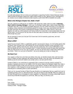
0021-7557/10/86-04/261
Jornal de Pediatria
Review Article
Copyright © 2010 by Sociedade Brasileira de Pediatria
Spinal muscular atrophy:
diagnosis, treatment and future prospects
Mariana T. C. Baioni,1 Celia R. Ambiel2
Abstract
Objective: To report on recent genetic and molecular discoveries and on future prospects for the treatment
of spinal muscular atrophy (SMA), thereby helping healthcare professionals to make a quick diagnosis and provide
appropriate and timely therapeutic support.
Sources: Information was collected from scientific articles published in the last 2 decades, retrieved from the
databases SciELO, PubMed, and MEDLINE.
Summary of the findings: SMA is a neurodegenerative disorder with autosomal recessive genetic heredity.
It is caused by a homozygous deletion of the survival motor neuron (SMN1) gene. This genetic alteration results
in reduced levels of the SMN protein, leading to degeneration of alpha motor neurons of the spinal cord and
resulting in muscle weakness and progressive symmetrical proximal paralysis. It is known that basic nutritional
and respiratory care and physiotherapy can be important to delaying disease progression and prolonging patients’
lives. Several drugs are being tested, some new, others, such as valproic acid, already known; paralysis can be
halted, but not reversed.
Conclusions: SMA is a difficult to diagnose disorder, because it is little known, and treatment is uncertain.
Pharmacological treatments and supportive therapies are not yet able to recover motor neurons or muscle cells
that have already been lost, but are aimed at delaying disease progression and improving patients’ residual muscle
function, as well as offering better quality of life and life expectancy.
J Pediatr (Rio J). 2010;86(4):261-270: Spinal muscular atrophy, motor neuron, therapy, SMN1 gene, SMN
protein, valproic acid.
Introduction
Spinal muscular atrophy (SMA) is a neurodegenerative
This genetic alteration to the SMN1 gene is responsible
disease with autosomal recessive heredity. After cystic
for a reduction in survival motor neuron (SMN) protein. The
fibrosis (1:6,000), SMA is the next most fatal disease with
SMN2 gene does not completely compensate for the absence
this genetic profile, with an incidence of 1:6,000 to 1:10,000
of SMN1 expression because it only produces 25% of SMN
births.1
protein.4 The lack of the SMN protein leads to degeneration
The frequency of carriers (heterozygotes) is one
in 40 to 60
of alpha (α) motor neurons located in the ventral horn of
people.2
the spinal cord, which leads to progressive and symmetrical
This disease is caused by a homozygous mutation or
muscle weakness and paralysis.2
deletion of the survival motor neuron gene 1 (SMN1), which
should be located in the telomeric region of chromosome
Clinical classification of SMA is based on age at onset
5q13. The principal determinant of severity is the number
and maximum motor function acquired, with the following
of copies of SMN2, a gene that is similar to SMN1 and is
categories: 1) severe (type I, severe SMA or Werdnig-
located in the centromeric region.3
Hoffmann disease); 2) intermediate (type II or chronic
1. Especialista, Fisiologia Humana, Universidade Estadual de Maringá (UEM), Maringá, PR, Brazil.
2. Doutora, Biologia Celular. Professora, Fisiologia Humana, UEM, Maringá, PR, Brazil.
No conflicts of interest declared concerning the publication of this article.
Suggested citation: Baioni MT, Ambiel CR. Spinal muscular atrophy: diagnosis, treatment and future prospects. J Pediatr (Rio J). 2010;86(4):261-270.
Manuscript submitted Aug 26 2009, accepted for publication Oct 14 2009.
doi:10.2223/JPED.1988
261
262 Jornal de Pediatria - Vol. 86, No. 4, 2010
Spinal muscular atrophy - Baioni MT & Ambiel CR
SMA); 3) mild (type III, juvenile SMA or Kugelberg-Welander
Type III SMA: (also known as juvenile SMA or Kugelberg-
disease); and 4) type IV (adult SMA).3 Other authors5-7
Welander syndrome) onset is after 18 months, but the actual
classify SMA into just three categories: severe, intermediate
age varies greatly. According to Wirth et al.,10 when the
and mild.
disease emerges before 3 years of age it is classified as Type
SMA is a difficult disorder to diagnose and treatment
IIIa SMA, whereas when onset is later, it is called Type IIIb
is uncertain. Diagnosis is based on evidence, both
SMA. The difference between the two is preservation of the
electrophysiological and histological, of denervation of the
ability to walk. Patients with Type IIIa are able to walk until
muscle.3 Nowadays, diagnosis is confirmed by molecular
they are 20, while Type IIIb patients will be able to walk for
analysis to demonstrate an absent SMN1 gene exon 7.2
their whole lives.11 Problems with swallowing, coughing or
Since this is a progressive neurodegenerative disease,
patients with SMA require special care, which can halt
disease progression and prolong their lives. The objective
of this bibliographic review article is to describe the clinical
and laboratory profile of SMA patients and report on recent
nocturnal hypoventilation are less common than in patients
with Type II, but may still be observed. As they age, these
patients may develop scoliosis. The principal characteristic
of these patients is that they are able to walk independently,
and life expectancy is indeterminate.3
genetic and molecular discoveries and the future prospects
Type IV SMA: there is no consensus on the age of onset
for treatment, thereby aiding health professionals to
of Type IV SMA. Russman3 reports that it emerges after 10
make rapid diagnoses and provide early and appropriate
years of age, whereas Wang et al.8 state that weakness
therapeutic support.
normally emerges during the second or third decade of life,
or at about 30 years of age. Motor function involvement
is mild and there are no problems with deglutition or
Development
respiration. These patients are able to walk normally and
Classification of SMA
have normal life expectancy.3,8
There are four SMA classifications, based on age at
disease onset and maximum motor function acquired.
Clinical features of SMA
Type I SMA: (also known as severe SMA, Werdnig-
Since only α motor neurons are lost progressively, only
Hoffmann disease or acute SMA) is characterized by early
motor function is compromised and sensory neurons are
onset (between 0 and 6 months of age), by a failure to
unaffected. This loss of function leads to weakness and to
acquire the ability to sit up and by very short life expectancy
progressive symmetrical atrophy of the proximal voluntary
(less than 2 years).3 Children diagnosed with this form have
muscles of the legs, arms and, sometimes, the trunk, as
very little control of their heads and cough and cry weakly.
the disease progresses.8
They lose the ability to swallow and feed before they reach
1 year of age. Trunk and limb weakness normally spreads
to the intercostal muscles, making it unlikely that a normal
respiratory cycle will develop. Although the intercostal
muscles are affected, the diaphragm is initially spared.
The risk of early mortality is usually associated with bulbar
dysfunction and respiratory complications.8 Historically,
these children have a short life expectancy (less than 2
years), but, thanks to improved clinical care, over recent
years survival has improved.9
Type II SMA: (or chronic SMA) generally becomes
symptomatic at around 6 to 18 months, but it may emerge
earlier. Some patients classified as having type II SMA
are able to sit up unaided while others can remain sitting
if they are positioned, but cannot sit up unaided.3 Better
developed patients are able to remain standing if provided
with support, but will nevertheless be unable to learn to walk.
Bulbar weakness, combined with swallowing difficulties, can
lead to reduced weight gain in some children. Furthermore,
these patients may have problems with coughing and
A number of unusual clinical features are observed in
SMA. One of these is the distribution of muscle weakness,
which is more compatible with a myopathic disorder than
with a neurogenic disorder.12 Proximal muscles are more
involved than distal muscles, legs are more affected
than arms and arms are more affected than the face and
diaphragm.8,12 In other words, muscle weakness and atrophy
does not have a homogeneous distribution. The severity
of muscle weakness is related to age at onset and children
with the most severe form of the disease (Type I SMA)
can appear normal at birth, but present muscle weakness
a few months later.8
Furthermore, the clinical course followed by SMA patients
who survive beyond childhood demonstrates that loss of
muscle strength is normally most evident at diseases onset
and that, after this, residual muscle power can remain stable
for months or years.12,13
Molecular genetic basis of SMA
with cleaning secretions from the trachea, may have fine
Genetic studies have shown that SMA is caused by
trembling (known as fasciculation) and can suffer from
absence of the SMN1 gene, which should be located in
scoliosis and contractures as they age.8 Life expectancy is
the telomeric region of chromosome 5.12,14,15 This gene
around 10 to 40
years.3,8
was identified in 1995 by Lefebvre et al.16 and has nine
Jornal de Pediatria - Vol. 86, No. 4, 2010 263
Spinal muscular atrophy - Baioni MT & Ambiel CR
exons that code for the SMN protein. All patients still
The SMN protein is widely distributed in all cells of
retain at least one copy of a very similar gene - SMN2
the body.18,22 It is found in the nuclei of cells and within
– which is located in the centromeric part of the same
the nucleus it binds to certain structures involved with
chromosome. The absence of SMN1 is caused either by
removing non-coding sequences (introns) from the pre-
a deletion or by a conversion that transforms SMN1 into
MRNA (messenger ribonucleic acid). Furthermore, it appears
SMN27 (Figure 1).
that SMN also participates in regulating transcription and
The SMN1 gene is responsible for complete synthesis
expression of certain genes.18,10
of the SMN protein. In contrast, the SMN2 gene is not
Despite advances in knowledge about the biochemistry
capable of completely synthesizing the protein, being only
of SMN, it is not yet clear how reduced levels in all different
responsible for a part of its production. SMN2 produces
cell types are specifically responsible for degeneration of
10 to 25% functional protein while the remaining 75%
α motor neurons. This raises the possibility that SMN has
produced is a protein that is truncated and unstable
(SMN∆7) and is rapidly degraded (Figure 1).10,15,17,18
an additional function that is restricted to these neurons.
Immunohistochemical studies have identified SMN in
Another important point that should be highlighted is
dendrites, implantation cones and axons of motor neurons,
that the number of intact copies of SMN2 is a determinant
suggesting it plays a role in transporting RNA along axons.10
of disease severity. Figure 2 illustrates the genotype
Furthermore, it has been observed that the motor neurons
of people who are affected and unaffected by SMA,
of SMN-knockout mice and zebrafish exhibit failure to
characterizing type I, II and III patients. It also illustrates
reach the motor end plate, having an axon with aberrant
the quantity of SMN protein synthesized by each genotype.
ramifications, indicating strong evidence that SMN plays an
As the number of copies of SMN2 increases, the quantity
important role in morphological development and axonal
of functional SMN protein increases and the severity of
migration.23 On the other hand, the SMN protein is also
the disease
reduces.5,7,10,19
The figure also demonstrates
found postsynaptically at the neuromuscular junction and in
that gene conversion events are responsible for the milder
the Z bands of striated muscle, indicating that pathogenesis
phenotypes, while deletion of the SMN1 gene leads to
does not exclusively involve the motor neuron cell body, but
more severe forms of the AME.7,20,21
can also affect the muscle fibers themselves.24
SNM = survival motor neuron;
MRNA = messenger ribonucleic acid.
Figure 1 - Structure of the SMN gene in chromosome 5
264 Jornal de Pediatria - Vol. 86, No. 4, 2010
Spinal muscular atrophy - Baioni MT & Ambiel CR
SMA = spinal muscular atrophy; SMN = survival motor neuron.
Figure 2 - Genotypes of people affected and unaffected by SMA
No cases have ever been reported of people with
Table 1 -
complete absence of the SMN protein, i.e., patients who
have SMA and also have an absent SMN2 gene. This is
probably because such a genotype would be incompatible
with life and the SMN protein may play an essential role
during embryo
development19,25,26
or may modulate
neuronal apoptosis.27
Diagnosis
Since SMA is a low-incidence neurological disorder,
diagnosis is difficult. Nevertheless, since SMA develops
progressively, rapidly establishing a precise diagnosis is
essential.
Children manifesting clinical signs characteristic of SMA,
such as hypotonia, paresis, areflexia and fasciculation,
should be investigated with care,1 since these clinical signs
can also be observed in other neuropathologies (Table 1).
Since neuromuscular diseases are the main causes
of childhood hypotonia28 and since the neuromuscular
diseases that most often affect children are SMA and the
dystrophies,28,29 Table 230 provides a summary of the
principal features that differentiate between the two groups.
Notwithstanding, it should be pointed out that not all of
the features described here will always be present in every
patient, since they vary according to the disease stage that
each patient is in at the time of assessment.
The most common causes of muscle hypotonia in
childhood
Principal causes of muscle hypotonia by age at onset
At birth
Neuromuscular diseases
Congenital myotonic dystrophy
Type I spinal muscular atrophy
Other causes
Systemic septicemia-induced diseases
Lung damage
Intracranial pathologies
Infections of the central nervous system
Disorders of the peripheral nerves
Diseases of the neuromuscular junction
Prader-Willi syndrome
Drug intoxication during pregnancy or delivery
After 6 months of age
Neuromuscular diseases
Spinal muscular atrophy, types II and III
Polyneuropathies
Childhood myasthenia gravis
Muscular dystrophies
Metabolic myopathy
Other causes
Congenital heart disease
Malnutrition
Rickets
Metabolic diseases
Nephropathies
Lung diseases
Jornal de Pediatria - Vol. 86, No. 4, 2010 265
Spinal muscular atrophy - Baioni MT & Ambiel CR
Table 2 -
Principal differences between spinal muscular atrophy and the muscular dystrophies
Clinical features
Spinal muscular atrophy
Muscular dystrophy
Symptoms
Weakness
Weakness
Signs
Muscle atrophy, lack of deep reflexes,
fasciculations, rapid and discrete involuntary
movements of muscles, such as trembling
Pseudo-hypertrophy of the calf, deep reflexes
may be normal, reduced or absent, depending on
the degree of muscle weakness
Test findings
Normal or reduced muscle enzymes,
neurogenic electroneuromyography results,
muscle biopsy with atrophic appearance
Very high muscle enzyme levels, myopathic
electroneuromyography results, muscle biopsy
with dystrophic appearance
Definitive diagnosis
Genetic test showing deletion of SMN1 gene
from chromosome 5
Genetic test showing deletion of the dystrophin
gene from the X chromosome or by absent or
deficient dystrophin in a biopsy (here we are only
referring to Duchenne and Becker dystrophies,
which are the most common types)
Disease mechanism
Degeneration of nerve cells in the spinal
ventral horn
Degeneration of muscle cells
Genetic heredity
Autosomal recessive (the disease can
manifest in both boys and girls, both parents
are carriers and each pregnancy has a 25%
chance of producing a child with the disease)
X-linked (the disease manifests in boys, mothers
are carriers and there is a 50% chance that male
children will have the disease or that female
children will be carriers)
Treatment
First line treatment is physiotherapy (more
details will be given later in the text)
First line treatment is physiotherapy (corticoids
may be prescribed)
Most common complications
Respiratory problems, scoliosis, contractures
Respiratory and cardiac problems, scoliosis and
contractures
Natural course of disease
SMA is a progressive disease; degeneration is
faster or slower depending on type
Dystrophies are progressive diseases;
degeneration is faster or slower depending on
type (Duchenne, Becker, etc.)
SMA = spinal muscular atrophy; SMN = survival motor neuron.
In general, diagnosis of SMA is made on the basis of
motor and sensory nerves.1,10 Fibrillation potentials are
evidence of muscle denervation, found on electromyography
observed at rest in cases of denervation, whether located
and in muscle biopsy.3 Diagnosis is confirmed by molecular
in the anterior horn or peripheral nerves, both duration and
analysis demonstrating that exon 7 of the SMN1 gene is
amplitude of motor unit potentials may be increased and
absent, irrespective of clinical classification.2
there may be a reduction in motor conduction velocity in
Creatine phosphokinase (CPK) may be normal or as
the earlier forms of AME.1
much as five times lower than normal.1 Serum CPK can
differentiate neurogenic diseases, of which SMA is one,
from myopathic diseases, such as dystrophies, in which
muscle damage raises CPK levels.
Muscle biopsy
A range of abnormal muscle features can be observed
in SMA patients. Certain histopathological findings are
characteristic, such as the presence of atrophic muscle
Electromyography
fibers, both type I and type II, hypertrophy of type I fibers
or fiber-type grouping.1,10 However, these findings can also
Electromyography can be used to determine whether the
be observed in other causes of denervation.31 Therefore, this
disease has affected motor neurons, nerve roots, peripheral
type of test does not confirm SMA, but provides additional
nerves, myoneural junction or muscle fibers.29
clinical data.
In SMA there is electrophysiological evidence of
In the slower-progressing forms, superimposition of
denervation, while conduction is found to be intact in
secondary myopathic abnormalities, such as angular fibers,
266 Jornal de Pediatria - Vol. 86, No. 4, 2010
Spinal muscular atrophy - Baioni MT & Ambiel CR
central nuclei, splits and myofibrillar disarrangement,
which exacerbate the muscle weakness (particularly of
increase as the disease progresses.32
the respiratory muscles),8 and can lead to atelectasis and
pulmonary collapse.24 Additionally, these children may suffer
from nocturnal hypoventilation and underdevelopment of
Genetic investigation
Molecular genetic tests provide definitive diagnosis
the lungs and chest wall.8,24
of SMA and could be the only tests performed. Genetic
Provision for these patients includes rapid access
investigation demonstrates that exon 7 of the SMN1 gene is
to special clinical intervention and respiratory support
completely absent (with or without a deletion of exon 8).2,8
when necessary (ranging from noninvasive ventilation to
Since the SMN2 gene does not have this exon, its absence
tracheostomy and mechanical ventilation). Techniques for
also demonstrates that the SMN1 gene is nullified.
cleaning the Airways and moving secretions are very useful,
If a patient suspected of having SMA does have a copy of
the SMN1 gene, then this copy should be investigated for mild
mutations such as point mutations, insertions and deletions
leading to a homozygous dysfunction of the
gene.8
Molecular genetic diagnosis is more precise and less
invasive than the other two tests described, but it is not
widely available in Brazil. It should be pointed out that
testing for deletions in the SMN gene can provide guidance
during cases in which diagnosis is
uncertain.1
Alternatively, Kolb et al.33 developed a technique
for measuring the SMN protein in mononuclear cells
(lymphocytes and monocytes), obtained from blood samples
from patients with SMA. As would be expected, SMN protein
levels were significantly reduced in the patients in comparison
including pulmonary physiotherapy and postural drainage.
These patients also need rapid access to antibiotic therapy
and should be on an immunization schedule including several
vaccines against agents that can cause severe pulmonary
infections.8,24,34
Both atrophy and hypotonia of the muscles have a direct
influence on the degree of compromise to respiratory and
motor function. In these situations it is the physiotherapist who
works to prevent and treat bone deformities and respiratory
disorders, contributing to preventing the disease from
progressing and to improving patients’ quality of life.13
b) Nutritional care:
Children with SMA can suffer from a variety of
stated that their test could
gastrointestinal problems, such as gastroesophageal reflux,
be used in the future to monitor clinical trials attempting to
constipation, abdominal distension and retarded gastric
increase levels of MRNA and/or the SMN protein itself, but
emptying.8,24 Reflux is a determinant of mortality and
that it is not the best choice for diagnosing SMA.
morbidity, because it can be associated with silent aspiration,
with the controls. The
authors33
which can lead to aspiration pneumonia, worsening the
Treatment
Since it is a progressive neurodegenerative pathology,
SMA patients require a range of special treatments that
can halt the disease’s progress and prolong their lives.
The majority of care revolves around supportive therapies
because, unfortunately, there is not yet any pharmacological
treatment.
Supportive therapies
A multidisciplinary team is responsible for prolonging
and improving the quality of patients’ lives.24 Care covers
respiratory and nutritional support in addition to orthopedic
and physiotherapeutic care, to avert postural problems.
situation even further. High fat foods should be avoided
because they delay gastric emptying and increase the risk
that reflux will occur.8
The origins of constipation are multifactorial and it may
be the result of abnormal gastrointestinal tract motility,
reduced intake of foods rich in fibers and of water and
also hypotonicity of muscles in the abdominal wall.8,34
Furthermore, in these patients reduced intestinal movement
can result in abdominal distension.8
Patient care includes pharmacological treatment for
gastroesophageal reflux, involving gastric acid neutralizers
and/or gastric secretion inhibitors, such as proton pump
inhibitors and histamine blockers,8 in addition to prokinetic
agents.24
Diet can be assessed by a nutritionist. It should be
a) Respiratory care:
remembered that SMA patients may have an acceptable
Pulmonary diseases are the principal cause of morbidity
fat mass for their body, but may be classified as below
and mortality among patients with SMA types I and II and
normal weight, on the weight/height criterion, because of
can affect a small number of patients with type III SMA.8
their reduced body mass. This could lead to inappropriate
The severity of muscle weakness and the fact that
dietary advice, which would in turn lead to obesity.8
these patients are either always lying down or get up very
In severe cases, where children are unable to feed
little mean that they have a limited capacity to cough
sufficiently enterally, parenteral calorie supplementation
and remove secretions from the lower respiratory tract.
should be considered in order to avoid muscle catabolism
As a result of this they are prone to recurrent infections
in children with low fat reserves.8
Jornal de Pediatria - Vol. 86, No. 4, 2010 267
Spinal muscular atrophy - Baioni MT & Ambiel CR
c) Orthopedic care:
treatment of SMA patients have as their therapeutic targets
The principal problem that result from the limited trunk
the SMN2 gene. Strategies that increase its transcription or
and limb motor function caused by the muscle weakness
that stabilize the protein it forms appear to be promising.37,38
include postural deformities (scoliosis), limitations to mobility
Some drugs based on this therapeutic perspective are
and the ability to carry out daily activities, increased risk of
described below.
pain, osteopenia and
fractures.4,8,24
Iannaccone34
reports
that scoliosis is rare before early childhood, and so is not
a) Histone deacetylase inhibitors:
normally seen in children with Type I SMA, but is common
This class of drugs has been investigated for treatment
in patients with Type II SMA and less common in those with
of SMA because of its ability to activate transcription of the
Type III. Weakness of the paraspinal muscles means that
SMN2 gene, because when histone is acetylated (inhibited
scoliosis progresses gradually and should be monitored
histone deacetylase), transcription factors become more
regularly.24
accessible to several genes (including SMN2), encouraging
Patients with Type I SMA also have difficulties related
their transcription.
to limited head control, posture and alignment. The
Clinically known drugs, such as valproic acid, sodium
principal problems among Type II patients are contractures,
butyrate and phenylbutyrate are examples of compounds
respiratory dysfunction and scoliosis. The combination of
that have an inhibitory action on the histone deacetylase
proximal muscle weakness and impaired balance means
enzyme. This property makes them potential candidates
that patients with Type III SMA fall frequently and suffer
for treatment of SMA, particularly valproic acid17,38,39 and
abnormal fatigue during physical activity.8
phenylbutyrate,40 which are better able to penetrate the
Interventions that can be employed to avoid worse
consequences are postural control, pain control and
CNS, in addition to the fact that their pharmacokinetics and
safety profiles have already been described.
contracture control, adaptation of daily activities, mobility
Valproic acid is the histone deacetylase inhibitor that
with a wheelchair or walking frame, ortheses for limbs
has been most investigated in preclinical and clinical trials
and therapies that encourage the development of mobility,
assessing its efficacy for treating SMA.17,38,39 The efficacy of
prolonging these children’s survival and alleviating the
valproic acid to induce an increase in the levels of the SMN
burden of the disease.8
protein has been demonstrated in cultures of fibroblasts
Regular exercise, such as swimming or other appropriate
from patients with SMA I,17,39 in hippocampus slices39 and
sports, is important to recover the self-esteem of these
in motor neuron cultures38 from an SMA I rat model. In
children, to introduce them into a social context and to
vivo, administration of valproic acid in the drinking water
report that
of a mouse model of SMA III, increased SMN protein
regular physical exercise can be helpful to develop muscles
levels in spinal marrow and also improved motor function,
and joints, increase bone density, improve intestinal mobility
with increased evoked motor potentials, reduced spinal
and promote a general sense of wellbeing.
marrow neuron degeneration and better innervation of
help maintain physical fitness. Swoboda et
al.13
Grondard et al.35 studied the benefits of regular exercise
in mutant rats with Type II SMA and found positive results.
the neuromuscular junction when compared with control
SMA animals.41
They noted that mutant rats that were forced to run in a
After these encouraging preclinical results, clinical
wheel exhibited an impressive increase in survival time,
trials were started. Non-randomized and non-placebo-
compared with non-exercised rats, and they also observed
controlled clinical studies by Tsai et al.42 and Weihl et al.43
a reduction in medullary motor neuron death.
These
with small numbers of patients demonstrated a modest
results suggest that regular physical exercises should be
improvement in muscle strength and subjective function.
combined with pharmacological treatment in order to test
These studies used the same dosage of valproic acid that
the possibility of cumulative protective effects against
is recommended for the treatment of epilepsy (15 to
disease progression.
50 mg/kg/day). Recently, Swoboda et al.44 published the
Finally, Oskoui & Kaufmann24 point out that modifications
need to be made to the homes of these children in order to
guarantee their safety and allow them independence.
first phase II clinical trial of valproic acid (15 to 50 mg/kg/
day), given to 42 patients with Type I, II or III SMA aged
from 2 to 31 years. The results are to a certain extent
inconclusive, probably because of the heterogeneous
nature of the sample, and it was not possible to detect a
Pharmacological
clear interference in disease progression caused by the
Sadly, there is currently no pharmacological treatment
drug. Some patients suffered weight gain and carnitine
available for SMA. However, on the basis of progress in
depletion. The authors themselves44 stressed the need for
understanding the genetic bases and the pathophysiology
randomized, placebo-controlled and double-blind clinical
of SMA, potential candidates to attempt to treat it are
trials that would be capable of a more accurate investigation
emerging.36,37 Some drugs that are being tested for
of the efficacy of valproic acid for SMA treatment.
268 Jornal de Pediatria - Vol. 86, No. 4, 2010
Spinal muscular atrophy - Baioni MT & Ambiel CR
A study conducted by Rak et al.,38 that is still in
having another child with the same disease. A positive
prepublication for the journal Neurology of Disease, showed
diagnosis of genetic disease should not be interpreted as
that cultures of motor neurons from mice with SMA I that
the determining factor for terminating a pregnancy, but as
were treated with valproic acid also increased SMN protein
a signal to introduce treatment before the baby develops
expression. However, unexpectedly, they observed a
symptoms related to the condition.
reduction in the excitability of axonal terminals, caused by
Therefore, it is also worth commenting that prenatal
an inhibitory effect of the drug on voltage dependent Ca+2
diagnosis is worthless if there is no adequate treatment that
channels and, possibly in other channels that contribute to
offers a positive response without provoking deleterious side-
motor neuron excitability. On the basis of these findings,
effects whether for the expectant mother or for her child.
the authors warn that valproic acid could aggravate certain
However, this will only be possible once there is a universal
symptoms of the disease in SMA patients.
plan for newborn infants, the necessary infrastructure to
Other drugs that are in preclinical or clinical phases
are hydroxyurea37,45 and the quinazolines,37 which have
enroll them on clinical trials and approved ethical regulations
for treatment of these presymptomatic children.24
the capacity to activate SMN2 gene transcription through
The use of stem cells is being studied as a promising
mechanisms that do not interfere in histone deacetylase
cellular source for treatment of disorders related to the
enzyme activity.
loss of these exclusive cells, as is the case of SMA.48 It is
important to remember that there are many obstacles that
b) Drugs to stabilize the SMN∆7 protein:
This group includes indoprofen46 (non-steroidal antiinflammatory) and some aminoglycosides antibiotics,
such as amikacin and tobramycin.47 These drugs have the
capacity to increase the efficiency of the translation process
of the protein derived from the SMN2 gene, leading to a
more stable protein. Unfortunately, both indoprofen and
aminoglycosides penetrate the CNS poorly.37 The synthesis
of compounds that retain the stabilizing property, but are
able to pass the hematoencephalic barrier is awaited with
great expectation.
researchers need to overcome in order to confirm effective
use of stem cells. Among these are: the production of a
large quantity of differentiated motor neurons from stem
cells49; survival of partially differentiated cells in the nervous
system after implantation; cells must have the capacity to
extend axons and create synapses; and, lastly, they must
all lead to significant functional recovery.24 Thus, in the not
so near future stem cells may be used for recuperation in
neuromuscular diseases.
Finally, substances with a neuroprotective action, (such
as cardiotrophin-1),50 and genetic conversion of the SMN2
gene into SMN1,51 are also the subject of therapeutic
Future prospects for diagnosis and treatment of
SMA
There are still several unknowns relating to SMA that
need to be elucidated. Wang et al.8 predicted that, based on
proposals being studied.
Final comments
recent therapeutic advances, it is possible that in the future
Both medical follow-up and palliative care are important
SMA will be treated more effectively in presymptomatic
throughout SMA patients’ entire lives. This care includes
patients, diagnosed as soon as the disease begins to
respiratory and nutritional support, and orthopedic and
develop, so that the clinical course is interrupted before
physiotherapeutic care to avoid postural disorders. In
muscle weakness becomes evident.
addition to this we can cite pharmacological treatments
Preliminary data from electrophysiological studies that
included estimation of motor units in children with SMA
that are still being studied, both employing new drugs and
drugs that are already well-known.
suggest that motor neuron loss is most significant during
It cannot be expected that the pharmacological
the postnatal period for the majority of patients.13 Therefore,
treatments currently being studied can recover motor
exams should be implemented for neonatal diagnosis, or
neurons or muscle cells that have already been lost to
even for prenatal detection, in order to anticipate access
atrophy. Rather, the objective is to retard the progress of
to special medical care. Wirth et al.10 report that families
the disease and improve residual muscle function. Sadly,
at risk of having a child with SMA can be offered a prenatal
paralysis can be halted, but it cannot be reversed. However,
diagnostic test based on analysis of chorionic villi (10th
through a combination of medical care and rehabilitation,
and 12th week) or amniotic fluid (14th and 16th week of
many patients with SMA can have fulfilling and productive
pregnancy).
lives and often have normal life expectancy.
In Brazil, the Centro de Estudos do Genoma Humano,
Finally, there is one other point it is important to
Universidade de São Paulo (CEGH-USP), offers prenatal
emphasize, which is that since SMA is a recessive genetic
diagnosis to couples who have already had one child with
disorder, genetic counseling is an essential component for
SMA, since at every pregnancy they run a 25% risk of
the families of these patients. Through genetic counseling,
Spinal muscular atrophy - Baioni MT & Ambiel CR
parents, who are carriers of SMA, should be encouraged
to be cautious when planning future pregnancies because
the risk of having children with the same heritage does
not go away.
References
1. Araújo AP, Ramos VG, Cabello PH. Dificuldades diagnósticas na
atrofia muscular espinhal. Arq Neuropsiquiatr. 2005;63:145-9.
2. Prior TW. Spinal muscular atrophy diagnostics. J Child Neurol.
2007;22:952-6. Review.
Jornal de Pediatria - Vol. 86, No. 4, 2010 269
20.Burghes AH. When is a deletion not a deletion? When it is converted.
Am J Hum Genet. 1997;61:9-15. Review.
21.McAndrew PE, Parsons DW, Simard LR, Rochette C, Ray PN, Mendell
JR, et al. Identification of proximal spinal muscular atrophy carriers
and patients by analysis of SMNT and SMNC gene copy number.
Am J Hum Genet. 1997;60:1411-22.
22.Coovert DD, Le TT, McAndrew PE, Strasswimmer J, Crawford TO,
Mendell JR, et al. The survival motor neuron protein in spinal
muscular atrophy. Hum Mol Genet. 1997;6:1205-14.
23.McWhorter ML, Monani UR, Burghes AH, Beattie CE. Knockdown
of the survival motor neuron (Smn) protein in zebrafish causes
defects in motor axon outgrowth and pathfinding. J Cell Biol.
2003;162:919-31.
3. Russman BS. Spinal muscular atrophy: clinical classifications and
disease heterogeneity. J Child Neurol. 2007;22:946-51.
24.Oskoui M, Kaufmann P. Spinal muscular atrophy. Neurotherapeutics.
2008;5:499-506. Review.
4. Shanmugarajan S, Swoboda KJ, Iannaccone ST, Ries WL, Maria BL,
Reddy SV. Congenital bone fractures in spinal muscular atrophy:
functional role for SMN protein in bone remodeling. J Child Neurol.
2007;22:967-73.
25.Kolb SJ, Battle DJ, Dreyfuss G. Molecular functions of the SMN
complex. J Child Neurol. 2007;22:990-4.
5. Feldkötter M, Schwarzer V, Wirth R, Wienker TF, Wirth B. Quantitative
analyses of SMN1 and SMN2 based on real-time lightCycler PCR:
fast and reliable carrier testing and prediction of severity of spinal
muscular atrophy. Am J Hum Genet. 2002;70:358‑68.
6. Chang JG, Hsieh-Li HM, Jong YJ, Wang NM, Tsai CH, Li H. Treatment
of spinal muscular atrophy by sodium butyrate. Proc Natl Acad
Sci U S A. 2001;98:9808-13.
7. Campbell L, Potter A, Ignatius J, Dubowitz V, Davies K. Genomic
variation and gene conversion in spinal muscular atrophy:
implications for disease process and clinical phenotype. Am J
Hum Genet. 1997;61:40-50.
8. Wang CH, Finkel RS, Bertini ES, Schroth M, Simonds A, Wong B,
et al. Consensus statement for standard of care in spinal muscular
atrophy. J Child Neurol. 2007;22:1027-49.
9. Oskoui M, Levy G, Garland CJ, Gray JM, O’Hagen J, De Vivo DC,
et al. The changing natural history of spinal muscular atrophy
type 1. Neurology. 2007;69:1931-6.
10.Wirth B, Brichta L, Hahnen E. Spinal muscular atrophy: from gene
to therapy. Semin Pediatr Neurol. 2006;13:121-31. Review.
11.Zerres K, Rudnik-Schöneborn S. Natural history in proximal
spinal muscular atrophy. Clinical analysis of 445 patients and
suggestions for a modification of existing classifications. Arch
Neurol. 1995;52:518-23.
12.Sumner CJ. Molecular mechanisms of spinal muscular atrophy. J
Child Neurol. 2007;22:979-89.
13.Swoboda KJ, Kissel JT, Crawford TO, Bromberg MB, Acsadi G,
D’Anjou G, et al. Perspectives on clinical trials in spinal muscular
atrophy. J Child Neurol. 2007;22:957-66.
14.Heier CR, Gogliotti RG, DiDonato CJ. SMN transcript stability: could
modulation of messenger RNA degradation provide a novel therapy
for spinal muscular atrophy? J Child Neurol. 2007;22:1013-8.
15.Meldrum C, Scott C, Swoboda KJ. Spinal muscular atrophy genetic
counseling access and genetic knowledge: parents’ perspestives.
J Child Neurol. 2007;22:1019-26.
16.Lefebvre S, Bürglen L, Reboullet S, Clermont O, Burlet P, Viollet
L, et al. Identification and characterization of a spinal muscular
atrophy-determining gene. Cell. 1995;80:155-65.
17.Sumner CJ, Huynh TN, Markowitz JA, Perhac JS, Hill B, Coovet
DD, et al. Valproic acid increases SMN levels in spinal muscular
atrophy patient cells. Ann Neurol. 2003;54:647-54.
18.Burghes AH, Beattie CE. Spinal muscular atrophy: why do low
levels of survival motor neuron protein make motor neurons sick?
Nat Rev Neurosci. 2009;10:597-609.
19.Monani UR, Sendtner M, Coovert DD, Parsons DW, Andreassi
C, Le TT, et al. The human centromeric survival motor neuron
gene (SMN2) rescues embryonic lethality in Smn(-/-) mice and
results in a mouse with spinal muscular atrophy. Hum Mol Genet.
2000;9:333-9.
26.Beattie CE, Carrel TL, Mcwhorter ML. Fishing for a mechanism:
using zebrafish to understand spinal muscular atrophy. J Child
Neurol. 2007;22:995-1003.
27.Kerr DA, Nery JP, Traystman RJ, Chau BN, Hardwick JM. Survival
motor neuron protein modulates neuron-specific apoptosis. Proc
Natl Acad Sci U S A. 2000;97:13312-17.
28.Diz MA, Diz MC. Hipotonia na infância. An Prod Acad Doc.
2007;1:184-9.
29.Reed UC. Doenças neuromusculares. J Pediatr (Rio J). 2002;78
Suppl 1:S89-S103.
30.ABRAME – Associação Brasileira de Amiotrofia Espinhal. Diferenças
entre Atrofias e Distrofias. http://www.atrofiaespinhal.org/oque_
atrofia_distrofia.php. Access: 12/05/2008.
31.Pons R, Andreetta F, Wang CH, Vu TH, Bonilla E, DiMauro S, et
al. Mitochondrial myopathy simulating spinal muscular atrophy.
Pediatr Neurol. 1996;15:153-8.
32.Mastaglia FL, Walton JN. Histological and histochemical changes
in skeletal muscle from cases of chronic juvenile and early adult
spinal muscular atrophy (the Kugelberg-Welander syndrome). J
Neurol Sci. 1971;12:15-44.
33.Kolb SJ, Gubitz AK, Olszewski RF Jr, Ottinger E, Sumner CJ,
Fischbeck KH, et al. A novel cell imunoassay to measure survival
of motor neurons protein in blood cells. BMC Neurol. 2006;6:6.
34.Iannaccone ST. Modern management of spinal muscular atrophy.
J Child Neurol. 2007;22:974-8.
35.Grondard C, Biondi O, Armand AS, Lécolle S, Della Gaspera BD,
Pariset C, et al. Regular exercise prolongs survival in a type 2 spinal
muscular atrophy model mouse. J Neurosci. 2005;25:7615-22.
36.Lunke S, El-Osta A. The emerging role of epigenetic modifications
and chromatin remodeling in spinal muscular atrophy. J Neurochem.
2009;109:1557-69.
37.Sumner CJ. Therapeutics development for spinal muscular atrophy.
NeuroRx. 2006;3:235-45. Review.
38.Rak K, Lechner BD, Schneider C, Drexl H, Sendtner M, Jablonka
S. Valproic acid blocks excitability in SMA type I mouse motor
neurons. Neurobiol Dis. 2009;36:477-87.
39.Brichta L, Hofmann Y, Hahnen E, Siebzehnrubl FA, Raschke H,
Blumcke I, et al. Valproic acid increases the SMN2 protein level:
a well-known drug as a potential therapy for spinal muscular
atrophy. Hum Mol Genet. 2003;12:2481-9.
40.Brahe C, Vitali T, Tiziano FD, Angelozzi C, Pinto AM, Borgo F, et al.
Phenylbutyrate increases SMN gene expression in spinal muscular
atrophy patients. Eur J Hum Genet. 2005;13:256-9.
41.Tsai LK, Tsai MS, Ting CH, Li H. Multiple therapeutic effects of
valproic acid in spinal muscular atrophy model mice. J Mol Med.
2008;86:1243-54.
42.Tsai LK, Yang CC, Hwu WL, Li H. Valproic acid treatment in six
patients with spinal muscular atrophy. Eur J Neurol. 2007;14:
e8-9.
270 Jornal de Pediatria - Vol. 86, No. 4, 2010
Spinal muscular atrophy - Baioni MT & Ambiel CR
43.Weihl CC, Connolly AM, Pestronk A. Valproate may improve
strength and function in patients with type III/IV spinal muscle
atrophy. Neurology. 2006;67:500-1.
49.Nayak MS, Kim YS, Goldman M, Keirstead HS, Kerr DA. Cellular
therapies in motor neuron diseases. Biochim Biophys Acta.
2006;1762:1128-38.
44.Swoboda KJ, Scott CB, Reyna SP, Prior TW, LaSalle B, Sorenson Sl,
et al. Phase II open label study of valproic acid in spinal muscular
atrophy. PLoS One. 2009;4:e5268.
50.Lesbordes JC, Cifuentes-Diaz C, Miroglio A, Joshi V, Bordet T, Kahn
A, et al. Therapeutic benefits of cardiotrophin-1 gene transfer
in a mouse model of spinal muscular atrophy. Hum Mol Genet.
2003;12:1233-9.
45.Liang WC, You CY, Chang JG, Chen YC, Chang YF, Wang HY, et al.
The effect of hydroxyurea in spinal muscular atrophy cells and
patients. J Neurol Sci. 2008;268:87-94.
46.Lunn MR, Root DE, Martino AM, Flaherty SP, Kelley BP, Coovert DD,
et al. Indoprofen upregulates the survival motor neuron protein
through a cyclooxygenase-independent mechanism. Chem Biol.
2004;11:1489-93.
47.Wolstencroft EC, Mattis V, Bajer AA, Young PJ, Lorson CL. A nonsequence-specific requirement for SMN protein activity: the role
of aminoglycosides in inducing elevated SMN protein levels. Hum
Mol Genet. 2005;14:1199-210.
48.Lee H, Shamy GA, Elkabetz Y, Schofield CM, Harrsion NL,
Panagiotakos G, et al. Directed differentiation and transplantation
of human embryonic stem cell-derived motoneurons. Stem Cells.
2007;25:1931-9.
51.DiMatteo D, Callahan S, Kmiec EB. Genetic conversion of an
SMN2 gene to SMN1: a novel approach to the treatment of spinal
muscular atrophy. Exp Cell Res. 2008;314:878-86.
Correspondence:
Celia Regina Ambiel
Rua Carlos Roberto Seghezzi 668
CEP 87140-000 - Paiçandu, PR - Brazil
E-mail: [email protected]


© Copyright 2026