
t
PRACTICE PARAMETERS Practice Parameters for the Management of Rectal Cancer (Revised) J. R. T. Monson, M.D. • M. R. Weiser, M.D. • W. D. Buie, M.D. • G. J. Chang, M.D. J. F. Rafferty, M.D.; Prepared by the Standards Practice Task Force of the American Society of Colon and Rectal Surgeons T he American Society of Colon and Rectal Surgeons is dedicated to ensuring high-quality patient care by advancing the science, prevention, and management of disorders and diseases of the colon, rectum, and anus. The Standards Committee is composed of Society members who are chosen because they have demonstrated expertise in the specialty of colon and rectal surgery. This Committee was created to lead international efforts in defining quality care for conditions related to the colon, rectum, and anus. This is accompanied by developing Clinical Practice Guidelines based on the best available evidence. These guidelines are inclusive, and not prescriptive. Their purpose is to provide information on which decisions can be made, rather than dictate a specific form of treatment. These guidelines are intended for the use of all practitioners, health care workers, and patients who desire information about the management of the conditions addressed by the topics covered in these guidelines. It should be recognized that these guidelines are not inclusive of all proper methods of care or exclusive of methods of care reasonably directed to obtaining the same results. The ultimate judgment regarding the propriety of any specific procedure must be made by the physician in light of all the circumstances presented by the individual patient. STATEMENT OF THE PROBLEM Colorectal carcinoma remains the second leading cause of cancer related deaths in Western countries with rectal carcinoma accounting for approximately 28% of cases arising from the large bowel. The estimated occurrence of new rectal cancer cases in the United States was projected to be 40,290 in 2012.1 Although the trend in incidence of new cases of colorectal carcinoma in the United States has decreased, there has been a significant increase in colorectal cancer incidence in economic transitioning countries worldwide.2 There have been significant changes in the management of rectal cancer over the past 10 to 15 years. A greater understanding of the disease process, more accurate radiological staging, multimodality therapeutic intervention, refined surgical techniques, and more detailed histopathological reporting have all contributed to improvements in the management and survival of patients. Management has become multidimensional and requires a coordinated effort on the part of physicians and surgeons. It is preferable that patients have the opportunity for a multidisciplinary discussion of their care before embarking on the treatment pathways outlined below. Input on the surgical management of rectal cancer should occur before beginning any treatment pathway for rectal cancer. METHODOLOGY These guidelines are built on the last set of the American Society of Colon and Rectal Surgeons Practice Parameters for treatment of rectal carcinoma published in 2005.3 An organized search of MEDLINE, PubMed, Embase, and the Cochrane Database of Collected Reviews was performed through February 2012. Key-word combinations included rectal cancer, total mesorectal excision (TME), radiotherapy, chemotherapy, endorectal ultrasound, magnetic resonance imaging (MRI), and enterostomy. Directed searches of the embedded references from the primary articles were also performed in selected circumstances. The final grade of recommendation was performed with the use of the Grades of Recommendation, Assessment, Development, and Evaluation (GRADE) system (Table 1).4 Defining the Rectum Contributing members of the Standards Practice Task Force of the American Society of Colon and Rectal Surgeons are listed in the Appendix. Dis Colon Rectum 2013; 56: 535–550 DOI: 10.1097/DCR.0b013e31828cb66c © The ASCRS 2013 Diseases of the Colon & Rectum Volume 56: 5 (2013) Anatomically the rectum is the distal portion of the bowel leading to the anal canal whose upper limit is defined by the end of the sigmoid mesocolon. Although this transition is anatomically placed where the taeniae coli splay and are no 535 536 Monson et al: Practice Parameters for the Management of Rectal Cancer (Revised) TABLE 1. The GRADE system-grading recommendationsa 1A Strong recommendation, high quality evidence Benefits clearly outweigh risk and burdens or vice versa RCTs without important limitations or overwhelming evidence from observational studies 1B Strong recommendation, moderate quality evidence Benefits clearly outweigh risk and burdens or vice versa 1C Strong recommendation, low or very low quality evidence 2A Weak recommendation, high quality evidence Benefits clearly outweigh risk and burdens or vice versa Benefits closely balanced with risks and burdens RCTs with important limitations (inconsistent results, methodological flaws, indirect or imprecise) or exceptionally strong evidence from observational studies Observational studies or case series 2B Weak recommendations, moderate quality evidence Benefits closely balanced with risks and burdens 2C Weak recommendation, low or very low quality evidence Uncertainty in the estimates of benefits, risks, and burden; benefits, risk, and burden may be closely balanced RCTs without important limitations or overwhelming evidence from observational studies RCTs with impo0rtant limitations (inconsistent results, methodological flaws, indirect or imprecise) or exceptionally strong evidence from observational studies Observational studies or case series Strong recommendation, can apply to most patients in most circumstances without reservation Strong recommendation, can apply to most patients in most circumstances without reservation Strong recommendation but may change when higher quality evidence becomes available Weak recommendation, best action may differ depending on circumstances or patients’ or societal values Weak recommendation, best action may differ depending on circumstances or patients’ or societal values Very weak recommendations; other alternatives may be equally reasonable RCT = randomized controlled trial. a Adapted from: Guyatt G, Gutterman D, Baumann MH, et al. Grading strength of recommendations and quality of evidence in clinical guidelines: report from an American College of Chest Physicians Task Force. Chest. 2006;129:174–181. Used with permission. longer distinctly identified, the sacral promontory is generally recognized as the transition point from a radiographic perspective. Preoperatively, a tumor whose distal margin is seen approximately 15 cm or less from the anal verge by using a rigid proctoscope should typically be classified as a rectal cancer.5 Although this provides a reproducible method for defining the level of the tumor, body habitus and sex must be taken into consideration in the final assessment of location (eg, the rectum is longer in taller patients). PREOPERATIVE ASSESSMENT A. Evaluation and Risk Assessment 1. A thorough disease history should be obtained eliciting disease-specific symptoms, associated symptoms, and family history. Routine laboratory values, including CEA levels should also be evaluated, as indicated. Grade of Recommendation: Strong recommendation based on moderate quality evidence, 1B. History and physical examination remain the cornerstone of the preoperative assessment aiding the clinician in determining the necessary preoperative investigations. A cancer-specific history can guide the surgeon to look for associated pathology or metastatic disease and initiate additional workup. Patients must also be assessed for their fitness to undergo surgery. There are several preoperative cardiac risk assessment systems that can be used to guide surgeons in preoperative management, although a more detailed discussion of perioperative risk stratification is beyond the scope of this guideline.6–8 A complete family medical history should be obtained to guide the surgeon to suspect hereditary cancer syndromes and look for associated pathology. Patients meeting clinical criteria for or having a family history of an increased susceptibility to colorectal cancer should be referred for genetic counseling for formal evaluation and possible testing. Detailed guidelines on the management of patients with dominantly inherited colorectal cancer have been previously published by the society.9 Routine laboratory examinations including complete blood cell counts, liver function tests, and chemistry panel should be performed based on patient comorbidities as indicated for preparation for general anesthesia. Carcinoembryonic antigen (CEA) levels should be assessed before elective treatment of rectal cancer for the establishment of baseline values and during the surveillance period to monitor for signs of recurrence.10 Although higher levels of CEA have been correlated with poorer prognosis, the data are insufficient to justify the use of a high preoperative CEA alone as an indication for adjuvant therapy.11,12 A confirmed rise in the CEA during the surveillance period should prompt further investigation for recurrent disease13. At present there is 537 Diseases of the Colon & Rectum Volume 56: 5 (2013) insufficient evidence to support the routine use of other tumor markers such as CA19-9 in the routine evaluation of patients with rectal cancer..11 clinical and posttreatment pathological stage. Grade of Recommendation: Strong recommendation based on moderate quality evidence, 1B. 2. As part of a full physical examination, proctosigmoidoscopy should be performed in conjunction with a digital rectal examination to determine the distance of the lesion from the anal verge, mobility, and to assess its position in relation to the sphincter complex. Grade of Recommendation: Strong recommendation based on low quality evidence, 1C. The TNM system, as defined by the American Joint Committee on Cancer, is the most commonly used system and is based on the depth of local tumor invasion (T stage), the extent of regional lymph node involvement (N stage), and the presence of distant metastasis (M stage) (Tables 2 and 3).23 Staging for rectal cancer should consider both the clinical stage (upon which subsequent treatment decisions are made) and the final pathological stage, which may represent the most important prognostic factor in rectal cancer.23 Although the overall TNM system was developed to stratify the prognosis of patients before the advent of neoadjuvant therapy and TME, current data suggest that, among patients receiving neoadjuvant therapy, final pathological stage stratifies disease-free survival.24 Increasing use of preoperative treatment has led to the requirement that the pathological staging may incorporate a "downstaging" effect and the prefix "y" is attached to the pathology report (designated "p") to reflect previous As part of a full physical examination, proctosigmoidoscopy should be performed in conjunction with a digital rectal examination (DRE) by the operating surgeon to determine the distance of the lesion from the anal verge. Clinical evaluation by DRE can be informative regarding the degree of tumor fixation and location and should be performed in conjunction with formal clinical staging by ultrasound or MRI. Proper identification of the tumor location also permits treatment stratification for sphincter preservation or for the assessment of treatment benefit from neoadjuvant therapy. 3. When possible, all patients with rectal cancer should undergo a full colonic evaluation with histological assessment of all colorectal lesions before treatment. Grade of Recommendation: Strong recommendation based on moderate quality evidence, 1B. Complete assessment of the colon should be performed (preoperatively or postoperatively) because the incidence of synchronous cancers is 1% to 3%, and the incidence of synchronous polyps is 30%.14–17 Colonoscopy is the preferred option because it offers the opportunity to confirm the diagnosis histologically and to endoscopically remove any synchronous polyps. An increasing number of patients may be diagnosed by alternative methods and referred for surgical therapy without having already undergone a complete endoluminal examination. In the case of an incomplete colonoscopy, a double-contrast barium enema18 or CT colonography may be used preoperatively.19–22 If preoperative colon evaluation is not feasible, early postoperative evaluation (within 3 to 6 months) is reasonable. Histological diagnosis should be confirmed before elective resection. This is particularly true if neoadjuvant therapy is being considered. For lesions amenable to local excision, with nondiagnostic initial biopsy results, information may be obtained at the time of transanal excision. Subsequent surgical management should be guided by the resultant histopathological findings. B. Staging 1. Rectal cancer staging should be routinely performed according to the American Joint Committee on Cancer TNM system with assignment of both pretreatment TABLE 2. AJCC TNM definitions (seventh edition) TNM Definitions Primary tumor (T) TX Primary tumor cannot be assessed T0 No evidence of primary tumor Tis Carcinoma in situ T1 Tumor invades the submucosa T2 Tumor invades the muscularis propria T3 Tumor invades the subserosa or into nonperitonealized perirectal tissues T4a Tumor penetrates to the surface of the visceral peritoneum T4b Tumor directly invades or is adherent to other organs or structures Regional lymph nodes (N) NX Regional lymph nodes cannot be assessed N0 No regional nodal metastasis N1 Metastasis in one to three regional lymph nodes N1a Metastasis in one regional lymph node N1b Metastasis in 2–3 regional lymph nodes N1c Tumor deposit(s) in the subserosa, mesentery, or nonperitonealized perirectal tissues without regional nodal metastasis N2 Metastasis in 4 or more regional lymph nodes N2a Metastasis in 4–6 regional lymph nodes N2b Metastasis in 7 or more regional lymph nodes Distant metastasis (M) M0 No distant metastasis M1 Distant metastasis M1a Metastasis confined to 1 organ or site M1b Metastasis in more than one organ/site or the peritoneum AJCC = American Joint Committee on Cancer. Used with the permission of the American Joint Committee on Cancer (AJCC), Chicago, Illinois. The original source for this material is the AJCC Cancer Staging Manual, Seventh Edition (2010) published by Springer Science and Business Media LLC, www.springer.com.23 538 Monson et al: Practice Parameters for the Management of Rectal Cancer (Revised) TABLE 3. AJCC stage groupings (seventh edition) Stage Stage 0 Stage 1 Stage IIA Stage IIB Stage IIC Stage IIIA Stage IIIB Stage IIIC Stage IVA Stage IVB T N M Tis T1, T2 T3 T4a T4b T1, T2 T3, T4aT2, T3T1,T2 T4aT3, T4aT4b Any T Any T N0 N0 N0 N0 N0 N1/N1c/N2a N1/N1cN2aN2b N2aN2bN1/N2 Any N Any N M0 M0 M0 M0 M0 M0 M0 M0 M1a M1b AJCC = American Joint Committee on Cancer. Used with the permission of the American Joint Committee on Cancer (AJCC), Chicago, Illinois. The original source for this material is the AJCC Cancer Staging Manual, Seventh Edition (2010) published by Springer Science and Business Media LLC, www.springer.com.23 multidisciplinary treatment.25 Preoperative staging should also be prefixed by the staging modality including c for clinical, u for ultrasound, mr for MRI, and ct for CT scan. 2. Clinical staging of the primary tumor by endorectal ultrasound (EUS) or dedicated high resolution rectal MRI should be performed. Grade of Recommendation: Strong recommendation based on moderate quality evidence, 1B. Endorectal ultrasound with rigid or flexible probes and MRI with either endorectal or increasingly phase array coils are the primary tumor-staging modalities of choice. There are advantages and disadvantages to each modality, and they can, therefore, be considered complementary, eg, EUS may be better for distinguishing between T1 and T2 tumors. Endorectal ultrasound is less accurate in the assessment of large bulky lesions (T4 stage accuracy of 44%–50%), and stenotic lesions can pose difficulties because the probe may be unable to traverse the lesion, leading to suboptimal staging.26,27 Accurate detection of involved lymph nodes remains a diagnostic challenge for all imaging modalities. Nodal staging is complicated by the fact that nodal size criteria are less well defined and, in general, are inaccurate because both benign and malignant nodes overlap to a great degree.28,29 In a meta-analysis, the sensitivities and specificities of imaging modalities for nodal staging were as follows: CT (55% and 74%), EUS (67% and 78%), and MRI (66% and 76%).30 However, staging accuracy has more recently improved based on the identification of specific features on MRI such as mixed signal intensity and irregular borders that identify malignant lymph nodes. Tumor circumferential margin (CRM) is defined as the shortest distance between the rectal tumor (including noncontiguous tumor) and the mesorectal fascia (TME).31 Although not incorporated in the TNM staging system, positive CRM status is an important prognostic factor and is strongly associated with an increased risk of local recurrence and decreased survival.31,32 Involvement of the mesorectal fascia by tumor increases the likelihood of local recurrence following TME by more than 4-fold.33 The definition of a positive margin in the TNM classification is 0 mm, but, in most cases, the CRM is considered positive when it is ≤1 mm.25 Magnetic resonance imaging is particularly useful in the evaluation of the CRM.34 The plane of the mesorectal fascia seen on MRI correlates with the fascia propria of the mesorectum resected with TME.34,35 Findings on pretreatment MRI can therefore be used for surgical planning. Although MRI is useful in the preoperative staging of rectal cancer, specific protocols have been developed for this utility. Standard pelvic MRI may not provide the same information that these protocols will.36 3. All patients with rectal cancer should have preoperative radiological staging to assess for metastatic disease. Grade of Recommendation: Strong recommendation based on moderate quality evidence, 1B. The liver and lungs are the most frequent sites of metastatic disease from rectal cancer. 37,38 Therefore, preoperative radiographic staging including a CT scan of the chest, abdomen, and pelvis should be routinely performed before the elective surgical resection of rectal cancer. This permits the detection and evaluation of local organ penetration or synchronous metastases, which may require a change in the treatment strategy, eg, chemotherapy rather than surgery first or potential simultaneous resection of both the primary tumor and the metastatic sites. A CT scan of the chest is more sensitive than a chest x-ray for detecting pulmonary metastases.39 Furthermore, a baseline pulmonary CT enables indeterminate lesions to be characterized with more confidence on follow-up.39 Alternative imaging strategies for patients with contrast dye allergies may include an MRI of the abdomen and pelvis with a non-contrast-enhanced chest CT or FDG-PET imaging. However, the role of FDG-PET/CT imaging is currently still evolving. Although PET has the 539 Diseases of the Colon & Rectum Volume 56: 5 (2013) potential to identify some occult lesions not demonstrated on conventional imaging, it is limited by the lack of intraluminal or intravenous contrast and low sensitivity with the potential for false-positive findings owing to inflammatory change. As such, its ultimate place in rectal cancer staging remains to be determined. C. Preparation for Surgery 1. When an ostomy is a consideration, preoperative counseling should be obtained with marking of the proposed ostomy site. Grade of Recommendation: Strong recommendation based on moderate quality evidence, 1B. The potential site of an ostomy should be marked preoperatively to ensure optimal fitting of the device. Preoperative assessment and ostomy site determination by an enterostomal therapist improves outcomes in patients who require a stoma.40,41 Intensive preoperative teaching has been shown to improve time to ostomy proficiency, reduce hospital length of stay, and realize a significant cost savings.42 Guidelines on appropriate stoma marking have been previously published jointly by The American Society of Colon and Rectal Surgeons and the Wound Ostomy Continence Nurses Society.43 TREATMENT Surgery should be performed by surgeons with special knowledge, training, and experience in the management of rectal cancer. Multiple articles have confirmed that survival in rectal cancer is improved and complication rates are decreased when specialty surgeons are involved in the care of these patients. It has also been shown that surgeons with specialty training in rectal cancer are more likely to perform restorative procedures, leading to fewer permanent ostomies.44,45 Treatment of rectal cancer is based on clinical disease stage. Patients with low-risk, early-stage disease are typically treated with primary surgical therapy. Treatment of locally advanced or high-risk disease requires a multidisciplinary approach to include neoadjuvant radiation or chemoradiation followed by surgery. A. Surgical Techniques and Operative Considerations Local Excision 1. Local excision is an appropriate treatment modality for carefully selected T1 rectal cancers without high-risk features. Grade of Recommendation: Weak recommendation based on moderate quality evidence, 2B. Local excision of early rectal cancer is an acceptable option in appropriately selected patients with favorable clinical and histological features or as a definitive treatment for patients with more advanced disease who are medically unfit for radical surgery.46 It can be performed with minimal morbidity and mortality either via transanal excision (Parks-type excision) or with a transanal endoscopic microsurgery approach. Accurate preoperative staging is essential in the selection of patients for local excision. The primary drawback of this approach is the inability to excise and stage mesorectal lymph nodes, because even T1 lesions have a 6% to 11% risk of harboring nodal metastasis depending on other histological features.26 Criteria for local treatment include well to moderately differentiated T1 cancer, the absence of lymphovascular or perineural invasion, and tumors less than 3 cm in diameter occupying less than one-third of the circumference of the bowel lumen.46 The technique involves a full-thickness excision of the lesion down to perirectal fat, with a macroscopically normal margin of 10 mm. The excised segment should be orientated for pathological examination. Although there is a paucity of well-designed randomized controlled trials (RCTs) on the topic, the transanal endoscopic microsurgery approach appears to be superior to the transanal approach in terms of visualization and resection of higher lesions.47–51, 52 Following local excision, the rate of local recurrence varies from 7% to 21% for T1 lesions and from 26% to 47% for T2 lesions.53–56 Local excision for T1 lesions can offer durable local control and acceptable overall survival in certain patient subgroups after sufficient patient counseling. With the exception of poor operative candidates, patients with T2 lesions should be recommended to undergo radical mesenteric excision. Local excision following neoadjuvant therapy for rectal cancer may be considered in a the setting of a clinical trial.57 Radical Excision 1. A thorough surgical exploration should be performed and the findings documented in the operative report. Grade of Recommendation: Strong recommendation based on low quality evidence, 1C. The surgical exploration includes a thorough assessment of the peritoneal cavity and the abdominal organs to detect or rule out synchronous lesions, more advanced malignant disease (carcinomatosis, adjacent organ involvement, occult metastasis), or coexisting pathology.58,59 These findings should be documented in the surgical report. 2. Total mesorectal excision should be used for curative resection of tumors of the middle and lower thirds of the rectum, either as part of low anterior or abdominoperineal resection. For tumors of the upper third of the rectum, a tumor-specific mesorectal excision should be used with the mesorectum divided ideally no less than 540 Monson et al: Practice Parameters for the Management of Rectal Cancer (Revised) 5 cm below the lower margin of the tumor. Grade of Recommendation: Strong recommendation based on high quality evidence, 1A. Appropriate surgical technique, including sharp mesorectal excision, is integral to optimizing oncological outcome and minimizing morbidity in rectal cancer surgery.60,61 Precise dissection between the visceral and parietal layers of the endopelvic fascia ensures en bloc removal of the primary rectal cancer and associated mesentery, lymphatics, and vascular and perineural tumor deposits. Mesorectal excision also preserves the autonomic nerves and reduces intraoperative bleeding.62 It is important to recognize that distal mesorectal spread often extends further than intramural spread, with deposits found up to 3 to 4 cm distal to the primary cancer.63,64 For tumors of the upper rectum, the mesorectal excision should extend 5 cm below the distal edge of the tumor, whereas a TME is required for tumors of the middle and lower rectum..46,63 Obtaining an adequate radial or CRM is critical for local control.31 A positive CRM is an independent predictor of local recurrence and decreased survival.65,66 Risk for CRM positivity increases with more advanced T and N stage. 31,65 The quality of surgery as identified by the proper plane of dissection also plays a key role in CRM positivity.31,65 For example, among patients registered in the CR07 study, 11% overall had involvement of the CRM, and, at 3 years, the estimated local recurrence rates were 4% for the group with a good plane of dissection compared to 13% for the poor group.67 Histological studies comparing TME from abdominoperineal resection (APR) and anterior resection specimens have reported significantly more positive CRMs and perforations in the APR specimens with the plane of resection lying within the sphincter muscle in more than onethird of the cases.68 Perforation during the resection of a rectal tumor is an adverse prognostic indicator and is associated with a significant increase in the risk of local recurrence and a reduction in 5-year survival.69,70 During APR, the levator ani muscle should be resected widely en bloc with the rectum and anal canal to avoid CRM involvement and decrease the perforation rate. This may be performed by either a transpelvic or an extended posterior perineal approach, often referred to as a cylindrical resection to facilitate complete tumor resection.71,72 3. A 2-cm distal mural margin is adequate for most rectal cancers when combined with a TME. For cancers located at or below the mesorectal margin, a 1-cm distal mural margin is acceptable. Grade of Recommendation: Strong recommendation based on moderate quality evidence, 1B. Distal intramural spread is uncommon and is found beyond 1 cm in only 4% to 10% of rectal cancers.73,74 Thus, a distal mural resection margin of 2 cm will remove all microscopic disease in the majority of cases.73 For tumors of the distal rectum at or below the mesorectal margin, a mural margin of 1 cm appears acceptable in conjunction with a TME in appropriately selected patients following local staging and preoperative counseling.73,75–77 4. Proximal vascular ligation at the origin of the superior rectal artery with resection of all associated lymphatic drainage is appropriate for most rectal cancer resections. Grade of Recommendation: Strong recommendation based on high quality evidence, 1A. An appropriate proximal lymphatic resection for rectal cancer is provided by the removal of the blood supply and lymphatics up to the level of the origin of the superior rectal artery, which is just caudal to the takeoff of the left colic artery (low tie).78,79 Although lymph node yield may be increased in procedures in which the inferior mesenteric artery (IMA) is ligated (high tie), no significant difference in survival has been found between the 2 techniques.80 However, in patients with clinically suspicious lymph nodes above this level, the resection should be extended proximally to include high ligation of the IMA. High ligation of the IMA at the origin at the aorta will likely provide superior mobilization for a tension-free coloanal anastomosis. Suspected periaortic lymph nodes should be biopsied; a more extended lymph node dissection can be performed at the discretion of the surgeon.58 5. In the absence of clinical involvement, extended lateral lymph node dissection is not necessary in addition to TME. Grade of Recommendation: Strong recommendation based on weak quality evidence, 1C. Advocates of lateral lymph node dissection (LLND), which includes removal of all nodal tissue along the common and internal iliac arteries, cite improved local control and survival.81 A meta-analysis comparing LLND with conventional surgery found that LLND did not confer a significant oncological benefit, but it was associated with increased urinary and sexual dysfunction.82 However, the lateral compartment is an area of significant concern for recurrent disease, and, when clinically evident disease is identified, it should be targeted for removal at the time of primary tumor resection irrespective of the use of neoadjuvant chemotherapy.83,84 6. Patients with an apparent complete clinical response to neoadjuvant therapy should be offered a definitive resection. Grade of Recommendation: Strong recommendation based upon moderate quality evidence, 1B. A complete pathological response without residual tumor cells has been reported in 8% to 16% of patients randomly assigned to preoperative chemoradiation in phase III trials. Although higher response rates of up to 30% have been reported in nonrandomized trials us- Diseases of the Colon & Rectum Volume 56: 5 (2013) ing alternative chemosensitizing regimens including capecitabine, oxaliplatin, or the targeted agents, these results could not be confirmed in the randomized setting.85–89 Conventional practice is to still offer such patients radical resection. Although some authors have questioned the need for radical excision, a major concern regarding this approach is the ability to accurately predict a complete pathological response. Neither clinical examination involving DRE nor current imaging modalities (MRI, CT, or PET scanning) can reliably predict pathological complete response such that radical surgery can be avoided.90–93 This issue will only be resolved by a randomized trial. At the present time a policy of observation should be reserved for patients who are not fit for or who refuse radical surgery. 7. After low anterior resection and TME, the formation of a colonic reservoir may be considered. Grade of Recommendation: Weak recommendation based on moderate quality evidence, 2B. Functional problems, including urgency, increased bowel frequency, clustering, and fecal incontinence, occur after a low anterior resection and are attributed, in part, to the loss of the reservoir function of the rectum. Various surgical techniques have been developed, including colonic J-pouch, transverse coloplasty, and the side-to-end anastomosis, to improve postoperative function. Metaanalyses have shown that the colonic J-pouch is superior to a straight coloanal anastomosis in terms of reduced bowel frequency and urgency up to 18 months postoperatively.94,95 There is less supportive evidence that either transverse coloplasty or side-to-end anastomosis can improve functional outcomes in comparison with a straight anastomosis.94,96 8. Intraoperative anastomotic leak testing should be performed to help identify an anastomosis at increased risk of a subsequent clinical leak. Grade of Recommendation: Strong recommendation based on moderate quality evidence, 1B. The incidence of anastomotic leak ranges from 3% to 32% with the range possibly accounted for by differences in patient populations, surgical technique, formation of a diverting ostomy, and use of radiological modalities to look for an anastomotic leak.97,98 Anastomotic leaks are associated with decreased survival and a significant increase in risk for local recurrence.99–101 Intraoperative anastomotic leak testing is accomplished by insufflating the rectum with air while submerging the anastomosis. In a cohort of 998 left-sided anastomoses, a positive leak test was observed in 65 of 825 tested anastomoses (7.9%).102 A subsequent clinical leak was observed in 7.7% of anastomoses with a positive leak test in comparison with 3.8% of anastomoses with a nega- 541 tive test and 8.1% of all untested anastomoses (p < 0.03). Options for intraoperative correction of the leak include suture repair, repeat anastomosis, or repair with proximal diversion. 9. A diverting ostomy should be considered for patients undergoing a TME for rectal cancer. Grade of Recommendation: Strong recommendation based on moderate quality evidence, 1B. A meta-analysis incorporating 4 RCTs and 21 nonrandomized studies with 11,429 participants showed a lower clinical anastomotic leak rate (risk ratio, 0.39; p < 0.001) and a lower re-operation rate (risk ratio, 0.29; p < 0.001) in the RCTs favoring the diverting ostomy group.103 A diverting ostomy can be either a diverting loop colostomy, typically of the transverse colon, or a diverting loop ileostomy. The loop ileostomy is preferred over loop colostomies because of the ease in reversal; however, loop ileostomies have been associated with an increased incidence of high stoma output and dehydration. Stoma prolapse was less frequent with a loop ileostomy in comparison with a loop colostomy.103 10. In patients undergoing a TME, an intraoperative rectal washout may be considered. Grade of Recommendation: Weak recommendation based on low quality evidence, 2C. Viable exfoliated malignant cells have been demonstrated in the lumen of patients with primary rectal cancer. Circular stapling devices for low colorectal anastomosis may provide a mechanism by which tumor cells are collected and subsequently implanted at the site of the anastomosis.104 Many surgeons undertake a rectal washout before stapling to reduce the number of exfoliated cells in the rectal lumen. However, overall, the level of evidence is poor, and 1 meta-analysis of only 342 patients without a clearly defined TME technique found no significant difference in the rate of local recurrence between patients who underwent a rectal washout and those who did not.105 11. In patients with T4 rectal cancers, resection of involved adjacent organs should be performed with an en bloc technique. Grade of Recommendation: Strong recommendation based on moderate quality evidence, 1B. The surgical objective should be to perform an en bloc resection with clear margins including any adjacent organs (R0 resection). Overall 5-year survival rates of up to 50% have been reported in patients with a R0 resection.106,107 Patients with such advanced disease should undergo a thorough preoperative evaluation to assess resectability and a role for neoadjuvant therapy. A strategy of induction chemotherapy followed by chemoradiotherapy may 542 Monson et al: Practice Parameters for the Management of Rectal Cancer (Revised) improve the rate of complete resection and reduce treatment-related toxicity.106,108–111 12. Current evidence indicates that laparoscopic TME can be performed with equivalent oncological outcomes in comparison with open TME when performed by experienced laparoscopic surgeons possessing the necessary technical expertise. Grade of Recommendation: Strong recommendation based on moderate quality evidence, 1B. Although mature data from large RCTs has established the safety and feasibility of laparoscopic colectomy in colon cancer with rates of recurrence equivalent to open surgery, an equivalent body of evidence does not currently exist for rectal cancer. Only the CLASICC trial has reported long-term data on patients with rectal cancer (n = 253) randomly assigned to a laparoscopic approach.112 A higher rate of radial margin involvement was reported in the CLASICC trial in the laparoscopic anterior resection group (12%) in comparison with the open anterior resection group (6%), although this was not statistically significant and did not translate into a difference in 5-year rates of local recurrence between the 2 groups.113 There was also a higher rate of erectile dysfunction in the laparoscopic arm. The COREAN RCT randomly assigned 170 patients per arm and identified no difference in the rate of CRM positivity between open (4.1%) and laparoscopic resection groups (2.9%) (p = 0.77) or in the rate of complete mesorectal resection (p = 0.414).114 A meta-analysis incorporating 17 trials found a small, but significant difference in the number of resected lymph nodes between the laparoscopic group (mean = 10) and the open group (n = 11) (p = 0.001), but no significant differences in radial, proximal, or distal margin status.115 Four prospective trials incorporating 886 patients have reported no significant difference in disease-free or overall survival between the laparoscopic and open groups with a follow-up ranging from 37 to 113 months, in accordance with the data from comparative studies.116–119 Recently the COLOR II trial reported its final results from 1103 randomly selected patients in abstract form. It identified no differences in rates of distal or CRMs or in number of lymph nodes recovered.120 Currently, a multicenter RCT is being conducted in the United States: ACOSOG-Z6051. It is designed to compare laparoscopic versus open resection following neoadjuvant chemoradiation for localized rectal cancer, and its results will provide further information on the oncological and functional safety of laparoscopic rectal cancer resection.121 Surgeons who plan to perform minimally invasive surgery for the treatment of rectal cancer should obtain the necessary technical expertise and experience before offering this to patients. Patients should be enrolled in a study or in an ongoing audit in which short- and long-term outcomes are recorded to ensure the highest quality of surgery. 13. Oophorectomy is advised for grossly abnormal ovaries or contiguous extension of a rectal cancer, but routine prophylactic oophorectomy is not necessary. Grade of Recommendation: Strong recommendation based on low quality evidence, 1C. The ovaries are the site for colorectal cancer metastasis in fewer than 15% of patients, but colorectal cancer metastases to the ovaries can reach a considerable size (Krukenberg tumor). At this time, there are insufficient data to support routine prophylactic oophorectomy at the time of colorectal resection; however, oophorectomy should be performed during resection of the primary tumor with curative intent in patients suspected or known to have ovarian involvement, either by direct extension or metastasis.122 If 1 ovary is involved with metastatic disease, a bilateral oophorectomy should be performed. Limited data exist regarding prophylactic oophorectomy in women with colorectal cancer without other risk factors for ovarian pathology such as hereditary nonpolyposis colorectal cancer or BRCA.123 Routine prophylactic oophorectomy of normal-appearing ovaries has not been associated with improved survival; however, there are insufficient data to recommend strongly for or against it.124 Oophorectomy may be considered in postmenopausal women after preoperative consultation, or in women at risk for ovarian cancer. B. Tumor-related Emergencies 1. In patients with large-bowel obstruction, an expanding stent is an acceptable treatment option in the palliative setting or as a bridge to definitive resection. Grade of Recommendation: Strong recommendation based on low quality evidence, 1C. Up to 20% of all patients with colorectal cancer present as emergencies, and the management of such patients is challenging with an operative mortality rate of up to 20%. 125–127 In the absence of perforation or life-threatening bleeding, a patient with large-bowel obstruction secondary to a rectal neoplasm may be considered for endoluminal therapy including ablation and stent placement where this expertise is available. Although successful stent deployment may be achieved, it is associated with a high risk for early failure due to stent migration, pain, or incontinence.128 An expanding stent can act as "a bridge" to surgery allowing for bowel decompression and primary anastomosis in selected cases or as a palliative adjunct in the metastatic setting.129 Endoluminal stenting of distal rectal cancers may not be appropriate because stents deployed in the low rectum can cause tenesmus and pain. Finally, the use of endoluminal stents, with their limited duration of patency, should be carefully considered in the current era of increasing survival among patients with unresectable colorectal cancer. Diseases of the Colon & Rectum Volume 56: 5 (2013) A proximal diverting ostomy is effective in relieving obstruction secondary to a rectal tumor in patients who are not candidates for stent placement, or in a center where it is not available. A diverting loop ostomy with a distal efferent limb should be used in a patient with complete obstruction to allow for distal venting. C. Multimodality Therapy Multimodality therapy has become standard for patients with locally advanced rectal cancers (T3-4/ Nx or Tx/ N1-2) especially if bulky, tethered, or fixed. Efficacy was initially demonstrated in the GISTG and NASBP trials where postoperative chemoradiotherapy reduced local recurrence from 55% to 33%, with significantly prolonged disease-free survival (DFS) in patients with locally advanced disease.130–132 These results were the basis for the National Cancer Institute consensus statement in 1990 recommending adjuvant therapy for stage II and III rectal cancer.133 Although historically multimodality therapy has been given postoperatively (adjuvant), there is overwhelming evidence that it is preferably delivered preoperatively (neoadjuvant) because of greater efficacy, lower toxicity, and better long-term outcomes.28 Neoadjuvant Therapy 1. Neoadjuvant therapy should be used for locally advanced cancers of the mid or distal rectum. Grade of Recommendation: Strong recommendation based on high quality evidence, 1A. There are 2 possible approaches to delivering neoadjuvant therapy: short-course radiotherapy (SCRT) using 5 Gray (Gy) daily over 5 days without chemotherapy followed by surgery within 1 week134 and "long-course" preoperative chemoradiotherapy (LCCRT) using conventional doses of 1.8 to 2 Gy per fraction over 5 to 6 weeks to a total dose of 45 to 50.4 Gy with concurrent administration of 5-fluorouracil-based chemotherapy followed by surgery 8 to 12 weeks later.86 There is good evidence to support both approaches. The Swedish Rectal Cancer Trial published in 1997 investigated SCRT randomly assigning 1168 patients to receive SCRT followed by surgery, or surgery alone. Compared with surgery alone, patients who received SCRT had reduced local recurrence (11% vs 27%, p < 0.001) and prolonged survival (5-year overall survival (OS) of 58% vs 48%, p = 0.004.135 At a median follow-up of 13 years, the benefits in terms of local recurrence (9% vs 26%, p < 0.001) and OS (38% vs 30%, p = 0.008) remained significant in patients who received SCRT.136 However, these patients did experience more GI complications and had a higher rate of hospitalization over the 6-month period following surgery.137 543 The benefit of neoadjuvant SCRT when combined with optimal mesorectal excision was demonstrated with the Dutch TME trial published in 2003. Of 1861 accrued patients, 924 and 937 were randomly assigned to receive either neoadjuvant SCRT followed by TME, or TME alone. Local recurrence was significantly lower in patients who received SCRT plus TME (2.4% vs 8.2%, p < 0.001), but there was no difference in OS.138 Long-term followup demonstrated lower recurrence rates in the SCRT arm, especially in the subgroups of patients with nodal involvement, patients with tumor located between 5 to 10 cm from the anal verge, and patients with negative CRMs.139 Patients with tumors in the upper rectum did not demonstrate additional benefit from SCRT. In addition, there was no long-term survival benefit for patients treated with SCRT. Although preoperative SCRT has been the favored treatment in Northern Europe and Scandinavia, in North America and in some European countries preoperative LCCRT has become the treatment of choice. The majority of patients receiving LCCRT obtain tumor downstaging, in which the final pathological stage at the time of surgery is lower than the initial clinical stage at the time of presentation.140,141 As many as 15% to 20% of patients will have a complete pathological response to treatment, with no viable tumor cells noted in the resected rectum. Tumor downsizing may facilitate complete tumor resection and, in the setting of low-lying tumors, may alter the surgical plan by making a sphincter-saving procedure possible.88,142 The efficacy of preoperative versus postoperative LCCRT was investigated in a trial published in 2004 by the German Rectal Cancer Study Group. This trial randomly assigned 823 patients with US/CT T3 or T4 and/or node-positive rectal cancer to either preoperative LCCRT or postoperative LCCRT.88 Chemoradiotherapy consisted of 5.4 Gy in 28 fractions with concurrent infusional fluorouracil (1000 mg/m2 per day for 5 days in the first and fifth week of radiation). Total mesorectal excision was performed in all patients according to a standardized technique, and all patients received an additional 4 cycles of 5-fluorouracil (5-FU)-based chemotherapy. The rate of local recurrence was 6% in the preoperative group versus 13% in the postoperative group (p = 0.006). Grade 3 or higher acute and long-term toxicity occurred significantly less frequently in patients who received neoadjuvant chemoradiation (p = 0.001 and 0.01). However, the rates of sphincter preservation, DFS, and OS did not differ between the 2 groups. Although postoperative LCCRT remains an option, based on this study, preoperative LCCRT has become the standard treatment for patients with locally advanced disease that requires downstaging. It has also become the standard of care to offer these patients additional cycles of chemotherapy postresection. 544 Monson et al: Practice Parameters for the Management of Rectal Cancer (Revised) A single small trial compared preoperative LCCRT with SCRT.143,144 The Polish trial randomly assigned 316 patients with T3/4 mid to low rectal cancer to either LCCRT or SCRT. Although rates of sphincter preservation were similar in both study groups, patients receiving LCCRT had a positive CRM rate of 4% at the time of surgery, compared with 13% in the SCRT group (p = 0.017). However, there was no significant difference in local recurrence, DFS, or OS. Complete pathological response was higher in patients receiving LCCRT in comparison with SCRT: 16% and 1% respectively. This result is not surprising because the SCRT protocol does not allow time for downstaging. The combination of neoadjuvant radiotherapy and TME surgery may result in significant long-term side effects including chronic bowel dysfunction, sphincter dysfunction, and sexual dysfunction. Thus, it is important to select patients for whom radiation affords maximum benefit. The recently reported MRC CR07 and NCICCTG C016 multicenter randomized study of 1350 patients compared the outcomes of preoperative SCRT followed by TME surgery versus initial TME surgery followed by selective postoperative LCCRT for patients with a positive CRM.134The primary outcome was local recurrence. This study demonstrated a significant decrease in local recurrence in patients receiving preoperative SCRT (HR 0.39, p < 0.0001), which was associated with a 6% absolute improvement in DFS at 3 years (p = 0.03) in comparison to the selective postoperative group. In summary, both preoperative LCCRT and SCRT followed by proper TME provide excellent local control for locally advanced tumors of the mid and lower third of the rectum. The advantages of LCCRT include tumor regression and downsizing, which may alter the surgical treatment plan in favor of a sphincter-preserving procedure. Short-course radiotherapy is typically used in patients whose tumor margin threatens the mesorectal fascia on imaging where tumor regression and downsizing would not improve resection or sphincter preservation. Shortcourse radiotherapy appears to be well tolerated by patients with less grade 3/4 acute toxicity and better compliance in comparison with LCCRT.142 On the other hand, SCRT may lead to more long-term complications secondary to higher dose per fraction. There are limited long-term data at present on the late functional results following LCCRT. A recent Cochrane review outlines the risks of increased surgical morbidity as well as late GI and sexual dysfunction associated with preoperative radiotherapy.145 At the present time in the United States, long-course chemoradiotherapy consisting of 5040 cGy, delivered concurrently with 5-FU chemotherapy, is the most common neoadjuvant regimen46 Ongoing clinical trials are addressing a number of questions including the role of newer chemotherapeutic agents such as oxaliplatin and capecitabine and whether radiotherapy can be used more selectively.85,146,147 In addition, the role of preoperative chemotherapy following SCRT is being investigated. F. Adjuvant Therapy 1. Adjuvant chemoradiotherapy should be recommended for select patients with stage III or high-risk stage II rectal cancer who have not received neoadjuvant therapy. Grade of recommendation: Strong recommendation based upon moderate quality evidence, 1B. Patients may be understaged by preoperative imaging and proceed straight to surgery only to be upstaged by pathological examination. In this situation, selected patients should be recommended for adjuvant chemoradiation. The primary disadvantages include increased toxicity to the small bowel in the radiation field, a potentially more radioresistant hypoxic postsurgical bed, and impaired healing of the perineal wound after APR.28 A number of RCTs have demonstrated the efficacy of adjuvant radiotherapy and chemotherapy in reducing local recurrence and cancer-related mortality.28,130,132,148 None of these trials were controlled for surgical technique or CRMs. Although there is good quality evidence that adjuvant CRT was beneficial in the pre TME era, there is currently no data showing a benefit following proper TME surgery for node positive or T3 tumours when the circumferential margins are pathologically clear (R0). Patients with a positive circumferential margin following TME surgery are at high risk for local recurrence and should be considered for additional treatment. Many patients do not benefit from conventional 5-FU therapy, and the encouraging results seen with newer chemotherapy regimens and biological agents in colon cancer have led to interest in the integration of new agents in the adjuvant treatment of rectal cancer. Despite the paucity of data on the role of oxaliplatin in this setting, FOLFOX is an approved regimen for adjuvant therapy of rectal cancer in guidelines from the National Comprehensive Cancer Network.46 This is based on the extrapolation of data from the MOSAIC and NSABP C-07 trials in which 5-FU/ leucovorin plus oxaliplatin (FOLFOX regimen) was associated with a significant improvement in DFS and OS compared with standard 5-FU therapy.149–151 Intuitively, patients who have rectal cancer with adverse prognostic features should derive treatment benefits similar to those demonstrated by patients with high-risk colon cancer. 2. Adjuvant chemotherapy should be recommended for patients with high-risk stage II and all stage III disease previously treated with neoadjuvant therapy. Grade of Recommendation: Strong recommendation based upon high quality evidence, 1A. 545 Diseases of the Colon & Rectum Volume 56: 5 (2013) The equivocal accuracy of preoperative staging and the frequent downstaging of both the primary tumor and regional lymph nodes can lead to uncertainty regarding the true original tumor stage.88,152 In the case of apparent downstaging following neoadjuvant chemoradiation, it is currently recommended to base adjuvant treatment decisions on the preoperative staging of the patient. The benefit of additional chemotherapy following preoperative chemoradiation may not be universal to all treatment subgroups. A subset analysis of the EORTC trial looked at the 785 patients who were assigned to receive postoperative chemotherapy and completed their assigned 4 cycles. Postoperative chemotherapy did not significantly improve DFS or OS for the total group. Multivariate analysis revealed that adjuvant chemotherapy was significantly associated with improved OS in those patients whose tumors were downstaged to ypT0–2 compared with stages ypT3–4.153 These findings may indicate that patients are more likely to benefit from adjuvant therapy if their disease can be downstaged by preoperative chemoradiation, but these data are preliminary. 2. Accurate, detailed, and consistent pathology reporting is integral in the estimation of patient prognosis and treatment planning in rectal cancer. It is recommended that the elements described in the College of American Pathologists guidelines on Protocol for the Examination of Specimens from Patients with Primary Carcinomas of the Colon and Rectum be reported. Grade of Recommendation: Strong recommendation based upon low quality evidence, 1C. The pathologist plays a key role in patient management: confirmation of the initial diagnosis, determination of final tumor stage, assessment of margin involvement, response to neoadjuvant therapy (CRC2007, 38).38 The surgeon should facilitate this process by ensuring that specimens are orientated correctly and delivered to the histopathology laboratory promptly, consistent with unit protocol. The American Society of Colon and Rectal Surgeons endorses and supports the College of American Pathologists guidelines on Protocol for the Examination of Specimens from Patients with Primary Carcinomas of the Colon and Rectum.25 The use of such structured protocols has been shown to improve the informational content of pathology reports.154 D. Documentation 1. The surgical report should include information regarding the diagnostic workup, intraoperative findings, and technical details of the procedure. Grade of Recommendation: Strong recommendation based on low quality evidence, 1C. The surgical report should clearly communicate the workup, intraoperative findings, and technical details of the procedure. Preoperative information should include comments on the histological confirmation of malignancy, the estimated stage of the tumor based on preoperative imaging, the estimated level of the tumor in the rectum, and confirmation that an ostomy site has been preoperatively marked. The report should also include a description of preoperative treatments. Relevant intraoperative factors should include confirmation that a thorough exploration for extrarectal disease was performed, including for the presence of synchronous metastases or gross involvement of mesenteric, periaortic, or lateral lymph nodes, tumor site, and adjacent organ involvement. Treatment details including type of incision, extent of bowel and mesenteric resection, anastomotic technique, anastomotic height, en bloc resection of contiguously involved organs, and an intraoperative assessment of the completeness of resection including margin status should also be described. Adverse events including tumor perforation should be clearly documented, because tumor perforation is associated with a significant increase in the risk of local recurrence and a reduction in 5-year survival.69,70 ACKNOWLEDGMENTS The ASCRS Standards Committee thanks Fergal Fleming, M.D., for assistance in literature search and preliminary outline. APPENDIX A: CONTRIBUTING MEMBERS OF THE ASCRS STANDARDS COMMITTEE Committee Members: W. Donald Buie, M.D., Chair, Janice Rafferty, M.D., Co-chair, Jose Guillem, M.D., Council Representative, Robin Boushey, M.D., George Chang, M.D., Daniel Feingold, M.D., Philip Fleshner, M.D., Jill Genua, M.D., Kerry Hammond, M.D., William Harb, M.D., Samantha Hendren, M.D., Daniel Herzig, M.D., Andreas Kaiser, M.D., David Larson, M.D., Sang Lee, M.D., James McCormick, D.O., Genevieve Melton-Meaux, M.D., Steven Mills, M.D., John Monson, M.D., Harvey Moore III, M.D., W. Brian Perry, M.D., P. Terry Phang, M.D., David Rivadeneira, M.D., Howard Ross, M.D., Scott Steele, M.D., Scott Strong, M.D., Charles Ternent, M.D., Madhulika Varma, M.D., Martin Weiser, M.D., Kirsten Wilkins, M.D. References 1.Siegel R, Naishadham D, Jemal A. Cancer statistics, 2012. CA Cancer J Clin. 2012;62:10–29. 2. Center MM, Jemal A, Ward E. International trends in colorectal cancer incidence rates. Cancer Epidemiol Biomarkers Prev. 2009;18:1688–1694. 546 Monson et al: Practice Parameters for the Management of Rectal Cancer (Revised) 3. Tjandra JJ, Kilkenny JW, Buie WD, et al; Standards Practice Task Force; American Society of Colon and Rectal Surgeons. Practice parameters for the management of rectal cancer (revised). Dis Colon Rectum. 2005;48:411–423. 4. Guyatt G, Gutterman D, Baumann MH, et al. Grading strength of recommendations and quality of evidence in clinical guidelines: report from an American College of Chest Physicians Task Force. Chest. 2006;129:174–181. 5. Lowry AC, Simmang CL, Boulos P, et al. Consensus statement of definitions for anorectal physiology and rectal cancer: report of the Tripartite Consensus Conference on Definitions for Anorectal Physiology and Rectal Cancer, Washington, D.C., May 1, 1999. Dis Colon Rectum. 2001;44:915–919. 6.Lee TH, Marcantonio ER, Mangione CM, et al. Derivation and prospective validation of a simple index for prediction of cardiac risk of major noncardiac surgery. Circulation. 1999;100:1043–1049. 7.Senagore AJ, Warmuth AJ, Delaney CP, Tekkis PP, Fazio VW. POSSUM, p-POSSUM, and Cr-POSSUM: implementation issues in a United States health care system for prediction of outcome for colon cancer resection. Dis Colon Rectum. 2004;47:1435–1441. 8. Cohen ME, Bilimoria KY, Ko CY, Hall BL. Development of an American College of Surgeons National Surgery Quality Improvement Program: morbidity and mortality risk calculator for colorectal surgery. J Am Coll Surg. 2009;208:1009–1016. 9. Church J, Simmang C; Standards Task Force; American Society of Colon and Rectal Surgeons; Collaborative Group of the Americas on Inherited Colorectal Cancer and the Standards Committee of The American Society of Colon and Rectal Surgeons. Practice parameters for the treatment of patients with dominantly inherited colorectal cancer (familial adenomatous polyposis and hereditary nonpolyposis colorectal cancer). Dis Colon Rectum. 2003;46:1001–1012. 10.Sturgeon CM, Duffy MJ, Stenman UH, et al; National Academy of Clinical Biochemistry. National Academy of Clinical Biochemistry laboratory medicine practice guidelines for use of tumor markers in testicular, prostate, colorectal, breast, and ovarian cancers. Clin Chem. 2008;54:e11–e79. 11. Locker GY, Hamilton S, Harris J, et al; ASCO. ASCO 2006 update of recommendations for the use of tumor markers in gastrointestinal cancer. J Clin Oncol. 2006;24:5313–5327. 12.Harrison LE, Guillem JG, Paty P, Cohen AM. Preoperative carcinoembryonic antigen predicts outcomes in node-negative colon cancer patients: a multivariate analysis of 572 patients. J Am Coll Surg. 1997;185:55–59. 13. Wiratkapun S, Kraemer M, Seow-Choen F, Ho YH, Eu KW. High preoperative serum carcinoembryonic antigen predicts metastatic recurrence in potentially curative colonic cancer: results of a five-year study. Dis Colon Rectum. 2001;44:231–235. 14. Barillari P, Ramacciato G, De Angelis R, et al. Effect of preoperative colonoscopy on the incidence of synchronous and metachronous neoplasms. Acta Chir Scand. 1990;156:163–166. 15. Adloff M, Arnaud JP, Bergamaschi R, Schloegel M. Synchronous carcinoma of the colon and rectum: prognostic and therapeutic implications. Am J Surg. 1989;157:299–302. 16. Bat L, Neumann G, Shemesh E. The association of synchronous neoplasms with occluding colorectal cancer. Dis Colon Rectum. 1985;28:149–151. 17. Isler JT, Brown PC, Lewis FG, Billingham RP. The role of preoperative colonoscopy in colorectal cancer. Dis Colon Rectum. 1987;30:435–439. 18.Sosna J, Sella T, Sy O, et al. Critical analysis of the performance of double-contrast barium enema for detecting colorectal polyps > or = 6 mm in the era of CT colonography. AJR Am J Roentgenol. 2008;190:374–385. 19. Fenlon HM, McAneny DB, Nunes DP, Clarke PD, Ferrucci JT. Occlusive colon carcinoma: virtual colonoscopy in the preoperative evaluation of the proximal colon. Radiology. 1999;210:423–428. 20. Macari M, Berman P, Dicker M, Milano A, Megibow AJ. Usefulness of CT colonography in patients with incomplete colonoscopy. AJR Am J Roentgenol. 1999;173:561–564. 21. Neri E, Giusti P, Battolla L, et al. Colorectal cancer: role of CT colonography in preoperative evaluation after incomplete colonoscopy. Radiology. 2002;223:615–619. 22. Sun L, Wu H, Guan YS. Colonography by CT, MRI and PET/CT combined with conventional colonoscopy in colorectal cancer screening and staging. World J Gastroenterol. 2008;14:853–863. 23. A Colon and Rectum. In: Edge SB, Byrd DR, Compton CC, eds. AJCC Cancer Staging Manual. 7th ed. New York, NY: Springer, 2010; 143–164. 24. Kuo LJ, Liu MC, Jian JJ, et al. Is final TNM staging a predictor for survival in locally advanced rectal cancer after preoperative chemoradiation therapy? Ann Surg Oncol. 2007;14:2766–2772. 25. Washington MK, Berlin J, Branton P, et al; Members of the Cancer Committee, College of American Pathologists. Protocol for the examination of specimens from patients with primary carcinoma of the colon and rectum. Arch Pathol Lab Med. 2009;133:1539–1551. 26. Garcia-Aguilar J, Pollack J, Lee SH, et al. Accuracy of endorectal ultrasonography in preoperative staging of rectal tumors. Dis Colon Rectum. 2002;45:10–15. 27.Marusch F, Koch A, Schmidt U, et al. Routine use of transrectal ultrasound in rectal carcinoma: results of a prospective multicenter study. Endoscopy. 2002;34:385–390. 28. Valentini V, Beets-Tan R, Borras JM, et al. Evidence and research in rectal cancer. Radiother Oncol. 2008;87:449–474. 29.Muthusamy VR, Chang KJ. Optimal methods for staging rectal cancer. Clin Cancer Res. 2007;13(22 pt 2):6877s–6884s. 30. Bipat S, Glas AS, Slors FJ, Zwinderman AH, Bossuyt PM, Stoker J. Rectal cancer: local staging and assessment of lymph node involvement with endoluminal US, CT, and MR imaging: a meta-analysis. Radiology. 2004;232:773–783. 31.Nagtegaal ID, Quirke P. What is the role for the circumferential margin in the modern treatment of rectal cancer? J Clin Oncol. 2008;26:303–312. 32.Nagtegaal ID, Marijnen CA, Kranenbarg EK, van de Velde CJ, van Krieken JH; Pathology Review Committee; Cooperative Clinical Investigators. Circumferential margin involvement is still an important predictor of local recurrence in rectal carcinoma: not one millimeter but two millimeters is the limit. Am J Surg Pathol. 2002;26:350–357. 33. Wibe A, Rendedal PR, Svensson E, et al. Prognostic significance of the circumferential resection margin following total mesorectal excision for rectal cancer. Br J Surg. 2002;89:327–334. 34.Lahaye MJ, Engelen SM, Nelemans PJ, et al. Imaging for predicting the risk factors–the circumferential resection margin Diseases of the Colon & Rectum Volume 56: 5 (2013) and nodal disease–of local recurrence in rectal cancer: a metaanalysis. Semin Ultrasound CT MR. 2005;26:259–268. 35.Mercury Study Group Diagnostic accuracy of preoperative magnetic resonance imaging in predicting curative resection of rectal cancer: prospective observational study. Br Med J. 2006;333:779. 36. Brown G, Daniels IR, Richardson C, Revell P, Peppercorn D, Bourne M. Techniques and trouble-shooting in high spatial resolution thin slice MRI for rectal cancer. Br J Radiol. 2005;78:245–251. 37.Scheele J, Stang R, Altendorf-Hofmann A, Paul M. Resection of colorectal liver metastases. World J Surg. 1995;19:59–71. 38.Mehta S, Johnson RJ, Schofield PF. Staging of colorectal cancer. Clin Radiol. 1994;49:515–523. 39. Colorectal Cancer (Contemporary Issues in Cancer Imaging). 1st ed. New York, NY: Cambridge University Press; 2007. 40. Bass EM, Del Pino A, Tan A, Pearl RK, Orsay CP, Abcarian H. Does preoperative stoma marking and education by the enterostomal therapist affect outcome? Dis Colon Rectum. 1997;40:440–442. 41. Crooks S. Foresight that leads to improved outcome: stoma care nurses’ role in siting stomas. Prof Nurse. 1994;10:89–92. 42. Chaudhri S, Brown L, Hassan I, Horgan AF. Preoperative intensive, community-based vs. traditional stoma education: a randomized, controlled trial. Dis Colon Rectum. 2005;48:504–509. 43.ASCRS and WOCN joint position statement on the value of preoperative stoma marking for patients undergoing fecal ostomy surgery. J Wound Ostomy Continence Nurs. 2007;34:627–628. 44. Read TE, Myerson RJ, Fleshman JW, et al. Surgeon specialty is associated with outcome in rectal cancer treatment. Dis Colon Rectum. 2002;45:904–914. 45. Ricciardi R, Roberts PL, Read TE, Baxter NN, Marcello PW, Schoetz DJ. Presence of specialty surgeons reduces the likelihood of colostomy after proctectomy for rectal cancer. Dis Colon Rectum. 2011;54:207–213. 46.NCCN Clinical Practice Guidelines in Oncology Rectal Cancer 3. 2012. Available at: http://www.nccn.org/professionals/physician_gls/pdf/rectal.pdf Accessed on May 21, 2012. 47.Langer C, Liersch T, Süss M, et al. Surgical cure for early rectal carcinoma and large adenoma: transanal endoscopic microsurgery (using ultrasound or electrosurgery) compared to conventional local and radical resection. Int J Colorectal Dis. 2003;18:222–229. 48. Christoforidis D, Cho HM, Dixon MR, Mellgren AF, Madoff RD, Finne CO. Transanal endoscopic microsurgery versus conventional transanal excision for patients with early rectal cancer. Ann Surg. 2009;249:776–782. 49. Doornebosch PG, Tollenaar RA, De Graaf EJ. Is the increasing role of transanal endoscopic microsurgery in curation for T1 rectal cancer justified? A systematic review. Acta Oncol. 2009;48:343–353. 50. Neary P, Makin GB, White TJ, et al. Transanal endoscopic microsurgery: a viable operative alternative in selected patients with rectal lesions. Ann Surg Oncol. 2003;10:1106–1111. 51. Gavagan JA, Whiteford MH, Swanstrom LL. Full-thickness intraperitoneal excision by transanal endoscopic microsurgery does not increase short-term complications. Am J Surg. 2004;187:630–634. 52. Guillem JG, Chessin DB, Jeong SY, Kim W, Fogarty JM. Contemporary applications of transanal endoscopic microsurgery: 547 technical innovations and limitations. Clin Colorectal Cancer. 2005;5:268–273. 53.Nash GM, Weiser MR, Guillem JG, et al. Long-term survival after transanal excision of T1 rectal cancer. Dis Colon Rectum. 2009;52:577–582. 54. Greenberg JA, Shibata D, Herndon JE 2nd, Steele GD Jr, Mayer R, Bleday R. Local excision of distal rectal cancer: an update of cancer and leukemia group B 8984. Dis Colon Rectum. 2008;51:1185–1191. 55. Bach SP, Hill J, Monson JR, et al; Association of Coloproctology of Great Britain and Ireland Transanal Endoscopic Microsurgery (TEM) Collaboration. A predictive model for local recurrence after transanal endoscopic microsurgery for rectal cancer. Br J Surg. 2009;96:280–290. 56. You YN, Baxter NN, Stewart A, Nelson H. Is the increasing rate of local excision for stage I rectal cancer in the United States justified?: a nationwide cohort study from the National Cancer Database. Ann Surg. 2007;245:726–733. 57. Garcia-Aguilar J, Shi Q, Thomas CR Jr, et al. A phase II trial of neoadjuvant chemoradiation and local excision for T2N0 rectal cancer: preliminary results of the ACOSOG Z6041 trial. Ann Surg Oncol. 2012;19:384–391. 58.Nelson H, Petrelli N, Carlin A, et al; National Cancer Institute Expert Panel. Guidelines 2000 for colon and rectal cancer surgery. J Natl Cancer Inst. 2001;93:583–596. 59. Association of Coloproctology of Great Britain and Ireland T. Guidelines for the Management of Colorectal Cancer. 3rd ed. London, UK: The Association of Coloproctology of Great Britain and Ireland at the Royal College of Surgeons of England; 2007. 60.Heald RJ, Ryall R. Recurrent cancer after restorative resection of the rectum. Br Med J (Clin Res Ed). 1982;284:826–827. 61. Martling AL, Holm T, Rutqvist LE, Moran BJ, Heald RJ, Cedemark B. Effect of a surgical training programme on outcome of rectal cancer in the County of Stockholm. Stockholm Colorectal Cancer Study Group, Basingstoke Bowel Cancer Research Project. Lancet. 2000;356:93–96. 62.Heald RJ, Husband EM, Ryall RD. The mesorectum in rectal cancer surgery–the clue to pelvic recurrence? Br J Surg. 1982;69:613–616. 63.Scott N, Jackson P, al-Jaberi T, Dixon MF, Quirke P, Finan PJ. Total mesorectal excision and local recurrence: a study of tumour spread in the mesorectum distal to rectal cancer. Br J Surg. 1995;82:1031–1033. 64. Hida J, Yasutomi M, Maruyama T, Fujimoto K, Uchida T, Okuno K. Lymph node metastases detected in the mesorectum distal to carcinoma of the rectum by the clearing method: justification of total mesorectal excision. J Am Coll Surg. 1997;184:584–588. 65. Quirke P, Durdey P, Dixon MF, Williams NS. Local recurrence of rectal adenocarcinoma due to inadequate surgical resection. Histopathological study of lateral tumour spread and surgical excision. Lancet. 1986;2:996–999. 66.Adam IJ, Mohamdee MO, Martin IG, et al. Role of circumferential margin involvement in the local recurrence of rectal cancer. Lancet. 1994;344:707–711. 67. Quirke P, Steele R, Monson J, et al; MRC CR07/NCIC-CTG CO16 Trial Investigators; NCRI Colorectal Cancer Study Group. Effect of the plane of surgery achieved on local recurrence in patients with operable rectal cancer: a prospective 548 Monson et al: Practice Parameters for the Management of Rectal Cancer (Revised) study using data from the MRC CR07 and NCIC-CTG CO16 randomised clinical trial. Lancet. 2009;373:821–828. 68.Nagtegaal ID, van de Velde CJ, Marijnen CA, van Krieken JH, Quirke P; Dutch Colorectal Cancer Group; Pathology Review Committee. Low rectal cancer: a call for a change of approach in abdominoperineal resection. J Clin Oncol. 2005; 23:9257–9264. 69. Porter GA, O’Keefe GE, Yakimets WW. Inadvertent perforation of the rectum during abdominoperineal resection. Am J Surg. 1996;172:324–327. 70.Slanetz CA Jr. The effect of inadvertent intraoperative perforation on survival and recurrence in colorectal cancer. Dis Colon Rectum. 1984;27:792–797. 71.Holm T, Ljung A, Häggmark T, Jurell G, Lagergren J. Extended abdominoperineal resection with gluteus maximus flap reconstruction of the pelvic floor for rectal cancer. Br J Surg. 2007;94:232–238. 72. West NP, Finan PJ, Anderin C, Lindholm J, Holm T, Quirke P. Evidence of the oncologic superiority of cylindrical abdominoperineal excision for low rectal cancer. J Clin Oncol. 2008;26:3517–3522. 73. Wolmark N, Fisher B. An analysis of survival and treatment failure following abdominoperineal and sphincter-saving resection in Dukes’ B and C rectal carcinoma. A report of the NSABP clinical trials. National Surgical Adjuvant Breast and Bowel Project. Ann Surg. 1986;204:480–489. 74. Williams NS, Dixon MF, Johnston D. Reappraisal of the 5 centimetre rule of distal excision for carcinoma of the rectum: a study of distal intramural spread and of patients’ survival. Br J Surg. 1983;70:150–154. 75. Andreola S, Leo E, Belli F, et al. Distal intramural spread in adenocarcinoma of the lower third of the rectum treated with total rectal resection and coloanal anastomosis. Dis Colon Rectum. 1997;40:25–29. 76. Kuvshinoff B, Maghfoor I, Miedema B, et al. Distal margin requirements after preoperative chemoradiotherapy for distal rectal carcinomas: are < or = 1 cm distal margins sufficient? Ann Surg Oncol. 2001;8:163–169. 77. Guillem JG, Chessin DB, Shia J, et al. A prospective pathologic analysis using whole-mount sections of rectal cancer following preoperative combined modality therapy: implications for sphincter preservation. Ann Surg. 2007;245:88–93. 78. Grinnell RS. Results of ligation of inferior mesenteric artery at the aorta in resections of carcinoma of the descending and sigmoid colon and rectum. Surg Gynecol Obstet. 1965;120:1031–1036. 79.Tjandra JJ, Fazio VW. Restorative resection for cancer of the rectum. Hepatogastroenterology. 1992;39:195–201. 80.Titu LV, Tweedle E, Rooney PS. High tie of the inferior mesenteric artery in curative surgery for left colonic and rectal cancers: a systematic review. Dig Surg. 2008;25:148–157. 81. Fujita S, Yamamoto S, Akasu T, Moriya Y. Lateral pelvic lymph node dissection for advanced lower rectal cancer. Br J Surg. 2003;90:1580–1585. 82. Georgiou P, Tan E, Gouvas N, et al. Extended lymphadenectomy versus conventional surgery for rectal cancer: a metaanalysis. Lancet Oncol. 2009;10:1053–1062. 83. Heriot AG, Byrne CM, Lee P, et al. Extended radical resection: the choice for locally recurrent rectal cancer. Dis Colon Rectum. 2008;51:284–291. 84.Shoup M, Guillem JG, Alektiar KM, et al. Predictors of survival in recurrent rectal cancer after resection and intraoperative radiotherapy. Dis Colon Rectum. 2002;45:585–592. 85. Gérard JP, Azria D, Gourgou-Bourgade S, et al. Comparison of two neoadjuvant chemoradiotherapy regimens for locally advanced rectal cancer: results of the phase III trial ACCORD 12/0405-Prodige 2. J Clin Oncol. 2010;28:1638–1644. 86. Bosset JF, Collette L, Calais G, et al; EORTC Radiotherapy Group Trial 22921. Chemotherapy with preoperative radiotherapy in rectal cancer. N Engl J Med. 2006;355:1114–1123. 87. Gambacorta MA, Valentini V, Coco C, et al. Chemoradiation with raltitrexed and oxaliplatin in preoperative treatment of stage II-III resectable rectal cancer: phase I and II studies. Int J Radiat Oncol Biol Phys. 2004;60:139–148. 88. Sauer R, Becker H, Hohenberger W, et al; German Rectal Cancer Study Group. Preoperative versus postoperative chemoradiotherapy for rectal cancer. N Engl J Med. 2004;351:1731–1740. 89. Crane CH, Eng C, Feig BW, et al. Phase II trial of neoadjuvant bevacizumab, capecitabine, and radiotherapy for locally advanced rectal cancer. Int J Radiat Oncol Biol Phys. 2010;76:824–830. 90.Allen SD, Padhani AR, Dzik-Jurasz AS, Glynne-Jones R. Rectal carcinoma: MRI with histologic correlation before and after chemoradiation therapy. AJR Am J Roentgenol. 2007;188:442–451. 91. Cascini GL, Avallone A, Delrio P, et al. 18F-FDG PET is an early predictor of pathologic tumor response to preoperative radiochemotherapy in locally advanced rectal cancer. J Nucl Med. 2006;47:1241–1248. 92. Capirci C, Rubello D, Chierichetti F, et al. Long-term prognostic value of 18F-FDG PET in patients with locally advanced rectal cancer previously treated with neoadjuvant radiochemotherapy. AJR Am J Roentgenol. 2006;187:W202–W208. 93. Vanagunas A, Lin DE, Stryker SJ. Accuracy of endoscopic ultrasound for restaging rectal cancer following neoadjuvant chemoradiation therapy. Am J Gastroenterol. 2004;99:109–112. 94. Brown CJ, Fenech DS, McLeod RS. Reconstructive techniques after rectal resection for rectal cancer. Cochrane Database Syst Rev. 2008:CD006040. 95. Liao C, Gao F, Cao Y, Tan A, Li X, Wu D. Meta-analysis of the colon J-pouch vs transverse coloplasty pouch after anterior resection for rectal cancer. Colorectal Dis. 2009;12:624–631. 96.Fazio VW, Zutshi M, Remzi FH, et al. A randomized multicenter trial to compare long-term functional outcome, quality of life, and complications of surgical procedures for low rectal cancers. Ann Surg. 2007;246:481–488. 97.Matthiessen P, Hallböök O, Rutegård J, Simert G, Sjödahl R. Defunctioning stoma reduces symptomatic anastomotic leakage after low anterior resection of the rectum for cancer: a randomized multicenter trial. Ann Surg. 2007;246:207–214. 98.Tsikitis VL, Larson DW, Poola VP, et al. Postoperative morbidity with diversion after low anterior resection in the era of neoadjuvant therapy: a single institution experience. J Am Coll Surg. 2009;209:114–118. 99. den Dulk M, Marijnen CA, Collette L, et al. Multicentre analysis of oncological and survival outcomes following anastomotic leakage after rectal cancer surgery. Br J Surg. 2009;96:1066–1075. 100.Matthiessen P, Hallböök O, Andersson M, Rutegård J, Sjödahl R. Risk factors for anastomotic leakage after anterior resection of the rectum. Colorectal Dis. 2004;6:462–469. Diseases of the Colon & Rectum Volume 56: 5 (2013) 101. Gendall KA, Raniga S, Kennedy R, Frizelle FA. The impact of obesity on outcome after major colorectal surgery. Dis Colon Rectum. 2007;50:2223–2237. 102.Ricciardi R, Roberts PL, Marcello PW, Hall JF, Read TE, Schoetz DJ. Anastomotic leak testing after colorectal resection: what are the data? Arch Surg. 2009;144:407–411. 103.Tan WS, Tang CL, Shi L, Eu KW. Meta-analysis of defunctioning stomas in low anterior resection for rectal cancer. Br J Surg. 2009;96:462–472. 104. Umpleby HC, Fermor B, Symes MO, Williamson RC. Viability of exfoliated colorectal carcinoma cells. Br J Surg. 1984;71:659–663. 105. Constantinides VA, Cheetham D, Nicholls RJ, Tekkis PP. Is rectal washout effective for preventing localized recurrence after anterior resection for rectal cancer? Dis Colon Rectum. 2008;51:1339–1344. 106. Gosens MJ, Klaassen RA, Tan-Go I, et al. Circumferential margin involvement is the crucial prognostic factor after multimodality treatment in patients with locally advanced rectal carcinoma. Clin Cancer Res. 2007;13(22 pt 1):6617–6623. 107.Lehnert T, Methner M, Pollok A, Schaible A, Hinz U, Herfarth C. Multivisceral resection for locally advanced primary colon and rectal cancer: an analysis of prognostic factors in 201 patients. Ann Surg. 2002;235:217–225. 108. Chua YJ, Barbachano Y, Cunningham D, et al. Neoadjuvant capecitabine and oxaliplatin before chemoradiotherapy and total mesorectal excision in MRI-defined poor-risk rectal cancer: a phase 2 trial. Lancet Oncol. 2010;11:241–248. 109.Medich D, McGinty J, Parda D, et al. Preoperative chemoradiotherapy and radical surgery for locally advanced distal rectal adenocarcinoma: pathologic findings and clinical implications. Dis Colon Rectum. 2001;44:1123–1128. 110.Nguyen NP, Sallah S, Karlsson U, et al. Combined preoperative chemotherapy and radiation for locally advanced rectal carcinoma. Am J Clin Oncol. 2000;23:442–448. 111.Fernández-Martos C, Pericay C, Aparicio J, et al. Phase II, randomized study of concomitant chemoradiotherapy followed by surgery and adjuvant capecitabine plus oxaliplatin (CAPOX) compared with induction CAPOX followed by concomitant chemoradiotherapy and surgery in magnetic resonance imaging-defined, locally advanced rectal cancer: Grupo cancer de recto 3 study. J Clin Oncol. 2010;28:859–865. 112. Guillou PJ, Quirke P, Thorpe H, et al; MRC CLASICC trial group. Short-term endpoints of conventional versus laparoscopic-assisted surgery in patients with colorectal cancer (MRC CLASICC trial): multicentre, randomised controlled trial. Lancet. 2005;365:1718–1726. 113. Jayne DG, Thorpe HC, Copeland J, Quirke P, Brown JM, Guillou PJ. Five-year follow-up of the Medical Research Council CLASICC trial of laparoscopically assisted versus open surgery for colorectal cancer. Br J Surg. 2010;97:1638–1645. 114. Kang SB, Park JW, Jeong SY, et al. Open versus laparoscopic surgery for mid or low rectal cancer after neoadjuvant chemoradiotherapy (COREAN trial): short-term outcomes of an open-label randomised controlled trial. Lancet Oncol. 2010;11:637–645. 115.Anderson C, Uman G, Pigazzi A. Oncologic outcomes of laparoscopic surgery for rectal cancer: a systematic review and meta-analysis of the literature. Eur J Surg Oncol. 2008;34:1135–1142. 549 116. Jayne DG, Guillou PJ, Thorpe H, et al; UK MRC CLASICC Trial Group. Randomized trial of laparoscopic-assisted resection of colorectal carcinoma: 3-year results of the UK MRC CLASICC Trial Group. J Clin Oncol. 2007;25:3061–3068. 117. Braga M, Frasson M, Vignali A, Zuliani W, Capretti G, Di Carlo V. Laparoscopic resection in rectal cancer patients: outcome and cost-benefit analysis. Dis Colon Rectum. 2007;50:464–471. 118.Hillingsø JG, Wille-Jørgensen P. Staged or simultaneous resection of synchronous liver metastases from colorectal cancer–a systematic review. Colorectal Dis. 2009;11:3–10. 119.Laurent C, Leblanc F, Wütrich P, Scheffler M, Rullier E. Laparoscopic versus open surgery for rectal cancer: long-term oncologic results. Ann Surg. 2009;250:54–61. 120. Bonjer HJ, Haglind E, Cuesta MA, et al. Laparoscopic surgery versus open surgery for rectal cancer: short-term outcomes of a randomised trial. Viganello-Lugano, Switzerland: European Society of Medical Oncology; 2011:LBA 2. 121.Fleshman J; American College of Surgeons Oncology Group (ACOSOG)-Z6051. A Phase III prospective randomized trial comparing laparoscopic-assisted resection versus open resection for rectal cancer. Available at: http://clinicaltrials.gov/ct2/ show/NCT00726622. Accessed on May 12, 2012 122. Sielezneff I, Salle E, Antoine K, Thirion X, Brunet C, Sastre B. Simultaneous bilateral oophorectomy does not improve prognosis of postmenopausal women undergoing colorectal resection for cancer. Dis Colon Rectum. 1997;40:1299–1302. 123. Banerjee S, Kapur S, Moran BJ. The role of prophylactic oophorectomy in women undergoing surgery for colorectal cancer. Colorectal Dis. 2005;7:214–217. 124. Young-Fadok TM, Wolff BG, Nivatvongs S, Metzger PP, Ilstrup DM. Prophylactic oophorectomy in colorectal carcinoma: preliminary results of a randomized, prospective trial. Dis Colon Rectum. 1998;41:277–83 283. 125.McArdle CS, Hole DJ. Emergency presentation of colorectal cancer is associated with poor 5-year survival. Br J Surg. 2004;91:605–609. 126.Smothers L, Hynan L, Fleming J, Turnage R, Simmang C, Anthony T. Emergency surgery for colon carcinoma. Dis Colon Rectum. 2003;46:24–30. 127. Diggs JC, Xu F, Diaz M, Cooper GS, Koroukian SM. Failure to screen: predictors and burden of emergency colorectal cancer resection. Am J Manag Care. 2007;13:157–164. 128. Hünerbein M, Krause M, Moesta KT, Rau B, Schlag PM. Palliation of malignant rectal obstruction with self-expanding metal stents. Surgery. 2005;137:42–47. 129. Watson AJ, Shanmugam V, Mackay I, et al. Outcomes after placement of colorectal stents. Colorectal Dis. 2005;7:70–73. 130. Fisher B, Wolmark N, Rockette H, et al. Postoperative adjuvant chemotherapy or radiation therapy for rectal cancer: results from NSABP protocol R-01. J Natl Cancer Inst. 1988;80:21–29. 131. Gastrointestinal Tumor Study Group. Prolongation of the disease-free interval in surgically treated rectal carcinoma. N Engl J Med. 1985;312:1465–1472. 132. Krook JE, Moertel CG, Gunderson LL, et al. Effective surgical adjuvant therapy for high-risk rectal carcinoma. N Engl J Med. 1991;324:709–715. 133.NIH Consensus Conference. Adjuvant therapy for patients with colon and rectal cancer. JAMA. 1990;264:1444–1450. 134.Sebag-Montefiore D, Stephens RJ, Steele R, et al. Preoperative radiotherapy versus selective postoperative chemoradiotherapy in 550 Monson et al: Practice Parameters for the Management of Rectal Cancer (Revised) patients with rectal cancer (MRC CR07 and NCIC-CTG C016): a multicentre, randomised trial. Lancet. 2009;373:811–820. 135.Improved survival with preoperative radiotherapy in resectable rectal cancer. Swedish Rectal Cancer Trial. N Engl J Med. 1997;336:980–987. 136. Folkesson J, Birgisson H, Pahlman L, Cedermark B, Glimelius B, Gunnarsson U. Swedish Rectal Cancer Trial: long lasting benefits from radiotherapy on survival and local recurrence rate. J Clin Oncol. 2005;23:5644–5650. 137. Birgisson H, Påhlman L, Gunnarsson U, Glimelius B; Swedish Rectal Cancer Trial Group. Adverse effects of preoperative radiation therapy for rectal cancer: long-term follow-up of the Swedish Rectal Cancer Trial. J Clin Oncol. 2005;23:8697–8705. 138. Kapiteijn E, Marijnen CA, Nagtegaal ID, et al; Dutch Colorectal Cancer Group. Preoperative radiotherapy combined with total mesorectal excision for resectable rectal cancer. N Engl J Med. 2001;345:638–646. 139. van Gijn W, Marijnen CA, Nagtegaal ID, et al; Dutch Colorectal Cancer Group. Preoperative radiotherapy combined with total mesorectal excision for resectable rectal cancer: 12-year follow-up of the multicentre, randomised controlled TME trial. Lancet Oncol. 2011;12:575–582. 140. Ceelen WP, Van Nieuwenhove Y, Fierens K. Preoperative chemoradiation versus radiation alone for stage II and III resectable rectal cancer. Cochrane Database Syst Rev. 2009:CD006041. 141. Quah HM, Chou JF, Gonen M, et al. Pathologic stage is most prognostic of disease-free survival in locally advanced rectal cancer patients after preoperative chemoradiation. Cancer. 2008;113:57–64. 142. Weiser MR, Quah HM, Shia J, et al. Sphincter preservation in low rectal cancer is facilitated by preoperative chemoradiation and intersphincteric dissection. Ann Surg. 2009;249:236–242. 143. Bujko K, Nowacki MP, Nasierowska-Guttmejer A, Michalski W, Bebenek M, Kryj M. Long-term results of a randomized trial comparing preoperative short-course radiotherapy with preoperative conventionally fractionated chemoradiation for rectal cancer. Br J Surg. 2006;93:1215–1223. 144. Bujko K, Nowacki MP, Nasierowska-Guttmejer A, et al. Sphincter preservation following preoperative radiotherapy for rectal cancer: report of a randomised trial comparing short-term radiotherapy vs. conventionally fractionated radiochemotherapy. Radiother Oncol. 2004;72:15–24. 145. Wong RK, Tnadan V, De Silva S, et al. Pre-operative radiotherapy and curative surgery for the management of localized rectal carcinoma. Cochrane Database Syst Rev. 2007:CD002102. 146. Carlomagno C, Farella A, Bucci L, et al. Neo-adjuvant treatment of rectal cancer with capecitabine and oxaliplatin in combination with radiotherapy: a phase II study. Ann Oncol. 2009;20:906–912. 147. Rödel C, Liersch T, Hermann RM, et al. Multicenter phase II trial of chemoradiation with oxaliplatin for rectal cancer. J Clin Oncol. 2007;25:110–117. 148. Quasar Collaborative Group, Gray G, Barnwell J, et al. Adjuvant chemotherapy versus observation in patients with colorectal cancer: a randomized study. Lancet 2007;370:2020–2029. 149. André T, Boni C, Mounedji-Boudiaf L, et al; Multicenter International Study of Oxaliplatin/5-Fluorouracil/Leucovorin in the Adjuvant Treatment of Colon Cancer (MOSAIC) Investigators. Oxaliplatin, fluorouracil, and leucovorin as adjuvant treatment for colon cancer. N Engl J Med. 2004; 350:2343–2351. 150. André T, Boni C, Navarro M, et al. Improved overall survival with oxaliplatin, fluorouracil, and leucovorin as adjuvant treatment in stage II or III colon cancer in the MOSAIC trial. J Clin Oncol. 2009;27:3109–3116. 151. Kuebler JP, Wieand HS, O’Connell MJ, et al. Oxaliplatin combined with weekly bolus fluoruoracil and leukovorin as surgical adjuvant chemotherapy for stage II and III colon cancer: results from NSABP C-07. J Clin Oncol. 2007;25:2198–2204. 152.Fietkau R, Klautke G. Adjuvant chemotherapy following neoadjuvant therapy of rectal cancer: the type of neoadjuvant therapy (chemoradiotherapy or radiotherapy) may be important for selection of patients. J Clin Oncol. 2008; 26:507–8 508. 153. Collette L, Bosset JF, den Dulk M, et al; European Organisation for Research and Treatment of Cancer Radiation Oncology Group. Patients with curative resection of cT3-4 rectal cancer after preoperative radiotherapy or radiochemotherapy: does anybody benefit from adjuvant fluorouracil-based chemotherapy? A trial of the European Organisation for Research and Treatment of Cancer Radiation Oncology Group. J Clin Oncol. 2007;25:4379–4386. 154. Cross SS, Feeley KM, Angel CA. The effect of four interventions on the informational content of histopathology reports of resected colorectal carcinomas. J Clin Pathol. 1998;51:481–482.


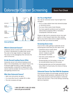


© Copyright 2026