
R E V I E W S
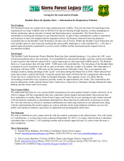
REVIEWS
Management of acute smoke inhalation injury
Michael H Toon, Marc O Maybauer, John E Greenwood,
Dirk M Maybauer and John F Fraser
Pulmonary injury from acute smoke inhalation is common in
burn patients. Smoke inhalation is present in about 22% of
all burn presentations, and in 60% where there are central
facial burns.1 Mortality data for smoke inhalation is disconCrit at
Care
Resusc
1441-2772
1 March
certing:
least
30%ISSN:
of burn
patients
with smoke inhala2010
12
1
53-61
tion injury die, compared with 2% of those without this
©Crit
Care
Resusc
2010
typewww.jficm.anzca.edu.au/aaccm/journal/publiof injury.2 Of all fire-related fatalities, 80%–90% are
cations.htm
attributed
to smoke inhalation.3 Overall, burns are a leading
Reviews
cause of accidental death, and smoke inhalation is now the
major contributor to the morbidity and mortality associated
with serious burns.2 Indeed, inhalation injury is a greater
contributor to overall morbidity and mortality than either
percentage of body surface area affected or age.4,5
Unfortunately, the improvements to other aspects of
burn care have not been mirrored in treatment of smoke
inhalation, and pulmonary complications have become an
increasingly important consideration in burn injuries.2 A
burn to 50% of the total body surface area (TBSA) with
accompanying smoke inhalation injury carries a 10% mortality risk, the same risk as a 73% TBSA burn with no
inhalation injury.6 Inhalation injury has a high incidence of
progression to serious clinical consequences: a 73% incidence of progression to respiratory failure (defined as
hypoxaemia, multiple pulmonary infections or prolonged
ventilatory support), and a 20% incidence of acute respiratory distress syndrome (ARDS).7 Clearly, advances in treatment of pulmonary injury sustained in fire exposure hold
great potential for improving patient outcomes.
Toxic smoke compounds
The components of smoke, including heat, differ with each
event, influencing the spectrum of effects.8 Smoke is a
heterogeneous compound unique to each fire in both
chemical composition and toxic features, depending on the
materials combusted and availability of oxygen. The components of smoke that cause damage are:
• Heat.
• Particulates. These are deposited in the airways according
to their size, with substances less than 1 μm diameter
able to reach the alveolar zone suspended in air. At that
site, they increase airway resistance, cause irritation and
interfere with pulmonary surfactant.8
• Systemic toxins. Toxins such as carbon monoxide and
cyanide adversely effect oxygen transport, availability and
utilisation.
ABSTRACT
Pulmonary injury from smoke inhalation is common in
burn victims, significantly contributing to the morbidity
and mortality of fire-related injuries. The impacts of
improvement in other aspects of burn care have not been
mirrored in treatment of smoke inhalation. Smoke is
heterogeneous and unique to each fire; it comprises
particulates, respiratory irritants and systemic toxins as well
as heat, all contributing to the pathological insult. Thermal
injury below the vocal cords is rare because of effective heat
dissipation in the upper airway. Particulate matter is the
chief contributor to the pathophysiology of smoke
inhalation injury, which has been extensively described.
Of paramount importance is the cascade of inflammatory
mediators following interaction of irritant substances with
lung parenchyma, leading to pulmonary oedema, cast
formation, airway obstruction, loss of hypoxic pulmonary
vasoconstriction and ventilation/perfusion mismatch.
Current treatment is based on supportive care, with airway
management, mechanical ventilation, humidification and
aggressive airway toilet the mainstays. Nebulisation of
β2-agonists, heparin and N-acetylcysteine have a role in
management, as does more specific treatment of carbon
monoxide or cyanide intoxication. Many promising
treatments are currently under investigation. The
therapeutic strategy of decontaminating the lungs early
after smoke exposure to prevent inhalation injury has
received little attention and may be of significant value.
This could potentially utilise amphoteric, hypertonic
chelating agents developed for topical and ocular
chemical exposures.
Crit Care Resusc 2010; 12: 53–61
• Respiratory irritants. Several components of smoke are
respiratory irritants and are thus substantially implicated in the high mortality rates. Water-soluble gases
such as ammonia and hydrogen chloride react with
water contained in mucous membranes and produce
strong alkalis and acids, which elicit intense and
prolonged inflammatory reactions. Lipid-soluble irritants (eg, oxides of nitrogen, phosgene and aldehydes)
exert effects more slowly as they dissolve into the
cellular membrane.8
Critical Care and Resuscitation • Volume 12 Number 1 • March 2010
53
REVIEWS
Heat. Burns to the nasal and oropharyngeal mucosa are
common in fire-exposed victims, but it is rare to encounter
thermal injury below the vocal cords.8 This is because heat
is effectively exchanged in the upper air passages, with the
temperature of inhaled air decreasing precipitously before
the lower airway is penetrated.8 In environments containing air, steam and smoke at sufficient temperatures to
cause thermal injury to the lower airway, the heat also
causes rapid oedema of the glottis, and the resultant
obstruction to the airway is rapidly fatal before the
sequelae of pulmonary burn becomes apparent.8
Systemic toxins. These are products of incomplete
combustion and include carbon monoxide and hydrogen
cyanide. Carbon monoxide intoxication, together with
heat incapacitation and a hypoxic environment, is the
most common immediate cause of death from fire.8
Carbon monoxide is an odourless, colourless gas that
binds with erythrocyte haemoglobin with about 250
times the affinity of oxygen. The resulting carboxyhaemoglobin molecule is inactive in oxygen transport and
shifts the oxygen-dissociation curve to the left, impairing
oxygen delivery at the tissue level. This results in a
marked reduction in oxygen-carrying capacity of the
blood.8 Furthermore, carbon monoxide competes with,
and inhibits, oxygen binding to cytochrome oxidase,
disrupting the aerobic metabolism chain and decreasing
the capacity for cellular respiration. 8 Thermal decomposition of nitrogen-containing polymers produces smoke
containing hydrogen cyanide. When inhaled, cyanide
combines with trivalent iron in the mitochondrial cytochrome a3 complex and thus inhibits electron transport
and cellular respiration.8
Particulates and irritants. The chief contributor to the
pathophysiology of smoke inhalation is particulate matter.
Carbonaceous particles (soot) impregnated with a variety
of toxins reach the alveolar level suspended in air.1 The
chemicals associated with these particles vary depending
on the products combusted but commonly include aldehydes from cellulosic materials such as wood and paper;
nitrogen oxides from fabric combustion; halogen acids
and sulfur dioxide from rubber; ammonia from wool, silk
and polyurethane; and phosgene from polyvinyl chloride.8,9 Water-soluble compounds are readily soluble in
airway mucus and interact freely with tissue at more
proximal levels of the respiratory system.9 Poorly watersoluble compounds (such as phosgene) penetrate the
airway mucosa deeply and may cause severe delayed
damage through late interaction with distal airway tissues,
up to 48 hours after exposure.9 This is an important
consideration when treating patients who present with
apparently mild clinical effects after smoke inhalation.
54
Pathophysiology of acute smoke inhalation injury
The pathophysiology of acute smoke inhalation injury has
been described in detail elsewhere.10-13 Here, I provide a brief
overview as a basis for understanding the options in clinical
management. As previously mentioned, above the oropharynx, thermal injury can produce significant inflammation, occluding the airway, but heat rarely causes damage
below the vocal cords because it is effectively dissipated.8
However, the other components of smoke — particulate
materials, systemic toxins and respiratory irritants8 — trigger
a cascade of events, resulting in pulmonary oedema and
ventilation/perfusion (V/Q) mismatch14,15 (Figure 1).
Of paramount importance is the cascade of inflammatory
mediators activated by the interaction of irritant substances
with the airway mucosa and lung parenchyma. Intrapulmonary leukocyte aggregation following activation of the
classic complement cascade releases even more chemokines
and cytokines, leading to the production of oxygen free
radicals.7
Nitric oxide (NO) synthase is induced by respiratory
epithelial cells and alveolar macrophages and produces NO,
a powerful vasodilator. The formation of NO increases
bronchial blood flow, decreases hypoxic pulmonary vasoconstriction in poorly ventilated areas of lung, and results in
V/Q mismatch. NO also combines with superoxide (O2-)
produced in large quantities by activated neutrophils to
form peroxynitrite (ONOO-).15 This reactive nitrogen species
leads to DNA damage and subsequent activation of poly
(ADP-ribose) polymerase, an important enzyme in DNA
repair. This activation and subsequent action requires a
large amount of chemical energy in the form of ATP and
NAD, the depletion of which causes necrotic cell death of
deprived energy-dependent tissues.15
The combination of these effects contributes to tissue
injury and increased pulmonary vascular permeability, leading to decreased diffusion, oedema and V/Q mismatch.15
Furthermore, neutrophil infiltration and fibrinogen activation by inflammatory mediators causes airway cast formation and widespread plugging. These casts obstruct the
airway, and subsequent efforts to mechanically ventilate the
lung can induce ventilator-induced barotrauma when the
patent lung becomes overstretched. The further tissue
injury and production of chemokines leads to a potent
accumulation of damage.15
Much of the study of smoke inhalation injuries in animal
models has focused on aspects of this pathophysiological
sequence. Attempts to manipulate and alter the chain of
effects experimentally have reinforced these theories, and
also suggested exciting treatment targets. Nevertheless,
few experimental treatments have proven to alter the
course of smoke inhalation injury or progressed to clinical
Critical Care and Resuscitation • Volume 12 Number 1 • March 2010
REVIEWS
Figure 1. Pathophysiology of acute smoke inhalation injury
Thermal injury
Above oropharynx
Tissue oedema
Rapid airway obstruction
Below larynx
Alveolar macrophages
+
Airway epithelia
Interacts with
Airway mucosa
+
Lung parenchyma
Irritant substance
Acts on
Classic complement cascade
Nitric oxide (NO)
Inflammatory mediators
Coagulation cascade
Intrapulmonary leukocyte aggregation
Fibrinogen activation
NAD + ATP
depletion
Increased airway
exudate
Airway casts
Cellular dysfunction
Inducible nitric
oxide synthase (iNOS)
Loss of hypoxic
pulmonary
vasoconstriction
V/Q mismatch
-)
Superoxide (O2
bronchial blood flow
Poly (ADP-ribose)
polymerase (PARP)
activation
DNA damage
trials, and the mainstay of treatment is still supportive
intensive care.
Current treatment of acute smoke inhalation
The first priority at the injury scene is rescue of the victim
from the source of fire to minimise exposure time. This is
usually the responsibility of fire-fighters. To reduce carboxyhaemoglobin levels as soon as possible, high-flow 100%
oxygen should be administered immediately via facemask.
The next step is a brief but careful body check to estimate
the extent of smoke inhalation and assess accompanying
injuries, such as burns and trauma. In addition, it is
important to determine whether the victim has been
exposed to an explosion, which can cause barotrauma to
the lung. If possible, information about comorbidities
should be obtained. Standard cardiopulmonary monitoring
(electrocardiography, pulse oximetry and non-invasive blood
pressure measurement) and intravenous access should be
established.16
Peroxynitrite (ONOO- )
Pulmonary oedema
Airway management
After these basic measures, it must be decided how best to
secure the airway. The risk of rapidly developing airway
oedema has to be taken into account, even if no dyspnoea
is present, but endotracheal intubation (especially at the
injury scene) carries risks, such as oesophageal intubation,
aspiration, barotrauma and laryngeal trauma. We consider
that prophylactic endotracheal intubation is not generally
advised in every patient and, depending on technical skill,
for every physician or paramedic. Nevertheless, it is important to realise that airway management will become more
difficult over time. Heat and smoke inhalation injury combined with extensive face or neck burns mandate intubation.16 In the case of oral burns with no inhalation injury, the
safest approach is to secure the airway early, but patients
with smoke inhalation injury but no facial or neck burns can
be carefully observed and intubated later, if it becomes
necessary.17
If the patient undergoes endotracheal intubation, the
tube should be carefully secured. It should be left uncut, as
Critical Care and Resuscitation • Volume 12 Number 1 • March 2010
55
REVIEWS
facial swelling in the next 24–48 hours can cause the end of
the endotracheal tube to retreat into the oropharynx,
necessitating re-intubation at the worst possible time.
Accidental removal of the endotracheal tube occurs easily
and may be fatal. In cases of vocal cord damage, tracheostomy may be necessary to prevent further damage.11 The
patient’s head should be elevated to minimise facial and
airway oedema. As a matter of course, haemodynamic
stability is a prerequisite. Aerosolised adrenaline or corticosteroids may be beneficial to reduce upper airway oedema,
but there is no conclusive evidence of their efficacy.
In the case of bronchospasm, nebulised administration of
bronchodilators, such as β2-agonists, improves respiratory
mechanics by decreasing airflow resistance and peak airway
pressures. This improves dynamic compliance. In addition,
β2-agonists have anti-inflammatory properties, decreasing
inflammatory mediators such as histamine, leukotrienes
and tumour necrosis factor-α. Finally, β2-agonists improve
airspace fluid clearance and stimulate mucosal repair.18-21
Treatment of specific intoxications
After initial stabilisation of the patient, information about
the fire source, combustion products and estimated duration of exposure10 should be sought. In cases of a specific
intoxication, appropriate therapies should be started. Carbon monoxide and cyanide toxicities have specific treatment strategies aimed at reducing serum levels of these
substances, which impair tissue oxygenation.
Carbon monoxide: In the presence of elevated carboxyhaemoglobin, high-flow 100% oxygen should be delivered via
facemask. Depending on the severity of injury and symptoms, the patient may need to be intubated and ventilated
with an FIo2 of 1.0. Rapid transportation to a facility that can
provide hyperbaric oxygenation may be considered, as oxygen delivered at 3 atmospheres is able to reduce the half-life
of carbon monoxide from 320 to 20 minutes.15 However,
data are lacking on the efficacy of this technique.
Cyanide: Cyanide intoxication can be treated either supportively or with antidotes, the choice of which remains controversial. Amyl nitrate and sodium thiosulfate are used to
oxidise haemoglobin to methaemoglobin, which preferentially binds cyanide. This is then able to dissociate freely and
undergo metabolism by liver enzymes.15 However, as these
substances are methaemoglobin-generators and can also
impair oxygen transport, they should be used only in cases of
proven diagnosis (increased plasma levels of cyanide) and
under continuous monitoring in an intensive care unit.
Methaemoglobin chelates cyanide to form cyanomethaemoglobin, which, as it dissociates, allows free cyanide to be
converted to thiocyanate by a liver mitochondrial enzyme
(rhodanase), using thiosulfate as a substrate. Thiocyanate is
then excreted into the urine.11
56
In contrast to these antidotes, hydroxocobalamin (vitamin
B12a), actively binds cyanide by forming cyanocobalamin,
which is directly excreted via the kidney. In case of intoxication
with 1 mg cyanide, hydroxocobalamin (50 mg/kg) is recommended. Because it averts methaemoglobin production,
hydroxocobalamin can be used even in the preclinical setting.
Accordingly, it represents the active compound of the
“cyanokit” used in prehospital management of smoke inhalation injury in Europe, with a reported decrease in mortality.22
Aggressive restoration of cardiopulmonary function augments the hepatic clearance of cyanide via rhodanase, and
has been reported to be successful in severe cyanide
poisoning (blood levels, 5.6–9 mg/L), as well as after ingestion, or smoke inhalation, even without the use of antidotes. The standard care of cyanide poisoning should
therefore combine aggressive supportive therapy with primary pharmaceutical treatment using hydroxocobalamin.23
Monitoring and investigations
Inhalation injury often develops with a latency of several
hours. Airway management and oxygenation status of the
patient, regardless of intubation status, need to be frequently re-evaluated to allow clinicians to react to the
dynamic development of smoke inhalation injury.11
After stabilisation of cardiopulmonary haemodynamics
and pulmonary gas exchange, the assumed diagnosis of
smoke inhalation injury needs to be verified. However, as
there are currently no uniform criteria, diagnosis is usually a
subjective decision based on the combination of history and
physical examination, which is confirmed by diagnostic
investigations. Symptoms of smoke inhalation injury that
raise the index of clinical suspicion are listed in Table 1.
Bronchoscopic examination of the airway represents the
gold standard to detect a pathognomonic mucosal hyperaemia. Chest radiographs may show signs of diffuse atelectases, pulmonary oedema or bronchopneumonia.23
The initial degree of injury is usually underestimated from
the chest x-ray, as the injury is confined mainly to the
airways.24 Neither a uniform algorithm for assessing inhalation injury nor a reliable indicator of progressive respiratory
failure in patients with smoke inhalation injury has yet been
Table 1. Symptoms of smoke inhalation injury
Facial burns
Burned lips and nasal hairs
Soot in sputum
Changed respiratory mechanics (hoarseness, coughing, stridor)
Dyspnoea
Cyanosis
Neurological deficits (current or anamnestic unconsciousness,
vertigo, nausea, vomiting)
Critical Care and Resuscitation • Volume 12 Number 1 • March 2010
REVIEWS
established. This is largely explained by the extreme heterogeneity of the clinical presentation. In addition, the delay in
the manifestation and development of acute lung injury as
a consequence of systemic inflammatory response syndrome, initiated by accompanying burns or trauma, complicate the evaluation of the isolated effects of smoke
inhalation. A useful algorithm for treatment is outlined in
Figure 2. Frequent blood gas and sputum analyses are
useful to monitor patients with smoke inhalation injury.11
Fluid resuscitation
Appropriate fluid resuscitation for patients with smoke
inhalation is still debated. It has been shown that fluid
requirements are increased in smoke inhalation injury in
patients with burns,25 but this does not inevitably mean
they are increased in isolated smoke inhalation injury. In
fact, over-resuscitation may increase pulmonary microvascular pressures and oedema formation under the highpermeability conditions in early lung injury.13 In a retrospective study of more than 2346 trauma patients, Plurad et al
found an increased incidence of late post-traumatic ARDS
with rising volumes of administered crystalloids and packed
red blood cells.26 Unfortunately, there is currently no evidence in the specific patient population with isolated smoke
inhalation injury. Thus, against the background of no
proven benefit and the potential risk of detrimental effects,
increased fluids should be avoided in patients with isolated
smoke inhalation injury. Instead, fluid resuscitation should
be guided by the urine output and haemodynamic parameters of the individual patient. Dynamic parameters (such as
changes in pulse pressure) rather than static parameters
(such as central venous or pulmonary artery occlusion
pressure) might be helpful.11
Mechanical ventilation
Management of lung injury due to smoke inhalation has, to
date, been largely supportive, with mechanical ventilation
providing the mainstay of care, along with humidification
and aggressive airway toilet.9 During the past two decades,
mechanical ventilation has been investigated extensively.
Low tidal volume ventilation with associated permissive
hypercapnia has been shown to effectively reduce ventila-
Figure 2. Treatment algorithm for smoke inhalation victims
Fire and smoke formation,
patient exposure
Rescue from danger zone
and decontaminate
Careful clinical examination:
- Unburned carbon in nose or pharynx ?
- Redness of mucosa (cherry red) ?
- Changes in awareness or neurological status ?
- Breathing difficulties (stridoror dyspnoea) ?
- Cardiac symptoms ?
- Secondary trauma ?
Suspected smoke inhalation injury
Yes
Basic arrangements:
- Oxygenation
- Airway management
- Monitoring of vital signs
- COHb
- PIV + blood sample
Yes
No
Additional symptoms or injuries:
- COHb> 15% ?
- Unconscious ?
- Neurological or psychomotor symptoms ?
- Cardiopulmonary impact or instability ?
- Pregnancy ?
Carbon monoxide
Cyanide
Mixed intoxication
(non-specific)
Secondary
trauma
No
Hyperbaric
oxygenation
ICU
Sodium thiosulfate
or Cyanokit
ICU
Hyperbaric
oxygenation
ICU
Emergency
department
ICU
Discharge
home
COHb = carboxyhaemoglobin. PIV = peripheral intravenous access.
Critical Care and Resuscitation • Volume 12 Number 1 • March 2010
57
REVIEWS
tor-induced lung injury.27 The Assessment of Low Tidal
Volume and Elevated End Expiratory Pressure to Obviate
Acute Lung Injury (ALVEOLI) trial revealed no effects of
higher positive end-expiratory pressure (PEEP) levels in ARDS
patients.28 In addition, prone positioning has been shown to
have no beneficial effect on mortality, despite a transient
improvement in oxygenation.29
Nebulisation of heparin and N-acetylcysteine in children
with massive burn injury and smoke inhalation injury
resulted in a significant decrease in incidence of re-intubation for progressive pulmonary failure, decreased incidence
of atelectasis, and reduced mortality.30 These data underscore the present treatment strategy for smoke inhalation
injury at the Shriners Burns Hospital in Galveston, USA,
which includes 5000 units of heparin and 3 mL of a 20%
solution of N-acetylcysteine aerosolised every 4 hours for
the first 7 days after the injury.
Experimental treatments
Treatment options remain limited despite interventional
studies targeting most levels of the pathological process,
particularly via the nebulised route.31 More invasive techniques to support patients with severe ARDS include extracorporeal membrane oxygenation and arteriovenous
carbon dioxide removal, but no single technique has made
a significant impact on current management.9
Vigorous airway toilet can be performed to counter
obstruction by casts.15 Nebulised heparin can be used to
prevent formation of fibrin casts, but heparin requires
antithrombin for efficacy, which is deficient after burn
injury.32 Consequently, concomitant administration of nebulised heparin and antithrombin has been studied, with
proven success.32 The utility of this method was demonstrated in 200832 after initial study of nebulised anticoagulants alone.33 The preliminary investigation appeared to
follow from the use of activated protein C in sepsis, and an
extension of this substance’s anticoagulant properties to
efficacy in combined smoke inhalation and sepsis.34 Highdose intravenous heparin was studied initially (with poor
effect35), followed by some improvement with the use of
intravenous tissue plasminogen activator,36 before nebulised
delivery of heparin showed encouraging results of efficacy.
Miller et al in April 2009 confirmed the preliminary results
by demonstrating a reduction in lung-injury scores in
patients with smoke inhalation injury administered nebulised heparin and the mucolytic N-acetylcysteine.7
Many drugs have proven effective in reducing the injury
to the lung parenchyma in animal models, but only a few
are in clinical use.9 These include the anticoagulants already
mentioned, as well as N-acetylcysteine and inhaled β2agonists such as albuterol.9 The Galveston group showed
58
potential for the use of albuterol in an ovine model in 2006
and called for further randomised clinical trials before
making recommendations for clinical use.37,38
Other drugs studied but not used extensively in clinical
practice have aimed to intervene at various levels of the
pathological process. Broadly, these have included antiinflammatory drugs, NO inhibitors and antioxidants. Methylprednisolone and the anti-epileptic drug phenytoin have also
been studied for their anti-inflammatory properties, with no
protective effect and modest protection, respectively.39,40
Because of the significant role of NO in the pathophysiology of smoke inhalation injury, modulating the production
and effects of this substance has held considerable interest
for investigators. Production of NO is catalysed by NO
synthase (NOS), three isoforms of which have been
described — two constitutive and one induced after injury
and inflammation. It was previously thought that inducible
NOS (iNOS) was largely responsible for the injurious effects
of NO, but the inhibition of neural NOS (nNOS) has been
the subject of promising research. Inhibition of both these
isoforms of NOS has been shown to restore hypoxic
pulmonary vasoconstriction in lungs impaired after the
activation of iNOS following tissue injury from smoke
inhalation.31,41,42 These strategies hold great potential but
have not as yet progressed past animal models.
Promising results have also been obtained with antioxidants, again administered by the nebulised route. Gammatocopherol (vitamin E) has been used in aerosolised form in
a sheep model to attenuate damage after smoke inhalation
caused by oxygen free radicals. Pulmonary function, as
measured by gas exchange, was improved in the treatment
group compared with the placebo group.43 Calls for further
investigation into antioxidants as a treatment modality are
warranted considering the positive results reported by
Wang et al a decade earlier in Arizona.44 This group
nebulised 21-aminosteroid in a rabbit model, in the hope
that its antioxidant effects would decrease smoke-induced
oxidative damage. Pulmonary insult as measured by wet/dry
lung weight and histological analysis as an indicator of
pulmonary oedema were decreased, again in the treatment
group. Although the rabbit model is not as widely reported,
these results add further weight to antioxidant administration directly to the lungs via nebulisation as a useful
treatment strategy in smoke inhalation injury.
Other strategies that have been investigated in an ovine
model with no success include the endothelin-I receptor
antagonist tezosentan (following the observation that the
powerful vasoconstrictor endothelin-I was associated with
lung injury in smoke inhalation)45 and blockade of Pselectin, a neutrophil-adhesion promoter.46
One of the more novel approaches with reported efficacy
in attenuating severe respiratory failure after smoke inhala-
Critical Care and Resuscitation • Volume 12 Number 1 • March 2010
REVIEWS
tion was reported in 1994 by LaLonde et al in Boston.47 This
group, again in an ovine model, aerosolised a complex of
deferoxamine (DFO; an iron chelator) and pentastarch, and
found this strategy had considerable efficacy after a standard smoke inhalation injury versus placebo and DFO alone.
The authors concluded that free iron release and oxidant
production in response to the injury were important in the
pathophysiology of acute lung injury. Interestingly, the DFOalone group showed no benefit versus the placebo group,
suggesting that the chelating ability of DFO was less
important than the antioxidant properties of the complex.
DFO is specific for iron, but this study is the only one to
explore the possibility of intervening early in the injury
process to prevent pulmonary catastrophe, rather than
reversing or minimising the injury after it has begun.
When signs of respiratory failure are present or imminent,
mechanical ventilation is used supportively to ensure adequate respiratory function.15 This aims to ensure adequate
ventilation to maintain alveolar patency without causing
alveolar distension. Plugging from fibrin clots and cellular
debris may result in barotrauma, causing further alveolar
damage with mechanical ventilation.15 Several different
modes are used, from conventional mechanical ventilation
using a pressure-controlled rather than volume-controlled
function to high-frequency percussive ventilation utilising
subtidal breaths at high frequency. The latter is associated
with decreased work of breathing, increased oxygenation,
decreased peak pressures and decreased incidence of pneumonia.15 This method has been reported by Reper et al in
Belgium, in respiratory failure following smoke inhalation in
human patients, and was compared with periods of conventional ventilation in the same subjects.48
Two randomised, controlled trials on “airway pressure
release ventilation” (APRV) in mechanically ventilated
patients — not patients with smoke inhalation injury —
revealed beneficial effects on oxygenation and lower endinflation pressures.49,50 However, there was no reduction in
mortality. Further research is necessary to determine
whether APRV represents a beneficial approach for ventilation of patients with smoke inhalation injury.
The volumetric diffusive ventilator (VDR; Percussionaire
Corp, Sandpoint, ID, USA) is a pneumatically powered,
pressure-limited ventilator that stacks oscillatory breaths to
a selected peak airway pressure by means of a sliding
venturi, Phasitron, resulting in low tidal volumes. In contrast
to APRV, the effects of the VDR have been studied in
patients with smoke inhalation injury. A prospective clinical
analysis revealed improved gas exchange and a decrease in
peak pressures.51 In addition, a retrospective study in 330
patients with inhalation injury reported a lower mortality
rate.52 Although these studies compared the VDR with
high-volume ventilatory strategies, data comparing it with
modern low tidal volume ventilation are still lacking. Therefore VDR cannot be recommended at present.
Concomitant pneumonia or septic complications should be
treated; intravenous gentamicin and ceftazidime have been
studied individually to evaluate their impact in smoke inhalation and pneumonia caused by Pseudomonas aeruginosa.53,54
When respiratory failure is severe, extracorporeal membrane oxygenation is an option, whereby the patient’s
circulation is bypassed through an extracorporeal circuit,
facilitating gas exchange through a semi-permeable membrane.15 A further, similarly invasive method of treating
respiratory failure is arteriovenous carbon dioxide removal,
which utilises an extracorporal circuit to remove that gas
and enables lower ventilatory pressures, with subsequent
reduction in barotrauma from mechanical ventilation.55
Future treatment options
The strategies discussed focus on ameliorating or reversing
the pathophysiological consequences of smoke inhalation
after significant damage has occurred, and are summarised in
Table 2. As a large proportion of lower airway injury after
smoke inhalation is a response to toxic chemicals following
Table 2. Treatment strategies for acute smoke
inhalation
Current
Under investigation
• Rescue victim from source
• Activated protein C
• High flow 100% O2
• Anti-inflammatory drugs:
• Body check
Methylprednisolone
• Intravenous access
Phenytoin
• ± Intubation (see Discussion)
• Nitric oxide synthase inhibitors
If upper airway oedema:
• Antioxidants:
γ -Tocopherol
• Nebulised adrenaline
• Nebulised corticosteroids
If bronchospasm:
Nebulised β2-agonists
If elevated carboxyhaemoglobin:
21-Aminosteroid
• Endothelin-I receptor
antagonist (tezosentan)
• P-selectin blockade
• High-flow 100% O2
• Nebulised deferoxaminepentastarch complex
• Hyperbaric oxygen
• Mechanical ventilation:
High-frequency percussive
ventilation
If cyanide Intoxication:
• Amyl nitrate
Airway pressure release
ventilation
• Sodium thiosulfate
• Hydroxocobalamin
(”cyanokit”)
• Mechanical ventilation:
Low tidal volume
• Nebulised heparin
• Nebulised N-acetylcysteine
Volumetric diffusive
ventilator
• Extracorporeal membrane
oxygenation
• Arteriovenous carbon dioxide
removal
• Pulmonary decontamination
with nebulised amphoteric
chelating agents
Critical Care and Resuscitation • Volume 12 Number 1 • March 2010
59
REVIEWS
their interaction with the respiratory mucosa, efforts to reduce
or abolish this process is a logical step. Such a therapeutic
strategy, administered early in the course of the exposure,
would essentially aim to decontaminate the lungs, analogous
to the administration of activated charcoal after a toxic
ingestion, and to the flushing of the cornea after an ocular
burn. As burns victims often present disproportionately well,
with a paucity of clinical signs considering the impending
respiratory compromise, or with considerable comorbid cutaneous burns, choosing treatment targets for early intervention is difficult if a delicate risk–benefit analysis is required.
A pulmonary decontamination compound and vehicle
therefore need to be minimally toxic and able to be administered to the acutely unwell and seriously compromised
patient alike. Ideally, such a treatment strategy would be:
• affordable and portable for use in emergency situations by
first aid personnel;
• able to decontaminate a wide variety of toxic substances
sufficient for empirical use;
• non-toxic or minimally toxic, sufficient for prophylaxis in
well patients and unlikely to further compromise seriously
unwell burns patients; and
• able to be nebulised for rapid and uncomplicated administration to the distal airways.
Products developed to reduce the damage incurred from
topical and ocular burns in industrial settings are currently on
the market and have demonstrably superior efficacy to water
in decontamination of such accidents. These products are
amphoteric, hypertonic chelating agents. Delivered directly to
the airways in the nebulised form, they have the potential to
decontaminate and thus prevent the serious sequelae of
smoke inhalation.56 Their binding effect minimises penetration of chemicals through the endothelial barrier, and their
hypertonic nature means they further reduce penetration of
toxic substances into tissues,56 thereby potentially having a
dual mechanism of action.
Importantly, their effects are non-specific, which is beneficial in the context of empirical treatment of a variety of
combustion products. As the components of smoke are
highly variable and unique to each fire, it is important that
early treatment options have potential efficacy across a broad
spectrum of toxic substances. The dual mechanism of action
is also of benefit for inhalation of substances that may not
necessarily form complexes readily, or neutralise appreciably.
Smoke inhalation injury in the context of burns continues
to be an important health problem. The prognosis for these
patients is long overdue for improvement, considering the
advances in treatment for other aspects of burn care. We are
too often reminded through modern warfare, terrorist incidents such as the 2002 Bali attacks, and other disasters such
as the BP oil refinery explosion in Texas, 2005, and the Black
Saturday bushfires in Victoria, 2009, what a threat fire
60
continues to be. Continuing investigation and development
of treatment strategies holds tremendous potential to
improve outcomes for victims of such tragedies.
Author details
Michael H Toon, MB BS Student1
Marc O Maybauer, Associate Professor,2 and Assistant Professor3
John E Greenwood, Associate Professor and Director4
Dirk M Maybauer, Associate Professor,2 and Assistant Professor3
John F Fraser, Director,5 and Adjunct Professor6
1 School of Medicine, University of Queensland, Brisbane, QLD.
2 Department of Anaesthesiology and Intensive Care, Philipps
University of Marburg, Marburg, Germany.
3 Department of Anesthesiology, Division of Critical Care Medicine,
University of Texas Medical Branch and Shriners Burns Hospital,
Galveston, USA.
4 Burns Unit, Royal Adelaide Hospital, Adelaide, SA.
5 Critical Care Research Group, University of Queensland, Prince
Charles Hospital, Brisbane, QLD.
6 School of Engineering Systems, Faculty of Built Environment and
Engineering, Queensland University of Technology, Brisbane, QLD.
Correspondence: [email protected]
References
1 Fraser JF, Venkatesh B. Recent advances in the management of burns.
Australas Anaesth 2005; 23-32.
2 Smith DL, Cairns BA, Ramadan F, et al. Effect of inhalation injury, burn
size, and age on mortality: a study of 1447 consecutive burn patients.
J Trauma 1994; 37: 655-9.
3 Kimmel EC, Still KR. Acute lung injury, acute respiratory distress
syndrome and inhalation injury: an overview. Drug Chem Toxicol 1999;
22: 91-128.
4 Maybauer MO, Maybauer DM, Herndon DN. Incidence and outcomes
of acute lung injury. N Engl J Med 2006; 354: 416-7.
5 Shirani KZ, Pruitt BA Jr, Mason AD Jr. The influence of inhalation injury
and pneumonia on burn mortality. Ann Surg 1987; 205: 82-7.
6 Endorf FW, Gamelli RL. Inhalation injury, pulmonary perturbations, and
fluid resuscitation. J Burn Care Res 2007; 28: 80-3.
7 Miller AC, Rivero A, Ziad S, et al. Influence of nebulized unfractionated
heparin and N-acetylcysteine in acute lung injury after smoke inhalation injury. J Burn Care Res 2009; 30: 249-56.
8 Prien T, Traber DL. Toxic smoke compounds and inhalation injury — a
review. Burns Incl Therm Inj 1988; 14: 451-60.
9 Nugen N, Herndon DN. Diagnosis and treatment of inhalation injury.
In: Herndon DN, ed. Total burn care. 2nd ed. London: WB Saunders,
2001: 262-70.
10 Maybauer MO, Rehberg S, Traber DL, et al. [Pathophysiology of acute
lung injury in severe burn and smoke inhalation injury] [German].
Anaesthesist 2009; 58: 805-812.
11 Maybauer DM, Traber DL, Radermacher P, et al. [Treatment strategies
for acute smoke inhalation injury] [German]. Anaesthesist 2006; 55:
980-2, 984-8.
12 Traber DL, Maybauer MO, Maybauer DM, et al. Inhalational and acute
lung injury. Shock 2005; 24 Suppl 1: 82-7.
13 Traber DL, Herndon DN, Enkhabaatar P, et al. The pathophysiology of
inhalation injury. In: Herndon DN, ed. Total burn care. 3rd ed. London:
Saunders, 2007: 248-61.
14 Maybauer MO, Rehberg S, Traber DL, et al. [Pathophysiology of acute
lung injury in severe burn and smoke inhalation injury] [German].
Anaesthesist 2009; 58: 805-12.
Critical Care and Resuscitation • Volume 12 Number 1 • March 2010
REVIEWS
15 Murakami K, Traber DL. Pathophysiological basis of smoke inhalation
injury. News Physiol Sci 2003; 18: 125-9.
16 Kafka G, Maybauer DM, Traber DL, Maybauer MO. [Treatment of
inhalation injury in preclinical emergency medicine] [German]. Notfall
Rettungsmed 2007; 10: 529-40.
17 Demling R. Smoke inhalation lung injury: an update. Eplasty 2008; 8:
e27.
18 Morina P, Herrera M, Venegas J, et al. Effects of nebulized salbutamol
on respiratory mechanics in adult respiratory distress syndrome.
Intensive Care Med 1997; 23: 58-64.
19 Zhang H, Kim YK, Govindarajan A, et al. Effect of adrenoceptors on
endotoxininduced cytokines and lipid peroxidation in lung explants.
Am J Respir Crit Care Med 1999; 160: 1703-10.
20 van der Poll T, Coyle SM, Barbosa K, et al. Epinephrine inhibits tumor
necrosis factor-atpha and potentiates interleukin 10 production
during human endotoxemia. J Clin Invest 1996; 97: 713-9.
21 Mcauley DF, Frank JA, Fang X, Matthay MA. Clinically relevant
concentrations of beta2-adrenergic agonists stimulate maximal cyclic
adenosine monophosphate-dependant airspace fluid clearance and
decrease pulmonary edema in experimental acid-induced lung injury.
Crit Care Med 2004; 32: 1470-6.
22 Fortin JL, Giocanti JP, Ruttimann M, Kowalski JJ. Prehospital administration of hydroxocobalamin for smoke inhalation associated cyanide
poisoning: 8 years of experience in the Paris Fire Brigade. Clin Toxicol
(Phila) 2006; 44 Suppl 1: 37-44.
23 Woodson LC. Diagnosis and grading of inhalation injury. J Burn Care
Res 2009; 30: 143-5.
24 Lee MJ, O’Connell DJ. The plain chest radiograph after acute smoke
inhalation. Clin Radiol 1988; 39: 33-7.
25 Cancio LC, Chaves S, Alvarado-Ortega M, et al. Predicting increased
fluid requirements during the resuscitation of thermally injured
patients. J Trauma 2004; 56: 404-13; discussion 413-14.
26 Plurad D, Martin M, Green D, et al. The decreasing incidence of late
posttraumatic acute respiratory distress syndrome: the potential role
of lung protective ventilation and conservative transfusion practice. J
Trauma 2007; 63: 1-7; discussion 8.
27 The Acute Respiratory Distress Syndrome Network. Ventilation with
lower tidal volumes for acute lung injury and the acute respiratory
distress syndrome. N Engl J Med 2000; 342: 1301-8.
28 Brower RG, Lanken PN, MacIntyre N, et al. Higher versus lower
positive end-expiratory pressures in patients with the acute respiratory distress syndrome. N Engl J Med 2004; 351: 327-36.
29 Gattinoni L, Tognoni G, Pesenti A, et al. Effect of prone positioning
on the survival of patients with acute respiratory failure. N Engl J Med
2001; 345: 568-73.
30 Desai MH, Mlcak R, Richardson J, et al. Reduction in mortality in
pediatric patients with inhalation injury with aerolized heparin/N-acetylcystine [correction of acetylcystine] therapy. J Burn Care Rehabil 1998;
19: 210-12. Erratum in: J Burn Care Rehabil 1999; 20 (1 Pt 1): 49.
31 Enkhbaatar P, Connelly R, Wang J, et al. Inhibition of neuronal nitric
oxide synthase in ovine model of acute lung injury. Crit Care Med
2009; 37: 208-14.
32 Enkhbaatar P, Esechie A, Wang J, et al. Combined anticoagulants
ameliorate acute lung injury in sheep after burn and smoke inhalation. Clin Sci (Lond) 2008; 114: 321-9.
33 Enkhbaatar P, Cox RA, Traber LD, et al. Aerosolized anticoagulants
ameliorate acute lung injury in sheep after exposure to burn and
smoke inhalation. Crit Care Med 2007; 35: 2805-10.
34 Maybauer MO, Maybauer DM, Fraser JF, et al. Recombinant human
activated protein C improves pulmonary function in ovine acute lung
injury resulting from smoke inhalation and sepsis. Crit Care Med
2006; 34: 2432-8.
35 Murakami K, Enkhabaatar P, Shimoda K, et al. High-dose heparin fails
to improve acute lung injury following smoke inhalation in sheep.
Clin Sci (Lond) 2003; 104: 349-56.
36 Enkhbaatar P, Murakami K, Cox R, et al. Aerosolized tissue plasminogen inhibitor improves pulmonary function in sheep with burn and
smoke inhalation. Shock 2004; 22: 70-5.
37 Palmieri TL, Enkhbaatar P, Bayliss R, et al. Continuous nebulized
albuterol attenuates acute lung injury in an ovine model of combined
burn and smoke inhalation. Crit Care Med 2006; 34: 1719-24.
38 Weiner-Kronish JP, Matthay MA. Beta-2-agonist treatment as a
potential therapy for acute inhalational lung injury. Crit Care Med
2006; 34: 1841-2.
39 Nieman GF, Clark WR, Hakim T Methylprednisolone does not protect
the lung from inhalation injury. Burns 1991; 17: 384-90.
40 Nishida K, Matsumoto N, Kikuchi Y, et al. Effect of phenytoin on
smoke inhalation injury in sheep. Shock 1995; 4: 211-15.
41 Westphal M, Enkhbaatar P, Schmalstieg FC, et al. Neuronal nitric
oxide synthase inhibition attenuates cardiopulmonary dysfunctions
after combined burn and smoke inhalation injury in sheep. Crit Care
Med 2008; 36: 1196-204.
42 Enkhbaatar P, Murakami K, Shimoda K, et al. The inducible nitric
oxide synthase inhibitor BBS-2 prevents acute lung injury in sheep
after burn and smoke inhalation injury. Am J Respir Crit Care Med
2003; 167: 1021-6.
43 Hamahata A, Enkhbaatar P, Kraft ER, et al. gamma-Tocopherol
nebulization by a lipid aerosolization device improves pulmonary
function in sheep with burn and smoke inhalation injury. Free Radic
Biol Med 2008; 45: 425-33.
44 Wang S, Lantz RC, Robledo RF, et al. Early alterations of lung injury
following acute smoke exposure and 21-aminosteroid treatment.
Toxicol Pathol 1999; 27: 334-41.
45 Cox RA, Enkhabaatar P, Burke AS, et al. Effects of a dual endothelin1 receptor antagonist on airway obstruction and acute lung injury in
sheep following smoke inhalation and burn injury. Clin Sci (Lond)
2005; 108: 265-72.
46 Chandra A, Katahira J, Schmalstieg FC, et al. P-selectin blockade fails to
improve acute lung injury in sheep. Clin Sci (Lond) 2003; 104: 313-21.
47 LaLonde C, Ikegami K, Demling R. Aerosolized deferoxamine prevents lung and systemic injury caused by smoke inhalation. J Appl
Physiol 1994; 77: 2057-64.
48 Reper P, Van Bos R, Van Loey K, et al. High frequency percussive
ventilation in burn patients: hemodynamics and gas exchange. Burns
2003; 29: 603-8.
49 Putensen C, Zech S, H W, et al. Long-term effects of spontaneous
breathing during ventilatory support in patients with acute lung
injury. Am J Respir Crit Care Med 2001; 164: 43-9.
50 Varpula T, Jousela I, Niemi R, et al. Combined effects of prone
positioning and airway pressure release ventilation on gas exchange
in patients with acute lung injury. Acta Anaesthesiol Scand 2003; 47:
516-24.
51 Carman B, Cahill T, Warden G, McCall J. A prospective, randomized
comparison of the volume diffusive respirator vs conventional ventilation for ventilation of burned children. J Burn Care Rehabil 2002; 23:
444-8.
52 Rue LWr, Cioffi WG, Mason AD, et al. Improved survival of burned
patients with inhalation injury. Arch Surg 1993; 128: 772-8; discussion 778-80.
53 Maybauer MO, Maybauer DM, Fraser JF, et al. Ceftazidime improves
hemodynamics and oxygenation in ovine smoke inhalation injury and
septic shock. Intensive Care Med 2007; 33: 1219-27.
54 Maybauer MO, Maybauer DM, Traber LD, et al. Gentamicin improves
hemodynamics in ovine septic shock after smoke inhalation injury.
Shock 2005; 24: 226-31.
55 Schmalstieg FC, Keeney SE, Rudloff HE, et al. Arteriovenous CO2
removal improves survival compared with high frequency percussive
and low tidal volume ventilation in a smoke/burn sheep acute
respiratory distress syndrome model. Ann Surg 2007; 246: 512-21.
56 Hall AH, Blomet J, Mathieu L. Diphoterine for emergent eye/skin
chemical splash decontamination: a review. Vet Hum Toxicol 2002;
❏
44: 228-31.
Critical Care and Resuscitation • Volume 12 Number 1 • March 2010
61


© Copyright 2026