
SUBCHORIONIC HEMATOMA OR SUBCHORIONIC CLOT
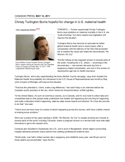
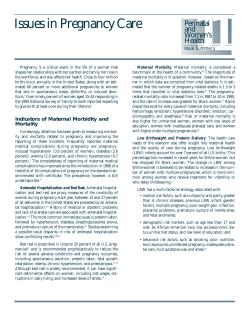
SUBCHORIONIC HEMATOMA OR SUBCHORIONIC CLOT
Val Catanzarite, MD, PhD
San Diego Perinatal Center
8010 Frost Street, Suite 300
San Diego, CA 92123
© 2008
What is a subchorionic hematoma or subchorionic clot? The “bag of waters” within the
uterus is composed of two layers, called the chorion and the amnion. The inner layer, closer
to the baby, is the amnion. The outer layer, which is normally against the uterine wall, is the
chorion. The term “subchorionic clot” or “subchorionic hematoma” describes a blood clot
between the bag of waters and the uterus.
How does a subchorionic hematoma look on ultrasound? We see subchorionic
hematomas or suspect subchorionic clots in perhaps 1% of pregnancies in the between 13
and 22 weeks. Most of these occur in women who have had vaginal bleeding. These must be
distinguished from regions of nonfusion of the membranes to the wall of the uterus, which
are very common prior to 16 weeks gestation. Findings which suggest a bleed or
hematoma rather than membrane separation include irregular texture to the material seen
beneath the membranes, a speckled rather than uniform appearance to the amniotic fluid.
The image at left shows a crescent shaped subchorionic clot, indicated by the arrows. The
image at right shows a larger, rounded subchorionic clot. Both women had experienced
bleeding episodes during the prior week, and had passed blood clots. On rare occasions, we
will be able to see the source of the bleeding beneath the membranes. Usually, we cannot.
This image is of a region of nonfusion of the membranes, also
called chorioamniotic separation. The space between the
membranes and uterine wall contains only fluid, and on scan
the membranes can be seen to move. Images like this are not
suggestive of blood beneath the membranes.
What causes subchorionic hematomas or subchorionic clots?
Usually, we cannot identify any cause for a subchorionic clot. On rare occasions, there will
be an underlying cause, such as a maternal coagulation disturbance, history of trauma,
severe maternal hypertension or early-onset preeclampsia, or maternal abuse of vasoactive
drugs such as cocaine or amphetamines. There is also a case report of a large subchorionic
hematoma occurring after thrombolytic therapy during pregnancy.
What problems can subchorionic hematomas or subchorinic clots cause? Subchorionic
bleeds do increase the chance pregnancy complications, particularly miscarriage and
preterm delivery. Sharma and colleagues described 129 pregnancies with a suspected
subchorinic clot; 5.4% were lost before 24 weeks' gestation and 18.6% of those
progressing beyond 24 weeks went on to preterm delivery. The preterm delivery rate
among those who experienced bleeding was 26.6%. In both Pearlstone's and Sharma's
reviews, there was an attempt made to relate the size or appearance of the subchorionic
bleed to specific pregnancy outcomes. The results do not provide us with sufficient
information to make predictions for any individual case.
Are there medications that can make a clot heal up? Many subchorionic hematomas will
slowly dissolve without treatment, just as a bruise under the skin dissolves. When the
occurs, Mother may experience dark red or brown vaginal discharge. Unfortunately, there
are not any medications which can aid in the healing of the clot, and while we can treat
some of the problems that can occur after a subchorionic clot or hematoma develops, such
as preterm contractions, we cannot predict whether these treatments will, or will not,
result in a successful pregnancy outcome.
What might happen if the baby is born early? Women who have a large subchorionic
clotor subchorionic hematoma diagnosed prior to 22 weeks' gestation must consider the
potential consequences of a decision to go forward with the pregnancy. They must accept
the possibility that they will go on to a very premature birth, and that a surviving infant
may not be neurologically or physically normal. For example, at 24 weeks' gestation, the
overall survival rate in our Newborn ICU is over 50%, but there is a high rate of long-term
neurologic or physical disabilities. Additionally, there is a possibility of fetal growth
problems prompting early delivery, again with potential for adverse neurologic and/or
physical outcomes.
What are the medical recommendations after a subchorionic clot is seen. When a
subchorionic bleed is suspected prior to 20 weeks' gestation, we recommend reduced
activity, recommend against any travel, and instruct mom to go to hospital should vaginal
bleeding, cramps or contractions occur. As the pregnancy progresses, recommendations
may include:
RhoGam, if Mother is Rh negative
Maternal education regarding signs and symptoms of preterm labor.
Sonograms to assess fetal growth, usually about once a month, but sometimes
more often, based on the appearance and size of the suspected bleed, and other
maternal factors.
Preterm labor treatment if contractions occur after 20-22 weeks.
Hospitalization, especially if bleeding occurs after 24 weeks.
Where can I learn more about subchorionic hematomas?
The two best articles which we have found in the literature regarding subchorionic
hematomas or subchorionic bleeds are
Pearlstone, M., and Baxi, L.: Subchorionic Hematoma A Review. Obstetrical & Gynecologic
Survey 1993;48:65-68 and Sharma, G., Kalish, R., and
Chasen, S.: Prognostic Factors Associated with Antenatal Subchorionic Echolucencies.
American Journal of Obstetrics & Gynecology 2003; 1989:994-6.
Maternal Fetal Medicine is a constantly changing field. The information given here may not
be current. Please check with one of the Maternal Fetal Medicine Specialists for up to date
information. Questions and comments are welcome. Please address these to:
Val Catanzarite, MD, PhD
San Diego Perinatal Center
8010 Frost Street, Suite 300
San Diego, CA 92123
©2008 by Val Catanzarite, MD, PhD. All rights reserved. This document may not be
reproduced or stored by any means without the expressed written consent of the author.
Last update: 5/30/2008.


© Copyright 2026