
Downloaded from by guest on September 9, 2014
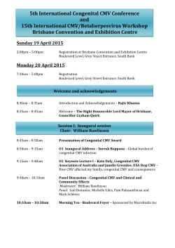
cm
1996;22 (June)
Brief Reports
1J J7
7. Goldschneider I, Gotschlich EC, Artenstein MS. Human immunity to the meningococcus. I. The role of humoral annbody. J Exp Moo 1969; 129:1307-26.
8. Keller AJ, Urbaniak SJ. Intensive plasma exchange on the cell separator:
effects on serum immunoglobulin and complement components. Br J
Haematoll978; 38:531-40.
9. Rodnitzky RL. Complications of plasma exchange in neurological patients.
Arch Neurol 1982;39:352-4.
Severe Acute Cytomegalovirus Sialadenitis in an
Immunocompetent Adult: Case Report
level of antinuclear antibodies was slightly elevated. The serum
angiotensin converting enzyme level was normal, and an anteroposterior radiograph of the chest did not show any signs of sarcoidosis or other diseases.
An ultrasonogram of the salivary glands confirmed diffuse enlargement ofboth the parotid and submandibular glands. Echogenicity was decreased, and there was no sign of sialolithiasis, neoplasms, cystic lesions, or abscesses. Examination of the abdomen
showed only a slightly increased echographic texture of the liver.
Examination of biopsy specimens from the left submandibular
gland and the minor salivary glands in the lower lip demonstrated
hyperplastic epithelium with predominantly lymphocytic infiltrate
CD8+, UCHLl +, and focal L26+ and some neutrophilic granulo-
In the majority of immunocompetent adults, cytomegalovirus
(CMV) causes a latent, asymptomatic infection. Occasionally, a
mild, febrile, self-limiting mononucleosis-like illness is observed.
However, CMV is an important human pathogen in utero and in
immunocompromised hosts; in such settings, CMV infection may
result in severe diseases including pneumonia, hepatitis, chorioretinitis, hematologic abnormalities, and involvement of the gastrointestinal tract and the CNS [I]. CMV has a well-established tropism
for the salivary glands, but even among patients with AIDS, CMV
sialadenitis is rare and usually presents as painful salivary gland
swelling [2]. We describe an immunocompetent adult with severe
acute sialadenitis of all head salivary glands. The patient recovered
after receiving symptomatic therapy for I month.
A 57-year-old man presented with a 4-day history of sore throat,
fever, chills, fatigue, arthralgia, and painful preauricular and submandibular swelling. Despite administration of antibiotics and
anti-inflammatory agents, the infection progressed, and he complained of increasing anorexia, exertional dyspnea, and aphagia
because of severe xerostomia. Physical examination on admission
revealed painful, pasty, and diffuse enlargement of both parotid
glands and submandibular glands (figure 1). The oral cavity was
extremely dry and erythematous. No saliva was expelled from the
salivary duct orifices when the salivary glands were massaged.
The tonsils were mildly hyperplastic, without purulent exudate.
The patient was sweaty and in mild respiratory distress, and he
had cervical lymphadenopathy. Findings on general physical examination were unremarkable. His temperature, pulse, and blood
pressure were normal.
The patient's medical history was remarkable only for a gastric
ulcer that was treated with a Billroth II procedure 20 years previously. During the previous 2 years, he had not left Germany.
Laboratory tests showed elevated levels ofaspartate aminotransferase (81 U/L) and alanine aminotransferase (49 U/L). The amylase
level was 177 U/L, and the WBC count was 22.6 X 10⁹/L with
81 % neutrophils and 8% lymphocytes. The erythrocyte sedimentation rate was elevated at 110 mm/h. Levels of extractable nuclear
antigens, cytoplasmic anti-neutrophil cytoplasmic antibodies, perinuclear anti-neutrophil cytoplasmic antibodies, double-stranded
DNA antibodies, and rheumatoid factor were normal. Only the
Reprints or correspondence: Dr. O. Guntinas-Lichius, Klinik fur Hals-, Nasen-, und Ohrenheilkunde der Universitiit zu Koln, Lindenthal, 0-50924 Cologne, Germany.
Clinical Infectious Diseases 1996;22:1117-8
© 1996 by The University of Chicago. All rights reserved.
1058-4838/9612206-0040$02.00
Figure l. Swelling of the submandibular and parotid glands of a
patient with CMV sialadenitis; the patient was noted to be pale and
sweaty on admission.
Downloaded from http://cid.oxfordjournals.org/ by guest on September 9, 2014
4. Rose HD, Lenz IE, Sheth NK. Meningococcal pneumonia: a source of
nosocomial infection. Arch Intern Med 1981; 141 :575-7.
5. Broome CV. The carrier state, Neisseria meningitidis. J Antimicrob
Chemother 1986; 18(supplA):25-34.
6. Goldschneider I, Gotschlich EC, Artenstein MS. Human immunity to the
meningococcus. II. The development of natural immunity J Exp Med
1%9; 129:1327-48.
1118
Brief Reports
1996; 22 (June)
Elevation of liver enzyme levels and leukocytosis have occurred
in adults with CMV mononucleosis [4]. The clinical course, laboratory findings, and spontaneous recovery of our patient resembled
the course ofCMV mononucleosis [1], even though he had symptomatic .CMV sialadenitis. Even among immunosuppressed hosts,
clinical manifestations of CMV infection of the salivary glands
are rare [1, 4, 5]; however, if clinically manifest infection occurs,
the parotid gland is affected exclusively [1]. In the present case,
treatment with ganciclovir or foscamet was not indicated [6], since
the patient was immunocompetent.
The biopsy specimens were not obtained at the climax of the
infection but at an early stage of recovery. Thus, only a few epithelial cells were positive for CMV when they were studied by IRC
techniques and ISH. Leukocytes had destroyed CMV-infected
cells, which were cleared through the ducts (detritus). Noninfected
epithelium was positive for PCNA; since PCNA may be positive
as well during DNA repair [7] or during proliferation ofepithelium
[8], staining for the presence of p53 antigen was performed to
detect significant prereparative cell-cycle arrest. Positive IRC and
ISH reactions in some epithelial cells showed remnants of fading
CMV infection. As expected, the patient fully recovered, without
major scarification, after 30 days with symptomatic treatment.
Orlando Guntinas-Lichius, Mathias Wagner,
Gerhard R. F. Krueger, Michael Streppel,
Martin Voessing, and Eberhard Stennert
Department of Otorhinolaryngology and the Immunopathology Section,
Institute of Pathology, University of Cologne, Cologne, Germany;
and the Department ofPathology and Laboratory Medicine,
University of Texas Medical School, Houston, Texas, USA
References
I. Alford CA, Britt WJ. Cytomegalovirus. In: Roizman B, Whitley RJ, Lopez
C, eds. The human herpesviruses. New York: Raven, 1993:227-55.
2. Wax TD, Layfield LJ, Zaleski S, et al. Cytomegalovirus sialadenitis in
patients with acquired immunodeficiency syndrome. Diagn Cytopathol
1994; 10:169-74.
3. Krueger GRF, Roewert J. Klinische Immunpathologie der Zytomegalievirus (CMV)-Infektion. AIDS Forschung 1988;5:243-57.
4. Oill PA, Fiala M, Schofferman J, Byfield PE, Guze LB. Cytomegalovirus
mononucleosis in a healthy adult. Am J Med 1977;62:413-7.
5. Schiodt M. mY-associated salivary gland disease: review. Oral Surg Oral
Med Oral Pathol1992;73:164-7.
6. Meyers JD. Management of cytomegalovirus infection. Am J Med 1988;
85(suppI2A):102-6.
7. Shivji KK, Kenny MK, Wood RD. Proliferating cell nuclear antigen
(PCNA) is required for DNA excision repair. Cell 1992; 69:367-74.
8. Mathews MB, Bernstein RM, Franza JR, Garrels n. Identity ofthe proliferating cell nuclear antigen and cyclin. Nature 1984;309:374-6.
Downloaded from http://cid.oxfordjournals.org/ by guest on September 9, 2014
cytes. The terminal ducts were full of purulent detritus. Further
immunohistochemical (IHC) investigations (by the alkaline phosphatase anti-alkaline phosphatase technique) and in situ
hybridization (ISH) revealed CMV in epithelial cells, without typical owl's eye cells; Epstein-Barr virus (EBV) and human herpesvirus 6 were not detected. The epithelium was highly positive for
proliferating cell nuclear antigen (PCNA) and negative for p53
antigen.
Virological examinations confirmed an acute CMV infection:
the titer of antibodies to CMV detected by CF was 1: > 160, and
an ELISA for IgM and IgG antibodies to CMV was positive.
These values were confirmed by retesting several blood samples.
Examinations for IgM antibodies to mumps virus, herpes simplex
virus, coxsackie B virus, varicella-zoster virus, and EBV were
negative, as was an ELISA for HIV 1, RIV 2, and HIV 1 p24
antigen. CMV chorioretinitis and CMV infection of the gastrointestinal tract and CNS were ruled out. Throughout his hospital
course, the patient had a normal CD4 cell count and a normal
CD8 cell count. Immunoelectrophoresis revealed only a transient
increase in IgM, with a level of 4.61 gIL during the first 3 weeks.
Levels of the other Ig subclasses and the complement factors C3
and C4 were normal.
After symptomatic treatment with amoxicillin/sulbactam and administration of iv fluids, parenteral nutrition, diclofenac suppository, and oral tramadol drops, the patient's xerostomia decreased
by day 14 after admission, and he started to take fluids orally.
Initial treatment with a 500-mg bolus ofmethylprednisolone failed,
and the drug was withdrawn after CMV infection was diagnosed.
Day by day, he continued to convalesce; an ELISA for IgM antibody to CMV was only weakly positive from day 14 onward, and
CF titers of antibody to CMV dropped to 1:40 after 14 days and
to 1:20 after 1 month. Liver enzyme levels and the WBC count
decreased daily and were nearly normal at demission.
After 18 days the patient started to eat, his salivary glands were
less painful, and massage of the glands on day 28 yielded scanty
saliva. After 30 days the patient left the hospital and was followed
up monthly; follow-up included blood tests. After 2 months, all
hematologic parameters had become normal. After 12 months,
serologic tests for antibodies to RIV remained negative, and follow-up ceased.
The diagnosis of fulminant primary CMV sialadenitis in this
immunocompentent adult was based on an elevated CF titer of
antibodies to CMV' a positive IgM titer, and detection of CMV
in a salivary gland. Patients whose sera are positive for heterophil
agglutinin or IgM antibody to EBV can have false-positive titers
ofIgM antibody to CMV [1, 3]; however, our patient's serum was
negative for both heterophil agglutinin and IgM antibody to EBV.
cm


© Copyright 2026