
The Diagnosis and Management of Soft Tissue Shoulder Injuries and Related Disorders
The Diagnosis and Management of Soft Tissue Shoulder Injuries and Related Disorders
Printed July 2004
•
(Print) ISBN 0478–27916–7
•
ACC1617
The Diagnosis and Management
of Soft Tissue Shoulder Injuries
and Related Disorders
B e s t p ra ct i ce e v i de n ce - b as e d g u i de l i n e
Statement of intent
Evidence-based best practice guidelines are produced to help health practitioners
and consumers make decisions about health care in specific clinical
circumstances. Research has shown that if properly developed, communicated
and implemented, guidelines can improve care. The advice on soft tissue injuries
of the shoulder given in this guideline is based on epidemiological and other
research evidence, supplemented where necessary by the consensus opinion of
the expert development team based on their own experience.
While guidelines represent a statement of best practice based on the latest
available evidence at the time of publishing, they are not intended to replace
the health practitioner’s judgment in each individual case.
The guideline is endorsed by:
NZ Association of Musculoskeletal Medicine
New Zealand Orthopaedic Association
New Zealand Society of Physiotherapists Inc.
The Royal Australian and New Zealand College of Radiologists – New Zealand
Royal New Zealand College of General Practitioners
Sports Medicine New Zealand Inc
The guideline is supported by:
Arthritis New Zealand
Contents
Purpose ...........................................................................................iii
About the guideline ......................................................................... v
Key Messages .................................................................................. xi
Algorithm ....................................................................................... xii
Summary table: clinical assessment of the shoulder .................. xv
Chapters
Background .......................................................................................................................1
Initial diagnosis and management .....................................................................................3
Rotator cuff disorders ......................................................................................................11
Frozen shoulder ..............................................................................................................19
Glenohumeral instabilities ..............................................................................................25
Acromioclavicular joint disorders ...................................................................................33
Sternoclavicular joint disorders.......................................................................................37
Cultural considerations ...................................................................................................39
Implementation ...............................................................................................................41
Further research..............................................................................................................43
Appendices
Appendix A: Evidence and Recommendation Grading System .......................................47
Appendix B: Guideline Methodology ..............................................................................51
Abbreviations ................................................................................. 55
Glossary ........................................................................................ 57
References ...................................................................................... 59
i
ii
Purpose
The purpose of the guideline is to provide an evidence-based summary of the diagnosis and
management options available for soft tissue shoulder injuries and related disorders to assist
health practitioners and consumers to make informed decisions to improve health outcomes.
iii
iv
About the guideline
Introduction
The Accident Compensation Corporation (ACC) commissioned and funded the New Zealand
Guidelines Group (NZGG) to develop an explicit evidence-based guideline in response to
evidence of variation in practice in the diagnosis and management of soft tissue shoulder
injuries.
The NZGG is a not-for-profit organisation established to promote effective health and
disability services. NZGG facilitates the development and implementation of guidelines across
all areas of health care in New Zealand. Guidelines make a contribution to this aim by sharing
the latest international studies and interpreting these in a practical way for adoption in the
New Zealand setting.
The development of the guideline was led and managed by the Effective Practice, Informatics
and Quality Improvement (EPIQ) Group under the auspices of the NZGG. The EPIQ group is
based in the School of Population Health in the Faculty of Medical and Health Sciences at the
University of Auckland. The centre is a collaboration of academics, clinicians and other health
sector professionals, who undertake training, research and development in evidence-based
practice, health informatics and quality improvement for the health and disability sector.
For more information on the NZGG visit www.nzgg.org.nz
Guideline development process
In February of 2003, NZGG convened a multidisciplinary team of stakeholder groups and
consumers to develop the guideline chaired by Associate Professor Bruce Arroll, (Dept of
General Practice and Primary Health Care, University of Auckland) with Gillian Robb (EPIQ,
University of Auckland) as full time project manager. Team members were nominated by
stakeholder groups and invited to take part. The team held two major meetings during the
year and several shorter meetings to discuss aspects of guideline development.
At the first meeting the Guideline Development Team defined the clinical questions and
scope of the guideline. Based on these questions, a systematic search of the literature was
undertaken to identify relevant studies. Inclusion and exclusion criteria were applied to select
the studies to be used in the guideline. For this guideline, only meta-analyses, systematic
reviews and randomised controlled trials were considered for treatment interventions.
Evidence grading system
The grading system used for this guideline involved three stages (refer to Appendix A for
further details).
1.
Individual studies were critically appraised and assessed for methodological quality
using the Generic Appraisal Tool for Epidemiology (GATE) appraisal tools (developed
by Professor Rod Jackson, University of Auckland, and available at: www.epiq.co.nz)
and assigned a quality rating.
v
2.
Each study was then assigned an evidence level using the Scottish Intercollegiate
Guidelines Network (SIGN) grading system, which incorporates the quality assessment
with the study type. Level of evidence [4] includes both published expert opinion and
the consensus of the Guideline Development Team. The SIGN grading system does not
include diagnostic tests, so we have included a separate system for identifying the level
of evidence for diagnostic tests. Throughout the guideline, the level of evidence has
been included alongside the references, formatted as: reference (level of evidence).
3.
The third step involved the development of evidence tables and the consideration of
the whole body of evidence ie, all the studies relevant to the issue, and this was used
to decide on a recommendation and an appropriate Recommendation Grade. The
Guideline Development Team agreed on the recommendations using the ‘Considered
Judgment’ Form. The NZGG Considered Judgment Form and Evidence Tables are
available at NZGG’s website (www.nzgg.org.nz - click on ‘Guidelines’, then title of the
Guideline, then ‘Supporting Materials’).
Grades of recommendations
The grades A to I are a measure of the strength of evidence underlying the recommendations
and should not be construed as an indication of the relative importance of the
recommendations.
In this guideline, Grade C refers to recommendations that were developed from published
expert opinion (eg, consensus documents). Expert opinion has only been cited where there
was no higher level of evidence.
The good practice points are the recommendations developed by the Guideline Development
Team in the absence of higher levels of evidence.
Consultation
A draft guideline was widely circulated to consumer groups, primary health care
organisations, professional colleges and organisations, expert reviewers, and other clinicians
for peer review, and this was modified where possible, as a result of their feedback. The expert
reviewers included:
vi
•
Steve Bentley, Specialist, Marinoto Clinic, Dunedin
•
Adam Dalgleish, Consultant, Middlemore Hospital, Counties Manukau
•
William Taylor, Senior Lecturer, University of Otago
•
Lisa Hansen, Private Practice, Hamilton
•
Pim Allen, Chief Medical Advisor, Waikato District Health Board
•
Chris Milne, Sports Physician, Sports Medicine NZ; NZ Medical Director Northern
Region, NZ Academy of Sport
•
Mary Magarey, Senior Lecturer, School of Health Sciences, University of South Australia
•
Mark Jones, Senior Lecturer, School of Health Sciences, University of South Australia
•
Memo Musa, Chief Executive Officer, Whanganui District Health Board
•
Steven Lillis, Senior Lecturer, Waikato Clinical School
•
Anthony Griffen, Clinical Leader Orthopaedic Department, Wellington Hospital
•
Richard Newsham-West, Vice President, Sports Medicine NZ
•
Kevin Morris, Corporate Medical Advisor, ACC
•
Cindy Farquhar, Postgraduate Professor of Obstetrics and Gynaecology, University of
Auckland National Women’s Hospital
•
Gary Collinson, General Practice, Auckland
•
John Grace, President, NZ Chiropractors Association
•
Brett Krause, Orthopaedic Surgeon, Hutt Valley District Health Board
Guideline development team
Chair: Bruce Arroll
MBChB, PhD, FRNZCGP, FAFPHM
Associate Professor General Practice, School of Population Health, University of Auckland
Project Manager: Gillian Robb
MPH (Hons), Dip Physiotherapy, Dip Ergonomics
Epidemiology and Biostatistics, School of Population Health, University of Auckland
Asstistant Project Manager: Bridget Kool
RGON, BHSc(Nursing), MPH(Hons)
Epidemiology and Biostatistics, School of Population Health, University of Auckland
Team members
Tim Astley
MBChB, FRACS
Orthopaedic Surgeon; President of the NZ Society of Shoulder and Elbow Surgeons, Auckland
vii
Craig Ball
MBChB, FRACS
Orthopaedic Surgeon; Shoulder and Elbow Specialist, Auckland
Mia Carroll
MHSc, BA, ADN, RGON
Director, NZGG Advisory Board; Course Coordinator, Bachelor of Nursing Programme, Te
Puna Whai Ora - School of Nursing and Health Studies, representing a consumer perspective,
Te Puke
Ra Durie
MBChB, FACSP, Dip Sports Med (Auckland)
Sports Physician. representing a Maori perspective, Palmerston North
Peter Gendall
MBChB, FRANZCR
Musculoskeletal Radiologist, Auckland
Chris Hanna
FACSP, MBChB (Otago), Dip Sports Med (Auckland)
Sports Physician, Auckland
Rob Kofoed
MBChB, Dip Sports Med (London), Dip Comm Emerg. Med (Auckland), FAMPA
Emergency Medicine Practitioner, Auckland
John Mayhew
BSc, MBChB, FRNZCGP, Dip Sports Med (London), Dip Obst, Dip Occ Med
Occupational and Sports Medicine Doctor, Auckland
Robert Moran
MHSc (Osteo), BSc, BSc (ClinSci)
Osteopath, Private Practice; Lecturer, School of Health and Community Studies, Unitec New
Zealand, Auckland
Duncan Reid
MHSc (Hons), PGD (Manip Physio), BSc, Dip Physiotherapy, Dip MT, MNZCP
Acting Head, Division of Rehabilitation and Occupation Studies, Auckland University of
Technology
James Watt
MBChB, MRCP (UK), FRNZCGP, FAFMM, Dip MSM, Dip Obst
Musculoskeletal Specialist, Auckland
Declaration of competing interests
None declared
viii
Acknowledgements
The Guideline Development Team would like to thank the members of the EPIQ Group for
their advice and guidance during the development of the guideline. Thanks are also due to
the NZGG team for their support, and for coordinating production of the guideline.
Updates
The guideline will be reviewed and updated every five years. NZGG will appoint an
appropriate person to carry out the review and will seek funding for this process from ACC. A
comprehensive literature search will be completed to identify any new evidence. Any changes
made to the guideline will be circulated to relevant people and organisations for review and
agreement. Agreed changes will be widely circulated to relevant health practitioner individuals
and groups.
ix
x
Key Messages
Initial diagnosis and management
•
•
•
•
Refer people with red flags immediately for specialist evaluation.
Refer people with displaced and/or unstable fractures, massive tears of the rotator
cuff, severe dislocations and failed attempts at reduction, urgently for specialist
evaluation.
If a significant rotator cuff tear is suspected, refer for diagnostic ultrasound.
If there is a suspected fracture, or a dislocation in a person aged >40 years, arrange
an x-ray.
Rotator cuff tendinosis, partial and full thickness tears
•
•
•
•
•
Use NSAIDs with caution. Simple analgesics may be sufficient.
Use subacromial corticosteroid injection with caution (provides short-term
symptomatic relief for people with tendinosis and partial thickness tears, but may
suppress healing).
A referral for a trial of supervised exercise is usually beneficial.
If a full thickness rotator cuff tear has not improved with non-operative
management by 4 – 6 weeks, refer the person to an orthopaedic specialist.
Refer those with tendinosis and partial thickness tears at 6 months if there is no
improvement with non-operative management.
Frozen shoulder
•
•
•
•
Typically presentation is pain and significant functional limitation in women aged
40 – 60 years.
Treat in the painful stage with an intra-articular corticosteroid injection performed
by a competent clinician. People with diabetes require blood sugar monitoring for
24 – 48 hours.
Initiate a gentle home exercise programme when the acute pain has settled.
Avoid aggressive mobilisation in the painful phase as this is likely to aggravate
symptoms.
Glenohumeral instabilities and dislocations
•
•
•
Attempt reduction of anterior and posterior dislocations, providing there is
appropriate expertise and adequate analgesia.
If reduction is successful, take x-rays in all those with an acute first-time
dislocation (to confirm reduction and assess for bony injury) and check
neurovascular function.
Refer to an orthopaedic specialist if there is insufficient expertise, 2 failed reduction
attempts, 2 or more traumatic dislocations or multidirectional instability.
Acromioclavicular joint dislocation
•
•
Grade I and II dislocations can be managed with a sling and analgesia. Heavy lifting
and contact sports should be avoided for 8 – 12 weeks.
Grade III dislocations are mostly managed conservatively but more severe
dislocations may require surgical management and should be referred early for
evaluation.
Sternoclavicular joint dislocation
•
•
•
Complete dislocation of the sternoclavicular joint (SC) requires urgent referral to
an orthopaedic specialist.
Consider pulmonary or vascular compromise in posterior dislocations.
X-rays of the SC joint can be difficult to interpret and CT may be preferred.
xi
Algorithm
xii
xiii
Algorithm notes
xiv
Summary table: clinical assessment
of the shoulder
xv
xvi
Background
The diagnosis and management of shoulder injuries is one of the most challenging areas of
musculoskeletal medicine. Pathologies and their clinical manifestations vary widely from
one person to another and pathologies often co-exist, further compounding the diagnostic
complexity.
The shoulder joint is the most mobile joint in the body and is therefore inherently unstable.
It is vulnerable to injuries from sport, falls and other accidents, as well as to injuries from
repetitive loading of the joint from overhead work or sport activities. Changes in the soft
tissues occurring with increasing age and genetic factors, such as skeletal design and
ligamentous laxity, also predispose the joint to injury and may also influence the outcome of
injury.
Differences in definition of injury result in considerable variation in reported incidence
and prevalence of shoulder disorders.1 They have been reported to be the third most
common musculoskeletal complaint in the general population and account for about 5% of
musculoskeletal consultations to general practitioners.2 The one year period prevalence is
reported to range from 20 – 50%, while the lifetime prevalence in the adult population has
been reported to be approximately 10%.1
New Zealand figures from the ACC indicate that there were 81,000 medical fee claims
(59,000 new claims and 22,000 ongoing claims) in 2002 for shoulder injuries, which
included soft tissue injuries, fractures and dislocations.3 The total annual cost for these
injuries was around 37 million dollars, with soft tissue injuries accounting for 74%,
representing a considerable cost to the public of New Zealand.
Scope
This guideline focuses on the diagnosis and management of injuries related to trauma in
the adolescent and adult population. These include rotator cuff disorders, frozen shoulder,
glenohumeral (GH) instabilities (acute dislocation and other types of instability), and
acromioclavicular (AC) and sternoclavicular (SC) injuries.
The guideline specifically excludes fractures, inflammatory and degenerative arthritic
conditions, endocrinological and neurological conditions, hemiplegic shoulder, and chronic
pain, including occupational overuse disorders.
The foundation of this document is evidence-based. However, there is a dearth of sound
evidence to guide the diagnosis and management of musculoskeletal conditions in general,
and shoulder injuries in particular. This guideline has included a distillation of the literature
of varying levels of evidence and where there is no evidence we have recorded the expert
opinion of the Guideline Development Team as Good Practice Points.
1
Definitions of shoulder disorders
For the purposes of this guideline, the following terms have been used together with the soft
tissue injuries to which they refer.
•
Rotator cuff disorders include impingement, subacromial bursitis, tendinosis, painful
arc syndrome, partial or full thickness and massive tear of the rotator cuff, long head of
biceps tendinosis or rupture and calcific tendinitis.
•
Frozen shoulder is also known as adhesive capsulitis.
•
Instabilities include acute and recurrent dislocation, and labral injury.
•
AC joint disorders include dislocation and stress osteolysis.
•
SC joint disorders include sprain and dislocation.
Figure 1:
Definitions of shoulder disorders
Anterolateral view
Coracoid process
Acromion
Glenoid
cavity
Anterior view
Labrum
Acromioclavicular
joint/ligament
Sternoclavicular
joint/ligament
Ribs
Inferoglenoid
tubercle
Coracoclavicular
ligament
Scapula
Coracoid
process
Scapulothoracic
articulation (joint)
Acromion
Clavicle
Sternum
Scapula
Glenohumeral
joint/ligament
(capsule)
Greater
tuberosity
Bicipital
groove
Lesser
tuberosity
Humerus
Thoracic ribs
Adapted from Glockner, S. Shoulder Pain: a diagnostic dilemma. American Family Physician
1995; 51(7).
2
Initial diagnosis and management
Overview
•
The presence of any red flags indicate an increased likelihood of serious pathology, and
the need for immediate referral.
•
Significant structural damage requires urgent referral.
•
Shoulder pain may be referred, or indicate a systemic disorder.
•
Radiology is an important diagnostic tool for shoulder pain, with special views being
preferred.
•
Diagnostic ultrasound is useful mainly for detecting full thickness rotator cuff tears.
•
Magnetic resonance imaging (MRI) is useful for excluding full thickness rotator cuff
tears.
•
Magnetic resonance arthrography (MR arthrography) is useful for detecting full and
partial thickness tears, as well as labral tears.
Recommendation
A
If a significant rotator cuff tear is suspected, refer for diagnostic ultrasound.
Grades indicate the strength of supporting evidence, rather than the importance of the recommendations – refer to page 49 for grading details.
3
Good practice points
✓
Diagnostic ultrasound should be undertaken by a radiologist with appropriate expertise.
Indications for radiography
• Strong suspicion of fracture
• Dislocation if aged >40 years or if clinically indicated
• Where surgery is being considered as a management option
Recommended views
✓
• AP glenoid fossa (Grashey) view
• Outlet or lateral scapular view
• Axial view
Plain films are best requested by a specialist, for people referred with shoulder problems that
have not responded to non-operative management or where surgery is being considered as a
management option.
✓
Refer people with red flags immediately for specialist evaluation.
✓
Refer people with displaced and/or unstable fractures, massive tears of the rotator cuff, severe
dislocations, and failed attempts at reduction urgently for specialist evaluation.
Recommended practice based on the professional experience of the Guideline Development Team where there is no other evidence.
4
History and physical examination
Try to establish an accurate and definitive diagnosis. Check for red flags and other significant
structural damage. Screen for extrinsic causes of shoulder pain. Include a neurological
examination if indicated.
History
Inquiry
Key Features
Consider
Age
>35 years
Rotator cuff
<35 years
Instability
Fall/direct trauma
Clavicle fracture
Fall onto point of shoulder
AC joint
Abduction/external rotation
Rotator cuff/dislocation
Head away (traction)
Brachial plexus
Above shoulder joint
AC joint
Upper arm/deltoid
Rotator cuff
Anterior upper arm
Biceps tendinitis
Below elbow (shooting)
Nerve/neck
Night pain
Rotator cuff disorder
Mechanism of injury
Pain location/
radiation
5
Physical examination
Action
Key features
Consider
Look
Asymmetry/deformity
Dislocation/fracture/AC joint dislocation
Wasting
Rotator cuff tear/nerve injury
Bruising
Dislocation/fracture
Scars
Previous injury/surgery
SC joint/clavicle/AC joint
Local tenderness/prominence
Long head biceps
Local tenderness bicipital groove
Greater tuberosity
Local tenderness/?fracture
Spine of scapula
Local tenderness/?fracture
Limited active/full passive
Rotator cuff disorder (impingement/tear)
Painful arc
Rotator cuff disorder
Limited active and passive
Frozen shoulder
Hypermobile
Instability
Feel
Test active ROM*
Test passive ROM*
Positive apprehension
Test
Special tests
*ROM: range of movement
6
Weak abduction/wasting deltoid
Axillary nerve injury (dislocation)
Weak abduction/external rotation
Rotator cuff tear
Weak internal rotation
Subscapularis/pectoralis major tear
There is no evidence that any specific test is both valid and reliable for the
diagnosis of shoulder injuries
Neurological examination
Motor
Sensory
Reflex
C5
Deltoid/biceps
Upper arm
Biceps
C6
Wrist extension
Thumb
Brachioradialis
C7
Wrist Flexion/finger extension
Middle finger
Triceps
C8
Finger grip
Fifth finger
None
T1
Hand intrinsics
Medial elbow
None
Referral
Indications for immediate referral: Red flags
Red flags are signs or symptoms that alert the clinician to serious pathology and the need for
immediate referral to an appropriate specialist. These include:
•
unexplained deformity or swelling
•
significant weakness not due to pain
•
suspected malignancy
•
fever/chills/malaise
•
significant/unexplained sensory/motor deficit
•
pulmonary or vascular compromise.
Indications for urgent referral
•
Displaced or unstable fracture
•
Failed attempted (x2) reduction of dislocated shoulder
•
Massive tear of the rotator cuff (>5 cm)
7
•
Severe dislocation of GH, AC or SC joint
•
Undiagnosed severe shoulder pain
Indications for early referral
•
Two or more traumatic dislocations
•
Recurrent posterior or other instabilities
•
Full thickness tear of the rotator cuff and no improvement after 4 – 6 weeks
•
Uncertain diagnosis
•
Failure to recover within expected timeframe
Extrinsic causes of shoulder pain
The site of pain may not be the source of the problem. Consider the following:
•
cervical spine disorders
•
nerve disorders
•
8
–
nerve root irritation
–
nerve compression/entrapment
–
brachial plexus injuries
–
neuralgic amyotrophy (Parsonage Turner Syndrome)
inflammatory disorders
–
rheumatoid arthritis
–
polymyalgia rheumatica
•
complex regional pain syndrome
•
myofascial pain syndrome
•
scapulo-thoracic articulation
•
thoracic and rib injuries
•
visceral disorders.
Provide appropriate treatment or refer to a specialist for further evaluation and
management.[4]
Imaging
Routine x-rays and diagnostic ultrasound are the imaging techniques commonly available to
primary health care providers. Referral to a specialist is required for other diagnostic imaging
procedures commonly used for soft tissue injuries of the shoulder, including MRI and MR
arthrography.
X-rays
No validated clinical decision rules were located to guide the use of x-rays in people
presenting with an acute shoulder injury to assess bony injury. However, standard
radiography is considered an important diagnostic tool in the evaluation of shoulder pain.4
Indications for x-ray [4]
•
Suspicion of fracture
•
Dislocation (for people aged >40 years, or where clinically indicated)
Where surgery is being considered as a management option, x-rays are best requested by the
orthopaedic surgeon to ensure appropriate views are obtained.[4]
Recommended views [4]
•
AP glenoid fossa (Grashey) view
•
Outlet or lateral scapular view
•
Axial view
Diagnostic ultrasound
Diagnostic ultrasound is a relatively simple and inexpensive imaging technique and there is
good evidence that it is a valid diagnostic tool in the diagnosis of full thickness rotator cuff
tears in a secondary care setting.5[DSR++]
The usefulness of diagnostic ultrasound in detecting rotator cuff tears in a primary care
setting has yet to be determined, although its value is well established in a secondary care
setting. In applying these results, primary care practitioners should be aware that due to the
lower prevalence of rotator cuff tears in the primary care setting, it is possible that diagnostic
ultrasound may not be as useful as in a secondary care setting.
9
The ability of diagnostic ultrasound to rule out rotator cuff disease with a negative test is yet
to be determined and there is no conclusive evidence for the validity of diagnostic ultrasound
in the diagnosis of partial tears.5[DSR++]
The effectiveness of diagnostic ultrasound varies depending on both the equipment and
operator.1,4[4] The Guideline Development Team recommended that diagnostic ultrasound
should be undertaken by a radiologist with appropriate expertise using equipment with
sufficient resolution (a transducer with a minimum frequency of at least 8 MHz).[4]
Indications for diagnostic ultrasound
•
Suspected rotator cuff damage where it is important to exclude a significant tear.
Magnetic resonance imaging
MRI and MR arthrography have become important techniques for evaluating shoulder
abnormalities.6
There is good evidence that MRI can rule out a full thickness rotator cuff tear, but the
evidence for MRI in ruling in rotator cuff tears has yet to be established.5[DSR++] There is
insufficient evidence to determine the validity of MRI in the detection of labral tears.
MR arthrography is a more recent technique involving the use of intra-articular contrast.
There are a few studies in the literature suggesting that it may be accurate in the detection
of full thickness rotator cuff tears and more accurate than diagnostic ultrasound and MRI in
detecting partial tears.5[DSR++]
There is some evidence that MR arthrography is valid for the detection of labral tears.7-10[D+]
10
Rotator cuff disorders
Overview
•
Rotator cuff disorders are the most common source of shoulder problems.
•
Tendinosis (tendinitis) is caused by collagen fibre fatigue and usually develops from
repetitive activity at, or above, shoulder height.
•
Rotator cuff tears usually occur as a result of trauma, and are rare in the young (age
<35 years) but common in older people, in whom the trauma may be minimal, and
tears may be asymptomatic.
•
Rotator cuff tears need to be distinguished from bicipital tendinosis, calcific tendinitis,
and an AC joint problem, although the latter often occurs with a rotator cuff tear.
Clinical tests are not that useful, although weakness not due to pain indicates a rotator
cuff tear.
•
NSAIDs have no demonstrated advantage over simple analgesics but have the potential
for significant adverse effects.
•
Tendinosis and partial thickness rotator cuff tears may benefit from subacromial steroid
injection and supervised exercise, but therapeutic ultrasound is of no benefit. If there is
no improvement after 6 months, operative management should be considered.
•
Full thickness tears need early repair, if occurring in those who are active and
physiologically young, while for others, subacromial steroid injection and supervised
exercise may be beneficial.
Recommendation
B
Prescribe NSAIDs with caution. They provide short-term symptomatic pain relief, but can have
serious consequences.
B
Use subacromial corticosteroid injection with caution. It provides short-term symptomatic relief
for people with tendinosis and partial thickness tears.
B
Provide a trial of supervised exercise by a recognised treatment provider for people with rotator
cuff disorders.
B
Avoid use of therapeutic ultrasound (no additional benefit over and above exercise alone).
Grades indicate the strength of supporting evidence, rather than the importance of the recommendations – refer to page 49 for grading details.
11
Good practice points
✓
Simple analgesics provide pain relief with less potential for serious side effects.
✓
Informed consent for subacromial steroid injection should include the risk of infection (very rare),
transient red face particularly in women, and sometimes ‘post-injection flare of pain’.
✓
Subacromial corticosteroid may be appropriate for full thickness tears as part of long-term
management where surgery is not being considered as a treatment option.
✓
If there is no significant improvement in those with a full thickness tear of the rotator cuff after 4
– 6 weeks of non-operative management, refer to an orthopaedic specialist.
✓
Early surgical management for a rotator cuff tear has the most to offer people with otherwise
healthy tissue and who are physiologically young and active.
Recommended practice based on the professional experience of the Guideline Development Team where there is no other evidence available
Background
Rotator cuff disorders are by far the most common source of shoulder problems, ranging from
mild strain causing impingement type symptoms, to massive tears with total absence of the
cuff and severe loss of function.11 They can occur in athletes, workers with repetitive overhead
activities and the elderly with years of use.12
Epidemiology
Rotator cuff tears are relatively rare before the age of 35 years. Partial tears of the rotator cuff
are more common than full thickness tears, but the incidence of full thickness tears increases
with increasing age and by the age of 70 or 80 years, cuff tears may be present in >50% of
people. These tears are usually compatible with normal painless functional activity. Traumatic
cuff tears in the older person are commonly associated with dislocation and may also involve
the subscapularis.13
Diagnosis
Rotator cuff disorders are commonly described as tendinosis, partial thickness tears, full
thickness tears and massive tears.
Note: ‘Tendinitis’ implies a pathology that is not strictly correct. Instead, one should use the
term ‘tendinosis’ which is not an inflammatory disorder.
•
12
Tendinosis describes a process involving fatigue failure of collagen fibres (degeneration)
frequently due to aging, microtrauma or vascular compromise.14
•
Partial thickness tears can occur on the bursal or articular side of the rotator cuff and
do not extend through the full thickness of the tendon.15
•
Full thickness tears extend through the full thickness of the tendon.13
•
Massive tears have been defined as tears >5 cm, but others prefer to classify these as
tears involving 2 or more tendons (usually supraspinatus and infraspinatus, but also
supraspinatus and subscapularis).16
AC joint problems are often associated with rotator cuff tears. Together with bicipital
tendinosis and calcific tendinitis, these injuries should be considered part of the differential
diagnosis.
History
Tendinosis usually develops as a consequence of repetitive activity, usually at shoulder height
or above.
Rotator cuff partial or full thickness tears are usually a consequence of trauma, but in the
older age group may happen with minimal trauma and are frequently asymptomatic.
The pain is typically felt around the lateral deltoid or the point of the shoulder. There may be
referral to the deltoid insertion and sometimes to the elbow.17
Activity-related and night pain are characteristic but non-specific. A painful arc is the classic
clinical manifestation of impingement.18
Paradoxically, tendinosis and associated impingement, or a small tear, in a young person, may
manifest symptomatically greater than a larger tear in an older person. A full tear may be painfree.
In the case of a major tear, people often report that ‘something serious’ has happened, or that
they have ‘torn something’.[4]
Physical examination
Various clinical tests for rotator cuff disorders have been described.19
No single clinical test or combination of tests is both reliable and valid for the diagnosis
of a partial or complete rotator cuff tear.5[DSR++] A clinical examination carried out by a
specialised clinician may be able to rule out a rotator cuff tear, but is less effective at detecting
tears.5[DSR++]
Weakness is the primary sign of loss of integrity of the rotator cuff. Weakness from pain
inhibition can be misleading and subacromial injection of local anaesthetic may clarify the
cause of the weakness.13
13
Investigations
In New Zealand, diagnostic ultrasound is the imaging technique most commonly used and is
a valid diagnostic test for full thickness rotator cuff tears.
Non-operative management
Due to lack of agreement in defining shoulder disorders, poor quality of studies, and
heterogeneity of studies with respect to participants, interventions, and outcomes, there
is little evidence to support or refute the efficacy of common interventions for shoulder
disorders in general,20,21[1++] and rotator cuff disorders in particular.12[1++]
The guideline team were in agreement however that the following description of the
management for each subgroup is appropriate.
Tendinosis/partial tears
•
Analgesia: Anti-inflammatory drugs provide short-term pain relief in acute shoulder
pain.22[1+] However, the advantage may be no more than the analgesic effect, and any
benefit must be balanced against possible adverse effects, including gastrointestinal
bleeding, alterations in renal and platelet function, hepatitis and bronchospasm.22,23
Simple analgesics such as paracetamol, may provide adequate analgesia and have less
potential for serious consequences.
•
Subacromial steroid injection: While there is controversy about the benefits of
subacromial steroid injection, there is some evidence that it provides short-term
symptomatic relief for people with shoulder pain including rotator cuff disorders.
20
[1++] 21[1++] 24[1++] 25[1+] There was insufficient evidence to determine the benefits
of steroid injection for people with confirmed partial or full thickness rotator cuff
tears.12[1++] The relative benefits of corticosteroid injection and physiotherapy remain
unclear. A Cochrane review based on 4 trials of limited quality found corticosteroids to
be superior to physiotherapy.20 In a recent high-quality pragmatic trial of physiotherapy
compared with corticosteroid injection for people with new episodes of shoulder pain,
no differences were found between groups.26[1+]
No reported adverse effects for subacromial injection for rotator cuff disorders were
located, although facial flushing was reported following the use of intra-articular
corticosteroid injection in people with adhesive capsulitis (RR=9.0, 95% CI 1.18
– 68.74).24,27 Other possible adverse effects include post-injection flare, thought to
be caused by preservatives in the injectable suspension. This is usually limited to 24
– 36 hours and can be relieved by the application of ice packs.28 Infection is rare and
should be avoided by use of sterile technique although clinicians should watch for
any significant increase in joint pain following joint injection. Tendon rupture can be
prevented by avoiding direct injection into the tendon. Hyperglycaemia is possible
in patients with diabetes.28 The long-term harms have not yet been established.
Subacromial steroid injection should therefore be used with caution. The Guideline
Development Team also felt it was important that practitioners document informed
consent for this procedure.
14
•
Rehabilitation: There is some evidence that supervised exercise is beneficial, and good
evidence that ultrasound is of no additional benefit over and above exercise alone.20
[1++]
•
Referral: Referral to an orthopaedic specialist is appropriate if there is failure to
improve at 6 months or longer, or at the person’s request.
Full thickness rotator cuff tear
•
Analgesia: Anti-inflammatory drugs provide short-term pain relief in acute shoulder
pain.22[1+] However, the advantage may be no more than the analgesic effect, which
can be achieved with simple analgesics with less potential for serious consequences.
•
Subacromial steroid injection: There is insufficient evidence to determine the
benefits or harms in full thickness rotator cuff tears.12[1++] However, the Guideline
Development Team were in agreement that for people with full thickness tears, steroid
injection is not appropriate if surgery may be considered as a future treatment option,
although it may be appropriate as part of long-term management where surgery is not
being considered.
•
Rehabilitation: A supervised exercise programme may be of limited benefit when the
acute symptoms have settled.[4]
•
Referral: Early referral (within 4 – 6 weeks) to an orthopaedic surgeon is indicated in
active people who are ‘physiologically young’ with significant structural damage due to
reasonably severe trauma. In older, more sedentary people, where cuff failure is largely
due to attrition, with tendon atrophy or retraction on imaging, operative management
may not be appropriate. However, where there is doubt about surgery as a treatment
option, it is better to refer people for an orthopaedic evaluation.
Massive tears
There is no agreement on the definition of a massive tear. It is generally accepted that a tear
>5 cm in diameter would be classified as a massive tear. However some prefer to classify a
massive tear as one involving two or more tendons.16
Massive tears usually involve all of supraspinatus and infraspinatus, or all of supraspinatus
and subscapularis. These tears are distinct from a smaller full thickness tear because of the
greater potential and rapidity of retraction and atrophy resulting in less favourable outcomes if
repair is delayed.
People with massive tears should be referred immediately to an orthopaedic specialist.[4]
Operative management of rotator cuff tears
There is insufficient evidence regarding the benefit of surgical versus non-operative
management of rotator cuff tears.12[1++] However, there is a consensus that early surgical
15
management for a massive tear has the most to offer people with otherwise healthy tissue and
who are ‘physiologically young’ and active.[4]
When evaluating the benefit of surgery, consider the functional demands of the person and
the likelihood of achieving treatment goals.16
Related disorders
Biceps tendon disorders
Although long head of biceps tendinosis may occur in relative isolation, it is part of the
spectrum of pathological processes of rotator cuff disorder. It is more common with increasing
age, and should be managed in the same way as rotator cuff tendinosis.
Isolated bicipital tendinitis is not common. Refer to an orthopaedic specialist where instability
(subluxation or dislocation) of the biceps tendon is suspected.
Rupture of the long head of biceps usually occurs in a degenerative tendon and is therefore
more common with increasing age.29 There is almost no indication for operative repair of
the acutely ruptured long head of biceps and the symptoms usually eventually subside
spontaneously. Management is symptomatic and should include an explanation of the nature
of the problem.
Calcific tendinitis
This is an enigmatic condition considered to be a form of dystrophic calcification. It occurs
spontaneously and is usually self-limiting, although the natural course is variable.30
The usual site of calcific tendinitis is in the supraspinatus tendon, affecting mainly
women aged 40 – 50 years. The reported incidence varies from 2.5 – 20% in people with
asymptomatic shoulders and up to 54% in people with shoulder disorders.31
Clinical symptoms vary, and many cases resolve spontaneously.31 An acute calcific tendinitis
is one of the causes of very severe pain (a ‘chemical boil’). At the other end of the spectrum,
deposits may reside quite asymptomatically and are often diagnosed as an incidental finding
at the time of injury.
Plain x-ray or ultrasound are the diagnostic tests of choice.[4]
Therapeutic ultrasound has been demonstrated to offer a benefit in terms of pain reduction
and improved quality of life at the end of treatment. However, the benefit was not maintained
at 9 months and there was insufficient information to determine how long the benefit lasted.
Radiological changes (improvement and resolution) were significant both at the end of
treatment and at the 9-month follow-up.32[1+]
Extracorporeal shock wave therapy (ESWT) has been used for the treatment of chronic
calcific tendinitis since the early 1990s.33 For people with failed non-operative management,
16
ESWT may offer an alternative to surgery for calcific tendinitis. There is weak evidence of
benefit but more high quality trials are needed to determine the effectiveness of this treatment
modality.30[1+] 34-36[1-]
People with severe pain and dysfunction may require urgent referral to an orthopaedic
specialist; otherwise referral is appropriate when non-operative treatment has failed, or at the
person’s request.
Isolated muscle tears
Isolated tears are rare, but occur most commonly in the subscapularis and pectoralis major.
Both require immediate referral for orthopaedic evaluation.
Subscapularis
Isolated tears of the subscapularis muscle are frequently missed. These injuries are typically
due to a traumatic event involving either a dislocation of the shoulder, or a forceful
hyperextension or violent external rotation of the adducted arm.37 38
Weakness of internal rotation, excessive external rotation and a positive ‘lift-off’ test (inability
to lift the internally rotated arm away from the small of the back) are indicative of a tear of the
subscapularis tendon.37
Pectoralis major
Rupture of pectoralis major is rare. It occurs most frequently in active younger men due
to work (manual labour) or sport (weight lifting) activities. It has also been reported in an
elderly population where it is most likely to have occurred during patient transfers.39
Initially swelling may make the diagnosis difficult, although people may report an audible
snap or a sensation of tearing. Examination should be repeated when the swelling and
haematoma resolve. An abnormal contour of the anterior axillary fold and weakness in the
muscle are indicative of a tear of the pectoralis major.
17
18
Frozen shoulder
Overview
•
Frozen shoulder (Adhesive capsulitis) is frequently poorly diagnosed and managed.
•
It is characterised by gradual and spontaneous onset of pain and global restriction of
movement of the GH joint, with pain lasting 2 – 3 months, stiffness lasting a further 3
– 12 months and then a gradual return of function over the next 6 – 12 months.
•
Diagnosis is clinical, and investigations are not usually necessary unless arthritis is
suspected.
•
Initial management involves provision of moderately strong analgesia, and intraarticular (glenohumeral) steroid, which may be technically difficult.
•
Exercise will exacerbate the pain in the early phase, but is of benefit once the acute
pain has settled.
•
Other treatment options that may be of benefit include laser therapy, and exercise plus
acupuncture.
•
Frozen shoulder occurs more frequently in those with diabetes, especially those with
Type 1 Diabetes (IDDM). Transient hyperglycaemia (24 – 48 hours) may develop
following intra-articular steroid injection and therefore monitoring and possibly
treatment are required.
Recommendation
B
Actively consider intra-articular corticosteroid injection performed by an experienced clinician, in
the painful phase of a frozen shoulder.
B
If required, offer supervised exercise by a recognised treatment provider to improve range of
movement after the acute pain has settled.
Grades indicate the strength of supporting evidence, rather than the importance of the recommendations – refer to page 49 for grading details.
19
Good practice points
✓
Informed consent for an intra-articular steroid injection should include likelihood of pain, the risk
of infection (very rare), transient red face particularly in women, and sometimes ‘post-injection
flare of pain’.
✓
People with diabetes should have their blood sugar levels monitored following corticosteroid
injection and there should be appropriate contingency plans in place if hyperglycaemia occurs.
✓
Avoid vigorous stretching in the early painful phase of a frozen shoulder as it will exacerbate pain.
✓
It is most important that people with a frozen shoulder understand the time it takes for this
condition to resolve.
Recommended practice based on the professional experience of the Guideline Development Team where there is no other evidence available
Background
Frozen shoulder is often poorly diagnosed and managed. This is partially due to a lack of
agreement about definitions and classification of this disorder, confusing terminology and
difficulty differentiating it from other conditions.40
The true frozen shoulder (primary or idiopathic frozen shoulder) is characterised by an
unknown aetiology, spontaneous and gradual onset of pain and a global restriction of
movement in the GH joint due to contracture and loss of compliance of the capsule.40,41
While the aetiology is typically unknown, there can be a history of minor trauma and
occasional significant injury.
Epidemiology
An accurate incidence of frozen shoulder in a general population has yet to be determined.40
It is more common in women than men, and typically presents between the ages of 40 and
60 years. Involvement of the contralateral shoulder occurs in up to 17% of cases, typically
sequentially rather than at the same time. Same-side recurrences are rare.42
The incidence of frozen shoulder is higher in people with diabetes (10 – 20%) and even
higher in people with diabetes who are insulin dependent (35%).40 It occurs at a younger age
in people with diabetes, is usually less painful, and does not respond as well to treatment.43
Both shoulders are often affected and residual pain and stiffness is more common.42
Diagnosis
The diagnosis of frozen shoulder is based on the medical history and clinical examination
and has been described as a ‘clinical diagnosis of exclusion’.44[4] The single alerting feature is
restriction of movement in all directions (‘global restriction’).
20
Phases
The clinical presentation of a primary frozen shoulder is classically described as involving
three phases: painful, stiff and resolving.40
Painful
There is an initial painful phase extending over 2 – 3 months characterised by the gradual
onset of aching pain which can become severe, and is worse at night with disturbed sleep.
During this active inflammatory stage, the shoulder is painful at rest and on movement, and
the range of motion becomes restricted rapidly. It is during this phase that an injection of
intra-articular corticosteroid into the GH joint is most likely to be effective.
Stiff
In this phase, lasting usually from 3 – 12 months, stiffness is the predominant feature. The
shoulder is usually painful when attempting to move it beyond the restricted range, but
movement within the available range is comfortable, and there is no pain at rest. The shoulder
may still be painful to lie on, but the constant aching night pain has subsided.
Resolving
The final ‘resolution’ phase is characterised by a gradual gain in range of movement with
less discomfort. This may take a further 6 – 12 months. In many cases, there is residual mild
restriction of the range of movement at the shoulder joint, although people do not report
functional limitation.45
History and physical examination
The key clinical difference between frozen shoulder and rotator cuff impingement or tear is
stiffness (limitation of passive range). In the initial stages when the stiffness is not necessarily
especially evident, clinical diagnosis can be difficult.
The pain characteristics, while not specific, are nevertheless typical. The pain can be severe
especially at night. It is felt in the same distribution as rotator cuff disorders but can radiate
down the arm, even below the elbow. It can be associated with ‘vague neurological symptoms’
which may be confusing. Sudden unguarded movements will produce a sharp severe pain in
the shoulder that will ‘stop the person in their tracks’.
Examination features include tenderness over the anterior capsule. Global limitation of
movement is present, but within the comfortable range there is seldom significant discomfort
on resisted movements and there is no weakness.
There is substantial functional limitation and people typically report problems with activities
of daily living such as dressing and reaching.46[4]
21
Investigations
Imaging studies are not routinely required for the diagnosis of idiopathic frozen shoulder
unless the history and physical examination suggest a need for additional investigation to rule
out other pathologies (arthritis).
X-rays and ultrasound for frozen shoulder are usually normal. Alterations in the normal joint
space on x-ray suggest arthritis.42
Blood tests are not usually performed.
Management
Pain associated with a frozen shoulder can be severe and disabling. A detailed explanation of
the nature and natural history of the condition is important at the outset so that people do not
develop unrealistic expectations.
Moderately strong analgesics are required, but seldom control the night pain adequately.
There is good evidence that intra-articular corticosteroid injection has a therapeutic
effect in the early management of frozen shoulder compared with placebo24[1++] or
physiotherapy.47[1+] Potential adverse effects should be discussed when obtaining informed
consent (see Chapter 3: Rotator cuff disorders: Tendinosis/partial tears).
The therapeutic effect is due to suppression of collageneogenesis (fibrosis). The injection
must be intra-articular (glenohumeral). Refer the patient unless it can be done with skill.
Fluoroscopic guidance should not ordinarily be necessary, but can be useful especially in
obese patients. Meticulous sterile technique is necessary, although infection is rare.
Exercise in the acutely painful phase of a frozen shoulder will exacerbate the pain and is
more appropriately initiated as the acute pain settles. When this happens, a simple home
programme of gentle exercise may be initiated, although a supervised exercise programme has
been found to lead to a faster improvement in the range of movement.47[1+]
Laser therapy may be beneficial in the treatment of frozen shoulder.20[1++] There is some
evidence that exercise and acupuncture, compared with exercise alone, may lead to better
outcomes.48[1+]
Mobilisation plus exercise was not found to be more effective than exercise alone.20[1++]
There is no evidence that hydrodilation (joint distension with sterile saline solution) is
effective.21[1++]
The optimal timing of the various treatment options is yet to be determined, although
aggressive mobilisation should be avoided during the severely painful phase as this is likely to
aggravate symptoms.42
22
Corticosteroid injection may increase blood sugar levels for 24 – 48 hours after the injection.
For people with diabetes and frozen shoulder, monitoring of blood sugar is recommended,
with contingency plans for elevated blood sugar levels.43 Generally, frozen shoulder in people
with diabetes is more difficult to treat compared to those without diabetes, with a lower rate
of spontaneous resolution.
23
24
Glenohumeral instabilities
Overview
Acute, first-time dislocation
•
This usually occurs as a result of trauma, although the mechanism of injury may not be
recalled.
•
Neurovascular damage is common, but often missed.
•
The diagnostic value of radiology is not clear.
•
Any method of reduction can be used by an appropriately skilled practitioner, with
relaxation being essential and analgesia provided as required. Reduction may cause
further damage (bony, neural, vascular), particularly in older people, and excessive
force must be avoided.
•
Following reduction, clinical evaluation (movement, neurovascular function) and xrays are required.
•
Surgical repair should be considered in young people engaged in demanding
physical activities, while for others, early mobility reduces subsequent stiffness, and
rehabilitation may reduce recurrence.
Recurrent dislocation
•
Manage as for an acute dislocation.
•
Surgical intervention should be considered after the second dislocation.
Instability
•
Includes anterior, posterior and multidirectional instability, as well as labral injuries.
•
Surgery may be an option and orthopaedic evaluation is required.
Recommendation
A
Young adults engaged in demanding physical activities, with a first traumatic shoulder dislocation
should be referred for orthopaedic evaluation.
Grades indicate the strength of supporting evidence, rather than the importance of the recommendations – refer to page 49 for grading details.
25
Good practice points
Investigations
✓
Pre-reduction x-ray is recommended in people aged >40 years.
Post-reduction x-ray is recommended for all people with an acute first time dislocation to confirm the
reduction and assess for bony injury.
X-ray is required for all people with a failed attempt at reduction.
X-ray is recommended for those with recurrent dislocation where surgical stabilisation may be a
management option.
Acute management
✓
Only clinicians with appropriate expertise should reduce anterior or posterior dislocations.
Relaxation is critical for successful reduction. Ensure adequate analgesia is given, if required, before
attempting reduction.
Attempt slow steady traction for at least 30 seconds.
Avoid excessive force while attempting to reduce a dislocated shoulder.
Urgent referral to an orthopaedic specialist is required when reduction is unsuccessful after two
attempts.
Post-reduction management: non-operative
✓
In people with a primary dislocation for whom non-operative management is appropriate, apply a sling,
provide analgesia and refer for a supervised exercise programme.
Following dislocation, people should not return to sport for at least 6 weeks, or when they have achieved
near normal muscle strength.
Recurrent dislocation
✓
People with recurrent dislocation (>2) should be referred to an orthopaedic specialist to evaluate the
need for surgical stabilisation.
Recommended practice based on the professional experience of the Guideline Development Team where there is no other evidence available
26
Background
Numerous classifications of clinical instability in the shoulder have been published and used,
but none have become universally accepted. Furthermore, there is confusion over definitions.
A comprehensive classification is therefore not proposed, but terms and definitions, which by
common usage are generally understood, will be used.
•
Laxity is the measured translation (linear displacement) of one articular surface in
relation to the other. (Laxity is not a pathological state unless it is qualified as such).
Instability is the symptomatic manifestation of pathological movement of one joint surface in
relation to the other. The two are not synonymous and it is for example possible to have a
‘very lax joint’ without instability and vice versa.49
Acute anterior dislocation
Acute anterior dislocation is also known as ‘acute primary anterior dislocation’ and ‘first time
dislocation’.
Epidemiology
The classic pathology is the Bankart lesion (the labrum is detached from the rim and the
capsule is stripped from the anterior glenoid neck) which occurs in about 85 – 90% of all
anterior dislocations.50 The younger the person, the higher the chance of a recurrent shoulder
dislocation. It has been reported that people aged <20 years have a 90% chance of recurrence,
while people aged >40 years have a 10% chance of recurrence.51
Traumatic dislocation is relatively common in the 6th decade of life.52 In the older person with
an acute dislocation, capsular tears are more likely than labral tears, and concomitant rotator
cuff tears should be considered a strong possibility.50
Associated pathologies
A Hill-Sachs lesion is a compression fracture of the humeral head, which occurs as the head
of the humerus encounters the sharp edge of the glenoid labrum, producing an impaction
posterosuperiorly in the humeral head.53
Axillary nerve damage associated with an anterior dislocation is common but frequently not
diagnosed. The reported incidence of axillary nerve injury after dislocation ranges from 5
– 54% and is more common in people aged >50 years.54
In older people with dislocation, associated rotator cuff tears are common.53 Vascular injuries
are rare but are also more likely in older people with fragile blood vessels. Urgent referral to a
vascular surgeon is required for evaluation and management.55
Clinicians should be aware that fracture, nerve and vascular injuries can also occur at the time
of reduction of the dislocation if attempts at reduction are too aggressive.53,56
27
Diagnosis
History & physical examination
•
Dislocations occur as a result of varying degrees of trauma depending on the
underlying laxity. People with shoulder injuries may not remember the mechanism of
injury but it typically involves a fall with the arm externally rotated and abducted.57
•
The classic attitude of the arm varies, but usually there is significant pain and loss of
the normal contour (the empty socket).
•
It is important to assess neurovascular function, especially the axillary nerve.
Investigations
•
The role of pre- and post-reduction x-rays has been questioned.58-61 As yet there is
insufficient evidence to determine the necessity of pre-reduction or post-reduction
radiography in the management of acute shoulder dislocation.
•
The Guideline Development Team recommends the following investigations.
–
Pre-reduction x-rays are indicated for people with a first time dislocation who
are aged >40 years.[4]
–
Post-reduction x-ray is recommended for all people with an acute first-time
dislocation to confirm the reduction and assess for bony injury.[4]
–
X-rays are required for all people with a failed attempt at reduction.[4]
–
X ray is recommended for people with recurrent dislocation where surgery is
being considered as a management option.[4]
Management
Attempts at reduction should be carried out as soon as is practically possible, and should be
painless, effective and not cause further injury.62
Use of medication
In some cases reduction can be achieved without the need for analgesia. The key is muscle
relaxation. A calm and reassuring manner is of great value! If ‘coaching’ does not work in
achieving satisfactory relaxation of the person and shoulder muscles, medication should be
considered.63 Inadequate relaxation and disimpaction may result in a fracture.
Analgesia or anaesthesia may be considered depending on facilities and the skills of the
clinician. There is weak evidence that intra-articular lidocaine (IAL) for the reduction of
28
shoulder dislocation can be as effective as IV sedation and reduces time in the emergency
department.64-65[1-] 66[1+] IAL is not widely used in the New Zealand setting and its use is not
recommended by the Guideline Development Team as a high level of expertise is required to
administer an intra-articular injection effectively.
Choice of reduction technique
There are numerous techniques described for reducing a dislocated shoulder, all of which
involve traction and disimpaction. There is no evidence that any one method of reduction is
more effective than any other. However there was agreement by the Guideline Development
Team that slow steady traction for at least 30 seconds is important. This can be achieved with
the arm hanging over a table or a bed.[4]
There are risks associated with reducing a dislocated shoulder, particularly in the older
osteoporotic person, including fracture, and nerve and vascular damage. Reduction should
therefore only be carried out by a person with appropriate knowledge, skill and experience.56
Avoid excessive force.
Where attempts at reduction are not successful (a maximum of 2 attempts) people should be
referred urgently to an orthopaedic surgeon.[4]
Post-reduction management
•
Check reduction
•
Check neurovascular function
•
Confirm position with x-ray
While controversial, there is no good evidence that immobilisation will reduce the incidence
of recurrence. It has recently been suggested that the traditional position of internal rotation
used with the application of a sling following an acute dislocation may contribute to the
rate of recurrent dislocation by allowing the labrum to displace. Placing the arm in external
rotation provides a greater contact force between the glenoid labrum and the glenoid. A
preliminary small randomised trial found that immobilisation in external rotation was
effective in preventing recurrent dislocation after 15 months.67 Further trials are necessary to
provide conclusive evidence.
Conventional management following dislocation includes sling, analgesics and a supervised
exercise programme. Post-reduction ultrasound may be indicated where there is suspicion of a
rotator cuff tear, which is commonly associated with dislocation in older age people.
Following dislocation, return to sport is not advised for at least 6 weeks until normal muscle
strength is achieved.[4]
29
Operative versus non-operative treatment of first-time dislocations
The management of acute first time dislocation is one of the most controversial areas in the
field of shoulder instability.68
A recent Cochrane review found that there is limited evidence to support primary surgery for
young adults, primarily male, engaged in demanding physical activities, who had sustained a
first traumatic shoulder dislocation.69[1++]
Non-operative management is more appropriate for the older person where recurrent
dislocation is less likely, but stiffness, more likely.70[4] The emphasis for these people is
on early mobility, and the strengthening aspect of the rehabilitation programme should be
tailored to the functional needs of the individual.70[4]
Recurrent anterior dislocation
After an initial dislocation, it has been reported that recurrent dislocation occurs in 94% of
those aged 11 – 20 years, 79% of those aged 20 – 30 years and 50% of those aged 30 – 40
years. Most redislocations occur within the first 2 years following the initial dislocation.50
Age has been found to be one of the most reliable predictors of recurrent dislocation.71 The
extent of the pathology that occurred at the time of injury may also play a role.72
Recent studies have suggested that the traditional position of internal rotation and adduction
may also contribute to increased rates of recurrent dislocation.73-75
Management is similar to that of a primary dislocation, although there is less need for
radiography. Refer to an orthopaedic specialist after the second dislocation.
Posterior dislocation
Acute traumatic posterior dislocation
Posterior dislocations represent <5% of all shoulder dislocations and are frequently
misdiagnosed radiologically. They can occur from a fall on the outstretched arm, direct trauma
to the anterior shoulder, or as a result of seizures or electric shock due to indirect muscle
forces.76 Management in the primary care setting is appropriate where the clinician has the
necessary experience and expertise, according to the same principles as for an acute anterior
dislocation. Recurrence is rare.
Recurrent posterior instability (including habitual instability)
While anterior instability is the most common presentation of instability, posterior instability
can also occur and is more often bidirectional (posterior and inferior instability) or
multidirectional rather than being unidirectional. It is usually the result of trauma although a
traumatic posterior instability can occur in individuals with generalised ligamentous laxity.77
30
Symptoms usually occur with the arm in a forward flexed, adducted and internally rotated
position (eg, rowing, the pull through phase of swimming strokes, and the follow through
phase of throwing). The reduction is the symptomatic complaint. Clinically, the posterior
stress test is positive and there may be posterior joint line tenderness.77
Some people can voluntarily slide their shoulder posteriorly and jump it back. The pathology
is a complex relationship between laxity and ‘abnormal muscle activity’. Refer for specialist
assessment.
Multidirectional instability
Multidirectional instability is frequently over-diagnosed. It is not well understood but is
essentially a global laxity of the shoulder joint which allows pathological movement in
several directions. The term ‘multidirectional instability’ was coined by Dr Charles Neer, to
distinguish this from ‘unidirectional instabilities’.78,79
Diagnosis, management and investigation should be done by an orthopaedic specialist
although there is much greater indication for non-operative management than, for example,
in recurrent anterior shoulder dislocations.
For people with multidirectional instability, a comprehensive rehabilitation programme
focusing on strengthening the scapular stabilisers and rotator cuff muscles may improve
function. Where such a programme fails to improve function by six months, surgical
intervention may be considered.
Other instabilities
Included under this heading are labral injuries, including the SLAP lesion (detachment of
the superior labrum either anteriorly, posteriorly or both). The posterior variant is essentially
posterosuperior instability and the pathology can be considered the equivalent of the
anteroinferior Bankart lesion.
Labral injuries are common in overhead athletes as a progressive failure of the labrum, or may
occur as a traumatic event in association with anterior dislocation.29,80
Diagnosis
There are no clinical tests that are both valid and reliable for the diagnosis of labral tears.
Diagnosis is by clinical suspicion and confirmed by MR arthrography.
Typically, there is a predominance of athletic males in their early to mid-twenties, who may
report activity-related sudden pains in the shoulder, or inability to perform an overhead throw
due to pain, in the absence of injury.81
The term ‘dead arm’ is commonly used to describe symptoms, which include a sudden sharp
paralysing pain or sense of subluxation associated with weakness, numbness and tingling.82
31
Others may be unaware of any sense of instability of the shoulder and report vague symptoms
of impingement associated with activity, or a particular aspect of an activity, or shoulder
fatigue, weakness or loss of endurance.81,83
Clicking and locking may also be a feature of instability.84 In SLAP lesions, pain is usually felt
in the anterior shoulder which increases with overhead activities and may also be associated
with a painful catching or popping sensation.85
Management
Orthopaedic referral is required.
32
Acromioclavicular joint disorders
Overview
•
AC joint injury mostly occurs as a result of a fall on the point of the shoulder, and
presents as localised AC joint tenderness and limited movement due to pain.
•
Radiology is only useful for assessing the degree of damage, as management is
determined by the severity of the injury.
•
–
Mild injuries are best managed non-operatively.
–
Moderate injuries are usually managed non-operatively for 3 months before
surgery is considered.
–
Severe injuries are usually best managed surgically.
Distal clavicular osteolysis may occur with trauma or repetitive activity, and presents
with dull AC joint pain and tenderness, inability to lie on the affected side at night,
and painful adduction. Activity modification is usually helpful, but specialist referral is
appropriate where this is not an option or not successful.
Good practice points
✓
Imaging
• If surgery is an option for an AC joint dislocation, perform x-rays to stage the degree of dislocation.
✓
Management
• People with Grade I and II sprains can be provided with a sling and analgesics for 5 – 7 days until
comfortable.
• Advise gradual return to activity as symptoms settle, and avoidance of heavy lifting and contact
sports for 8 – 12 weeks.
• People with Grade III AC joint sprains can also be managed non-operatively but if this is not successful
after 3 months, consider referral to a specialist for further evaluation.
• More serious AC joint dislocations require referral for orthopaedic evaluation.
Recommended practice based on the professional experience of the Guideline Development Team where there is no other evidence available
Background
AC joint injuries are more common in men and typically occur between the second and
fourth decades of life, from a fall on the point of the shoulder (eg, rugby, skiing, cycling).86
AC joint injuries are classified according to the extent of the damage.86,87
33
•
Grade I: Intact joint with minor tear of the AC ligaments.
•
Grade II: Up to 50% vertical subluxation of the clavicle with rupture of the AC
ligament and stretching of the coracoclavicular ligaments.
•
Grade III: >50% vertical subluxation of the clavicle with complete rupture of both AC
and coracoclavicular ligaments.
Diagnosis
Local tenderness on palpation of the AC joint and limited range of motion secondary to pain
are indicative of involvement of the AC joint.88
Asymmetry of the contours of the painful shoulder compared with the unaffected shoulder
alerts the clinician to the presence of Grade II or greater AC joint injury. However, deformity
may only become apparent after several weeks when pain and muscle spasm diminish,
allowing the weight of the arm to sublux the AC joint.
The cross-arm horizontal adduction test and the active compression test (O’Brien’s test) have
been described for diagnosis of AC joint pathology, but there is no evidence that either test is
both reliable and valid.89
Imaging
Rockwood states that ‘plain radiography continues to be the most readily available, costeffective method for routine investigation of injuries to the AC joint’.86
Weighted views have been recommended for the diagnosis of occult Grade III AC joint
dislocations, although there is weak evidence to suggest that they are not effective.87[D+]
A recent survey of 112 practicing members of the American Shoulder and Elbow Surgeons
in the USA and Canada found that 57% did not use weighted views, and of those that did
(43%), the results were not used to determine surgical intervention.90
Management
Grade I & II
Most people with a Grade I or II AC joint sprains will recover full painless function with
non-operative management. Various methods of taping are used in the early management
of these injuries but there is a lack of evidence to support this. These injuries are generally
treated with a sling for 5 – 7 days until the person is comfortable.91[4] Ice and analgesics may
provide some initial pain relief, while gentle mobilisation and strengthening can be initiated
when symptoms settle.91[4] Return to usual activity can be expected within 2 – 3 weeks.91[4]
Heavy lifting and contact sports should be avoided for 8 – 12 weeks.86[4]
34
Grade III
There is controversy about the management of Grade III AC joint dislocations. There is
insufficient evidence to recommend either non-operative or operative management, but there
is general agreement in the literature that non-operative management of these injuries is
appropriate.92[1+] 86[4] Manage these injuries as for Grade I and II sprains. If a trial of nonoperative management is not successful after 3 months, consider referral to a specialist for
further evaluation.[4] Some subgroups may benefit from surgical stabilisation (eg, athletes
involved in overhead sports and people involved in heavy manual labour). If there is any
doubt, early referral to an orthopaedic specialist is appropriate.
Serious dislocations (>Grade III)
There is general agreement that in most cases, people with more serious AC joint dislocation
should be managed surgically. Refer for specialist evaluation.
Distal clavicular osteolysis
Clavicular osteolysis refers to the resorption of bone from the distal end of the clavicle. This
may occur in response to an acute injury (traumatic osteolysis) or in response to repetitive
stress on the shoulder, as in weight lifting (atraumatic osteolysis).86
Clinical features include a dull ache over the AC joint and inability to lie on the affected
side at night. As it becomes more severe, activities of daily living may become more painful.
Physical examination reveals tenderness over the AC joint which is more marked on
horizontal adduction. Range of movement may be full, but associated with impingement
signs.93
Radiographic features include apparent widening of the joint space, and distal clavicular
erosion.94
Most people with distal clavicular osteolysis will respond to non-operative management,
the most important component being activity modification. People who do not respond to
non-operative management, or who are unable to modify their activities, need a referral for
specialist evaluation.93
35
36
Sternoclavicular joint disorders
Overview
•
Injuries to the SC joint are uncommon, occurring mainly as a result of compression
forces to the chest in motor vehicle accidents and sport (falls, crush injuries, heavy
lifting).
•
Local SC joint pain and tenderness occur, with a variable gap in the SC joint depending
on the degree of damage.
•
Mild and moderate sprains can be managed non-operatively.
•
Severe sprains (dislocations) require specialist evaluation.
–
Posterior dislocation may damage major blood vessels, trachea and/or
oesophagus, causing pulmonary and/or vascular compromise.
Good practice points
✓
Although rare, clinicians should watch for pulmonary or vascular compromise due to a posterior
dislocation of the SC joint usually resulting from severe compression trauma. Immediate referral to an
appropriate specialist is indicated.
✓
Most injuries of the SC joint are mild sprains and can be managed with a sling, analgesics and return to
activity as tolerated.
Recommended practice based on the professional experience of the Guideline Development Team where there is no other evidence available
Background
SC joint disorders can be traumatic (involving sprains and dislocations), or atraumatic, the
latter not being common and outside the scope of this guideline.
SC dislocations are rare. The most common causes of injury is a motor vehicle accident,
followed by sporting injuries.95 Falls, crush injuries and heavy lifting have also been reported
as causes of SC joint injuries.96
Sprains of the SC joint are described as mild, moderate or severe. In a mild sprain, the
ligaments are intact and the joint is stable. In a moderate sprain, the joint is subluxed and
ligaments may be partially disrupted. A severe sprain involves dislocation of the joint.95
Anterior dislocations are more common than posterior dislocations. In the latter, it is
important to remember the proximity of the major vessels, trachea and oesophagus.97
37
Diagnosis
An SC joint injury should be suspected with any history of a direct blow to the anterior chest
or indirect trauma to the shoulder girdle resulting in tenderness over the SC joint.96
A mild sprain of the SC joint is the most common injury and is characterised by local
tenderness and swelling with no evidence of instability.
In a more severe injury, pain is typically severe and localised, with the arm held across the
chest. A palpable gap in the joint may be present.
People with a complete dislocation may exhibit signs of pulmonary or vascular compromise.98
Breathing difficulties, shortness of breath, or a feeling of suffocation or hoarseness should
alert the clinician to the possibility of tracheal compression. Oesophageal compression may
manifest as a difficulty in swallowing or a tight feeling in the throat.96
Vascular compromise in the upper limb is suggestive of an injury to the inominate artery, or
the subclavian artery or vein.96 Any evidence of compromise of the airway or blood supply
requires immediate referral to a specialist.
Observation for deformity and direct palpation for tenderness and swelling are the most usual
ways of determining an SC joint injury.96
Imaging
Routine x-rays of the SC joint are difficult to interpret. Various views have been suggested,
but none are ideal. CT may be the best radiological technique for the diagnosis of SC joint
pathologies.95-97[4]
Management
Acute sprains of the SC joint may benefit from local application of ice and a sling for a few
days with early active movement as tolerated.95[4] Mild sprains resolve within 7 – 10 days,
while a moderate sprain may take 3 – 6 weeks.96[4] Management is difficult, as once the disc
is damaged, there is a high likelihood of pain and subsequent arthritis.
People with complete dislocations of the joint associated with significant trauma should be
referred for specialist evaluation and treatment. Pulmonary or vascular complications require
immediate referral.95[4]
38
Cultural considerations
An increasing cultural diversity in New Zealand provides an opportunity for health
practitioners to embrace other cultures so that health gains are not compromised.99 The
Treaty of Waitangi provides for a special relationship between Maori and the Crown in
all aspects of health care, based on the three principles of partnership, participation and
protection.
Health care practitioners should be aware that people from different cultures may have
different belief systems that sometimes means they view the health system and its treatments
differently from those with scientifically-based practices. Recognition and respect for other
knowledge systems is central to the concept of cultural competence and practitioners should
look for opportunities for collaboration which include both approaches.99
Maori
Maori are more likely than non-Maori to be injured at work. This reflects the types of
occupations in which Maori are more likely to be involved, such as manual and trade
occupations. A high involvement in sport is another reason for higher injury rates in Maori
adults. Nearly half (47%) of Maori aged 15 – 24 years and 31% of Maori aged 25 – 44
years reported being injured in a sport/game setting in the New Zealand Health Survey
1996/97.100 While there are no specific data, it is likely that a significant proportion of these
injuries are shoulder injuries that require management at the primary care level. Access to
appropriate care is a concern. It has been documented that Maori receive less care than nonMaori for ankle injuries and there is no reason to believe this would be different for shoulder
injuries.101 Further research is required to verify this and to identify other specific issues
relating to access to appropriate care for Maori with a shoulder injury.
Pacific peoples
An understanding of Pacific peoples’ diversity and culture and the way in which this can
influence a Pacific person’s viewpoint on health is necessary. Such an understanding will help
the health practitioner gain the Pacific person’s confidence and facilitate the history taking,
physical examination, diagnosis and management of soft tissue injuries of the shoulder
resulting in better outcomes.
Problems of access to primary care for Pacific peoples have been well documented.102 Pacific
peoples may benefit from resources to help with transport to and from the primary and
secondary health care providers, or from resources to enable them to be managed within the
community.
For both Maori and Pacific peoples with a shoulder injury, commonsense, courtesy and
respect go a long way to cross cultural barriers that may exist. Most cultural differences can
be overcome if treatment options are explained thoroughly in a language that is understood.
Providing educational material in the person’s own language may also improve the quality of
care for Maori and Pacific peoples.
39
Recommendation
C
Health care practitioners providing care for M ori and Pacific peoples should be sensitive to their
particular needs.
Grades indicate the strength of supporting evidence, rather than the importance of the recommendations – refer to page 49 for grading details.
40
Implementation
Two initial strategies will be undertaken to disseminate and implement the guideline.
1.
The completed guideline and supporting material will be posted on the NZGG and
ACC websites.
2.
A laminated summary version of the guideline will be circulated to all groups involved
in the diagnosis and management of soft tissue shoulder injuries. It will contain:
i.
key messages
ii.
diagnostic and management flowchart
iii.
a basic shoulder examination.
The Guideline Development Team suggests that ACC, NZGG and other relevant parties
develop a detailed implementation plan.
41
42
Further research
Research topics that would improve the management of shoulder injuries in primary care
have been identified below.
•
Evaluation of the effectiveness of the algorithm in improving diagnosis and
management of shoulder injuries in the primary care setting.
•
Development and validation of clinical decision rules for the use of diagnostic
ultrasound and radiography for shoulder injuries.
•
Evaluation of the appropriateness of referrals to the various specialist groups and
development and validation of referral guidelines.
•
Development and validation of indicators to improve the quality of care for
musculoskeletal injuries in primary care.
•
Performance of a randomised trial to investigate the effectiveness of primary surgical
repair in young physically active people with a first acute traumatic dislocation.
•
Performance of randomised trials to investigate the effectiveness of various
rehabilitation interventions for people with rotator cuff disorders, frozen shoulder and
instability.
43
44
Appendices
A.
Evidence and recommendation grading system
B.
Guideline methodology
45
46
Appendix a:
Evidence and recommendation grading system
For this guideline, grading has only been applied to studies involving diagnostic procedures
or therapy interventions. References used for the background text have not been graded, but
provide some context for the recommendations.
Quality
The quality of each study was assessed using the Generic Appraisal Tool for Epidemiology
(GATE) tools.
http://www.health.auckland.ac.nz/population-health/epidemiology-biostats/epiq/index.html).
Each study was graded according to the following criteria:
+
Good: Very low risk of bias; met all criteria
~
Fair: Low risk of bias
x
Poor: Risk of bias; most criteria not met
Levels of evidence for therapy
The Guideline Development Team ranked the evidence according to the revised system of
the Scottish Intercollegiate Guidelines Network (SIGN). Evidence statements relating to
interventions have been assigned a grading according to the ‘strength’ of the supporting
evidence where 1 is the best quality evidence and 4 is expert opinion.
47
Adapted SIGN Grading System used to identify level of evidence
1++
High quality meta-analyses, systematic reviews of RCTs, or RCTs with a very low risk of bias
1+
Well conducted meta-analysis, systematic reviews of RCTs or RCTs with a low risk of bias
1-
Well conducted meta-analysis, systematic reviews of RCTs or RCTs with a high risk of bias
2++
High quality systematic reviews of case-control or cohort studies
High quality case-control or cohort studies with a very low risk of confounding, bias, or chance and
a high probability that the relationship is causal
2+
Well conducted case control or cohort studies with a low risk of confounding, bias, or chance and a
moderate probability that the relationship is causal
2-
Case control or cohort studies with a high risk of confounding, bias, or chance and a significant
risk that the relationship is not causal
3
Non-analytic studies, eg, case reports, case series
4
Expert Opinion eg, narrative reviews, expert panel
Note: Level of evidence [4] in this guideline includes both published expert opinion and the
consensus of the Guideline Development Team.
Levels of evidence for diagnostic tests
Inclusion criteria
The key criteria used to evaluate the quality of diagnostic test studies were:
•
number of participants >35
•
blind independent assessment of the new test and the reference standard
•
comparison of a reference test with the new test in >90% of people
•
an appropriate spectrum of people or a defined clinical group.
Only studies with a level of evidence D++ or D+ were included in this guideline.
48
Levels of evidence
Single diagnostic studies
D++
Good: All four criteria met
D+
Fair: One or two of the criteria not met
D-
Poor: None of the criteria met.
Diagnostic Systematic Reviews
DSR++
High quality meta-analysis or systematic review of diagnostic studies
DSR+
Fair quality meta-analysis or systematic review of diagnostic studies
DSR-
Poor quality meta-analysis or systematic review of diagnostic studies.
Grading
The final step in grading is to consider the WHOLE BODY OF EVIDENCE ie, all the studies
relevant to the issue, and decide on a recommendation using a considered judgment process.
Grades of recommendation
A
The recommendation is supported by good evidence
B
The recommendation is supported by fair evidence
C
The recommendation is supported by expert opinion only eg, published consensus document
I
No recommendation can be made because the evidence is insufficient ie, evidence is lacking, of
poor quality or conflicting and the balance of benefits and harms cannot be determined.
49
Good practice points
✓
Recommended practice based on the professional experience of the Guideline Development Team where
there is no other evidence
The grades are not listed in hierarchical order. Instead, the most important recommendation
will be listed first and so on.
50
Appendix b: guideline methodology
Target group
The target group for the guideline is primary care practitioners. This group includes primary
care medical practitioners, physiotherapists and osteopaths who are able to assess soft tissue
injuries of the shoulder and decide initial management.
Consumers are also an important target group. A focus group meeting was held to gain a
more in-depth understanding from the consumer perspective of issues in the diagnosis and
management of shoulder injuries. Consumers were also consulted during the development of
the Consumer Information Brochure.
Scope of the guideline
Inclusions
This guideline addresses the diagnosis and management of people with the following shoulder
injuries:
•
rotator cuff disorders
•
frozen shoulder
•
GH instabilities (acute and recurrent dislocation, other instabilities)
•
AC and SC injuries.
Exclusions
•
Fractures
•
Inflammatory conditions
•
Degenerative conditions
•
Endocrinological and neurological conditions
•
Hemiplegic shoulder
•
Chronic shoulder pain including occupational overuse disorders
Note: There is currently insufficient information to consider the risks and benefits of
diagnostic procedures and treatment interventions to complete a balance sheet for shoulder
injuries.
51
Criteria used to locate and include studies
Types of Studies
•
Systematic reviews, meta-analyses and randomised trials were included
•
Only published studies in the English language were considered for inclusion
Participants
•
Adults with injuries of the shoulder and related disorders
•
Adolescents for instabilities only, given that dislocation and recurrent dislocation are
more common
Types of interventions
Diagnosis
•
Clinical tests
•
Radiography
•
Diagnostic ultrasound
•
MRI
Treatment
•
Medication (simple analgesics, NSAIDs)
•
Corticosteroid injection
•
Physiotherapy interventions (electrotherapy, exercise therapy)
•
Acupuncture
Search strategy
Comprehensive searching was undertaken:
52
•
General databases: Medline, CINAHL, EMBASE, AMED, SPORTDiscus, Current
Contents
•
Cochrane Library [Systematic Reviews, Controlled Trials Register, Database of Abstracts
of Reviews of Effectiveness (DARE)]
Selected internet sites
•
PEDRo (Physiotherapy Evidence Database) www.ptwww.chs.usyd.edu.as/pedro/
•
NHS Clinical Trials
•
Health Technology Assessment for NHS www.hta.nhsweb.nhs.uk/
•
NHS Centre for Reviews and Dissemination www.york.ac.uk/inst/crd/listong.htm
•
New Zealand Health Technology Assessment www.nzhta.chmeds.ac.nz
•
Bandolier www.jr2.ox.ac.uk/bandolier/
•
National Guideline Clearing House www.guideline.gov/index/asp
•
American Association of Orthopaedic specialists (AAOS) http://www.aaos.org/
Reference lists of included studies and relevant reviews were checked for further trials.
Key words used in the search strategy are listed in the evidence document (available from
the NZGG website www.nzgg.org.nz ) which also contains evidence tables for the included
studies.
53
54
Abbreviations
ACC
Accident Compensation Corporation
AC
Acromioclavicular
CT
Computed Tomography Imaging
EPIQ
Effective Practice, Informatics & Quality Improvement
ESWT
Extracorporeal shock wave therapy
GH
Glenohumeral
IAL
Intra-articular lidocaine
IV
Intra-venous
MRI
Magnetic Resonance Imaging
NSAID
Non-steroidal anti-inflammatory drug
ROM
Range of movement
NZGG
New Zealand Guidelines Group
SLAP
Superior labrum anterior to posterior
SC
Sternoclavicular
55
56
Glossary
Amyotrophy:
See Neuralgic amyotrophy
Arthrography:
An x-ray technique in which a dye is injected into a joint allowing better
visualisation of damage to the structures of the joint.
Collageneogenesis: Regeneration of collagen tissue.
Complex Regional Pain Syndrome:
(CRPS) Used to be described as reflex sympathetic dystrophy. It is a
chronic pain condition believed to be due to the result of dysfunction in
the central or peripheral nervous system, characterised by severe pain,
changes in the colour and temperature of the skin, sweating and swelling.
It can be triggered by tissue injury or nerve injury.
Disability:
A limitation in performance of socially defined roles and tasks within a
sociocultural and physical environment (eg, inability to perform in sport).
Dystrophic calcification:
Calcification occurring in degenerated or necrotic tissue.
Electrotherapy:
Electrophysical modalities used for therapeutic purposes by
physiotherapists.
Extracorporeal Shock Wave Therapy (ESWT):
Focused sonic shock waves delivered to the body to break up calcium
deposits in the shoulder. Also used for renal and gallstones.
Glenoid fossa:
The ‘socket’ part of the shoulder joint.
Hemiplegic shoulder:
a person who has had a stroke and is paralysed on one side of the body
often suffers from a painful shoulder due to a lack of muscle control to
protect the shoulder.
Hydrodilation:
A technique used to stretch the capsule in a frozen shoulder by distending
it with fluid.
Impairment:
The loss of, or abnormality in anatomic or physiologic structure or
function at the level of an organ (eg, loss of range of movement or
strength).
Instability:
A sense of instability or dislocation of the shoulder experienced by the
person usually during activity.
Laser:
A concentrated light beam of the electromagnetic spectrum between 630
and 1300 nm, used for the treatment of pain in musculoskeletal injuries.
The physiological effects are unclear.
57
Laxity:
An excess range of movement in the shoulder joint not associated with
symptoms.
Labrum:
The labrum is a rim of soft fibrous tissue that deepens the shoulder socket
particularly in the inferior half.
Myofascial pain:
A group of muscle disorders characterised by the presence of
hypersensitive points, called trigger points associated with pain, muscle
spasm, tenderness, stiffness, limitation of motion, weakness, and
occasionally autonomic dysfunction.
Neuralgic amyotrophy:
This condition, also known as Parsonage Turners Syndrome or brachial
neuritis, is characterised by severe pain lasting about 2 weeks followed by
motor weakness with or without sensory loss.
Polymyalgia rheumatica:
An inflammatory disorder causing pain and stiffness in the neck and
shoulder area, typically affecting people in their 60s and 70s.
Osteophyte:
An outgrowth of bone usually associated with joint degeneration.
Os Acromionale:
An ossification centre occurring at the end of the acromion which may
resemble a fracture of the acromion. It may be associated with pain due to
impingement of the rotator cuff.
Red flags:
Signs and symptoms which alert the clinician that more serious pathology
may exist, and usually require prompt attention.
Rehabilitation:
A process that involves any procedure designed to maximise function
after injury or illness.
Rotator cuff:
The rotator cuff is a group of four muscles providing stability at the
shoulder joint. It includes subscapularis, supraspinatus, infraspinatus and
teres minor.
Scapulothoracic articulation:
The area where the scapula, which is embedded in muscle, glides over the
rib cage.
Subacromial:
A space in the shoulder joint beneath the acromion.
Subluxation:
Partial dislocation.
Zygoapophyseal joint:
A spinal facet joint.
58
References
1.
van der Heijden GJ. Shoulder disorders: state of the art review. Baillieres Clinical
Rheumatology 1999; 13:287-309.
2.
Stevenson JH, Trojian T. Evaluation of shoulder pain. Journal of Family Practice 2002;
51(7):605-11.
3.
Zhang Z, Bennett S, Wang J, Byrne K, Mallett S. The general description of shoulder
injuries using routinely collected ACC data. Wellington: Accident Compensation
Corporation, 2003.
4.
Stiles RG, Otte MT. Imaging of the shoulder. Radiology 1993; 188:603-613.
5.
Dinnes J, Loveman E, McIntyre LF, Waugh N. The effectiveness of diagnostic tests for
the assessment of shoulder pain due to soft tissue disorders: a systematic review. Health
Technology Assessment 2003; 7(29).
6.
Singh RB, Hunter JC, Smith KL. MRI of shoulder instability: state of the art. Current
Problems in Diagnostic Radiology 2003; 32(3):127-134.
7.
Palmer WE, Brown JH, Rosenthal DI. Labral-ligamentous complex of the shoulder:
Evaluation with MR arthrography. Radiology 1994; 190(3):645-651.
8.
Palmer WE, Caslowitz PL. Anterior shoulder instability: diagnostic criteria determined
from prospective analysis of 121 MR arthrograms. Radiology 1995; 197(3):819-25.
9.
Willemsen UF, Wiedemann E, Brunner U, Scheck R, Pfluger T, Kueffer G, et al.
Prospective evaluation of MR arthrography performed with high-volume intraarticular
saline enhancement in patients with recurrent anterior dislocations of the shoulder.
American Journal of Roentgenology 1998; 170(1):79-84.
10.
Bencardino JT, Beltran J, Rosenberg ZS, Rokito A, Schmahmann S, Mota J, et al.
Superior labrum anterior-posterior lesions: diagnosis with MR arthrography of the
shoulder. Radiology 2000; 214(1):267-71.
11.
Sher JS. Anatomy, biomechanics and pathophysiology of rotator cuff disease.
In: Iannotti JP, Williams GR, Jr., editors. Disorders of the Shoulder: Diagnosis and
Management. Philadelphia: Lippincott, Williams & Wilkins, 1999.
12.
Ejnisman B, Carrera EF, Fallopa F, Peccin MS, Cohen M. Interventions for tears of the
rotator cuff in adults. Cochrane Library 2004 (Issue 1).
13.
Matsen FA, Arntz CT, Lippitt S. Rotator Cuff. In: Rockwood CA, Matsen FA, editors.
The Shoulder. 2nd ed. Philadelphia: W.B. Saunders, 1998.
14.
Khan KM, Cook JL, Taunton JE, Bonar F. Overuse tendinosis: not tendinitis. Part
1: a new paradigm for a difficult clinical problem. Physician & Sportsmedicine 2000;
28(5):31-46.
59
60
15.
Fukuda H. The management of partial-thickness tears of the rotator cuff. Journal of Bone
and Joint Surgery - Series B 2003; 85(1):3-11.
16.
Gerber C. Massive rotator cuff tears. In: Iannotti JP, Williams GR, Jr., editors. Disorders
of the Shoulder: Diagnosis and Management. Philadelphia: Lippincott, Williams and
Wilkins, 1999.
17.
Arroyo JS, Flatow EL. Management of rotator cuff disease: intact and repairable cuff.
In: Iannotti JP, Williams GR, Jr., editors. Disorders of the Shoulder: Diagnosis and
Management. Philadelphia: Lippincott, Williams and Wilkins, 1999.
18.
Tytherleigh-Strong G, Hirahara A, Miniaci A. Rotator cuff disease. Current Opinion in
Rheumatology 2001; 13(2):135-145.
19.
Tennent TD, Beach WR, Meyers JF. A review of the special tests associated with
shoulder examination; part I: the rotator cuff tests. American Journal of Sports Medicine
2003; 31(1):154-160.
20.
Green A. Physiotherapy interventions for shoulder pain. Cochrane Database of
Systematic Reviews 2003(3).
21.
Green S, Buchbinder R, Glazier R, Forbes A. Interventions for shoulder pain. Cochrane
Library 2004(Issue 1).
22.
van der Windt DA, van der Heijden GJ, Scholten RJ, Koes BW, Bouter LM. The efficacy
of non-steroidal anti-inflammatory drugs (NSAIDS) for shoulder complaints. Journal of
Clinical Epidemiology 1995; 48:691-704.
23.
Anonymous. Acute pain management: scientific evidence: National Health and Medical
Research Council, 1999.
24.
Buchbinder R, Green S, Youd JM. Corticosteroid injections for shoulder pain. Cochrane
Database of Systematic Reviews 2003(1):CD004016.
25.
Arroll B, Goodyear-Smith F. Corticosteroid injections for painful shoulder: a metaanalysis. Wellington: Accident Compensation Corporation, 2003.
26.
Hay EM, Thomas E, Paterson SM, Dziedzic K, Croft PR. A pragmatic randomised trial
of local corticosteroid injection and physiotherapy for the treatment of new episodes
of unilateral shoulder pain in primary care. Annals of Rheumatic Diseases 2003; 62:394399.
27.
van der Windt DA, Koes BW, Deville W, Boeke AJ, de Jong BA, Bouter LM. Effectiveness
of corticosteroid injections versus physiotherapy for treatment of painful stiff shoulder
in primary care: randomised trial. BMJ 1998; 317(7168):1292-6.
28.
Cardone DA, Tallia AF. Joint and soft tissue injection. American Family Physician 2002;
66(2):283-288.
29.
Yamaguchi K, Bindra R. Disorders of the biceps tendon. In: Iannotti JP, Williams
GR, Jr., editors. Disorders of the Shoulder: Diagnosis and Management. Philadelphia:
Lippincott, Williams and Wilkins, 1999.
30.
Gerdesmeyer L, Wagenpfeil S, Haake M, Maier M, Loew M, Wortler K, et al.
Extracorporeal shock wave therapy for the treatment of chronic calcifying tendonitis
of the rotator cuff: a randomised controlled trial. Journal of the American Medical
Association 2003; 290(19):2573-2580.
31.
Haake M, Deike B, Thon A, Schmitt J. Exact focusing of extracorporeal shock wave
therapy for calcifying tendinopathy. Clinical Orthopaedics & Related Research 2002;
397:323-331.
32.
Ebenbichler GR, Erdogmus CB, Resch KL, Funovics MA, Kainberger F, Barisani G, et
al. Ultrasound therapy for calcific tendinitis of the shoulder [comment]. New England
Journal of Medicine 1999; 340(20):1533-8.
33.
Thiel M. Application of Shock Waves in Medicine. Clinical Orthopaedics & Related
Research 2001; 387(18-21).
34.
Pan P, Chou CL, Chiou HJ, Ma HL, Lee HC, Chan RC. Extracorporeal shock wave
therapy for chronic calcific tendinitis of the shoulders: a functional and sonographic
study. Archives of Physical Medicine & Rehabilitation 2003; 84:988-993.
35.
Cosentino R, De Stefano R, Selvi E, Frati E, Manca S, Frediani B, et al. Extracorporeal
shock wave therapy for chronic calcific tendinitis of the shoulder: single blind study.
Annals of the Rheumatic Diseases 2003; 62(3):248-250.
36.
Loew M, Daecke W, Kusnierczak D, Rahmanzadeh M, Ewerbeck V. Shock-wave therapy
is effective for chronic calcifying tendinitis of the shoulder. Journal of Bone & Joint
Surgery - British Volume 1999; 81(5):863-7.
37.
Warner JJ, Answorth AA. Diagnosis and management of subscpularis tendon tears.
Techniques in Orthopaedics 1994; 9:116-125.
38.
Caughey MA, Welsh P. Muscle ruptures affecting the shoulder girdle. In: Rockwood
CA, Matsen FA, editors. The Shoulder. 2nd ed. Philadelphia: W.B. Saunders, 1998.
39.
Dodds SD, Wolfe SW. Injuries to the pectoralis major. Sports Medicine 2002;
32(14):945-52.
40.
Harryman DT, Lazarus JM, Rozencwaig R. The stiff shoulder. In: Rockwood CA, Matsen
FA, editors. The Shoulder. 2nd ed. Philadelphia: W.B. Saunders, 1998.
41.
Rizk TE, Pinals RS. Frozen shoulder. Seminars in Arthritis & Rheumatism 1982;
11(4):440-452.
61
62
42.
Noel E, Thomas T, Schaeverbeke T, Thomas P, Bonjean M, Revel M. Frozen shoulder.
Joint, Bone, Spine: Revue du Rhumatisme 2000; 67(5):393-400.
43.
Smith L, Burnet SP, McNeil JD. Musculoskeletal manifestations of diabetes mellitus.
British Journal of Sports Medicine 2003; 37:30-35.
44.
Grubbs N. Frozen shoulder syndrome: a review of literature. Journal of Orthopaedic &
Sports Physical Therapy 1993; 18(3):479-87.
45.
Cuomo F. Disorders of the Shoulder: Diagnosis and Management. In: Iannotti
JP, Williams GR, Jr., editors. Disorders of the Shoulder: Diagnosis and Management.
Philadelphia: Lippincott, Williams & Wilkins, 1999.
46.
Neviaser RJ, Neviaser TJ. The frozen shoulder; diagnosis and management. Clinical
Orthopaedics & Related Research Issue 1987; 223:59-64.
47.
Carette S, Moffet H, Tardif J, Bessette L, Morin F, Fremont P, et al. Intra-articular
corticosteroids, supervised physiotherapy, or a combination of the two in the
treatment of adhesive capsulitis of the shoulder: a placebo-controlled trial. Arthritis and
Rheumatism 2003; 48(3):829-838.
48.
Sun KO, Chan KC, Lo SL, Fong DY. Acupuncture for frozen shoulder. Hong Kong
Medical Journal 2001; 7(4):381-91.
49.
Cole BJ, Warner JJ. Anatomy, biomechanics, and pathophysiology of glenohumeral
instability. In: Iannotti JP, Williams GR, Jr., editors. Disorders of the Shoulder: Diagnosis
and Management. Philadelphia: Lippincott, Williams & Wilkins, 1999.
50.
Liu SH, Mark H. Anterior shoulder instability: current review. Clinical Orthopaedics &
Related Research 1996; 323:327-337.
51.
Matsen FA, Thomas SC, Rockwood CA, Wirth CJ. Glenohumeral instability. In:
Rockwood CA, Matsen FA, editors. The Shoulder. 2nd ed. Philadelphia: W.B. Saunders,
1998.
52.
Hayes K, Callanan M, Walton J, Paxinos A, Murrell GA. Shoulder instability:
management and rehabilitation. Journal of Orthopaedic & Sports Physical Therapy 2002;
32(10):497-509.
53.
Beeson MS. Complications of shoulder dislocation. American Journal of Emergency
Medicine 1999; 17(3):288-95.
54.
Perlmutter GS. Axillary nerve injury. Clinical Orthopaedics & Related Research 1999;
368:28-36.
55.
Perron AD, Ingerski MS, Brady WJ, Erling BF, Ullman EA. Acute complications
associated with shoulder dislocation at an academic emergency department. Journal of
Emergency Medicine 2003; 24(2):141-145.
56.
Visser CPJ, Coene NJ, Brand R, Tavy DJ. The incidence of nerve injury in anterior
dislocation of the shoulder and its influence on functional recovery. Journal of Bone &
Joint Surgery - British Volume 1999; 81(4):679-85.
57.
Matthews LS, Pavlovich LJ. Anterior and anteroinferior instability: diagnosis and
management. In: Iannotti JP, Williams GR, Jr., editors. Disorders of the Shoulder:
Diagnosis and Management. Philadelphia: Lippincott, Williams & Wilkins, 1999.
58.
Hendey GW, Kinlaw K. Clinically significant abnormalities in postreduction
radiographs after anterior shoulder dislocation [comment]. Annals of Emergency
Medicine 1996; 28(4):399-402.
59.
Hendey G. Necessity of radiographs in the emergency department management of
shoulder disolocations. Annals of Emergency Medicine 2000; 36(2):108-113.
60.
Shuster M, Abu-Laban RB, Boyd J. Prereduction radiographs in clinically evident
anterior shoulder dislocation. American Journal of Emergency Medicine 1999; 17(7):6538.
61.
Harvey RA, Trabulsy ME, Roe L. Are post-reduction anteroposterior and scapular Y
views useful in anterior shoulder dislocations? American Journal of Emergency Medicine
1992; 10:149-151.
62.
Beattie TF, Steedman DJ, McGowan A, Robertson CE. A comparison of the Milch
and Kocher techniques for acute anterior dislocation of the shoulder. Injury 1986;
17(5):349-52.
63.
Schubert H. Emergency case: reducing anterior shoulder dislocation. Easy is good.
Canadian Family Physician 2002; 48:469-472.
64.
Orlinsky M, Shon S, Chiang C, Chan L, Carter P. Comparative study of intra-articular
lidocaine and intravenous meperidine/diazepam for shoulder dislocations. Journal of
Emergency Medicine 2002; 22(3):241-245.
65.
Matthews DE, Roberts T, McInerney V. Intraarticular lidocaine versus intravenous
analgesic for reduction of acute anterior shoulder dislocations. A prospective
randomized study. American Journal of Sports Medicine 1995; 23(1):54-58.
66.
Kosnik J, Shamsa F, Raphael E, Huang R, Malachias Z, Georgiadis GM. Anesthetic
methods for reduction of acute shoulder dislocations: a prospective randomized study
comparing intraarticular lidocaine with intravenous analgesia and sedation. American
Journal of Emergency Medicine 1999; 17(6):566-70.
67.
Itoi E, Hatakeyama Y, Kido T. A new method of immobilization after traumatic anterior
dislocation of the shoulder: a preliminary study. Journal of Shoulder & Elbow Surgery
2003; 12(5):413-415.
63
64
68.
Hawkins RJ, Mohtadi NG. Controversy in anterior shoulder instability. Clinical
Orthopaedics & Related Research 1991(272):152-61.
69.
Handoll HHG, Almaiyah MA, Rangan A. Surgical versus non-surgical treatment for
acute anterior shoulder dislocation. Cochrane Database of Systematic Reviews 2004 (Issue
1).
70.
Jackins S, Matsen FA, 3rd. Management of shoulder instability. Journal of Hand Therapy
1994; 7(2):99-106.
71.
Kralinger FS, Golser K, Wischatta R, Wambacher M, Sperner G. Predicting recurrence
after primary anterior shoulder dislocation. American Journal of Sports Medicine 2002;
30(1):116-20.
72.
Dines DM, Levinson M. The conservative management of the unstable shoulder
including rehabilitation. Clinics in Sports Medicine 1995; 14(4):797-816.
73.
Itoi E, Hatakeyama Y, Urayama M, Pradhan RL, Kido T, Sato K. Position of
immobilization after dislocation of the shoulder: a cadaveric study. Journal of Bone and
Joint Surgery (Series A) 1999; 81(3):385-390.
74.
Should acute anterior dislocation of the shoulder be treated in external rotation? 48th
Annual Meeting of the Orthopaedic Research Society; 2002; Dallas, Texas. Available at
www.ors.org/Transactions/48/0830.pdf [Accessed January 2004].
75.
Murrell GA. Treatment of shoulder dislocation: is a sling appropriate? Medical Journal of
Australia 2003; 179:370-371.
76.
Ramsey ML, Klimkiewicz JJ. Posterior instability: diagnosis and management.
In: Iannotti JP, Williams GR, Jr., editors. Disorders of the Shoulder: Diagnosis and
Management. Philadelphia: Lippincott, Williams & Wilkins, 1999.
77.
Pollock RG, Biglani LU. Recurrent posterior shoulder instability. Clinical Orthopaedics &
Related Research 1993; 291:85-96.
78.
Schenk TJ, Brems JJ. Multidirectional instability of the shoulder: pathophysiology,
diagnosis, and management. Journal of the American Academy of Orthopaedic Surgeons
1998; 6(1):65-72.
79.
An YH, Friedman RJ. Multidirectional instability of the glenohumeral joint. Orthopedic
Clinics of North America 2000; 31(2):275-85.
80.
Parentis MA, Mohr KJ, El Attrache NS. Disorders of the superior labrum: review and
treatment guidelines. Clinical Orthopaedics & Related Research 2002(400):77-87.
81.
Kvitne RS, Jobe FW. The diagnosis and treatment of anterior instability in the throwing
athlete. Clinical Orthopaedics & Related Research 1993(291):107-23.
82.
Rowe CR. Recurrent transient anterior subluxation of the shoulder. Clinical Orthopaedics
& Related Research 1987; 223:11-19.
83.
Rames RD, Karzel RP. Injuries to the glenoid labrum, including SLAP lesions.
Orthopedic Clinics of North America 1993; 24(1):45-53.
84.
Magarey M, Jones MA, Grant ER. Biomedical considerations and clinical patterns
related to disorders of the glenoid labrum in the predominantly stable glenohumeral
joint. Manual Therapy 1996; 1(5):242-249.
85.
Snyder SJ, Karzel RP, Del Pizzo W. SLAP lesions of the shoulder. Arthroscopy 1990;
6:274-279.
86.
Rockwood CA, Williams GR, Young DC. Disorders of the acromioclavicular joint. In:
Rockwood CA, Matsen FA, editors. The Shoulder. 2nd ed. Philadelphia: W.B. Saunders,
1998.
87.
Bossart PJ, Jouce SM, Manaster BJ, Packer SM. Lack of efficacy of “weighted”
radiographs in diagnosing acute acromioclavicular separation. Annals of Emergency
Medicine 1988; 17(1):20-24.
88.
Harris TG, Lynch SA. Acromioclavicular joint separations: update, diagnosis,
classification and treatment. Current Opinion in Orthopedics 2003; 14:255-261.
89.
O’Brien SJ, Pagnani MJ, Fealy S, McGlynn SR, Wilson JB. The active compression
test: a new and effective test for diagnosing labral tears and acromioclavicular joint
abnormality. American Journal of Sports Medicine 1998; 26(5):610-3.
90.
Yap JL, Curl LA, Kvitne RS, McFarland EG. The value of weighted views of the
acromioclavicular joint. American Journal of Sports Medicine 1999; 27(6):806-809.
91.
Beim GM. Acromioclavicular joint injuries. Journal of Athletic Training 2000; 35(3):261267.
92.
Larsen E, Bjerg-Nielsen A, Christensen P. Conservative or surgical treatment of
acromioclavicular dislocation. A prospective, controlled, randomized study. Journal of
Bone & Joint Surgery - American Volume 1986; 68(4):552-5.
93.
Stephens M, Wolin PM, Tarbet JA, Alkhayarin M. Osteolysis of the distal clavicle:readily
detected and treated shoulder pain. Physician & Sportsmedicine 2000; 28(12):35-6, 39,
43-4.
94.
Shaffer BS. Painful conditions of the acromioclavicular joint. Journal of the American
Academy of Orthopaedic Surgeons 1999; 7(3):176-88.
95.
Wirth MA, Rockwood CA. Disorders of the sternoclavicular joint: pathophysiology,
diagnosis and management. In: Iannotti JP, Williams GR, Jr., editors. Disorders of the
Shoulder: Diagnosis and Management. Philadelphia: Lippincott, Williams and Wilkins,
1999.
65
96.
Yeh GL, Williams GR, Jr. Conservative management of sternoclavicular injuries.
Orthopedic Clinics of North America 2000; 31(2):189-203.
97.
Rockwood CA, Wirth MA. Disorders of the sternoclavicular joint. In: Rockwood CA,
Matsen FA, editors. The Shoulder. 2nd ed. Philadelphia: W.B. Saunders, 1999.
98.
Szalay EA, Rockwood CA, Jr. Injuries of the shoulder and arm. Emergency Medicine
Clinics of North America 1984; 2(2):279-94.
99.
Durie M. Cultural Competence and Medical Practice in New Zealand. Wellington,
2001.
100. Te Puni Kokiri. Ministry of Maori Development. Tikanga Oranga Hauora. Whakapakari
2000(4).
101. Broad J, Robb G, Ameratunga S, Larmer P, Jackson R. Managing acute soft tissue
injuries of the ankle. ACC report: University of Auckland, 2001.
102. Barnett R. Household and vocational variations in the survival strategies of the urban
poor. Health and Place 2001; 7:141-157.
66
Statement of intent
Evidence-based best practice guidelines are produced to help health practitioners
and consumers make decisions about health care in specific clinical
circumstances. Research has shown that if properly developed, communicated
and implemented, guidelines can improve care. The advice on soft tissue injuries
of the shoulder given in this guideline is based on epidemiological and other
research evidence, supplemented where necessary by the consensus opinion of
the expert development team based on their own experience.
While guidelines represent a statement of best practice based on the latest
available evidence at the time of publishing, they are not intended to replace
the health practitioner’s judgment in each individual case.
The guideline is endorsed by:
NZ Association of Musculoskeletal Medicine
New Zealand Orthopaedic Association
New Zealand Society of Physiotherapists Inc.
The Royal Australian and New Zealand College of Radiologists – New Zealand
Royal New Zealand College of General Practitioners
Sports Medicine New Zealand Inc
The guideline is supported by:
Arthritis New Zealand
The Diagnosis and Management of Soft Tissue Shoulder Injuries and Related Disorders
Printed July 2004
•
(Print) ISBN 0478–27916–7
•
ACC1617
The Diagnosis and Management
of Soft Tissue Shoulder Injuries
and Related Disorders
B e s t p ra ct i ce e v i de n ce - b as e d g u i de l i n e


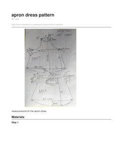


© Copyright 2026