
CONSENSUS TREATMENT RECOMMENDATIONS FOR LATE-ONSET POMPE DISEASE AANEM PRACTICE TOPIC
AANEM PRACTICE TOPIC
CONSENSUS TREATMENT RECOMMENDATIONS FOR LATE-ONSET
POMPE DISEASE
EDWARD J. CUPLER, MD,1 KENNETH I. BERGER, MD,2 ROBERT T. LESHNER, MD,3 GIL I. WOLFE, MD,4 JAY J. HAN, MD,5
RICHARD J. BAROHN, MD,6 and JOHN T. KISSEL, MD,7 of the AANEM CONSENSUS COMMITTEE
ON LATE-ONSET POMPE DISEASE
1
Department of Neurology, Oregon Health & Science University, Portland, Oregon, USA
2
Division of Pulmonary and Critical Care Medicine, Department of Medicine and Department of Physiology and Neuroscience, New
York University School of Medicine, New York, New York, USA
3
Department of Neurosciences, University of California San Diego, San Diego, California, USA
4
Department of Neurology, University of Texas Southwestern Medical School, Dallas, Texas, USA
5
Department of Physical Medicine and Rehabilitation, University of California Davis, Sacramento, California, USA
6
Department of Neurology, University of Kansas Medical Center, Kansas City, Kansas, USA
7
Department of Neurology, Division of Neuromuscular Medicine, Ohio State University, Columbus, Ohio, USA
Accepted 14 October 2011
ABSTRACT: Introduction: Pompe disease is a rare, autosomal
recessive disorder caused by deficiency of the glycogen-degrading lysosomal enzyme acid alpha-glucosidase. Late-onset Pompe
disease is a multisystem condition, with a heterogeneous clinical
presentation that mimics other neuromuscular disorders. Methods: Objective is to propose consensus-based treatment and
management recommendations for late-onset Pompe disease.
Methods: A systematic review of the literature by a panel of specialists with expertise in Pompe disease was undertaken. Conclusions: A multidisciplinary team should be involved to properly
treat the pulmonary, neuromuscular, orthopedic, and gastrointestinal elements of late-onset Pompe disease. Presymptomatic
patients with subtle objective signs of Pompe disease (and
patients symptomatic at diagnosis) should begin treatment with
enzyme replacement therapy (ERT) immediately; presymptomatic
patients without symptoms or signs should be observed without
Abbreviations: 6MWT, 6-minute walk test; AANEM, American Association of Neuromuscular & Electrodiagnostic Medicine; AFOs, ankle-foot
orthoses; BiPAP, bilevel positive airway pressure; CPAP, continuous positive airway pressure; DEXA, dual-energy X-ray absorptiometry; EMBASSY,
Exploratory Muscle Biopsy, Biomarker, and Imaging Assessment Study;
EMG, electromyography; ERT, enzyme replacement therapy; FDA, Food
and Drug Administration; FVC, forced vital capacity; GAA, acid alpha-glucosidase; LOTS, Late-Onset Treatment Study; MEP, maximal expiratory
pressure; MIP, maximal inspiratory pressure; rhGAA, recombinant human
acid alpha-glucosidase; SDB, sleep-disordered breathing; SF-36; 36-item
Short Form Health Survey Questionnaire
Key words: acid alpha-glucosidase; acid maltase deficiency; lysosomal
storage disorder; neuromuscular disease; Pompe disease
Disclaimer: This article was prepared and reviewed by the AANEM and
did not undergo the separate review process of Muscle & Nerve.
Approved by the American Association of Neuromuscular &
Electrodiagnostic Medicine (AANEM) Board, September 2011.
Disclosures: Task force co-chairs: Dr. Barohn has nothing to disclose.
Dr. Kissel serves as a consultant for Alexion Pharmaceuticals for a
clinical trial in myasthenia gravis. Task force members: Dr. Han has
nothing to disclose. Dr. Berger has served as a consultant to Genzyme
Corporation for clinical trials in MPS I and Pompe disease, and he has
served as a consultant to BioMarin Corporation for clinical trials in MPS
IV and MPS VI. Dr. Cupler is a member of the speaker bureau for
Genzyme Corporation and for Athena Diagnostics, Inc., and is a member
of the Pompe Registry North American Board of Advisors for Genzyme
Corporation. Dr. Leshner is a member of the Genzyme Global Advisory
Board for Pompe disease and has received honoraria for speaking
engagements and research support from Genzyme Corporation. Dr.
Wolfe has served on the speaker bureau for Genzyme Corporation.
None of the authors of this article is an inventor, and no authors are
receiving royalty payments. All authors received travel support and
honoraria for the initial consensus development meeting from MedLogix
Communications, LLC, and the AANEM, through an educational grant
from Genzyme Corporation.
Correspondence to: C. French, AANEM, 2621 Superior Drive NW,
Rochester, MN 55901
C 2011 Wiley Periodicals, Inc.
V
Published online 15 December 2011 in Wiley
(wileyonlinelibrary.com). DOI 10.1002/mus.22329
AAEM Practice Topic: Late-Onset Pompe Disease
Online
Library
use of ERT. After 1 year of ERT, patients’ condition should be
reevaluated to determine whether ERT should be continued.
Muscle Nerve 45: 319–333, 2012
Consensus statements are important for developing
treatment recommendations when evidence-based
medical guidelines and treatment recommendations
based on controlled trials are sparse or nonexistent. In
the case of late-onset Pompe disease, it is an appropriate time to assemble a group of experts in the field to
review and analyze the medical literature and create
recommendations for disease management (based
on the best available evidence) until additional data
from controlled studies become available to enable
further recommendations. The American Association of Neuromuscular & Electrodiagnostic Medicine
(AANEM) convened a consensus committee of specialists with expertise in the diagnosis and treatment
of Pompe disease and, using a modified consensus
development conference method,1 the committee
worked to create consensus-based recommendations
for the treatment of late-onset Pompe disease.
OVERVIEW OF POMPE DISEASE
History of Pompe Disease. Pompe disease, also
referred to as acid maltase deficiency, is a rare, autosomal recessive disorder that was first described in
1932 in a 7-month-old girl who died of cardiomyopathy. The disease was identified as a glycogen storage
disorder in which glycogen had accumulated within
vacuoles of all the examined tissue.2,3 In 1954, the
disease was classified as glycogen storage disease type II,4
and in 1963 it became the first identified lysosomal
storage disorder.5,6 During the 1960s and 1970s,
cases of a milder phenotype that occurred in older
patients (late-onset Pompe disease) were described
by Engel and colleagues.7 The reported frequency
of infantile-onset Pompe disease ranges from
approximately 1 in 35,000 to 1 in 138,000 among
Taiwanese and Dutch populations, respectively.8,9
The estimated frequency of late-onset Pompe
MUSCLE & NERVE
March 2012
319
FIGURE 1. Timeline for Pompe disease: 75 years from description to availability of disease-modifying agent. [Color figure can be
viewed in the online issue, which is available at wileyonlinelibrary.com.]
disease is 1 in 57,000.10 The actual frequency of lateonset Pompe disease in the USA is not known, but
one study suggested that the frequency of all forms
may be as high as 1 in 40,000.11
There was no disease-specific treatment for Pompe
disease until enzyme replacement therapy (ERT) was
first attempted in 1973. Highly purified, placentaderived acid alpha-glucosidase (GAA) enzyme was
administered by intravenous infusion, taking advantage of the ability of lysosomes to internalize exogenous proteins by endocytosis.12 Initial attempts at
ERT encountered problems of immunogenicity, and
there was limited availability of purified GAA for
enzyme delivery.13,14 In 1979, the 28-kb gene for GAA
was identified on chromosome 17,15 and in the 1990s
new recombinant technology became available, enabling the production of enough recombinant human
acid alpha-glucosidase (rhGAA) to allow ERT clinical
trials to be conducted. Figure 1 show the timeline for
the development of ERT for Pompe disease.
In 2006, alglucosidase alfa (Myozyme; Genzyme
Corporation, Cambridge, Massachusetts)16 was
approved by the U.S. Food and Drug Administration
(FDA) and the European Medicines Agency and
became the first disease-specific treatment for Pompe
disease. The approval was based on the results of a
pivotal trial of ERT with alglucosidase alfa in 18
infants.17 The results of the first randomized, double-blind, placebo-controlled study of ERT, known as
the Late-Onset Treatment Study (LOTS),18 led to
the approval in 2010 of Lumizyme (Genzyme Corporation)19 for the treatment of late-onset Pompe disease in the USA. Along with its approval, the FDA
320
AAEM Practice Topic: Late-Onset Pompe Disease
mandated postmarketing surveillance of alglucosidase alfa. The manufacturer will conduct studies to
evaluate the tests used for drug profiling and monitoring and for qualification, quantification, specification, and stability of alglucosidase alfa. Moreover, the
FDA required that additional immunogenicity, safety,
efficacy, pharmacokinetics, and long-term follow-up
studies be conducted to better understand the role
of alglucosidase alfa in the treatment of Pompe disease. These studies are currently ongoing.
Pathophysiology of Pompe Disease. Lysosomal GAA
catalyzes the breakdown of glycogen into glucose.
Acid alpha-glucosidase deficiency in Pompe disease
results in the accumulation of lysosomal and
FIGURE 2. Pathophysiology of Pompe disease. GAA, acid
alpha-glucosidase; UDP, uridine diphosphate. Based on Raben
et al.108 with permission. [Color figure can be viewed in the
online issue, which is available at wileyonlinelibrary.com.]
MUSCLE & NERVE
March 2012
nonlysosomal glycogen in multiple tissues20–22
(Fig. 2). In infantile-onset Pompe disease, GAA
enzyme activity is either completely or nearly completely absent (typically <1% of normal activity in
skin fibroblasts). Some residual enzyme activity
(approximately 2–40% of normal activity in skin
fibroblasts) is present in most children and adults
with the late-onset form.23,24 Recent evidence has
shown a failure of productive autophagy in patients
with Pompe disease and the progressive accumulation of autophagosomes that disrupt the contractile
apparatus in muscle fibers.25 Moreover, currently
unknown abnormalities in a subset of lysosomes
interfere with the recycling of autophagosomes and
their contents, which leads to skeletal muscle damage.25 The GAA gene is highly pleomorphic; 289
sequence variations have been reported to date that
include 197 proven pathogenic mutations, 67 nonpathogenic mutations, and 25 sequence variants
with unknown functional effect.26
Clinical Presentation and Disease Progression. Infantile-onset Pompe disease is a progressive, multisystem disorder that causes hypotonia and feeding
difficulties in the first year of life. The disease
affects cardiac, skeletal and smooth muscles, the
pulmonary and gastrointestinal systems, and anterior horn cells. Death due to cardiorespiratory failure typically occurs in the first year of life.3,24,27
Late-onset Pompe disease is also a multisystem disease; it may manifest at any time after 12 months of
age. Its clinical presentation includes: progressive
muscle weakness, especially in the trunk and lower
limbs; respiratory symptoms; and progression to respiratory insufficiency due to diaphragmatic and intercostal muscle weakness.3,24 Respiratory complications are the most frequent cause of death in lateonset Pompe disease. To date, evidence regarding
cardiac involvement in late-onset Pompe disease is
inconclusive; however, Wolff–Parkinson–White syndrome has been reported.28
Diagnosis of Late-Onset Pompe Disease. The diagnosis of Pompe disease, particularly the late-onset
form, is often difficult because it can clinically resemble a myriad of other neuromuscular disorders. A
high level of clinical suspicion is necessary for a
timely and accurate diagnosis.24 A complete discussion of the clinical presentation and diagnostic
guidelines for late-onset Pompe disease was published in 2009.20 Briefly, physical examination usually
reveals more proximal than distal weakness. The pelvic girdle is affected to a greater degree than the
shoulder girdle, and weakness of the abdominal
muscles and diaphragm may be present early on.
Scapular winging, paraspinal muscle atrophy, scapuloperoneal weakness,29 and facial involvement with
weakness or ptosis (unilateral or bilateral) have also
AAEM Practice Topic: Late-Onset Pompe Disease
been observed.30,31 Clinical myotonia is absent, but
electrophysiological myotonia, especially in the paraspinal muscles, is detected frequently.20 Fatigue of
the jaw muscles and difficulties with chewing and
swallowing often result in inadequate intake of protein, calories, vitamins, and minerals, which leads to
endogenous muscle protein breakdown.24 Swallowing dysfunction can be diagnosed in patients with
late-onset Pompe disease by a videofluoroscopic swallowing assessment.24 Sensory, cerebellar, and cognitive involvement have not been reported.20
Laboratory testing usually reveals nonspecific
elevation of serum creatine kinase.10 Needle electromyography (EMG) studies often indicate myopathic potentials with increased muscle membrane
irritability and myotonia.20 Of note, even when
needle EMG findings in limb muscles are normal,
abnormalities may still be found in the paraspinal
muscles alone.29 Pulmonary function testing may
reveal decreased forced vital capacity (FVC); a
detailed discussion of pulmonary involvement is
provided in what follows.
Although the clinical phenotype of late-onset
Pompe disease varies, genetic analysis of the GAA
gene,24 or a determination of the level of GAA
enzyme activity in blood, fibroblasts, or muscle tissue, can provide a definitive diagnosis.32 Although
muscle biopsy often reveals a vacuolar myopathy
with increased amounts of glycogen,31,33 a normal
muscle biopsy does not exclude late-onset Pompe
disease.20
METHODS
A literature search was conducted in the PubMed
and Embase databases for clinical trials and relevant articles published before June 2010. The
search terms included Pompe disease, acid alpha-glucosidase deficiency, acid maltase deficiency, glycogen storage disease type II, and glycogenosis type II. Panel
members reviewed these articles individually and
discussed them in small group sessions. Subsequently, the panel convened for further discussion
and consensus development. In addition, articles
published after the consensus meeting was held,
references cited by the articles produced by the literature search, and references suggested by the
authors are included in this article. Each panel
member was assigned a section to write and review.
Writing contributions are listed in the Appendix.
RESULTS
The literature search produced 105 articles relevant
to Pompe disease. The AANEM consensus panel,
consisting of several neuromuscular specialists and
a pulmonologist, reviewed all articles. The most
frequent signs and symptoms of late-onset Pompe
MUSCLE & NERVE
March 2012
321
disease are presented in what follows as musculoskeletal, pulmonary, and gastrointestinal tract manifestations. In addition, treatment recommendations
for these signs and symptoms are discussed.
Musculoskeletal Manifestations and Management of
Late-Onset Pompe Disease. Musculoskeletal Manifestations. Musculoskeletal involvement in late-onset
Pompe disease is dominated by progressive muscle
weakness affecting proximal more than distal
muscles.24,34 In a recent observational study, weakness of the proximal extremities was the presenting
symptom in 93% of patients with late-onset Pompe
disease.35 In addition, the study demonstrated that
strength assessed over a 1-year period by using
quantitative muscle testing declined by 7.1% in the
lower limbs and by 4% in the upper limbs.35 Subsequent secondary musculoskeletal complications
include contractures, limb and spinal deformities,
and osteopenia/osteoporosis.24 Scoliosis was found
in approximately half of late-onset Pompe disease
patients.36 Gross and fine motor functions required
for daily living and ambulation are affected by primary and secondary musculoskeletal impairments,
as well as by pulmonary (more than cardiac) dysfunction.20,37,38 In addition, general fatigue and
poor endurance are common concerns, reported by
50–76% of patients,20,37,38 with pulmonary compromise as a likely contributing factor. Muscle pain
and soreness are early symptoms that were reported
in 46% of patients.37 Therefore, management of
patients with late-onset Pompe disease should be
individualized to account for the variations in musculoskeletal
impairment
observed
between
patients.38
Overall Management of Musculoskeletal Issues. General clinical management and rehabilitation principles to guide the care of patients with late-onset
Pompe disease include preserving or optimizing
motor function, preventing or minimizing secondary complications, maximizing the benefits of ERT
or other therapies as they become available, promoting overall health while managing disease-associated symptoms, and improving quality of life.
Rehabilitation and management efforts in lateonset Pompe disease should be comprehensive
and preventive, based on individual assessment
and appreciation of disease progression.24 To
address quality-of-life issues, rehabilitation services
should include physical therapy, occupational therapy, respiratory therapy, speech therapy, dysphagia
evaluation and nutritional support, orthotics, and
assistive technology, as well as vocational/avocational,
mental health, and social work services.
Strengthening and Therapeutic Exercise. There
are no established guidelines for muscle strengthening or therapeutic exercise for individuals with
322
AAEM Practice Topic: Late-Onset Pompe Disease
late-onset Pompe disease. Studies of the effects
and treatment roles of exercise and nutrition in
late-onset Pompe disease have been few and have
had small sample sizes.34,39,40 Although more information is needed, these studies suggest that submaximal aerobic exercise may increase muscle
strength and function through improved clearance
of accumulated glycogen in the muscle cytosol.41
In contrast, there is insufficient evidence that
resistance training improves strength in late-onset
Pompe disease. Traditionally, excessively strenuous
resistance exercises have been discouraged in muscle disorders because of the potential for exacerbating muscle degeneration,42–44 and in Pompe
disease there is additional theoretical concern that
excessive muscle contraction might lead to
increased leakage of glycogen from lysosomes or
cause lysosomal rupture, thereby hastening muscle
damage.45 The favorable pattern of response in
the strength of proximal and respiratory muscles
in ERT studies of late-onset Pompe disease is
encouraging, but further studies are needed.18,36
Therefore, at this time the authors recommend that
the general precautions regarding strengthening
exercises that are followed for other degenerative
muscle diseases should be applied to late-onset
Pompe disease46,47 (Table 1). Therapeutic exercise
in late-onset Pompe disease may pose a risk for cardiopulmonary compromise in this population. Consequently, a pulmonologist should evaluate the
patient before initiation of an exercise program.24
Therapeutic exercise should start slowly, allowing for periods of rest, followed by a gradual
increase in exercise intensity from mild to moderate, reaching aerobic levels of about 60–70% of
maximal effort at a frequency of 3–5 days per
week.24,40,43 Optimally, an experienced physical
therapist should develop a structured program,
monitor the patient, and remain in contact with
the prescribing physician. Pulse oximetry, heart
rate, and perceived exertional effort (e.g., Borg
scale) should be monitored initially to guide progression of the program. Moreover, the physical
therapist should teach the patient how to monitor
heart rate and correlate it with perceived exertional effort and oxygen saturation information to
prepare the patient for a self-monitored, homebased exercise program. A preliminary report of
one late-onset Pompe disease patient demonstrated that side-alternating vibration training produced sustained improvement in mobility at 1 year
of follow-up.48 Larger studies are needed before
this form of training can be recommended.
Contracture and Limb Deformity. Because no
established guidelines exist for management of secondary musculoskeletal impairments, including
contracture and deformity, in late-onset Pompe
MUSCLE & NERVE
March 2012
Table 1. Treatment recommendations for the musculoskeletal element of late-onset Pompe disease.
Provide patient with information on the following resources:
Muscular Dystrophy Association, Acid Maltase Deficiency Association, Pompe Registry, Association for Glycogen Storage Disease,
International Pompe Association
Physical examination and assessments
Patients should be examined by a cardiologist and pulmonologist before beginning an exercise program
Screen all patients diagnosed with Pompe disease, regardless of age and wheelchair use, with dual-energy x-ray absorptiometry (DEXA);
follow-ups can be considered on a yearly basis
Patients with late-onset Pompe disease and reduced bone density should undergo medical evaluation, including laboratory testing
and medication review by an endocrinologist or bone density specialist
Conduct fall risk assessment followed by a formal evaluation for balance and safe gait training for patients at increased risk for
osteoporosis and falls
Recommend adaptive equipment, such as a cane or walker, to reduce risk of falls
Physical/occupational therapy
A physical or occupational therapist should develop an exercise program that may include one or more of the following: walking,
treadmill, cycling, pool-based program, swimming, submaximal aerobic exercise, or muscle strengthening, that follows the
guidelines for other degenerative muscle diseases
Avoid overwork weakness, excessive fatigue, disuse, strenuous exercises, and eccentric contractions
Emphasize submaximal aerobic exercise
Incorporate functional activities when possible
Teach patient to monitor heart rate and breathing in relation to exertion
Integrate energy conservation techniques and biomechanical advantages
A preventive stretching regimen should be started early and performed as part of the daily routine to prevent or slow the development
of muscle contractures and deformities
Management of contractures
Manage contractures by using orthotic devices, appropriate seating position in the wheelchair, and standing supports
Surgical intervention
Surgical intervention should be considered for scoliosis when the Cobb angle is between 30 and 40
Vitamins and mineral supplements
Recommend vitamin D, calcium, and bisphosphonates, following the guidelines for other neuromuscular disorders
disease, general principles established for the management of other neuromuscular disorders can be
applied.49 These general principles include limiting contracture and deformity by gentle daily
stretching, correction of improper positioning,
judicious and timely use of splints and orthotic
interventions, and provision of adequate support
in all positions, including sitting and supported
standing.49 Written instructional materials for the
family are more effective than verbal instruction.49
Prevention of contracture and deformity is critical
to preserve function and limit other secondary
complications, such as skin breakdown and
chronic musculoskeletal pain. A preventive stretching regimen should be implemented early and be
performed as part of the daily routine. Such regimens are better tolerated and accepted if they are
initiated before muscle tendon tightness and contractures develop, a point at which stretching often
becomes painful. Aggressive stretching should be
approached cautiously because, at least in severely
affected children, a tendency for pathologic fracture
has been observed.50
Adaptive Equipment and Orthotic Interventions. A
variety of orthotic interventions are available for
contracture management, such as ankle–foot
orthoses (AFOs) to prevent plantarflexion contractures, thigh binders to prevent iliotibial band
contractures, knee splints to prevent knee flexion
AAEM Practice Topic: Late-Onset Pompe Disease
contractures, resting wrist/hand/finger splints to
prevent wrist and finger flexor contractures over
multiple joints,34 and lumbar corsets for management of back pain. Nocturnal resting splints or
AFOs to prevent plantarflexion contractures have
not been adequately studied in Pompe disease.49,51
Wheeled walkers or quad canes may help with
ambulation, depending on the degree and pattern
of muscle weakness. As the disease progresses,
adaptive equipment for bathroom needs and transfers will improve safety and independence while
decreasing complications. A summary of rehabilitation strategies for musculoskeletal complications in
Pompe disease is presented in Table 1. Other physical modalities (e.g., heat therapy) that have not
been adequately studied in Pompe disease are not
recommended. Appropriate seating systems for
wheelchairs are important in the prevention of
limb and spinal contractures and deformity. Prescribed equipment could include a head support,
solid seat and back, lateral trunk support, and a
leg rest/foot plate.52 Supported standing could
promote dual treatment goals: it may achieve
weight-activated stretching in multiple lower limb
joints and may also provide benefit in the management of osteoporosis.53 Various supine, prone, vertical, or hydraulic standers are available, in addition to
power standing functions on motorized wheelchairs.
These measures can be considered after evaluation
MUSCLE & NERVE
March 2012
323
by a physical or occupational therapist.24 Prophylactic surgery for contracture management (e.g., tenotomy and tendon transfer) is rarely used and generally does not appear to improve function.49
Scoliosis and Spinal Deformity. Limited evidence
is available to guide the management of scoliosis
and spinal deformity in late-onset Pompe disease.
As a consequence of proximal limb and truncal
weakness, and paraspinal muscle atrophy, some
patients with late-onset Pompe disease adopt a
compensatory lumbar lordosis while standing,
whereas others may experience rigid spine syndrome.54,55 Scoliosis progression can be monitored
with serial radiography once transition to wheelchair use has occurred. The frequency of scoliosis
screening depends on the degree and rate of disease progression and on the patient’s age.
Spinal deformities in Pompe disease may
become very severe, and they have the potential to
produce several problems. Severe scoliosis and pelvic obliquity lead to pain, poor sitting balance,
more difficulty in attendant-provided care, skin
ulcers, and potential exacerbation of restrictive
lung disease.56 Information is limited regarding
spinal bracing in Pompe disease, but in other neuromuscular disorders spinal bracing does not
change the natural history of scoliosis.51,57,58
Recent studies in Duchenne dystrophy suggest that
corticosteroids slow the progression of scoliosis59,60; it is unclear, however, whether ERT results
in a similar benefit in late-onset Pompe disease.
Although scoliosis was a common feature in an
open-label ERT trial, it was not followed as an outcome measure.36
At present, surgical intervention remains the
only effective treatment for progressive spinal
deformity in neuromuscular disease. Drawing from
experience in other neuromuscular disorders—
particularly Duchenne dystrophy—the consensus is
that surgical intervention is indicated when the
Cobb angle is between 30 and 40 .60–64 As no outcome data are available for spinal corrective surgery in late-onset Pompe disease, the authors recommend that the general clinical guidelines for
spinal intervention in Duchenne dystrophy be followed in late-onset Pompe disease and that these
decisions be guided by the individual situation.
Preoperative pulmonary function testing is essential, although a recent Duchenne dystrophy study
showed no significant increase in operative and
postoperative complications for patients with FVC
>30% of predicted.65 Careful preoperative risk
assessment and aggressive postoperative ventilatory
support are recommended to minimize morbidity.
In those patients at risk, preoperative mask-fitting
and initiation of nocturnal noninvasive positive
pressure ventilation can facilitate postoperative re324
AAEM Practice Topic: Late-Onset Pompe Disease
spiratory recovery.65 Postoperative management after corrective spinal surgery should focus on the
early institution of physical therapies, mobilization
when clinically stable, pain control, ventilatory support, and pulmonary hygiene. It remains unclear
whether spinal surgery leads to sustained improvement in pulmonary function. It is important to
keep in mind that a primary goal of either prevention or correction of scoliosis is to provide the
patient with improved sitting balance; this, in itself,
can help maintain use of a wheelchair, facilitate
nursing care, and enhance quality of life.
Osteopenia and Osteoporosis. Low bone mineral
density (osteoporosis) is a common feature in
patients with Pompe disease. A recent study demonstrated that 67% of the patients tested had a
bone mineral density z-score of 1 and that the
decrease in bone density was present in both the
infantile- and late-onset forms of Pompe disease.66
Therefore, the authors recommend that individuals with late-onset Pompe disease, including all
children and adults who are wheelchair- or ventilator-dependent or who have decreasing muscle
strength, be screened annually with dual-energy
X-ray absorptiometry (DEXA).24,66 On the basis of
a recent study showing a high prevalence of osteoporosis in patients with Pompe disease,66 the
authors recommend that all patients with Pompe
disease undergo fall risk assessment, followed by a
more formal evaluation for balance testing and
gait training in those individuals at increased risk
of falls. Adaptive equipment such as a cane or
walker may be recommended to reduce the risk of
falls, and education for patients and family members regarding safe ambulation strategies should
be provided. Vitamin D and calcium supplements
should be taken by all patients with abnormal
DEXA z-scores. Bisphosphonate use in late-onset
Pompe patients should follow the same guidelines
used for the general population. Pompe disease
patients with reduced bone density should
undergo routine medical evaluation by an endocrinologist or bone density specialist that includes
laboratory testing and medication review. Standing
frame and weight-bearing exercises can be incorporated into therapy sessions for bone density
building.
Pulmonary Manifestations and Management. The respiratory concerns raised by Pompe disease center
around progressive weakness of the diaphragm
and the accessory muscles of respiration. In lateonset Pompe disease, trunk muscles and proximal
muscles in the lower limbs are usually affected
first, followed by involvement of the diaphragm
and other respiratory muscles. This leads to pulmonary
insufficiency
and
sleep-disordered
MUSCLE & NERVE
March 2012
breathing (SDB).24,67–69 Involvement of the respiratory system can be overlooked when overt respiratory symptoms are absent.67 Frequently, the earliest
manifestation
of
respiratory
system
involvement is repeated episodes of tracheobronchitis and pneumonia due to expiratory muscle
weakness and impaired cough.3 As the disease progresses, all respiratory muscles weaken, leading to
lower-than-normal lung volumes.35,70 Sleep-disordered breathing may occur, including nocturnal
hypoventilation and oxygen desaturation71 and,
consequently, an increased need for ventilatory support.35 Although respiratory insufficiency is manageable with ventilatory assistance,68,69 respiratory insufficiency is the most common cause of death in
patients with late-onset Pompe disease.67
Pulmonary Function Tests. Because of the high
prevalence of respiratory complications and the
progressive nature of Pompe disease, assessment of
respiratory function is imperative and should be
conducted on a regular basis.20,24,72 Objective
assessment of respiratory muscle function is typically obtained by measurements of FVC and maximal inspiratory and expiratory muscle pressures
(MIP and MEP, respectively).73,74 It is important to
note that, even before an overt reduction in
upright FVC occurs, diaphragmatic weakness may
be detected by measurement of FVC in both the
supine and seated positions.75 Diaphragmatic weakness is suggested if there is a 10% decrease of
FVC in the supine compared with the upright position; a 30% decrease indicates severe weakness.74
In a recent observational study of patients with
late-onset Pompe disease, approximately 65% of
patients demonstrated reduction in upright FVC
and, in the remaining patients, respiratory muscle
dysfunction was identified either by reduction in
FVC in the supine position or by reduction in MIP
and MEP.35,76
Sleep-Disordered
Breathing. Sleep-disordered
breathing may occur in many neuromuscular disorders because of impaired respiratory muscle function in the supine position and alterations in respiratory control mechanisms during sleep.69,71,73
Ventilatory abnormalities during sleep include
obstructive sleep apnea and sustained hypoventilation.68,69,71,73,77 Nocturnal hypoventilation may
occur in parallel to or may precede daytime respiratory insufficiency. In neuromuscular disorders,
SDB may occur when FVC falls below 30–50% predicted73,78; however, SDB in patients with Pompe
disease may occur when upright FVC is only moderately abnormal, because of the disproportionate
diaphragmatic involvement.71 Symptoms suggestive
of SDB, such as apneas, gasping respirations, and
restless sleep24,68 should be assessed as part of the
clinical evaluation of all patients with Pompe disAAEM Practice Topic: Late-Onset Pompe Disease
Table 2. Treatment recommendations for the respiratory element
of late-onset Pompe disease.
Involve a pulmonologist experienced in managing patients with
neuromuscular diseases
Up-to-date vaccinations, including vaccination against
pneumococcus and influenza
Early and aggressive treatment of bacterial and viral infections
Clear secretions from airways (e.g., cough assist
device, suction)
Train/educate patients and families to use assisted cough
and inspiratory muscle techniques
Treat sleep-disordered breathing with continuous positive airway
pressure (CPAP) or bilevel nocturnal noninvasive ventilation (BiPAP)
In the absence of sleep studies, consider BiPAP ventilation if
arterial PCO2 is 45 mm Hg, supine forced vital capacity is
<50% of predicted, negative inspiratory force is <60 cm H2O,
or oxygen saturation falls to <88% for 5 continuous minutes
during sleep
Treat concomitant conditions, such as asthma
or cardiomyopathy
Consider enzyme replacement therapy
ease. Until recently, full polysomnography was the
preferred method for evaluation of upper airway
obstruction; however, other diagnostic modalities
are now available for respiratory disorders.78,79
Limited overnight monitoring with pulse oximetry
to identify nocturnal hypoxemia and/or capnography to identify hypoventilation coupled with a sensitive airflow detector may be used. Capnography is
a noninvasive test that can be used to evaluate
hypercapnia; however, false-negative results may be
obtained in patients at greatest risk for respiratory
insufficiency (e.g., when either tidal volume is
reduced or concomitant obstructive lung disease is
present).80 Identification of ventilatory abnormalities during sleep is important, and it leads to
timely initiation of the appropriate therapy; therefore, it is advisable that the managing physician
order sleep studies if there is doubt that the
patient might have SDB. Table 2 summarizes the
treatment recommendations for the pulmonary
aspects of late-onset Pompe disease.
Respiratory Insufficiency. Respiratory insufficiency in patients with late-onset Pompe disease
develops because of progressive respiratory muscle
weakness, with or without SDB. Because diaphragmatic involvement is an early and prominent finding, respiratory insufficiency can develop while
patients are still ambulatory and, in rare cases,
it can be the initial manifestation of the disease.28,71,72,81–86 Although assessment for carbon
dioxide retention is best accomplished by analysis
of arterial blood gases, this test may not be readily
available in some outpatient settings. Alternatively,
capnography may be used as a surrogate measure,
or elevation of serum bicarbonate concentration in
venous blood can be used as a screening test for
MUSCLE & NERVE
March 2012
325
chronic hypoventilation; an elevated serum bicarbonate concentration (>27 mEq/L) may be a
marker of renal compensation for chronic carbon
dioxide retention.87 Chronic respiratory insufficiency may progress to development of cor pulmonale if left untreated.
Treatment of Respiratory Issues. Maintenance of
optimal functional status in patients with Pompe
disease requires multiple interventions.24,88 Therapy should be titrated on the basis of the patient’s
underlying abnormalities and pulmonary status.
Patients benefit from the involvement of a pulmonologist experienced in managing patients with
neuromuscular disorders to monitor and treat respiratory manifestations. A cardiologist should be
consulted if the patient shows signs or symptoms
of heart involvement, which are usually rare in
late-onset Pompe disease.89 Chest radiographs
should be obtained at baseline and as clinically
indicated thereafter. All patients should receive
regular vaccinations, including pneumococcus and
influenza vaccinations.24
Impairment in secretion clearance due to weakened cough can be addressed by both manual and
mechanical techniques.90 Inhaled bronchodilators
can be used in conjunction with airway clearance
techniques, assisted coughing maneuvers, and
inspiratory muscle training.90 Bronchodilators and
corticosteroids (inhaled and/or oral) may be useful if concurrent asthma is present. Treatment of
both viral and bacterial infections should be early
and aggressive.24,90 Treatment of SDB should target the underlying respiratory disorder before supplemental oxygen is given. For patients with obstructive sleep apnea, treatment can be limited to
continuous positive airway pressure (CPAP); for
patients with nocturnal hypoventilation, treatment
with nocturnal noninvasive bilevel positive airway
pressure (BiPAP) ventilation should be used.68,71
Pulmonary and sleep consultants are recommended. In the absence of sleep studies, BiPAP
should be considered if arterial partial pressure of
carbon dioxide is 45 mm Hg, supine FVC is
<50% of predicted, negative inspiratory force is
<60 cm H2O, or oxygen saturation falls to <88%
for 5 continuous minutes during sleep.91,92 Supplemental oxygen can be used if these methods do not
fully correct the hypoxia, but careful titration of the
rate of oxygen administration coupled with close
monitoring of arterial carbon dioxide levels are
required to prevent oxygen-induced hypercapnia.93
Finally, results from the LOTS trial suggest that
ERT may help to improve pulmonary function18;
however, until the long-term effect of ERT on
respiratory function is known, noninvasive ventilatory
support should be pursued whenever the patient’s
clinical history suggests SDB. Specific consideration
326
AAEM Practice Topic: Late-Onset Pompe Disease
Table 3. Treatment recommendations for the gastrointestinal
element of late-onset Pompe disease.24
Coordinate treatment through a multidisciplinary neuromuscular
clinic
Involve an experienced dietitian
Obtain videofluoroscopic swallowing assessment and evaluation for gastroesophageal reflux to guide management of feeding either orally or through a feeding tube
Monitor growth parameters carefully
Provide adequate nutrition consisting of high protein diet (20–
25%) with attention to vitamins and minerals
Educate patients about the appropriate use of over-the-counter medications
Patients receiving enzyme replacement therapy should be
monitored for IgG antibodies every 3 months for 2 years and
then annually thereafter
should be given to patients with severely limited ventilatory reserve who are not able to sustain spontaneous
breathing during wakefulness for even short periods.
Although attention to pulmonary toilet combined
with noninvasive ventilatory support may be successful
for a prolonged time in selected patients,69 many
patients who require continuous ventilatory support
will need a tracheostomy to ensure a reliable connection to the airway and successful ventilation.24 Because
of the morbidity and subsequent loss of cough associated with tracheostomy, this decision often presents as
a therapeutic dilemma.
Gastrointestinal and General Medical Considerations
for Late-Onset Pompe Disease. Patients with lateonset Pompe disease develop feeding and swallowing difficulties that can be attributed to facial
hypotonia, facial muscle fatigue, tongue weakness
and enlargement, fatigue and reduced range of
motion of muscles of mastication, reduced tongue
capping, and reduced lip seal. The difficulty in
feeding and swallowing leads to inadequate intake
of proteins and, consequently, endogenous protein
breakdown.24 Swallowing dysfunction can be diagnosed by a videofluoroscopic swallowing test.24 An
experienced dietitian, preferably one who specializes in metabolic disorders, should be involved in
the development and management of a feeding
plan for patients with Pompe disease. At this time,
there are inadequate data to recommend any specific dietary regimen.40,94 However, a combination
high-protein and low-carbohydrate diet has been
suggested, which was shown to improve muscle
strength in patients with late-onset Pompe disease.95 In addition, ERT may resolve gastrointestinal symptoms in patients with late-onset Pompe
disease; however, additional studies are needed to
further document the frequency and severity of
gastrointestinal symptoms in patients and their
rapid amelioration in response to ERT.96 If the
risk of aspiration becomes high, oral feeding
MUSCLE & NERVE
March 2012
ducted in 90 patients 8 years of age (range 10–70
years). The two coprimary endpoints were distance
walked during a 6MWT and the percent of predicted FVC in the upright position. At week 78,
statistically significant findings in favor of alglucosidase alfa were noted in both the 6MWT and percent of predicted upright FVC results (Fig. 4A and
B). The increase in 6MWT distance occurred in
the first 26 weeks of treatment and was maintained
for the next 52 weeks (Fig. 4A). All of the secondary and tertiary endpoints favored the treatment
group, but only the MEP results reached statistical
significance.18 Patients in the treatment and placebo groups had similar frequencies of adverse
events, serious adverse events, and treatmentrelated adverse events. Anaphylactic reactions
occurred only in patients who received alglucosidase
FIGURE 3. Multidisciplinary approach for the management and
care of Pompe disease. Based on Kishnani et al.24 [Color figure
can be viewed in the online issue, which is available at
wileyonlinelibrary.com.]
should be discontinued, and installation of a feeding tube becomes necessary.24 Table 3 lists the recommendations for treatment of the gastrointestinal aspects of late-onset Pompe disease.
DISCUSSION
Late-onset Pompe disease is a multisystem disorder
that requires the involvement of a multidisciplinary24 team for the proper management and treatment of the pulmonary, neuromuscular, and gastrointestinal elements of the disease (Fig. 3).
The Late-Onset Treatment Study. The success of
alglucosidase alfa in the treatment of infantile-onset
Pompe disease97 and the reported benefits in several
small, open-label studies in late-onset Pompe disease36,98–101 led to the design and implementation of
the LOTS trial in patients diagnosed with late-onset
Pompe disease.18 Inclusion criteria included the ability to walk 40 meters during the 6-minute walk test
(6MWT; with assistive devices permitted) and reproducible FVC 30–80% of predicted in the upright
position with a postural drop in FVC of 10% after
repositioning from upright to supine. Patients were
excluded if they required any invasive ventilation or
if they needed noninvasive ventilation while awake
and upright.18
Those patients who qualified were randomly
assigned in a ratio of 2:1 to receive biweekly infusions of alglucosidase alfa (20 mg/kg, based on
body weight) or placebo. Patients were stratified by
baseline distance walked on the 6MWT (<300 or
>300 meters) and by baseline values recorded as
percentage of predicted FVC in the upright position (<55% or >55%). The LOTS trial was conAAEM Practice Topic: Late-Onset Pompe Disease
FIGURE 4. Changes from baseline in distance walked and in
forced vital capacity according to study group. Based on van
der Ploeg et al.14 (A) and van der Ploeg et al.18 (B) with permission. [Color figure can be viewed in the online issue, which
is available at wileyonlinelibrary.com.]
MUSCLE & NERVE
March 2012
327
Table 4. Treatment warning for alglucosidase alfa (Lumizyme;
Genzyme Corporation, Cambridge, Massachusetts).19
Warning
Life-threatening anaphylactic reactions, severe
allergic reactions, and immune-mediated reactions
have been observed in some patients during
Lumizyme infusions.18 Therefore, appropriate
medical support should be readily available
when Lumizyme is administered.
alfa (3 of 60 treated patients, or 5%). All alglucosidase alfa recipients tested negative for immunoglobulin G (IgG) anti-GAA antibodies at baseline,
and all patients seroconverted by week 12. The
data indicate that alglucosidase alfa has a positive
effect on the disease process or processes that produce impaired ambulation and respiratory insufficiency in late-onset Pompe disease, but treatment
carries a risk of serious potential complications,
including anaphylactic reactions.18,19
Perspectives on the LOTS Trial. Currently, ERT
with alglucosidase alfa is the only specific treatment
available for late-onset Pompe disease in adults and
children. The greatest gains in all endpoints in the
treatment group occurred during the first 26 weeks
of therapy with alglucosidase alfa. Those gains were
maintained during the 78 weeks of the study.18 The
overall clinical response observed in the LOTS trial
suggests that prevention of further loss of muscle
function is the target treatment goal for ERT. A very
modest, but statistically significant, benefit in the
6MWT was reported; however, it is not clear that
this improvement is functionally significant. The
6MWT, although useful in clinical trials, does not
reflect the routine functional walking employed by
most individuals.102 This may explain why the
improvements in 6MWT results documented in this
study were not associated with subjective improvements in the quality-of-life measure (36-item Short
Form [SF-36] Health Survey Questionnaire) that
was employed in the same study. Of note, it is possible that the alternate-week, 20-mg/kg dose of alglucosidase alfa, which substantially altered the course
of the disease in infantile-onset Pompe disease,97
might be insufficient for patients with the late-onset
form of the disease. A longer-term study of alglucosidase alfa in children and adults with late-onset
Pompe disease is warranted to fully understand the
long-term potential of ERT in this population.
Anaphylactic reactions, a serious potential complication of treatment with any recombinant
human protein, have been reported to occur with
alglucosidase alfa.97 Because 5% of patients in the
treatment group of the LOTS trial developed an
anaphylactic reaction, the authors recommend caution during home-based infusion and that patients
treated with alglucosidase alfa who have persis328
AAEM Practice Topic: Late-Onset Pompe Disease
tently high antibody titers should be closely monitored until the effect of the antibodies is more
fully understood (Table 4).19
Practical Applications
ERT. The decision
of
the
LOTS
Trial
and
making process to treat
patients with late-onset Pompe disease may not
completely mirror the strict inclusion criteria for
the LOTS trial. More information is needed
regarding the course and effect of long-term treatment of Pompe disease with alglucosidase alfa in
children and adults. However, until such additional information is available, the authors have
compiled a series of recommendations, as shown
in Table 5.
Presymptomatic
Patients
without
Objective
Signs. Enzyme replacement therapy is not currently recommended for patients who have no
symptoms or objective signs (proximal muscle
weakness or reduced FVC in either upright or
supine position) of Pompe disease. When a presymptomatic patient is confirmed to have Pompe
disease through newborn screening, sibling screening, or another screening program, it is recommended that such patients be examined every 6
months and that muscle strength and pulmonary
function (including FVC in both upright and
supine positions) be monitored at these visits.
Enzyme replacement therapy with alglucosidase
alfa is recommended at the earliest onset of symptoms or objective signs of Pompe disease.103
Presymptomatic Patients with Objective Signs. Presymptomatic patients who were confirmed to have
Pompe disease through any of the aforementioned
screening programs and who have proximal muscle weakness detectable on the Medical Research
Council scale or reduction in respiratory parameters, as evidenced by reduced FVC in either the
upright or the supine position, should be treated
with alglucosidase alfa.
Symptomatic Patients. Enzyme replacement
therapy with alglucosidase alfa is recommended for
patients who have symptoms or signs of Pompe disease, with demonstrable muscle weakness on physical examination or reduction in pulmonary parameters on pulmonary function testing. Treatment is
recommended regardless of the use of noninvasive
ventilation and irrespective of whether the patient
meets the LOTS trial’s inclusion criteria of FVC
<80% of predicted and lower extremity weakness.
In addition, ERT is recommended for patients who
are very weak physically (e.g., having difficulties
completing activities of daily living).
Patients with Severe Symptoms. No randomized
clinical trials have provided evidence regarding the
effectiveness of ERT in patients who have severe
signs and symptoms of Pompe disease and who use
MUSCLE & NERVE
March 2012
Table 5. Treatment recommendations based on the stage and severity of Pompe disease.
Condition
Presymptomatic patients
without objective signs
Presymptomatic patients
with objective signs
Symptomatic patients
Severe symptoms
Length of ERT
Monitoring
Recommendations
Patients should be examined every 6 months for proximal muscle weakness and
pulmonary function
Enzyme replacement therapy (ERT) should be started at:
– Onset of symptoms
– Onset of detectable proximal muscle weakness or reduced forced vital capacity in either
upright or supine position
ERT should be started if:
– Presymptomatic patients have proximal muscle weakness detectable on the Medical
Research Council scale or reduced forced vital capacity in either upright or supine position
ERT should be started if:
– There is either reduction in forced vital capacity in either upright or supine position or
increased limb weakness
– Patient has difficulty completing activities of daily living
– Is or is not using noninvasive ventilation
If the patient is confined to a wheelchair and is using invasive ventilation during the day and at night:
– ERT is recommended for 1 year, followed by evaluation of the effectiveness of therapy
– After one year, ERT is recommended on a case-by-case basis for patients who require
continuous invasive ventilation, using the collective information acquired by the multispecialty team
– Continue ERT if severe signs and symptoms are stabilized or improved
One year followed by reassessment to consider whether to continue the treatment
Patients receiving enzyme replacement therapy should be monitored for IgG antibodies
every 3 months for 2 years, then annually thereafter
a wheelchair and require invasive ventilation during the day and at night. Until such evidence is
available, the authors suggest that ERT with alglucosidase alfa be administered for at least 1 year to
these patients. After 1 year, decisions regarding
the continuation of ERT should be made on a
case-by-case basis. Further studies are warranted to
determine the utility of ERT in this patient population. Enzyme replacement therapy should be continued if, after 1 year of treatment, patients show
no decline in signs and symptoms of Pompe disease or an alteration in the pretreatment course of
clinical decline (an indication of prevention, stabilization, reversal, or slowing of disease progression)
during routine neuromuscular clinic visits.
Length of ERT. The assumption is that ERT is
a long-term, perhaps lifelong, commitment once a
patient starts this therapy. Because the LOTS trial
showed only modest improvement in the primary
endpoints, it might be difficult for a patient or
physician to perceive that the patient’s condition
has stabilized or improved with ERT. In addition,
because of the significant cost of ERT, and because
the drug was FDA approved for LOTS on the basis
of only one randomized, controlled study, it is reasonable to treat the patient with ERT for 1 year,
after which the physician should discuss with the
patient whether to continue ERT.
Lumizyme ACE Program. In the USA, Lumizyme is
available only through a restricted distribution program—the Lumizyme ACE (Alglucosidase Alfa
Control and Education) Program.104 The program
is a risk evaluation and mitigation strategy program
AAEM Practice Topic: Late-Onset Pompe Disease
designed to mitigate the potential risk of rapid disease progression in infantile-onset Pompe disease
patients and patients with late-onset disease who
are <8 years of age, for whom the safety and efficacy of Lumizyme have not been evaluated in
randomized, controlled studies. The Lumizyme
ACE Program acts to ensure that …‘‘the known
risks of anaphylaxis and severe allergic reactions
associated with the use of alglucosidase alfa are
communicated to patients and prescribers and to
ensure that potential risks of severe cutaneous and
systemic immunomediated reactions to alglucosidase alfa are communicated to patients and prescribers.’’104 Moreover, prescribers, health-care
facilities, and patients treated in the USA must
enroll in the Lumizyme ACE Program before alglucosidase alfa will be authorized for shipment, and
prescribers and health-care facilities at which ERT
infusions will be conducted must complete the
Lumizyme ACE Program online certification (www.
lumizyme.com/ace/default.asp).104
Newborn Screening Program. In the USA, the Newborn Screening Saves Lives Act was signed into law
on April 24, 2008. The law allows for expansion of
screenings for newborns and authorization of a
grant program to increase funding to state and
local health agencies to provide screening, counseling, and health-care services to newborns and children who have or are at risk of developing Pompe
disease. Between 2007 and 2010, newborn screening programs for Pompe disease have been mandated by state legislatures in Illinois, Missouri, New
Mexico, and New York. The legislation mandates
MUSCLE & NERVE
March 2012
329
that screening be performed for a panel of lysosomal storage disorders including Pompe, Gaucher,
Fabry, Niemann–Pick, and Krabbe diseases. To
date, no state has a fully functional program. The
two main issues slowing the addition of Pompe disease screening to states’ newborn screening panels
are cost and lack of a universal assay. Currently,
four assays are being considered: tandem mass
spectrometric, fluorometric, immunologic, and
digital microfluidic assays. A pilot study of a digital
microfluidic chip assay began in Illinois in November 2010.105
A study on the benefits of newborn screening
in Taiwan showed that, between October 1, 2005
and December 31, 2007, 45% of all newborns had
GAA activity measured in dried blood spots
through the newborn screening program.106 Of
206,088 newborns screened, 6 cases of Pompe disease were diagnosed and treated immediately with
ERT. After 14–32 months of treatment, infants
who had early cardiac involvement demonstrated
normalization of cardiac size, with normal physical
growth and age-appropriate gains in motor development. The 1 infant without cardiac involvement
also achieved normal motor development with
treatment.106
RECOMMENDATIONS FOR FUTURE RESEARCH AND
MANAGEMENT
The completion of the LOTS trial and the FDA
approval of alglucosidase alfa as ERT for late-onset
Pompe disease are tremendous achievements, with
clear implications for other genetic metabolic disorders. However, much critical work remains to be
completed in several areas, especially in relation to
late-onset Pompe disease. Because Lumizyme was
FDA approved on the basis of only one clinical
trial, phase 4 studies are necessary to further evaluate the effect of this agent in the adult population.
Additional clinical studies of alternative ERT strategies are urgently needed. Basic research is also
needed to establish the links between GAA enzyme
deficiency, glycogen deposition, lysosomal function, and skeletal muscle dysfunction. In addition,
future research might build on this success and
extend treatment benefits to all people living with
Pompe disease. Potential studies may involve smallmolecule pharmacological chaperones, gene therapy, and new-generation ERT.
Chaperone therapy explores the concept of
protecting nascent but deficient enzyme from
being targeted for degradation. Chaperone therapy
is currently in clinical trials for a variety of lysosomal-storage diseases. Clinical trials of gene therapy
for patients with Pompe disease have begun that
use a recombinant adeno-associated virus GAA construct. Proof-of-concept trials performed in a mouse
330
AAEM Practice Topic: Late-Onset Pompe Disease
Table 6. Treatment recommendations based on the experience
in Taiwan.106
The authors recommend the implementation of newborn
screening programs in all states to diagnose and properly
treat infants with or at risk of developing infantile-onset or
late-onset Pompe disease.
model for Pompe disease showed significant
improvements in diaphragmatic function and respiratory parameters.107 Proof-of-concept trials that
involve mannose-6-phosphate receptors or that target sarcolemmal endocytic lysosomal uptake, or that
do both, may help to develop strategies that
increase the benefits of alglucosidase alfa therapy.
From a clinical perspective, comprehensive
screening studies are needed to confirm what initial studies have suggested—that Pompe disease,
and especially late-onset Pompe disease, may be
more common than previously thought. As discussed here, the clinical presentation of late-onset
Pompe disease is so variable that prospective analyses of large groups of patients who have idiopathic
myopathies with or without other organ involvement will be required to determine the frequency,
as well as the phenotypic limits, of late-onset
Pompe disease. Inherent in the goal of identifying
all patients with Pompe disease are a precise determination of the sensitivity and specificity of currently used testing methodologies and a refinement and validation of currently recommended
diagnostic algorithms.
Second, there is a need for the development of
reliable newborn screening programs for Pompe
disease that can be applied universally. Such programs will be invaluable for identifying, at the earliest stage possible, infants who have multiple signs
of Pompe disease and are already suspected of having the disorder, as well as presymptomatic infants
who can be monitored closely, so that ERT can be
administered immediately or at the earliest sign of
clinical involvement. Table 6 lists the recommendations for newborn screening programs. Accurate
ascertainment programs, especially in late-onset
Pompe disease, will also facilitate more robust natural history studies to define the progression and
scope of the skeletal muscle and other organ
involvement in the disease.
We suggest that the current information on
managing the pulmonary, cardiac, and other general medical complications of Pompe disease is
inadequate. Future studies should define the optimal methods for detecting early pulmonary
involvement in patients by evaluating: (1) supine
versus upright pulmonary function testing to assess
diaphragmatic weakness; (2) the roles of nocturnal
oximetry, capnography, and polysomnography;
MUSCLE & NERVE
March 2012
and (3) the optimal management of any detected
respiratory abnormalities. Moreover, it is clear that
additional studies are needed to better define the
indications for ERT in late-onset Pompe disease, as
well as the optimal ERT dose and the length of
treatment needed in these cases. Currently, the
Exploratory Muscle Biopsy, Biomarker, and Imaging Assessment Study (EMBASSY) of patients with
late-onset Pompe disease treated with alglucosidase
alfa is being developed and should provide useful
information in this regard. However, the effects of
ERT on strength, motor function, pulmonary and
cardiac status, and quality-of-life measures in lateonset Pompe patients, at varying degrees of disease
severity, need to be more fully documented.
The authors thank the AANEM and MedLogix Communications,
LLC, for editorial support. This article was sponsored by an unrestricted grant from Genzyme Corporation.
APPENDIX
Author Contributions. Dr. Barohn and Dr. Kissel
were the co-chairs of the AANEM Task Force. Dr.
Barohn led the discussion of recommendations for
future research. Dr. Berger led the discussion on
pulmonary manifestations, pulmonary testing, and
management in Pompe disease. Dr. Cupler provided expert opinion on the history, pathophysiology, and diagnosis (including a newborn screening
program) of Pompe disease. Dr. Han and Dr.
Wolfe led the discussion on the musculoskeletal
component of Pompe disease. Dr. Kissel, along
with Dr. Barohn, provided the recommendations
for future research in Pompe disease and provided
insightful practical applications of the LOTS trial
data. Dr. Leshner provided detailed description
and analysis of the LOTS trial and Lumizyme. All
authors drafted, reviewed, and edited their designated section(s); provided critical revision of the
full article; and approved the final draft of the article. Recommendations were developed by the
authors based on a consensus of the group.
Authors’ Note. As recently reported in JAMA (May
11, 2011, Vol. 305, No. 18), providing ‘‘trustworthy
guidelines’’ is important to society. The AANEM
began the process of working on this report in
early 2010 before the release of the Institute of
Medicine’s (IOM’s) two reports creating standards
for guideline development. The authors have
addressed some of the eight recommendations in
the IOM paper, which are included for the purpose of full disclosure. This paper was written by
an AANEM Task Force. Dr. Barohn and Dr. Kissel
were the co-chairs of the task force. They were chosen as chairs because they were free from any conflict. Every attempt was made to include authors
AAEM Practice Topic: Late-Onset Pompe Disease
without a conflict. Due to several physicians being
unable to attend the initial meeting, the final
panel involved three physicians without a conflict
and four with a conflict (KIB, EJC, TRL, GIW).
The papers underwent peer review by the Professional Practice Committee and the AANEM Board.
It was believed that those with a conflict were important to include in the creation of the recommendations paper because they had the most experience with Pompe patients. The AANEM
reviews the organization’s papers at least every 5
years. In addition, papers are updated when new
evidence suggests the need for modification of recommendations included in the paper.
REFERENCES
1. Murphy MK, Black NA, Lamping DL, McKee CM, Sanderson CF,
Askham J, et al. Consensus development methods, and their use in
clinical guideline development. Health Technol Assess 1998;2:i–iv,
1–88.
2. Pompe JC. Over idioptische hypertrophie van het hart. Ned Tidschr
Geneeskd 1932;76:304–311.
3. Kishnani PS, Howell RR. Pompe disease in infants and children.
J Pediatr 2004;144(suppl):S35–43.
4. Cori GT. Biochemical aspects of glycogen deposition disease. Mod
Probl Paediat 1957;3:344–358.
5. Hers HG. Alpha-glucosidase deficiency in generalized glycogen-storage disease (Pompe’s disease). Biochem J 1963;86:11–16.
6. de Duve C, Pressman B, Gianetto T, Wattiaux D, Appelmans F. Tissue fractionation studies. Biochem J 1955;60:604–617.
7. Engel AG, Seybold ME, Lambert EH, Gomez MR. Acid maltase deficiency: comparison of infantile, childhood, and adult types. Neurology 1970;20:382.
8. Chien YH, Chiang SC, Zhang XK, Keutzer J, Lee NC, Huang AC,
et al. Early detection of Pompe disease by newborn screening is feasible: results from the Taiwan screening program. Pediatrics 2008;
122:e39–45.
9. Ausems MG, Verbiest J, Hermans MP, Kroos MA, Beemer FA,
Wokke JH, et al. Frequency of glycogen storage disease type II in
The Netherlands: implications for diagnosis and genetic counselling. Eur J Hum Genet 1999;7:713–716.
10. Ausems MG, Lochman P, van Diggelen OP, Ploos van Amstel HK,
Reuser AJ, Wokke JH. A diagnostic protocol for adult-onset glycogen storage disease type II. Neurology 1999;52:851–853.
11. Martiniuk F, Chen A, Mack A, Arvanitopoulos E, Chen Y, Rom WN,
et al. Carrier frequency for glycogen storage disease type II in New
York and estimates of affected individuals born with the disease.
Am J Med Genet 1998;79:69–72.
12. de Duve C, Wattiaux R. Functions of lysosomes. Annu Rev Physiol
1966;28:435–492.
13. de Barsy T, Jacquemin P, van Hoof F, Hers HG. Enzyme replacement in Pompe disease: an attempt with purified human acid
alpha-glucosidase. Birth Defects Orig Artic Ser 1973;9:184–190.
14. Van den Hout JM, Kamphoven JH, Winkel LP, Arts WF, de Klerk
JB, Loonen MC, et al. Long-term intravenous treatment of Pompe
disease with recombinant human alpha-glucosidase from milk.
Pediatrics 2004;113:e448–457.
15. Honig J, Martiniuk F, D’Eustachio P, Zamfirescu C, Desnick R, Hirschhorn K, et al. Confirmation of the regional localization of the genes
for human acid alpha-glucosidase (GAA) and adenosine deaminase
(ADA) by somatic cell hybridization. Ann Hum Genet 1984;48:49–56.
16. Myozyme [package insert]. Cambridge, MA: Genzyme Corporation;
2010.
17. Kishnani PS, Nicolino M, Voit T, Rogers RC, Tsai AC, Waterson J,
et al. Chinese hamster ovary cell-derived recombinant human acid
alpha-glucosidase in infantile-onset Pompe disease. J Pediatr 2006;
149:89–97.
18. van der Ploeg AT, Clemens P, Corzo D, Escolar D, Florence J, Groeneveld G, et al. A randomized study of alglucosidase alfa in lateonset Pompe’s disease. N Engl J Med 2010;362:1396–1406.
19. Lumizyme [package insert]. Cambridge, MA: Genzyme Corporation; 2010.
20. American Association of Neuromuscular & Electrodiagnostic Medicine (AANEM). Diagnostic criteria for late-onset (childhood and
adult) Pompe disease. Muscle Nerve 2009;40:149–160.
21. Raben N, Roberts A, Plotz PH. Role of autophagy in the pathogenesis of Pompe disease. Acta Myol 2007;26:45–48.
MUSCLE & NERVE
March 2012
331
22. Reuser AJ, Drost MR. Lysosomal dysfunction, cellular pathology
and clinical symptoms: basic principles. Acta Paediatr Suppl 2006;
95:77–82.
23. Hirschhorn R, Reuser AJ. Glycogen storage disease type II: acid
a-glucosidase (acid maltase) deficiency. Scriver’s OMMBID website.
http://www.ommbid.com//OMMBID/the_online_metabolic_and_
molecular_bases_of_inherited_disease/b/abstract/part16/ch135
(accessed August 9, 2011).
24. Kishnani PS, Steiner RD, Bali D, Berger K, Byrne BJ, Case LE, et al.
Pompe disease diagnosis and management guideline. Genet Med
2006;8:267–288.
25. Raben N, Takikita S, Pittis MG, Bembi B, Marie SK, Roberts A,
et al. Deconstructing Pompe disease by analyzing single muscle
fibers: to see a world in a grain of sand. Autophagy 2007;3:546–552.
26. Kroos M, Pomponio RJ, van Vliet L, Palmer RE, Phipps M, van der
Helm R, et al. Update of the Pompe disease mutation database
with 107 sequence variants and a format for severity rating. Hum
Mutat 2008;29:E13–26.
27. Kishnani PS, Hwu WL, Mandel H, Nicolino M, Yong F, Corzo D. A
retrospective, multinational, multicenter study on the natural history of infantile-onset Pompe disease. J Pediatr 2006;148:671–676.
28. Mu¨ller-Felber W, Horvath R, Gempel K, Podskarbi T, Shin Y, Pongratz D, et al. Late onset Pompe disease: clinical and neurophysiological spectrum of 38 patients including long-term follow-up in 18
patients. Neuromuscul Disord 2007;17:698–706.
29. Barohn RJ, McVey AL, DiMauro S. Adult acid maltase deficiency.
Muscle Nerve 1993;16:672–676.
30. Barnes D, Hughes RA, Spencer GT. Adult-onset acid maltase deficiency with prominent bulbar involvement and ptosis. J R Soc Med
1993;86:50.
31. Amato AA. Acid maltase deficiency and related myopathies. Neurol
Clin 2000;18:151–165.
32. Kallwass H, Carr C, Gerrein J, Titlow M, Pomponio R, Bali D, et al.
Rapid diagnosis of late-onset Pompe disease by fluorometric assay
of alpha-glucosidase activities in dried blood spots. Mol Genet
Metab 2007;90:449–452.
33. Engel AG, Hirschhorn R, Huie M. Acid maltase deficiency. In:
Engel AG, Franzini AC, editors. Myology, 3rd ed. New York:
McGraw-Hill; 2004. p 1559–1586.
34. Case LE, Kishnani PS. Physical therapy management of Pompe disease. Genet Med 2006;8:318–327.
35. Wokke JH, Escolar DM, Pestronk A, Jaffe KM, Carter GT, van den
Berg LH, et al. Clinical features of late-onset Pompe disease: a prospective cohort study. Muscle Nerve 2008;38:1236–1245.
36. Strothotte S, Strigl-Pill N, Grunert B, Kornblum C, Eger K, Wessig
C, et al. Enzyme replacement therapy with alglucosidase alfa in 44
patients with late-onset glycogen storage disease type 2: 12-month
results of an observational clinical trial. J Neurol 2010;257:91–97.
37. Hagemans ML, Winkel LP, van Doorn PA, Hop WJ, Loonen MC,
Reuser AJ, et al. Clinical manifestation and natural course of lateonset Pompe’s disease in 54 Dutch patients. Brain 2005;128:671–677.
38. van der Beek NA, Hagemans ML, Reuser AJ, Hop WC, van der
Ploeg AT, van Doorn PA, et al. Rate of disease progression during
long-term follow-up of patients with late-onset Pompe disease. Neuromuscul Disord 2009;19:113–117.
39. Slonim AE, Bulone L, Minikes J, Hays AP, Shanske S, Tsujino S,
et al. Benign course of glycogen storage disease type IIb in two
brothers: nature or nurture? Muscle Nerve 2006;33:571–574.
40. Slonim AE, Bulone L, Goldberg T, Minikes J, Slonim E, Galanko J, et al.
Modification of the natural history of adult-onset acid maltase deficiency
by nutrition and exercise therapy. Muscle Nerve 2007;35:70–77.
41. Bembi B, Ciana G, Martini C, Benettoni A, Gombacci A, Deganuto
M, et al. Efficacy of multidisciplinary approach in the treatment of
two cases of nonclassical infantile glycogenosis type II. J Inherit
Metab Dis 2003;26:675–681.
42. Sayers SP, Clarkson PM, Rouzier PA, Kamen G. Adverse events associated with eccentric exercise protocols: six case studies. Med Sci
Sports Exerc 1999;31:1697–1702.
43. Abresch RT, Han JJ, Carter GT. Rehabilitation management of neuromuscular disease: the role of exercise training. J Clin Neuromuscul Dis 2009;11:7–21.
44. Clarkson PM. Exercise-induced muscle damage—animal and
human models. Med Sci Sports Exerc 1992;24:510–511.
45. Griffin J. Infantile acid maltase deficiency. I. Muscle fiber destruction after lysosomal rupture. Virchows Arch B Cell Pathol Incl Mol
Pathol 1984;45:23–36.
46. Fowler WM Jr. Role of physical activity and exercise training in
neuromuscular diseases. Am J Phys Med Rehabil 2002;81(suppl):
S187–195.
47. Eagle M. Report on the muscular dystrophy campaign workshop:
exercise in neuromuscular diseases Newcastle, January 2002. Neuromuscul Disord 2002;12:975–983.
48. Khan A, Ramage B, Robu I, Benard L. Side-alternating vibration
training improves muscle performance in a patient with late-onset
Pompe disease. Case Report Med 2009;741087.
332
AAEM Practice Topic: Late-Onset Pompe Disease
49. McDonald CM. Limb contractures in progressive neuromuscular
disease and the role of stretching, orthotics, and surgery. Phys Med
Rehabil Clin N Am 1998;9:187–211.
50. Case LE, Hanna R, Frush DP, Krishnamurthy V, DeArmey S,
Mackey J, et al. Fractures in children with Pompe disease: a potential long-term complication. Pediatr Radiol 2007;37:437–445.
51. Brooke MH, Fenichel GM, Griggs RC, Mendell JR, Moxley R, Florence
J, et al. Duchenne muscular dystrophy: patterns of clinical progression
and effects of supportive therapy. Neurology 1989;39:475–481.
52. Sabol TP, Haley ES. Wheelchair evaluation for the older adult. Clin
Geriatr Med 2006;22:355–375.
53. Ward K, Alsop C, Caulton J, Rubin C, Adams J, Mughal Z. Low
magnitude mechanical loading is osteogenic in children with disabling conditions. J Bone Miner Res 2004;19:360–369.
54. Laforeˆt P, Doppler V, Caillaud C, Laloui K, Claeys KG, Richard P,
et al. Rigid spine syndrome revealing late-onset Pompe disease.
Neuromuscul Disord 2010;20:128–130.
55. Kostera-Pruszczyk A, Opuchlik A, Lugowska A, Nadaj A, Bojakowski
J, Tylki-Szymanska A, et al. Juvenile onset acid maltase deficiency
presenting as a rigid spine syndrome. Neuromuscul Disord 2006;16:
282–285.
56. Hart DA, McDonald CM. Spinal deformity in progressive neuromuscular disease. Natural history and management. Phys Med Rehabil
Clin N Am 1998;9:213–232.
57. Cambridge W, Drennan JC. Scoliosis associated with Duchenne
muscular dystrophy. J Pediatr Orthop 1987;7:436–440.
58. Merlini L, Granata C, Bonfiglioli S, Marini ML, Cervellati S, Savini
R. Scoliosis in spinal muscular atrophy: natural history and management. Dev Med Child Neurol 1989;31:501–508.
59. Alman BA, Raza SN, Biggar WD. Steroid treatment and the development of scoliosis in males with Duchenne muscular dystrophy.
J Bone Joint Surg Am 2004;86-A:519–524.
60. Kinali M, Messina S, Mercuri E, Lehovsky J, Edge G, Manzur AY, et al.
Management of scoliosis in Duchenne muscular dystrophy: a large
10-year retrospective study. Dev Med Child Neurol 2006;48:513–518.
61. Cervellati S, Bettini N, Moscato M, Gusella A, Dema E, Maresi R.
Surgical treatment of spinal deformities in Duchenne muscular dystrophy: a long term follow-up study. Eur Spine J 2004;13:441–448.
62. Oda T, Shimizu N, Yonenobu K, Ono K, Nabeshima T, Kyoh S.
Longitudinal study of spinal deformity in Duchenne muscular dystrophy. J Pediatr Orthop 1993;13:478–488.
63. Granata C, Merlini L, Cervellati S, Ballestrazzi A, Giannini S, Corbascio M, et al. Long-term results of spine surgery in Duchenne
muscular dystrophy. Neuromuscul Disord 1996;6:61–68.
64. Miller F, Moseley CF, Koreska J. Spinal fusion in Duchenne muscular dystrophy. Dev Med Child Neurol 1992;34:775–786.
65. Harper CM, Ambler G, Edge G. The prognostic value of pre-operative predicted forced vital capacity in corrective spinal surgery for
Duchenne’s muscular dystrophy. Anaesthesia 2004;59:1160–1162.
66. van den Berg LE, Zandbergen AA, van Capelle CI, de Vries JM,
Hop WC, Van den Hout JM, et al. Low bone mass in Pompe disease: muscular strength as a predictor of bone mineral density.
Bone 2010;47:643–649.
67. Katzin LW, Amato AA. Pompe disease: a review of the current diagnosis and treatment recommendations in the era of enzyme
replacement therapy. J Clin Neuromuscul Dis 2008;9:421–431.
68. Mellies U, Stehling F, Dohna-Schwake C, Ragette R, Teschler H,
Voit T. Respiratory failure in Pompe disease: treatment with noninvasive ventilation. Neurology 2005;64:1465–1467.
69. Perrin C, D’Ambrosio C, White A, Hill NS. Sleep in restrictive and
neuromuscular respiratory disorders. Semin Respir Crit Care Med
2005;26:117–130.
70. Laforeˆt P, Nicolino M, Eymard PB, Puech JP, Caillaud C, Poenaru
L, et al. Juvenile and adult-onset acid maltase deficiency in France:
genotype–phenotype correlation. Neurology 2000;55:1122–1128.
71. Mellies U, Ragette R, Schwake C, Baethmann M, Voit T, Teschler
H. Sleep-disordered breathing and respiratory failure in acid maltase deficiency. Neurology 2001;57:1290–1295.
72. Pellegrini N, Laforeˆt P, Orlikowski D, Pellegrini M, Caillaud C,
Eymard B, et al. Respiratory insufficiency and limb muscle weakness
in adults with Pompe’s disease. Eur Respir J 2005;26:1024–1031.
73. Ward NS, Hill NS. Pulmonary function testing in neuromuscular
disease. Clin Chest Med 2001;22:769–781.
74. American Thoracic Society/European Respiratory Society (ATS/
ERS) statement on respiratory muscle testing. Am J Respir Crit
Care Med 2002;166:518–624.
75. van der Ploeg AT. Monitoring of pulmonary function in Pompe disease: a muscle disease with new therapeutic perspectives. Eur Respir
J 2005;26:984–985.
76. Berger KI, Skrinar A, Norman RG, et al. Ventilatory dysfunction in
late onset Pompe disease. Proc Am Thorac Soc 2005;171:A788.
77. Margolis ML, Howlett P, Goldberg R, Eftychiadis A, Levine S.
Obstructive sleep apnea syndrome in acid maltase deficiency. Chest
1994;105:947–949.
MUSCLE & NERVE
March 2012
78. Mellies U, Dohna-Schwake C, Voit T. Respiratory function assessment and intervention in neuromuscular disorders. Curr Opin
Neurol 2005;18:543–547.
79. Sleep-related breathing disorders in adults: recommendations for
syndrome definition and measurement techniques in clinical
research. The report of an American Academy of Sleep Medicine
Task Force. Sleep 1999;22:667–689.
80. Gay PC. Sleep and sleep-disordered breathing in the hospitalized
patient. Respir Care 2010;55:1240–1254.
81. Moufarrej NA, Bertorini TE. Respiratory insufficiency in adult-type
acid maltase deficiency. South Med J 1993;86:560–567.
82. Keunen RW, Lambregts PC, Op de Coul AA, Joosten EM. Respiratory failure as initial symptom of acid maltase deficiency. J Neurol
Neurosurg Psychiatry 1984;47:549–552.
83. Kim DG, Jung K, Lee MK, Hyun IG, Lim HJ, Song HG, et al. A
case of juvenile form Pompe’s disease manifested as chronic alveolar hypoventilation. J Korean Med Sci 1993;8:221–224.
84. Trikalinos TA, Ip S, Raman G, Cepeda MS, Balk EM, D’Ambrosio
C, et al. Home diagnosis of obstructive sleep apnea–hypopnea syndrome. Rockville, MD: Agency for Healthcare Research and Quality;
2007. p 1–127.
85. Winkel LP, Hagemans ML, van Doorn PA, Loonen MC, Hop WJ,
Reuser AJ, et al. The natural course of non-classic Pompe’s disease;
a review of 225 published cases. J Neurol 2005;252:875–884.
86. Rosenow EC, Engel AG. Acid maltase deficiency in adults presenting as respiratory failure. Am J Med 1978;64:485–491.
87. Mokhlesi B, Tulaimat A, Faibussowitsch I, Wang Y, Evans AT. Obesity
hypoventilation syndrome: prevalence and predictors in patients with
obstructive sleep apnea. Sleep Breath 2007;11:117–124.
88. Mellies U, Lofaso F. Pompe disease: a neuromuscular disease with
respiratory muscle involvement. Respir Med 2009;103:477–484.
89. Soliman OI, van der Beek NA, van Doorn PA, Vletter WB, Nemes
A, van Dalen BM, et al. Cardiac involvement in adults with Pompe
disease. J Intern Med 2008;264:333–339.
90. Finder JD, Birnkrant D, Carl J, Farber HJ, Gozal D, Iannaccone ST,
et al. Respiratory care of the patient with Duchenne muscular dystrophy: ATS consensus statement. Am J Respir Crit Care Med 2004;
170:456–465.
91. LCD for Respiratory Assist Devices (L27228). National Government
Services website. http://apps.ngsmedicare.com/applications/Content.
aspx?DOCID¼21448&CatID¼3&RegID¼51&ContentID¼34363. Vol.
2011; 2010.
92. Clinical indications for noninvasive positive pressure ventilation in
chronic respiratory failure due to restrictive lung disease, COPD,
and nocturnal hypoventilation—a consensus conference report.
Chest 1999;116:521–534.
93. Aubier M, Murciano D, Milic-Emili J, Touaty E, Daghfous J, Pariente
R, et al. Effects of the administration of O2 on ventilation and blood
AAEM Practice Topic: Late-Onset Pompe Disease
94.
95.
96.
97.
98.
99.
100.
101.
102.
103.
104.
105.
106.
107.
108.
gases in patients with chronic obstructive pulmonary disease during
acute respiratory failure. Am Rev Respir Dis 1980;122:747–754.
Ravaglia S, Pichiecchio A, Rossi M, Filippi PD, Minelli A, Moglia A,
et al. Dietary treatment in adult-onset type II glycogenosis. J Inherit
Metab Dis 2006;29:590.
Schoser B, Hill V, Raben N. Therapeutic approaches in glycogen
storage disease type II/Pompe disease. Neurotherapeutics 2008;5:
569–578.
Bernstein DL, Bialer MG, Mehta L, Desnick RJ. Pompe disease: dramatic improvement in gastrointestinal function following enzyme
replacement therapy. A report of three later-onset patients. Mol
Genet Metab 2010;101:130–133.
Kishnani PS, Corzo D, Nicolino M, Byrne B, Mandel H, Hwu WL,
et al. Recombinant human acid [alpha]-glucosidase: major clinical
benefits in infantile-onset Pompe disease. Neurology 2007;68:99–109.
Winkel LP, Van den Hout JM, Kamphoven JH, Disseldorp JA,
Remmerswaal M, Arts WF, et al. Enzyme replacement therapy in
late-onset Pompe’s disease: a three-year follow-up. Ann Neurol
2004;55:495–502.
van Capelle CI, Winkel LP, Hagemans ML, Shapira SK, Arts WF,
van Doorn PA, et al. Eight years experience with enzyme replacement therapy in two children and one adult with Pompe disease.
Neuromuscul Disord 2008;18:447–452.
Ravaglia S, Moglia A, Costa A, Repetto A, Danesino C. Enzyme
replacement therapy in late-onset type II glycogenosis. Eur J Neurol
2009;16:e125.
Merk T, Wibmer T, Schumann C, Kruger S. Glycogen storage disease type II (Pompe disease)—influence of enzyme replacement
therapy in adults. Eur J Neurol 2009;16:274–277.
Orendurff MS, Schoen JA, Bernatz GC, Segal AD, Klute GK. How
humans walk: bout duration, steps per bout, and rest duration.
J Rehabil Res Dev 2008;45:1077–1089.
Laloui K, Wary C, Carlier RY, Hogrel JY, Caillaud C, Laforeˆt P. Making diagnosis of Pompe disease at a presymptomatic stage: to treat
or not to treat? Neurology 2011;77:594–595.
Genzyme. Lumizyme ACE Program. Vol. 2011. Cambridge, MA:
Genzyme Corporation; 2011.
Millington DS, Sista R, Eckhardt A, Rouse J, Bali D, Goldberg R,
et al. Digital microfluidics: a future technology in the newborn
screening laboratory? Semin Perinatol 2010;34:163–169.
Chien YH, Lee NC, Thurberg BL, Chiang SC, Zhang XK, Keutzer J,
et al. Pompe disease in infants: improving the prognosis by newborn
screening and early treatment. Pediatrics 2009;124:e1116–1125.
Mah CS, Falk DJ, Germain SA, Kelley JS, Lewis MA, Cloutier DA,
et al. Gel-mediated delivery of AAV1 vectors corrects ventilatory
function in Pompe mice with established disease. Mol Ther 2010;
18:502–510.
Raben N, Plotz P, Byrne BJ. Acid alpha-glucosidase deficiency (glycogenosis type II, Pompe disease). Curr Mol Med 2002;2:145–166.
MUSCLE & NERVE
March 2012
333


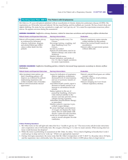
© Copyright 2026