
Design of Drug Delivery Methods for the Brain and Central
Design of Drug Delivery Methods for the
Brain and Central Nervous System
BY
ERIC LUESHEN
B.S., University of Nebraska-Lincoln, 2007
THESIS
Submitted as partial fulfillment of the requirements
for the degree of Doctor of Philosophy in Bioengineering
in the Graduate College of the
University of Illinois at Chicago, 2015
Chicago, IL
Defense Committee:
Prof. Andreas Linninger, Chair and Advisor
Dr. Marvin Rossi, Neurology, Rush University Medical Center
Prof. Urmila Diwekar, Bioengineering
Prof. James Patton, Bioengineering
Prof. Michael Cho, Bioengineering
ACKNOWLEDGEMENTS
First of all, I want to express my gratitude and sincere thanks to my advisor
Prof. Andreas Linninger for his guidance and motivation throughout my research. I gained
a wealth of knowledge through his tutelage in and out of his classroom and lab. I admire
him for his work ethic and passion for the field of bioengineering.
I would also like to thank all of my thesis committee members for serving on my
committee and for your valuable input during my preliminary exam. I would like to thank
Dr. Marvin Rossi for sharing with me his knowledge of the brain and central nervous
system, as well as for access to COMSOL software. I would like to thank Prof. Michael Cho,
Prof. Urmila Diwekar, and Prof. James Patton for the immense amount of knowledge I
gained through taking their courses.
I also extend my gratitude to all of my current and past lab colleagues: Indu,
Sebastian, Kevin, Ian, Chih-Yang, Ying, Cierra, Sukhi, Brian, Nikhil, Seon, Andrej, Madhawa,
Jeonghwa, Minh, Jacek, Joe, and Tejen. They provided valuable suggestions and critical
reviews of my work especially during group meetings and private discussions.
I would like to acknowledge the invaluable support Indu, Joe, and Tejen provided
me with in the intrathecal magnetic drug targeting research. Indu’s knowledge of
nanoparticle synthesis was necessary to the success of the project. Tejen and Joe helped
with reproducing experiments and rebuilding more models. Without all of their help the
research would not have progressed as quickly as it did.
I would also like to acknowledge the help I received from Andrej and Cierra
throughout the pharmacokinetics research. These two were pivotal in our project’s success.
Andrej’s programming of the parameter estimation, and Cierra’s research into
ii
pharmacokinetic scaling laws were major contributions to the success of this project. I
would also like to extend my thanks to our collaborators at the Technical University of
enmark,
artina eit ig, rof. a i ul ani, and rof. urkan Sin, for their help with the
computer-aided framework for facilitating the development of PBPK models.
Special thanks to Dr. Ankit Mehta, Dr. Bakhtiar Yamini, and Dr. Richard Penn for
their valuable advice in convection-enhanced delivery. Also, thank you to Dr. Ali Alaraj for
his input and guidance in my intrathecal magnetic drug targeting research.
I thank Prof. Gayle Woloschak, Prof. Tatjana Paunesku, and Prof. Wendelin Stark for
offering their expertise in nanoparticle synthesis and characterization.
Lastly, I am extremely grateful to all of my family and friends whose invaluable
support helped keep me motivated even in the most difficult of times throughout this
process. Specifically, I would like to thank my grandma Pat, mom, dad, my two sisters
Brandy and Cassie, Dave, and Justin. I would not be where I am today without your love,
guidance, and support.
iii
TABLE OF CONTENTS
CHAPTER
1.
2.
PAGE
INTRODUCTION ..............................................................................................................................
1.1.
Summary ..........................................................................................................................
1.2.
Motivation........................................................................................................................
1.2.1. Drug delivery to the brain and CNS .......................................................................
1.2.1.1. Convection-enhanced delivery ................................................................................
1.2.1.1. Intrathecal magnetic drug targeting .....................................................................
1.2.2. Physiologically-based pharmacokinetic models...............................................
1.2.2.1. Mechanistic PBPK models with empirical scaling laws .................................
1.2.2.2. Multiscale mechanistic PBPK model with mechanistic intra- and
inter-species scaling ....................................................................................................
1.3.
Specific Aims ...................................................................................................................
1.3.1. Engineer a First Principles Physiologically-Based Pharmacokinetic
Modeling Framework ..................................................................................................
1.3.2. Transport Phenomena of Convection-Enhanced Delivery and Design of
Backflow-Free Catheters ............................................................................................
1.3.3. Development of Intrathecal Magnetic Drug Targeting Methods ................
FIRST PRINCIPLES PHYSIOLOGICALLY-BASED PHARMACOKINETIC
MODELING.........................................................................................................................................
2.1.
Abstract.............................................................................................................................
2.2.
Introduction ....................................................................................................................
2.3.
Mathematical Formulation of Whole Body Pharmacokinetics ...................
2.3.1. Steady state model .......................................................................................................
2.3.1.1. Blood flow ........................................................................................................................
2.3.1.2. Systemic circulation network ..................................................................................
2.3.2. Transient drug biotransport.....................................................................................
2.3.2.1. Cyclosporin pharmacokinetics ................................................................................
2.3.2.2. PBPK model assumptions ..........................................................................................
2.3.2.3. Pharmacokinetics in blood compartments .........................................................
2.3.2.4. Pharmacokinetics in organs and tissues ..............................................................
2.3.2.5. Cellular level reaction kinetics.................................................................................
2.3.3. Kinetic inversion problem and solution ..............................................................
2.3.3.1. Non-linear programming algorithm for parameter estimation .................
2.3.3.2. Approximation of confidence intervals for implicit systems .......................
2.4.
Results: Case Study on Cyclosporin .......................................................................
2.4.1. Whole body circulation model .................................................................................
2.4.2. Pharmacokinetic model on Cyclosporin bolus injection into a rat ............
iv
1
1
1
1
2
4
6
7
8
9
10
11
11
13
13
14
17
20
20
21
21
24
25
27
28
29
30
30
32
34
35
36
2.4.3.
3.
4.
Mechanistic parameter estimation for a therapeutic Cyclosporin
intravenous administration ......................................................................................
2.5.
Discussion ........................................................................................................................
2.6.
Conclusions .....................................................................................................................
INTERSPECIES SCALING IN PHARMACOKINETICS: DISCOVERING DRUG
BIODISTRIBUTION MECHANISMS IN VIVO ..........................................................................
3.1.
Abstract.............................................................................................................................
3.2.
Introduction ....................................................................................................................
3.2.1. From classical to mechanistic whole body pharmacokinetic models ......
3.2.2. Mechanistic PBPK models with empirical scaling laws .................................
3.2.3. Multiscale mechanistic PBPK model with mechanistic intra- and
inter-species scaling ....................................................................................................
3.3.
Interspecies Pharmacokinetic Modeling Framework.....................................
3.3.1. Overview of the methods ...........................................................................................
3.3.2. Whole body circulation network ............................................................................
3.3.3. Mathematical formulation of Cyclosporin pharmacokinetics .....................
3.3.4. Parameter estimation - kinetic inversion problem and solution ...............
3.3.5. Interspecies scaling laws for pharmacokinetics ...............................................
3.4.
Results: From Pharmacokinetic Rat Trials to Predictions in Monkeys,
Pigs, and Humans ..........................................................................................................
3.4.1. Systemic blood circulation model for the mouse, rat, monkey, pig,
and human .......................................................................................................................
3.4.2. Pharmacokinetic model of a Cyclosporin bolus injection into the rat .....
3.4.3. Mechanistic parameter estimation for a therapeutic IV Cyclosporin
administration in the rat ............................................................................................
3.4.4. Interspecies scaling of pharmacokinetics: scaling from rats to
monkeys, pigs, and humans ......................................................................................
3.4.4.1. Scaling of pharmacokinetic parameters in organs...........................................
3.4.4.2. Rhesus monkey ..............................................................................................................
3.4.4.3. Pig .......................................................................................................................................
3.4.4.4. Human ...............................................................................................................................
3.5.
Discussion ........................................................................................................................
3.5.1. Scaling of pharmacokinetics between different species ................................
3.5.2. Prediction-quality assessment of interspecies scaling ..................................
3.5.3. Pharmacokinetic simulations for human models.............................................
3.6.
Conclusions .....................................................................................................................
STUDYING THE VARIABLE DISTRIBUTION GEOMETRY OF CONVECTIONENHANCED DRUG DELIVERY BOTH IN SILICO AND IN VIVO........................................
4.1.
Abstract.............................................................................................................................
4.2.
Introduction ....................................................................................................................
v
36
41
51
53
53
54
54
56
57
59
59
60
61
63
64
67
67
70
71
73
74
76
77
79
80
80
81
83
85
89
89
90
5.
6.
4.3.
Materials and Methods ...............................................................................................
4.3.1. Computer simulations .................................................................................................
4.3.2. Nanoparticle infusions ................................................................................................
4.3.3. Sphericity .........................................................................................................................
4.4.
Results ...............................................................................................................................
4.5.
Discussion ........................................................................................................................
4.6.
Conclusions .....................................................................................................................
TRANSPORT PHENOMENA OF CONVECTION-ENHANCED DELIVERY AND
DESIGN OF BACKFLOW-FREE CATHETERS .........................................................................
5.1.
Abstract.............................................................................................................................
5.2.
Introduction ....................................................................................................................
5.2.1. Background .....................................................................................................................
5.3.
Materials and Methods ...............................................................................................
5.3.1. In vitro experiments.....................................................................................................
5.3.2. Computer simulations .................................................................................................
5.4.
Results ...............................................................................................................................
5.4.1. Channel-inducing catheter ........................................................................................
5.4.2. Simulations of channel-inducing catheter experiments ................................
5.4.3. Testing and analysis of channel-inducing catheter .........................................
5.4.4. Dual-action backflow-free catheter systems ......................................................
5.4.5. Dual-action catheter prototype designs...............................................................
5.4.6. Testing and analysis of dual-action catheters ...................................................
5.5.
Discussion ........................................................................................................................
5.6.
Conclusions .....................................................................................................................
INTRATHECAL MAGNETIC DRUG TARGETING ..................................................................
6.1.
Abstract.............................................................................................................................
6.2.
Introduction ....................................................................................................................
6.3.
Materials and Methods ...............................................................................................
6.3.1. Synthesis of gold-coated magnetite nanoparticles (Fe3O4@Au MNPs) ...
6.3.2. In vitro human spine model ......................................................................................
6.3.2.1. Geometry and anatomy ..............................................................................................
6.3.2.2. CSF pulsations ................................................................................................................
6.3.2.3. Nanoparticle injection.................................................................................................
6.3.2.4. Magnetic targeting........................................................................................................
6.3.3. Multiphysics simulation-based determination of optimum
magnetic field .................................................................................................................
6.3.4. Experimental procedure to determine MNP collection efficiency.............
6.4.
Results ...............................................................................................................................
6.4.1
Nanoparticle characterization .................................................................................
6.4.2. Collection efficiency as a function of magnet strength ...................................
vi
91
91
92
93
94
97
101
103
103
104
107
110
110
111
111
111
113
115
118
118
119
121
123
125
125
125
128
128
130
130
131
132
132
133
135
136
136
137
6.4.3.
6.4.4.
7.
8.
Collection efficiency as a function of time ...........................................................
Collection efficiency as a function of magnet location along
spine model .....................................................................................................................
6.4.5. Magnetic guidance at physiological distance & implant-assisted
guidance ...........................................................................................................................
6.5.
Discussion ........................................................................................................................
6.6.
Conclusions .....................................................................................................................
IMPLANT-ASSISTED INTRATHECAL MAGNETIC DRUG TARGETING .......................
7.1.
Abstract.............................................................................................................................
7.2.
Introduction ....................................................................................................................
7.3.
Materials and Methods ...............................................................................................
7.3.1. Synthesis of gold-coated magnetite nanoparticles (Fe3O4@Au MNPs) ...
7.3.1.1. Preparation of the iron stock solution ..................................................................
7.3.1.2. Synthesis of the Fe3O4 nanoparticles ....................................................................
7.3.1.3. Coating the Fe3O4 nanoparticles with gold (Au) ...............................................
7.3.2. In vitro human spine model & experimental setup .........................................
7.3.3. Experimental procedure to determine MNP collection efficiency .............
7.4.
Results ...............................................................................................................................
7.4.1. Computer-aided design of a high gradient magnetic field ............................
7.4.2. In vitro IT-MDT experiments at a physiological distance:
implant vs. no implant ................................................................................................
7.5.
Discussion ........................................................................................................................
7.6.
Conclusions .....................................................................................................................
CONCLUSIONS AND FUTURE WORK ......................................................................................
8.1
Summary ..........................................................................................................................
8.2.
Contributions of This Dissertation .........................................................................
8.3.
Future Work ....................................................................................................................
140
142
144
147
151
153
153
154
157
158
160
160
160
161
163
164
164
167
170
173
174
174
174
181
CITED LITERATURE ...................................................................................................................... 185
APPENDICES
APPENDIX A .................................................................................................................... 204
APPENDIX B .................................................................................................................... 211
VITA ..................................................................................................................................................... 213
vii
LIST OF TABLES
TABLE
1.
2.
3.
4.
5.
6.
7.
8.
9.
10.
PAGE
List of symbols used ......................................................................................................................
Physiological data for Sprague-Dawley rats. Fraction of blood in organ was
obtained from (1), the blood flow rate, organ volume and volume fraction
of total body from (2,3) and the fractions of cells and interstitium in
organs from (4)................................................................................................................................
Optimal values of kinetic rates of binding (k1, ..., k4), mass transfer coefficients
(k5, ..., k28), kinetic clearance rates (k29, k30), are shown, where (+) denotes mass
transfer or binding into, (-) out of the compartment and (*) the clearance terms.
Standard deviation was estimated for 95.0% individual confidence interval. The
least square fitting objective function value is 3.140. ......................................................
Biochemical and Physiological Scaling from Rat (I) to Larger Species (J) ................
Organ volumes for the reference mouse, rat, rhesus monkey, pig, and human. ....
Blood volumes for the reference mouse, rat, rhesus monkey, pig, and human. .....
Blood flow rates for the reference mouse, rat, rhesus monkey, pig and human. ...
Optimal parameter estimates of kinetic rates of binding (k1…k4), mass transfer
rates (k5…k28) and metabolic rates (k29, k30), are shown, where (+) denotes
binding into, (-) out of compartment, (M) the metabolic terms and UA the mass
transfer rates. The fraction of free drug in each organ, which is available for
mass transfer, kQ, correlates the drug concentration in the interstitial fluid and
in entire organ. Deviations were estimated for 95.0 % individual confidence
level. The least square fitting objective function value is 3.140. Parameters,
which have been scaled from rat to other species based on physiological
values are marked with (*). ........................................................................................................
Comparison of experimental and predicted Cyclosporin binding and
pharmacokinetic parameters in individual stem cell transplantation patients
after a continuous, 24 h iv administration. Experimental data on Cyclosporin
dose, concentration and 12 h AUC has been obtained from (5). The data
obtained from literature did not specify the weight of each patient or exact
dose data; therefore, the predicted values are based on the simulated 24 h
injection into a 73 kg reference human with pharmacokinetic parameters
obtained by scaling from the male Sprague-Dawley rat trial with 6 mg/kg
iv bolus administration. Had the exact weight of each patient and dose data
been available, prediction errors would have been reduced and a plot with
error bars would have been included. ....................................................................................
Comparison of backflow-free and single-port catheters. ................................................
viii
19
35
37
66
68
69
69
72
80
121
LIST OF FIGURES
FIGURE
1.
2.
3.
4.
5.
6.
7.
8.
9.
10.
11.
12.
PAGE
Overview of the “pharmacokinetic modeling framework” and its components ....
Diagram of a circulatory system model consisting of arterial, venous and
capillary segments in a Sprague-Dawley rat ........................................................................
A compartmental model example of the Cyclosporin (CyA) physical mass
transfer from unbound plasma fraction (cP) through the capillary membrane
into the interstitial fluid and the tissue cells........................................................................
Distribution of Cyclosporin in selected organs, tissues, and blood compartments
after an intravenous bolus administration of 6 mg/kg into a Sprague-Dawley rat
obtained experimentally (crosses), estimated from literature (circles) and
predicted by our PBPK model (lines) .....................................................................................
Total mass of Cyclosporin in vivo measured (crosses) and predicted by our
mechanistic model (lines) as a function of time .................................................................
Model results predicting the in vivo evolution of the mass fraction of Cyclosporin
in tissues and organs of a rat ......................................................................................................
Model results predicting the evolution of total mass of Cyclosporin in vivo in
tissues and organs of a rat ...........................................................................................................
Dose-response curves in selected organs of a rat, based on unique parameter set
obtained only from a single therapeutic dose experiment (squares) at 6.0 mg/kg
IV for two minutes ..........................................................................................................................
Model matrix showing a set of simulations based on the calculated optimal
parameters for Cyclosporin injection into a rat. The middle figure shows the
possibility of dose variation calculations in selected organs, based on the set of
parameters obtained solely from the therapeutic (6.0 mg/kg) set of experiments.
The left figure shows the results from rats with different pathological conditions,
obesity (red line) compared to cirrhosis (black dashed line), in liver and fat
tissues. The figure on the right depicts the variation of injection time from bolus
to 12 h continuous administration ..........................................................................................
A generic framework for PBPK model development and validation (left), and its
computer-aided implementation as a modeling tool (right) .........................................
Predicted Cyclosporin concentration profile (a) in a rat's pericardium after (b)
continuous 12 h injection of 7 mg/kg in contrast to (d) a controlled flow rate of
7 mg/kg over the same time period. The aim of the therapy design case study is
to maintain a constant concentration of 1 μg/ml in the heart muscle for a period
of 11 h as seen in (c) ......................................................................................................................
(a) A collection of vertebrates has been compiled for demonstrating interspecies
scaling of pharmacokinetics. (b) The topology of the vasculature network is
ix
18
22
23
39
40
40
41
43
45
47
49
13.
14.
15.
16.
17.
18.
19.
20.
21.
22.
23.
24.
25.
26.
shown on the right with twelve organs including the venous (left), capillary
(middle), and arterial (right) parts… (c) The blood compartment consisting of
red blood cells (Hct), plasma (unbound and bound drug fractions), and
organs/tissues is shown on the left ......................................................................................... 60
Scaling of mass transfer parameters. Mass transfer rates of Cyclosporin from
blood to selected organs in rat, monkey, pig, and human ............................................... 73
Key physiology library for laboratory animals and human ........................................... 76
Pharmacokinetic simulations of Cyclosporin administration in monkeys, pigs,
and humans based on a single laboratory experiment in rats, and the proposed
mechanistic model of biodistribution ..................................................................................... 78
Simulation of population pharmacokinetics on Cyclosporin administered i.v. to
five human subjects over 2.5 h at a dose of 4 mg/kg ........................................................ 84
Sphericity (S) provides a simple, objective measure of the conformity of
distribution geometry to an idealized rough spherical distribution calculated
from 2 dimensional slices (A) and an idealized spherical distribution calculated
from 3 dimensional data (B) ...................................................................................................... 94
Computer simulations of the CED of 20 µL FPNPs infused at a rate of 0.5 µL/min
into the right caudate putamen. Rat DTI data were used to model the anatomy and
anisotropy of brain tissue ............................................................................................................ 95
Representative histological sample following 0.5 µL/min CED of 20 µL FPNPs
into the right caudate putamen (A). In this slice, volume of distribution is
outlined (B) and length and width are measured to calculate sphericity (C) .......... 96
Representative individual distribution geometries (boxed) were compared
with corresponding relative volume of distribution (dark bars) and sphericity
(light bars). Infusions of 20 µL FPNPs were performed in 4 animals at a rate
of 0.5 µL/min .................................................................................................................................... 97
Unpredictable and non uniform distribution geometries from in silico (A and C)
and in vivo (B and D) CED experiments even though very low volumetric flow
rate of 0.5 μL/min were used. (Adapted from our previous work.(9)) ..................... 107
Schematic of experimental setup for catheter testing by CED infusion
experiments ...................................................................................................................................... 111
Schematic of novel, backflow-free channel-inducing catheter ..................................... 113
Simulations comparing the channel-inducing and common single-port
catheters ............................................................................................................................................. 115
Photographs of channel-inducing (a, b, c, g, h, and i) vs. single-port (d, e, f, j, k,
and l); catheter experiments at flow rate 1.0 µl/min (a-f) and 3.0 µl/min (g-l) .... 116
Photographs of channel-inducing catheter at 5.0 µL/min at (a) 0 min, (b) 50
min, (c) 100 min, (d) 150 min, and then flow rate increased to 10 µL/min for
an additional 50 minutes (e) 200 min .................................................................................... 117
x
27.
28.
29.
30.
31.
32.
33.
34.
35.
36.
37.
38.
Schematic drawing of both the coaxial and series dual-action novel
backflow-free catheters................................................................................................................
Photographs of coaxial dual-action catheter at 3.0 µL/min at (a) 10 min,
(b) 30 min, (c) 50 min, (d) 70 min, and (e) 90 min ...........................................................
Photographs of series dual-action catheter at 3.0 µL/min at (a) 30 min,
(b) 60 min, (c) 90 min, (d) 120 min, and (e) 150 min ......................................................
(A) Schematic of Fe3O4@Au nanoparticle; (B) TEM image of nanoparticles
showing hydrodynamic diameter to be between 20-25 nm; (C) TEM image
showing a partially gold-coated Fe3O4 core which confirms the core diameter
to be around 8-12 nm; (D) The energy density spectrum of the gold-coated
magnetite nanoparticles determined using EDS which indicates the presence
of elements Au, Fe and O ..............................................................................................................
Schematic of entire experimental setup with the in vitro human spine model
which clearly shows the three different zones (injection, targeting, and
barrier zones)...................................................................................................................................
Magnetic field lines produced by (A) in vitro human spine model used in our
experiments with the 0.528 T surface field strength targeting magnet and the
1.05 T surface field strength barrier magnet and by (B) spine model in which
both magnets are of equal 1.05 T surface field strength .................................................
Magnetic field produced by targeting magnet and barrier magnet (shown
in xz-plane)........................................................................................................................................
Plot of magnetization vs. magnetic field, obtained by SQUID magnetometry, for
the Fe3O4@Au magnetic nanoparticles used in our in vitro intrathecal magnetic
drug targeting experiments ........................................................................................................
Graph showing the nanoparticle collection efficiency (CE) as a function of
magnet strength at the targeting zone ...................................................................................
Graph showing the nanoparticle collection efficiency (CE) as a function of time .
Graph showing the variation of nanoparticle collection efficiency (CE) at
different target zones in the in vitro human spine model as a function of the
distance of the targeting magnetic field from the injection site ...................................
(A) Patient MRI showing the 4 cm physiological distance between intrathecal
119
120
121
130
131
134
134
137
139
141
143
space and epidermis; (B) Simulated magnetic field produced by the 0.528 T
surface field, 1045 lb pull force strength magnet placed at a 4 cm distance away
from the spinal canal; magnetic field within the spinal canal is within a
0.116-0.160 T range; (C) Simulated field when two ferrous implants were
placed within epidural space; high gradient magnetic field created within the
spinal canal is within a 0.067-0.626 T range ....................................................................... 145
xi
39.
40.
41.
42.
43.
44.
(A) TEM image of nanoparticles showing hydrodynamic diameter to be
between 20-25 nm; (B) TEM image showing a partially gold-coated Fe3O4
core which confirms the core diameter to be around 8-12 nm .................................... 158
Schematic of entire experimental setup with the in vitro human spine model
which clearly shows the three different zones (injection, targeting, and barrier
zones), as well as the magnetically susceptible implant (spiral) and the targeting
magnet placed at a 4 cm physiological distance away from the spinal canal .......... 162
3D wireframe images built in COMSOL 4.3a of both the (A) spiral and (B) mesh
implants inside the subarachnoid space of the in vitro human spine model which
were used in our experiments ................................................................................................... 163
(A) Patient MRI showing the 4 cm physiological distance between intrathecal
space and epidermis; (B) Simulated magnetic field produced by the 0.528 T
surface field, 1045 lb pull force strength magnet placed at a 4 cm distance away
from the spinal canal; magnetic field within the spinal canal is within a
0.116-0.248 T range (magnetic field gradient ranging from 0.356-0.480 T/cm);
(C) Simulated field when a spiral implant was placed within subarachnoid space;
high gradient magnetic field created inside the spinal canal with field values
between 0.062-12.027 T (magnetic field gradient ranging from 6.129-26.346
T/cm); (D) Simulated field when a mesh implant was placed within
subarachnoid space creating a high gradient magnetic field inside the spinal
canal with field values between 0.101-28.316 T (magnetic field gradient
ranging from 25.676-150.222 T/cm)...................................................................................... 166
Graph showing the high gradient magnetic fields around both implants, which
have field strengths varying from 0.101-28.316 T for the mesh implant and
from 0.062-12.027 T for the spiral implant.......................................................................... 167
Graph showing a comparison of the nanoparticle collection efficiencies from
three different types of experiments: (i) control, (ii) magnet at a physiological
distance, and (iii) magnet at a physiological distance while ferromagnetic
implant, either spiral or mesh, is within subarachnoid space....................................... 169
xii
LIST OF ABBREVIATIONS
AD
Al heimer’s disease
BBB
Blood-brain barrier
CED
Convection-enhanced delivery
CNS
Central nervous system
CSF
Cerebrospinal fluid
DTI
Diffusion tensor imaging
EDS
Energy dispersive x-ray spectroscopy
IA-IT-MDT
Implant-assisted intrathecal magnetic drug targeting
IT
Intrathecal
IT-MDT
Intrathecal magnetic drug targeting
MNP
Magnetic nanoparticle
PBPK
Physiologically-based pharmacokinetics
PD
arkinson’s disease
PK
Pharmacokinetics
SQUID
Superconducting quantum interference device magnetometry
TEM
Transmission electron microscopy
xiii
SUMMARY
Due to the impermeability of the blood-brain barrier (BBB) to macromolecules
delivered systemically, drug delivery to the brain and central nervous system (CNS) is quite
difficult and has become an area of intense research. Techniques such as convectionenhanced intraparenchymal delivery and intrathecal magnetic drug targeting offer a means
of circumventing the blood-brain barrier for targeted delivery of therapeutics. This
dissertation focuses on three aspects of drug delivery: pharmacokinetics, convectionenhanced delivery, and intrathecal magnetic drug targeting.
Classical pharmacokinetics mainly uses black-box curve fitting techniques without
biochemical or biological basis. This dissertation advances the state-of-the-art of
pharmacokinetics and pharmacodynamics by incorporating first principles and
biochemical/biotransport mechanisms in the prediction of drug fate in vivo. A whole body
physiologically-based pharmacokinetics (PBPK) modeling framework is engineered which
creates multiscale mathematical models for entire organisms composed of organs, tissues,
and a detailed vasculature network to predict drug bioaccumulation and to rigorously
determine kinetic parameters. These models can be specialized to account for species,
weight, gender, age, and pathology. Systematic individual therapy design using the
proposed mechanistic PBPK modeling framework is also a possibility.
Biochemical, anatomical, and physiological scaling laws are also developed to
accurately project drug kinetics in humans from small animal experiments. Our promising
results demonstrate that the whole-body mechanistic PBPK modeling approach not only
elucidates drug mechanisms from a biochemical standpoint, but offers better scaling
precision. Better models can substantially accelerate the introduction of drug leads to
xiv
SUMMARY (continued)
clinical trials and eventually to the market by offering more understanding of the drug
mechanisms, aiding in therapy design, and serving as an accurate dosing tool.
Convection-enhanced drug delivery (CED) is a technique used to bypass the BBB via
direct intracranial injection using a catheter driven by a positive pressure gradient from an
infusion pump. Although CED boasts the advantage of achieving larger drug distribution
volumes compared to diffusion driven methods, difficulty in predicting drug spread and
preventing backflow along the catheter shaft commonly occur. In this dissertation, a
method for predicting drug distributions in the brain using diffusion tensor imaging (DTI)
data is employed to show how small variations in catheter placement can lead to drastically
different volumes of drug distribution in vivo. The impact that microfluid flow has on
deformable brain phantom gel is studied in order to elucidate the causes of backflow, and
the results are used to develop backflow-free catheters with safe volumetric flow rates up
to 10 μl/min. Through implementation of our backflow-free catheter designs, physicians
will be able to target specific regions of the brain with improved accuracy, increased drug
concentration, and larger drug distribution geometries.
Intrathecal (IT) drug delivery involves direct drug infusion into the spinal canal and
has become standard practice for treating many CNS diseases. Although IT drug delivery
boasts the advantage of reduced systemic toxicity compared to oral and intravenous
techniques, current IT delivery protocols lack a means of sufficient drug targeting at
specific locations of interest within the CNS. In this dissertation, the method of intrathecal
magnetic drug targeting (IT-MDT) is developed to overcome the limited targeting
capabilities of standard IT drug delivery protocols. The basic idea behind IT-MDT is to
guide intrathecally injected, drug-functionalized magnetic nanoparticles (MNPs) using an
xv
SUMMARY (continued)
external magnetic field to diseased regions within the spinal canal. Cerebrospinal fluid
(CSF) transport phenomena are studied, and in vitro human spine surrogates are built.
Experiments are run on the in vitro human spine model to determine the feasibility of
IT-MDT and to develop novel treatment therapies. Computer simulations are performed to
optimize magnetic field placement and/or implant design for generating high gradient
magnetic fields, as well as to study how these fields aid in therapeutic nanoparticle
localization.
Large collection efficiencies of MNPs were achieved during in vitro IT-MDT and
implant-assisted IT-MDT experiments with concentration levels nearly nine times that of
the control when no magnetic field was present. Testing different magnetizable implants
showed that implant design is a key factor in achieving the largest MNP collection efficiency
within the targeting region. Knowledge gained from the in vitro IT-MDT experiments and
simulations will be used in the future to develop IT-MDT methods in animals and humans.
xvi
1. INTRODUCTION
1.1.
Summary
Drug delivery to the brain and central nervous system (CNS) is an area of intense
research due to the difficulty of delivering large molecular weight therapeutics across the
blood-brain barrier (BBB) when administered systemically. Although many drugs for
treating neurodegenerative and other diseases have been proven effective in vitro,
physicians have a hard time proving drug efficacy in vivo because reaching therapeutic
levels within the brain and CNS is greatly hindered by the BBB. This has become an
engineering problem, and improved methods of drug delivery to the brain and CNS are
warranted. This dissertation focuses on three aspects of drug delivery: pharmacokinetics,
convection-enhanced delivery, and intrathecal magnetic drug targeting. The outcomes of
this research aim to provide more efficient methods for the treatment of
neurodegenerative disorders, brain cancer, spasticity, chronic pain, and other diseases.
1.2.
Motivation
1.2.1. Drug delivery to the brain and CNS
More than 600 disorders afflict the central nervous system (6). These disorders are
often associated with atrophy of nervous system structures, and they affect millions of
people worldwide. The two most common types of neurodegenerative disorders are
Al heimer’s disease and arkinson’s disease (7). Primary brain and CNS tumors are also
becoming more prevalent.
Al heimer’s isease (A ) and arkinson’s isease (
) collectively effect 6 million
people within the United States, and around 42 million people worldwide (8–10). These
1
numbers are expected to grow substantially as the average age of the population increases.
The current estimated cost of providing care for both AD and PD patients in the U.S. is $215
billion per year (11). It is also estimated that more than 688,000 people are currently living
with a diagnosis of a primary brain or CNS tumor in the United States, and 68,470 new
cases are expected to be diagnosed in 2015 (12,13).
Drug administration to the central nervous system for treatment of the above
mentioned diseases is a challenge due to the highly selective and efficient barrier
protecting the brain known as the blood-brain barrier (BBB). Almost 100% of largemolecule drugs and more than 98% of all small-molecule drug do not penetrate the BBB
(14). There are two important characteristics for small-molecule drugs to potentially cross
the BBB: high lipophilicity and a molecular mass no greater than 500 Da (15). Many
neurodegenerative diseases remain under-treated by effective therapies due to the
inability of commonly used drugs to cross the BBB; not because there is a lack of candidate
drugs to treat the diseases (16). Therefore, alternative methods to effectively deliver
neurotherapeutics and target specific regions of the brain and CNS are necessary.
1.2.1.1. Convection-enhanced delivery
Convection-enhanced delivery (CED) is a localized drug delivery technique used to
target specific regions of the brain for the treatment of cancer and neurodegenerative
diseases that bypasses the blood-brain barrier. An intracerebral catheter is placed with a
continuous infusion of therapeutic molecules that are propelled to the site of drug delivery
within the parenchyma through a pump. CED directly distributes therapeutic agents to a
specific target area at dramatically increased doses and has been shown to achieve much
larger drug distribution volumes compared to diffusion driven methods (99,126). These
2
major advantages of CED offer great potential for more efficient treatment of
neurodegenerative diseases such as Al heimer’s and
arkinson’s, as well as for the
treatment of brain tumors.
Even with all of the advantages CED has to offer, clinical trials treating gliomas that
utilized CED, such as the TransMID and PRECISE trials, have failed to demonstrate
statistically significant improvement in survival (17) despite the proven efficacy of
therapeutic agents in animal models (18,19). Retrospective analysis of the PRECISE trial
found that infusate distribution was highly variable among patients; therefore, any
potential efficacy of drugs delivered by CED may be hindered by ineffective delivery (20–
22). One of the inherent engineering challenges involved with CED is known as backflow,
which has a large impact on infusate distribution volumes and geometries. Backflow is
characterized by fluid discharge along the catheter shaft instead of distally into the soft
tissue. In order to prevent the possibility of backflow, low volumetric flow rates are applied
which limit the achievable drug distribution volumes from CED. This can render CED
treatment ineffective since a small convective flow rate produces a narrow drug
distribution inside the treatment region. The causes of backflow need to be determined,
and more efficient catheter designs and CED protocols are needed to expand the drug
distribution inside the treatment region. This is especially important when administering
toxic chemotherapeutics which could adversely affect other brain regions when backflow
occurs, as well as not reach a therapeutically efficacious concentration within the target
region due to backflow.
3
1.2.1.2. Intrathecal magnetic drug targeting
Intrathecal (IT) drug delivery is a technique which involves the direct infusion of
therapeutic molecules into the cerebrospinal fluid (CSF) filled space within the spinal canal.
IT delivery is advantageous because the drugs bypass the blood-brain barrier.
Furthermore, drugs experience a longer half-life within the CSF since they encounter
minimal protein binding and are not exposed to the same enzymatic activities which
systemically administered drugs face within the blood (213). Current implications for IT
drug delivery include leptomeningeal metastases (149–151), spasticity (152), pain
management (153), and spinal anesthesia (154,155). For example, clinical studies on pain
management show that IT delivery provides ideal pain control with fewer side effects,
while using only a small fraction of the dose required when the drug is administered orally
or intravenously (159,214). The IT delivery of neurotrophic factors has also been deemed a
promising treatment for neurodegenerative diseases such as amyotrophic lateral sclerosis
(ALS),
untington’s disease, and
arkinson’s disease (156,157,215). Upon IT drug
administration, the drugs are rapidly dispersed within the spinal canal by both molecular
diffusion and by the pulsatile motion of CSF (157,178).
Standard intrathecal drug delivery methods rely mainly on empirical observations.
The use of current IT guidelines cannot guarantee reproducible outcomes or desired
therapeutic concentrations in specific target regions of the spinal canal (157,231). Due to
variability in patients’ spinal anatomy, as well as the fre uency and magnitude of their CSF
pulsations, maintaining a therapeutic dose within the targeted region while attempting to
avoid systemic toxicity is a major challenge and disadvantage of standard IT delivery
methods (178). Current IT protocols require continuous drug infusion in order to maintain
4
a sufficiently high drug concentration at the target site despite drug distribution
throughout the entire central nervous system leading to harmful side effects. For many CNS
diseases like leptomeningeal metastases, the effected tissue is located in specific regions of
the spinal canal; therefore, a method to target these regions is warranted.
To maximize the therapeutic effect without excessive risk of toxicity in non-targeted
zones, the intrathecally injected drugs should be concentrated at specific locations within
the spinal canal. At this point there is no delivery technique to confine drugs locally, where
it is needed in high concentrations, without having large amounts of drug spread
throughout the entire brain and spinal canal, which may cause side effects.
We propose the ideas of intrathecal magnetic drug targeting (IT-MDT) and implantassisted intrathecal magnetic drug targeting (IA-IT-MDT) to achieve localized, high
concentrations of magnetic nanocarriers which can be functionalized to different drugs to
overcome the limitations of standard IT drug delivery. IT-MDT builds on the method of
magnetic drug targeting (MDT), which is a type of active drug targeting method utilizing
drug-functionalized magnetic nanoparticles localized to a target region by use of an
external magnetic field. The most notable benefits of MDT reside in local drug action and
minimization of systemic side effects. Until now, MDT methods have been mainly used to
target drugs delivered systemically through blood vessels and have shown great promise
(161–169). The novelty of our approach lies in applying the methods of MDT within the
CSF-filled spinal canal, not in blood vessels, in order to achieve a localized therapeutic
effect using much smaller drug doses and substantially reducing systemic toxicity for novel
treatments of CNS diseases.
5
1.2.2. Physiologically-based pharmacokinetic models
Pharmacokinetic (PK) models aim to establish relationships between drug
administration, bioaccumulation, and elimination from dose-response measurements
in vivo. Typical PK models fit parametric functions with multiple adjustable constants or
exponential coefficients (18–20). Non-mechanistic parameters such as volume of
distribution, area under the curve (AUC), and the intrinsic clearance rates are usually
computed. These classical PK models derive little information about drug reaction kinetics
and biotransport phenomena; they also do not satisfy conservation laws, so that drug
species balances are not necessarily closed. There is a critical need in the pharmaceutical
industry for methods which link modeling, simulation, drug approval, and rigorous
experimental data analysis (26).
Several
authors
have
since
proposed
whole
body
physiologically-based
pharmacokinetic (PBPK) prediction and modeling techniques (2,23–25). PBPK models
include several biological subsystems such as blood, the lymphatic and the central nervous
system, tissues and organs, which can further be subdivided into multiple phases including
cells and interstitial fluid. PBPK models are accepted as a recommended approach for interand intra-species extrapolations and to simulate pharmacokinetic profiles for various
administration modes and dose regimes. These models incorporate data from many
sources such as biochemical, physiological, and drug-dependant parameters for various
species, individuals, or with pathological changes (31). In the literature, compartmental
PBPK models are typically stipulated as a set of interconnected vessels with ideal mixing,
where both biochemical and transport mechanisms are given as black-box, empirical
relations. The kinetics of novel drugs can be studied more systematically with mechanistic
6
biochemical models in entire organisms. Prediction accuracy in PK models could also be
greatly improved by the incorporation of conservation laws, and fundamental transport
and biochemical reaction mechanisms, which is beyond the scope of black-box approaches.
Several authors have recently used first principles modeling to elucidate the biochemical
reaction mechanisms of new drugs in vivo (29–36). Their model topology does not account
for the physiologically consistent blood or lymph perfusion, in the arteries, capillaries, and
veins of individual organs. Consequently, simulation of blood sampling techniques is
imprecise, involving significant errors of pharmacokinetic parameter estimates. These
errors limit the fidelity of the previous PBPK models for the extrapolation of information
from small animals to larger ones, from animals to humans, or for the prediction of drug
fate for varying dosing regimes.
1.2.2.1. Mechanistic PBPK models with empirical scaling laws
Traditional interspecies scaling laws in pharmacokinetics often deploy simple
polynomial relationships between properties of interest such as the intrinsic clearance. The
allometric approach is empirical, and rests on the assumption that the underlying
physiological processes such as cardiac output, heartbeat frequency, breath duration, are
only related to body mass (72). Typical empirical relations for interspecies scaling include:
clearance versus body weight, the product of clearance and maximum life-span potential
versus body weight, the product of clearance and brain weight versus body weight, and the
application of a fixed exponent to clearance (72). Interesting research from Geoffrey West
and colleagues about allometric scaling laws in biology shows early attempts to
incorporate first principles into determining whole organism metabolic rates in different
animals. In particular, West’s group developed a model to explain the origin of uarter7
power scaling laws and there use in determining metabolic rates when organism body
sizes vary over large orders of magnitude (73,74). Unfortunately, interspecies
extrapolation with these simple scaling laws is not satisfactory because it does not account
for fundamental biochemical mechanisms, but merely incorporates weight or size factors.
It is difficult to predict drug fate for different dosing regimes using non-mechanistic PK
models (22). Due to the limitation in predictive capabilities, expensive, and timeconsuming dose-response data have to be acquired in extensive animal trials in rats, then
dogs and monkeys, until finally arriving at reasonably safe specifications for human trials.
Consequently, there is an urgent need in the pharmaceutical industry to develop
mechanistic pharmacokinetic (PK) models able to both expedite knowledge gain from
experimental trials and, simultaneously, address safety concerns.
1.2.2.2. Multiscale mechanistic PBPK model with mechanistic intra- and inter-species
scaling
We propose a multiscale biological system model to describe the drug fate in: cells,
tissues, organs, whole body, among individual subjects, and across species. Our model
solves the problem of rigorously estimating unknown biochemical reaction and transport
parameters from in vivo datasets and identifying whole body physiologically-based
pharmacokinetic (PBPK) models. These parameters may be scaled according to
fundamental chemical and physical principles to create a consistent and rigorous
pharmacokinetic model with better predictive capabilities than classical black-box
pharmacokinetics.
Our approach uses a first principles PBPK model as a starting point, enforcing the
conservation laws and reaction and mass transfer mechanisms based on the scaled
8
parameters. Therefore, the prediction of the bioaccumulation will not result from simple
algebraic scaling but will be the outcome of solving the entire network using the scaled
parameters for each category. Such a model is capable of addressing both physiological as
well as biochemical and transport variations between each species with much higher
accuracy than can be expected from simple allometric scaling laws. Better models like the
one we propose can substantially accelerate the introduction of drug leads to clinical trials
and eventually to the market by offering more understanding of the drug mechanisms,
aiding in therapy design, and serving as an accurate dosing tool.
1.3.
Specific Aims
In debilitating diseases of the central nervous system (CNS), treatment protocols
which use systemic drug delivery are limited due to the impermeability of the blood-brain
barrier (BBB). Therefore, improved methods of targeted drug delivery are warranted. The
outcomes of this dissertation will be as follows: a first principles pharmacokinetic modeling
framework with novel interspecies scaling laws, backflow-free catheters for more efficient
convection-enhanced intraparenchymal drug delivery, and an intrathecal magnetic drug
targeting protocol for improved treatment of CNS diseases. The overall vision of the project
aims at improved therapy options for patients suffering from diseases such as brain cancer,
spasticity, chronic pain, and other neurodegenerative diseases including Al heimer’s
isease and arkinson’s isease. The specific aims are as follows:
9
1.3.1. Engineer a first principles physiologically-based pharmacokinetic modeling
framework
A novel whole body physiologically-based pharmacokinetics (PK) modeling
framework is developed in order to advance the prior black-box PK models by
incorporating first principles, conservation of mass, species and momentum, and
biochemical/biotransport mechanisms for the prediction of drug fate in vivo. A rat blood
circulation model was constructed and combined with biotransport, biochemical reactions
and metabolism of the immunosuppressant Cyclosporin. The proposed methodology was
demonstrated on a case study in male Sprague Dawley rats. A rigorous parameter
estimation technique was used to derive the unknown rate parameters from actual drug
dose–response experiments in animals. A mathematical programming technique was used
for solving the transport and kinetic inversion problem. Key PK parameters were
determined to establish drug biodistribution dynamics in all organs and tissues. Prediction
of organ drug bioaccumulation as a function of cardiac output, physiology, pathology or
administration route is possible with the proposed PBPK framework. Biochemical,
anatomical and physiological scaling laws were developed which yield more reliable
interspecies and intraspecies extrapolation of drug biodistribution. This work, which
resulted in two refereed journal publications (23,24) and three refereed conference
proceedings articles (25–27), demonstrated how the whole-body PK modeling approach
not only elucidates drug mechanisms from a biochemical standpoint, but offers better
scaling precision.
10
1.3.2. Transport phenomena of convection-enhanced delivery and design of
backflow-free catheters
Another research phase of this dissertation was devoted to studying the transport
phenomena and drug distribution variability which occurs during convection-enhanced
intraparenchymal drug delivery. Physiological and geometric properties of heterogeneous
and anisotropic rat brain tissue were obtained from diffusion tensor imaging (DTI)
datasets. These DTI datasets helped to construct physiologically consistent transport
tensors for nanoparticle and bulk fluid transport within the brain tissue. Computer-assisted
image
reconstruction
was
conducted,
and
simulations
were
performed
using
computational fluid dynamics software. In vitro CED of trypan blue dye into agarose gel
brain phantoms was performed to aid in the understanding of CED transport phenomena.
In vivo CED of fluorescent nanoparticles into rat striatum was performed, and the
distribution geometry was examined histologically with fluorescence microscopy.
Knowledge gained from in silico, in vitro, and in vivo CED experiments was then used to
design and test novel backflow-free catheters. This work, which resulted in four refereed
journal publications (20,28–30), aided in understanding the transport phenomena during
CED as well as in the engineering of several novel catheter designs.
1.3.3. Development of intrathecal magnetic drug targeting methods
The final research phase of this dissertation aims at developing the methods of
intrathecal magnetic drug targeting (IT-MDT) and implant-assisted IT-MDT (IA-IT-MDT) in
order to increase the targeting capabilities of intrathecally administered drugs for more
efficient treatment of CNS diseases. Intrathecal drug transport phenomena were studied,
11
in vitro human spine surrogates were built, magnetic nanoparticles were synthesized, and
in vitro experiments were performed. Optimization of magnetic field placement and
implant design was also studied computationally. This work, which resulted in two
refereed journal publications (31,32) and one refereed conference proceedings article (33),
offers great promise for improved treatment methods of CNS diseases upon further
validation in vivo.
12
2. FIRST PRINCIPLES PHYSIOLOGICALLY-BASED PHARMACOKINETIC MODELING
2.1.
Abstract
Unknown dose regimes are typically assessed on animals prior to clinical trials.
ecent advances in the evaluation of new leads’ efficacy have been achieved by
pharmacokinetic modeling. Further improvements, including determination of the drug’s
mechanism of action and organism biodistribution, require an effective methodology for
solving parameter estimation challenges. This research solves the problem of rigorously
estimating unknown biochemical reaction and transport parameters from in vivo datasets
and identifying whole-body physiologically based pharmacokinetic (PBPK) models.
A rat blood circulation model was combined with biotransport, biochemical
reactions and metabolism of the immunosuppressant Cyclosporin. We demonstrate the
proposed methodology on a case study in Sprague-Dawley rats by bolus iv injections of 1.2,
6 and 30 mg/kg. Key pharmacokinetic parameters were determined, including renal and
hepatic clearances, elimination half-life, and mass transfer coefficients, to establish drug
biodistribution dynamics in all organs and tissues. This multi-scale model satisfies first
principles and conservation of mass, species and momentum.
Prediction of organ drug bioaccumulation as a function of cardiac output,
physiology, pathology or administration route may be possible with the proposed PBPK
framework. Successful application of our model-based drug development method may lead
to more efficient preclinical trials, accelerated knowledge gain from animal experiments,
and shortened time-to-market of new drugs.
13
2.2.
Introduction
The effect of novel drugs on targeted organs is typically studied in animal drug
dosing trials. Pharmacokinetic models establish relationships between drug dosage,
bioaccumulation
and
clearance
utilizing
dose-response
measurements.
Classical
pharmacokinetic (PK) models relate anatomy and physiology parametrically to doseresponse data while fitting exponential functions with multiple adjustable constants or
exponential coefficients (34–36). The resulting black-box formula permit the computation
of the area under the curve (AUC), the plasma half-life of elimination (t1/2) or the intrinsic
clearance rates. Limitations are observed when fitted relations are used to extrapolate drug
concentration profiles for different doses (37).
Non-mechanistic, classical PK methods derive very little information about reaction
kinetics and biotransport phenomena. It is reportedly difficult to scale or extrapolate
information among laboratory animals or to predict drug fate for different dosing regimes
(38). Hence, large sets of dose-response data have to be acquired in extensive animal trials
in rats, then dogs and monkeys, until finally arriving at reasonably safe specifications for
human trials. Prediction accuracy in PK models could be greatly improved by the
incorporation of conservation laws, and fundamental transport and biochemical reaction
mechanisms, which is beyond the scope of black-box approaches.
The efficacy of novel drugs can be studied more systematically with mechanistic
biochemical models. Several authors have proposed whole body physiologically-based
pharmacokinetic (PBPK) prediction and modeling techniques (2,39–41). A thorough
review of whole body physiologically-based pharmacokinetic models (42) recognizes the
need for methods linking modeling, simulation, drug approval and rigorous experimental
14
data analysis. Previous work commonly deployed compartments, typically encompassing
several biological subsystems such as blood, plasma, red blood cells, interstitial fluid, the
lymphatic system, the central nervous system, tissues and organs (43).
One possible scenario for drug fate modeling in the whole body is to determine the
drug transport parameters by in vitro measurements, for instance with tissue-to-plasma
partitioning coefficients (4,44). However, these compartmental models typically do not
account for the physiologically consistent blood perfusion or lymphatic fluid exchange
patterns.
Several authors have recently used first principles modeling to elucidate the
biochemical reaction mechanisms of new drugs in vivo (45–52). Although mechanistic, the
underlying algorithms commonly infer systemic circulation with a set of continuity and
conservation differential equations. Every part of the whole body flow network has to be
entered manually. This soon leads to infeasibility if multiple animal models need to be
tested.
In contrast, our improved workflow ensures that for arbitrarily complex PBPK
networks with hundreds of biological “compartments”, corresponding differential
equations are automatically generated and validated. This allows for testing various
circulatory system configurations with greater detail as more data become available on the
drug-organ interactions. Given solid data describing individual pharmacokinetic processes,
the selection of an accurate but general mechanistic model still remains a scientific
challenge.
In this chapter a rigorous engineering approach based on first principles of mass,
species and momentum conservation is proposed to build upon the advances in classical
15
PK modeling. The presented work aims at determining drug reaction kinetics and transport
phenomena from actual experimental dose-response measurements. To scientifically
examine drug fate in living organisms, we emphasize the need for a closed loop iterative
methodological approach: i) obtaining experimental data, ii) constructing first principle
models iii) estimating parameters and iv) gaining insights from comparing working
hypotheses with experimental sets.
We will demonstrate the advantages of our methodology with a case study on the
immunosuppressant Cyclosporin. Our advanced mechanistic model results in a rigorous
analysis of biodistribution after a bolus iv injection into a rat. The model will also be used
to assess different administration regimes, which in the case of Cyclosporin have been
shown to enormously affect interactions with physiology, cardiovascular dynamics and
pharmacology (5,39,53,54). We hope to contribute an improved level of understanding in
such a complex topic.
This chapter is organized as follows. Section Mathematical Formulation of Whole
Body Pharmacokinetics lays out the conceptual foundation: (i) steady state systemic blood
circulation model, (ii) mechanistic transport, mass transfer and biochemical reaction
parameters, and (iii) a parameter estimation technique for determining the unknown
model parameters from actual animal experiments. The application of this concept is
demonstrated with a case study on Cyclosporin in section 2.4. Results are discussed in
section 2.5.
16
2.3.
Mathematical Formulation of Whole Body Pharmacokinetics
This section introduces our PBPK model with inherent first principles intraspecies
scaling, an overview of which is shown in Figure 1. We present a living system model which
predicts the drug biodistribution among individual subjects of the same species, in an
organism, organs, tissues, and cells.
1. Intraspecies scaling is accomplished by application of physiological and
morphological differences onto the underlying first principles PBPK model.
2. Whole body drug dynamics results from application of initial and boundary
conditions onto the underlying system, for instance dose regime or weight of
subject.
3. Biodistribution in organs is given by physiology, blood circulation, fat and muscle
content. These parameters are obtained experimentally.
4. Drug transiency in tissues is characterized by biotransport through capillary
walls into the interstitium. Biotransport is a function of the size of mass transfer
area while the specific mass transfer rate of blood-organ interface may be
constant. By solving for unknown specific mass transfer rates, tissue:plasma
partition coefficients can be calculated.
5. Drug fate on the cellular level is determined by selected biochemical reactions.
Metabolism will be scaled according to chemical principles. Metabolic action has
to be studied independently and is usually observed in renal cells, hepatocytes
or on cytochromes.
17
We will introduce a rigorous parameter estimation technique for deriving the unknown rate
parameters from actual drug dose-response experiments in animals. A mathematical
programming technique for solving the transport and kinetic inversion problem will be
discussed.
Figure 1. Overview of the “pharmacokinetic modeling framework” and its components. This
physiologically based pharmacokinetic model seeks to determine the drug fate in the entire
organism through three mechanisms: Systemic blood circulation based on anatomical and
physiological facts, biotransport and mass transfer between blood, extracellular space and cytosol,
and biochemical reactions accounting for the metabolism of specialized organ cell lines.
18
Table 1. List of symbols used.
Symbol
Variables and parameters
c
ĉ
t
F
V
k
kA+
kAkH+
kHkQ+
kQkM
fP
fA
fH
Fin
Fout
I
Ṙ
t1/2
m
y
A
U
S
Φ
Λ
Ψ
τ
Indices
B
P
A
H
Q
M
IF
TC
0
i
j
k
in
out
+
-
Units
Description
Type
(μg/ml)
(μg/ml)
(h)
(ml/h)
(ml)
Modeled drug concentration, cH, cP, cA, cQ, cM
Experimental drug concentration
Time
Volumetric flow rate
Volume
Vector of kinetic rates
Kinetic rate of binding from plasma unbound to plasma bound
Kinetic rate of unbinding from plasma bound to plasma unbound
Kinetic rate of binding from plasma unbound to hematocrit
Kinetic rate of unbinding from hematocrit to plasma unbound
Kinetic rate of mass transfer from plasma unbound to organ
Kinetic rate of mass transfer from organ to plasma unbound
Kinetic rate of drug elimination
Optimal set of kinetic rate parameters
Volume fraction of unbound plasma compartment in plasma
Volume fraction of bound plasma compartment in plasma
Volume fraction of red blood cells in blood (hematocrit)
Blood flow into a vascular segment
Blood flow out of a vascular segment
Drug delivery rate
Reaction term
Half-life of elimination
Mass of drug
Vector of state variables
Surface of mass exchange
Mass transfer coefficient from plasma
Sensitivity matrix
Objective function value
Variance-covariance matrix
Functional matrix of physiological parameters
Student’s t-function
State variable
State variable
Independent var.
State variable
Parameter
Design variable
Design variable
Design variable
Design variable
Design variable
Design variable
Design variable
Design variable
Design variable
Design variable
Design variable
Design variable
Design variable
Design variable
Design variable
Design variable
State variable
State variable
State variable
State variable
State variable
State variable
State variable
State variable
Function
Function
Blood
(Volume) fraction of unbound drug in plasma
(Volume) fraction of bound drug in plasma
Hematocrit (red blood cells)
Organ, tissue
Metabolites
Interstitial fluid in organ or tissue
Tissue cells
Subscript for initial conditions
Index over a vasculature segment
Index over an organ
Index over a clearance term
Inflow into a vascular segment
Outflow from vascular segment
Superscript, denoting increase of concentration in compartment
Superscript, denoting decrease of concentration in compartment
Subscript
Subscript
Subscript
Subscript
Subscript
Subscript
Subscript
Subscript
Index
Index
Index
Index
Index
Index
Index
Index
(h-1)
(h-1)
(h-1)
(h-1)
(m3h1)
(m3h1)
(h-1)
(vol/vol)
(vol/vol)
(vol/vol)
(ml/h)
(ml/h)
(μg/h)
(μg/h)
(h)
(μg)
(m2)
(m/s)
19
2.3.1. Steady state model
Rapid systemic drug distribution is mainly due to the convection of blood, lymph or
cerebrospinal fluid. The fluid circulation can be mathematically modeled as directed, cyclic
graphs (55). To introduce the principle, the first layer of our multiscale pharmacokinetic
model elaborates only a systemic blood circulation network disregarding the lymph or
cerebrospinal fluid flow for which the same algorithm would be applied. The network
graph holds blood pressures at the vertices and computed blood flow rates at the edges, as
described by fluid momentum equations. The solution of the steady state equations gives
the volumetric blood perfusion rates and blood pressures.
The blood circulation network has given pressure boundaries for the heart
represented by two atria and two ventricles. The large vessels of the vascular system are
composed of simple cylindrical segments, with known radii, lengths and flow resistances.
The organs’ blood supply is maintained by segments with calculated e uivalent hydraulic
properties from Eq. (1), so that physiologically consistent perfusion rates (1) and pressure
drops are ensured. Each organ’s blood supply is further divided into arterial, capillary and
venous sections. The distribution of blood flow resistances to these three parts is estimated
according to known physiological data (1,3).
2.3.1.1. Blood flow
The blood flow in large arteries and veins may be approximated by the Hagen–
Poiseuille equation, as a function of radius (r), length (L) and flow resistance (α) as in
Eq. (1). We assume this model for all blood vessels in the body.
(1)
20
Here,
is the pressure drop along a segment, F is the volumetric blood flow rate, and µ is
the dynamic viscosity of blood.
2.3.1.2. Systemic circulation network
To complete the systemic circulatory model and close the mass balances, flow rates
through the different organs and tissues, pressure drops and physiological blood perfusion
rates, as well as the systemic resistances for tissues and organs of juvenile, adult and aged
animals were incorporated (1,3). Figure 2 depicts the steady state systemic blood
circulation model designed for the Sprague-Dawley rat based on experimentally validated
physiological values for twelve main organs to match total cardiac output, arterial, capillary
and venous blood pressures and organ perfusion rates.
2.3.2. Transient drug biotransport
Drug biodistribution is dynamic with regard to time and space. The differential
equations of the species transfer between the blood, plasma and organs are described in
the next section and are outlined in Figure 3. What we create does not need to be an exact
representation but rather a useful one that leads us with the least effort to correct
decisions, thus improving the drug fate prediction capabilities.
21
Figure 2. Diagram of a circulatory system model
consisting of arterial, venous and capillary segments in a
Sprague-Dawley rat. The circles denote constant
pressure boundary conditions at heart ventricles, the
arrows represent vascular segments, the numbers mark
nodes of a segment. Twelve organs/tissues are included
in the model with corresponding weights and volumes.
The drug delivery sites can be defined arbitrarily as
sources in the model equations. Drug elimination and
excretion takes place in liver and kidneys. Each organ is
further modeled with several blood and tissue
compartments as depicted in Figure 3.
22
Figure 3. A compartmental model example of the Cyclosporin (CyA) physical mass transfer
from unbound plasma fraction (cP) through the capillary membrane into the interstitial fluid
and the tissue cells. It is assumed that cyclophilin ligands (L) bind with the drug reversibly by
an equilibrium reaction to form a ligand-protein complex (CyA.L). The drug is only available for
mass transfer in blood or interstitial fluid while in the unbound state (CyA). In plasma, it binds
reversibly to red blood cells, with concentration cH, and to the plasmic proteins/lipids, termed
“drug bound in plasma” with concentration cA. The convective transfer of drug into the
capillaries is mediated by blood flow with the inflow concentrations of unbound drug in plasma
cP,in, cA,in, cH,in and the outflow concentrations cP,out, cA,out, cH,out. The concentration of drug in the
organ, cQ needs to be determined from the unknown overall mass transfer rate (U) and the total
mass transfer area (A). VB is the volume of the blood segment, fP the fraction of unbound plasma
compartment, VQ the volume of organ or tissue and mCyA is the concentration of the metabolite.
23
2.3.2.1. Cyclosporin pharmacokinetics
Cyclosporin is an immunosuppressive exogenous drug and is described as a cyclic
nonribosomal peptide of eleven amino acids, with relatively low molecular mass (1202.61
g/mol) and high lipophilicity (log Poctanol:water = 2.92) (56). It may be administered to
suppress a patient’s immune response in allograft transplantations. The exact action
mechanism of Cyclosporin is not known. Experimental evidence suggests that
immunosuppression is due to specific and reversible inhibition of immunocompetent
lymphocytes in the G0 or G1 phase of the cell cycle (57). T-lymphocytes are preferentially
inhibited. Cyclosporine also inhibits the production and release of lymphokines including
interleukin-2 (IL-2) and T-cell growth factor (TCGF). The presence of membraneassociated proteins which stereospecifically bind Cyclosporin and react with anticyclophilin antibodies has been documented in rat and human tissues in vitro (57–59).
Because the main route of Cyclosporin elimination in animals and humans is
through hepatic metabolism, and since less than 1% of an administered dose is excreted in
the urine unchanged, the systemic clearance can be said to equal metabolic clearance and
possibly hepatic clearance as well (60). Overall Cyclosporin mass excreted through the GI
tract of rats over a period of 24 hours is only a minute fraction; therefore, it can be
neglected (40,61).
Several authors point out that Cyclosporin also metabolizes in the gastrointestinal
tract mainly through Cytochrome P450. We assume that the fraction of these GI tract
metabolites is minimal when compared to hepatic and renal excretion and other tissues’
metabolites (62,63).
24
2.3.2.2. PBPK model assumptions
It is assumed that the drug is delivered intravenously, and therefore mathematically
modeled as a source term in the free blood plasma portion. Drug elimination can be
described as a sink term, and for our purposes arbitrarily chosen in the kidneys and liver.
No irreversible reactions with surrounding organs, tissues, or blood occur. We assume that
the pure Cyclosporin excretion is only a minute fraction.
Next, we describe the assumptions on the cellular level. For a peptide, as is the case
with Cyclosporin, peptide-ligand binding will occur in organs and tissues (57–59), which
could be described by a set of biochemical reactions:
(2)
(3)
where the drug (CyA) binds with ligand (L) to form a bound complex (CyA.L), which is then
modeled as an elimination by reaction into a metabolite (mCyA). The action mechanism of
the immunosuppressant in an organism is depicted schematically in Figure 3.
The kinetic rates in these reactions are tissue and ligand concentration dependent.
Additional experiments would be needed for the determination of the ligand-drug complex
concentrations. Although the specific binding data may be available in literature (34,40),
we assume equilibrium for the reaction characterized by an equilibrium constant, kEQ, as in
Eq. (4):
(4)
We further assume, based on transport mechanisms suggested previously (45), that
physical mass exchange is the primary mechanism of drug transport through the capillary
25
endothelium and that the interstitial fluid concentration is in equilibrium with the liganddrug complex as given in Eq. , where cIF is the concentration in the interstitial fluid and cTC
is the concentration in tissue cells.
(5)
Because the interstitial fluid concentration, cIF, is hard to observe experimentally
(4), it will be eliminated through the following substitution steps. We obtain Eq. (6) which
expresses the drug concentration in the interstitial fluid volume VIF as being proportional
to the concentration of drug in the entire organ volume VQ. The unknown parameter kq is
introduced to replace the expression containing the equilibrium constant kEQ:
(6)
The mass transfer between unbound plasma and the organs is assumed to be driven
by physical concentration gradient, thus eliminating cIF from the Eq. (7):
(7)
This assumption needs to be carefully revised for other drugs. We finally introduce new
unknowns
, which can be found in Table 1 and are kinetic rates of mass
transfer, to avoid fitting unobservable quantities like UA and for simplification of the
notation.
The following indexing scheme is defined: vascular segments (i), the organs (j) and
metabolites (l). Here, the capillary segment of the vascular network with index i is
connected to the organ with index j.
26
2.3.2.3. Pharmacokinetics in blood compartments
In the proposed multi-phase model, blood is divided into (1) red blood cells
(hematocrit, H), (2) plasma lipoproteins, A, and (3) the aqueous part of plasma, P. The
hematocrit level, fH, is assumed constant over the duration of the experiment, although it
may vary in certain diseases (34,40). The relative volume fractions of A and P are also
constant throughout the transient simulation. The plasma lipoprotein fraction
compartment will be referred to as plasma bound compartment with volume fraction fA.
The drug dissolved in the aqueous plasma is free to transfer into organs and is named
unbound drug in plasma compartment; its volume fraction is fP. The blood fractions are
related to each other as in Eq. (8):
(8)
The species accumulation in the compartment entitled plasma fraction of unbound
drug in blood, dcP/dt, is balanced by drug entering and exiting through convection in the
blood,
, mass transfer of free drug to the organ, UA(cP-cIF), and a
chemical binding reaction to red blood cells, ṘH, and plasma lipoproteins, ṘA. The reaction
terms can be given as:
(9)
(10)
Pharmacokinetics in unbound plasma compartment. The change of the
concentration in unbound plasma compartment cP,i over time can be expressed as in Eq. :
(11)
27
where U is the overall mass transfer coefficient, A is the physical mass transfer exchange
area specific to each organ j, cP is the concentration in the plasma unbound fraction,
and
are the drug’s inflow and outflow for a cylindrical segment. The new
unknown parameters
represent the kinetic rates of mass transfer between plasma
unbound and an organ. This unknown fraction is determined by the parameter estimation
technique described in Section 2.3.3. We also assume that this fraction is constant between
different species. In addition, Ij accounts for the drug infusion into the tissue, for example,
in intramuscular administration, and Ii from Eq. (11) is the drug infusion rate into the
plasma.
Pharmacokinetics in bound plasma compartment and erythrocytes. The change
of the concentration in bound plasma compartment is given in Eq. (12). The change of the
concentration in erythrocytes can be described by Eq. (13):
(12)
(13)
2.3.2.4. Pharmacokinetics in organs and tissues
Let indices Q represent an organ, and P the unbound plasma compartment in the
capillary segment i of the vascular network. Each organ has interstitial fluid and tissue
cells. The organ volume can be expressed as the sum of the interstitial volume, VIF, and the
tissue cell volume, VTC. These volumina are considered to be constant for the duration of
the simulations. The drug concentration change in organs and tissues cQ,j over time can be
expressed as in Eq. (14):
28
(14)
where VQ is the volume of organ j,
and
are the mass transfer rates of drug from
plasma into organ interior, cQ is the average concentration of drug in the given organ, ṘM is
the metabolic and elimination rate of drug from the organ as defined in Eq. (15) and Ij is the
drug injection term into the organ.
2.3.2.5. Cellular level reaction kinetics
We define clearance in terms of the biochemical PK reactions in the PBPK model.
The drug elimination in selected organs is computed with the help of Eq. (15), where
symbol cM,l denotes the l-th metabolite concentration in the given organ, j, and kM,l are the
metabolic and elimination rate constants assuming linear first order kinetics.
(15)
In summary, Eqs. (8)-(15) constitute the dynamic mathematical model based on
first principles of drug dispersion in the entire body. This system of ordinary differential
equations can be written in compact form as stated conceptually in Eq. (16). The functional
matrix Ψ(k,t) accounts for physiological and anatomical parameters of the reference animal
and depends on the unknown kinetics and transport parameters, k. The initial conditions,
c(t0) = c0, specify known initial drug concentrations. The unknown coefficients, k, that
parameterize this model can be determined from experiments by an inversion technique
described next.
(16)
29
2.3.3. Kinetic inversion problem and solution
A set of experimentally obtained concentration measurements is needed for each
subsystem such as the plasma, blood, and tissues. The optimal parameters for the
mechanistic PBPK model are determined from the minimum least squares distance
between experimental and predicted drug concentrations. The objective function measures
the fit of the first principles model with the measured data set. We propose a slight
modification to the least squares fitting objective function. Eq. (17) accounts for the
differences in the number of samples for each measured tissue concentration
by using a
weight wi = 1/Ni, where Ni is the number of discrete time measurements for organ j and Nj
is the number of measured tissue concentration profiles:
(17)
The concentration profiles, c, are obtained by numerically integrating a system of
first-order ordinary differential equations (ODEs) in Eqs. (8)-(15). A total of 183 ODEs is
automatically generated by our custom-made software.
2.3.3.1. Non-linear programming algorithm for parameter estimation
The general form of the pharmacokinetic model can be written as in Eq. (18):
(18)
where c is the modeled state and its derivation , is the experimental measurement of the
state dependent on parameters k and the functional matrix of physiological parameters
Ψ(k,t). Among several numeric integrators, we recommend the DASSL software package be
30
used with numerical forward estimation of the ODE system derivatives (64). To solve the
minimization problem, a large-scale globally convergent algorithm is needed. Nonlinear
programming algorithms have been successfully used previously (65–69). The family of
Trust Region (TR) Algorithms (70–73) has been extensively applied for large-scale
parameter estimation problems over the last two decades; therefore, they were used in our
case study. The optimization was performed at a supervisory level by a Trust Region
Algorithm. The trust region algorithm guides the implicit integration performed in the
DASSL software package which solves the differential algebraic equation system with a
variable time step.
The choice of initial parameters has implications on the speed of convergence and
on robustness. Faster convergence is usually of minor importance, but proper scaling and
transformations of kinetic rates as described by Tang et al. (74) can help both the
convergence as well as transforming the “cliffs” and “valleys” of the objective space. To
ensure robustness, the best approach is to use global optimization techniques (75). For an
initial estimate of the parameter vector, k, which contains the mass transfer coefficients,
the rates of binding and the clearance rates, the initial objective function value from Eq. ()
is calculated. The method then evaluates the sensitivity matrix, S, which is a derivative of
the objective function with regard to the vector k as shown below:
(19)
This step is performed by forward numeric differentiation. The solution of the
parameter estimation gives the desired kinetic constants and drug clearance for each organ
in the organism.
31
2.3.3.2. Approximation of confidence intervals for implicit systems
This section present a method to assess the confidence in the model choice and its
parameter estimates. Quantitative metrics of model fidelity are useful to assess whether
the experimental observations are sufficient to justify the model. Selected mechanistic
parameter ranges are also necessary to quantify the variability of parameter estimates.
Approximations of the probability distribution functions for the parameter
realizations will be addressed by confidence intervals. To set realistic parameter ranges
with finite bounds, the method of confidence intervals introduces the concept of a
confidence level. A confidence level of
corresponds to the portion of the uncertain
space containing 95% of all possible parameter realizations (76–79). The computation of
confidence intervals requires construction of a probability density function of the uncertain
parameters from the variance-covariance matrix.
Linear models like in Eq. (20) relate the predicted variables y to the parameter set,
k, and the
matrix of inputs, X. The scalar n refers to the dimension of the y vector; p is
the number of unknown parameters.
(20)
(21)
Minimizing the square modeling error eTe under the assumption of normal
distribution yields the optimal parameter set, , as given in Eq. (22).
(22)
The square
variance-covariance matrix Λk is defined in Eq. (23). Its diagonal
contains the individual parameters’ variances (σii). The off-diagonal elements represent the
cross-covariances of the estimated parameters (σij, i ≠ j).
32
(23)
With Eq. (22), the entries of Λk for the optimal parameters, , can be computed
according to Eq. (24):
(24)
The variances, σii, and the covariances, σij, can be used to construct the parameter
probability distribution functions. For distributed systems, we propose the use of
individual confidence regions (ICR). Individual confidence regions ignore the crosscovariances among parameters (80,81), thus the confidence intervals for each parameter
are calculated independently. Eq. (25) yields the width of the ICR parameter range as a
function of a Student’s t-test (τ) for a specified γ-confidence level. ICR defined by Eq. (25)
are represented by p-dimensional hyper-cubes in the probabilistic parameter space k.
(25)
Unfortunately the predicted concentration profiles resulting from our PBPK model
in Eq. (17) are not accessible explicitly; therefore, an implicit approach for parameter
confidence estimation is needed. In implicit models like transport and kinetic parameter
inversion, the least-squares estimation cannot be solved in closed form. The variable
vector, y, can be approximated by a multi-variable Taylor expansion about the optimal
parameter set
, where the first terms
information. The matrix
signify its parameter sensitivity
is the Jacobian of the model. The Taylor expansion permits
the approximate solution of the non-linear parameter estimation problems. It also provides
33
the basis for a first-order approximation of the variance-covariance matrix about the
optimal parameter set as given in Eq. (26).
Given the recipe for computing
(26)
, the individual confidence regions for the
parameters (k) could thus be constructed according to Eq. (27) for implicit systems.
(27)
More details on the computation of the individual confidence intervals can be found
in (73).
2.4.
Results: Case Study on Cyclosporin
This section presents the case study on Cyclosporin. It shows the systemic blood
circulation model of a rat followed by formulation of the mechanistic PBPK transport and
biochemical reaction model. Finally, the nonlinear parameter estimation technique will be
used to determine the unknown PBPK model parameters with individual confidence
interval estimates to assess model quality. Specifically, the results demonstrate a parameter
estimation technique for therapeutic bolus injection of 6 mg/kg Cyclosporin into a rat. We
will also demonstrate the prediction of the drug fate in the entire organism for various
doses, dosing times, and animal pathologies.
An ample set of experimental dose-response data is available with measured
Cyclosporin concentration profiles in blood, plasma proteins, unbound plasma, and twelve
organs after injection of the drug into Sprague-Dawley rats (40). The first experimental
dataset concerns intravenous injection of Cyclosporin solution into the femoral vein. Bolus
injections over a period of two minutes were administered in subtherapeutic 1.2 mg/kg,
34
therapeutic 6.00 mg/kg, and supertherapeutic 30 mg/kg doses into male rats 277 g
15 g
in weight. Organ concentrations at specific time instants were recorded and measured by
radioimmunoassay after decapitation, bleeding, freezing of samples at -20°C, thawing, and
homogenization (2,34,40). The sampling frequency was about 5 rats at each time point, 50
rats in total. The application of the mechanistic modeling framework described in section
2.3 will be presented next.
2.4.1. Whole body circulation model
A whole body circulatory model for the 277 g Sprague-Dawley rat based on
experimentally obtained physiological values for twelve main organs was constructed as
depicted in Figure 2. Key parameters, such as organ and tissue weight, density,
physiological blood perfusion rates, the pressure drops, the nominal blood volume, and
blood parameters, are summarized in Table 2.
Table 2. Physiological data for Sprague-Dawley rats. Fraction of blood in organ was obtained from
(1), the blood flow rate, organ volume and volume fraction of total body from (2,3) and the fractions
of cells and interstitium in organs from (4).
Organ
Liver
Kidneys
Brain
Bone
Fat
Guts
Pericardium
Lungs
Muscle
Skin
Spleen
Thymus
Blood
Fraction of
blood in organ
(vol/vol) (1)
0.21
0.16
0.03
0.04
0.02
0.12
0.26
0.36
0.04
0.02
0.22
0.18
--
Blood
volume
(ml) (2)
2.05
0.40
0.34
0.69
0.22
1.30
0.23
0.39
5.24
0.87
0.14
0.14
--
Blood flow
rate (ml/min)
(2,3)
26.10
20.40
4.42
5.60
0.88
16.60
8.66
85.04
4.50
12.90
1.40
1.55
--
Organ
volume
(ml)
11.2
2.5
11.2
17.1
10.8
10.8
0.9
1.1
132.3
43.4
0.7
0.8
20.7
Organ volume
fraction
(vol/vol) (1)
0.040
0.009
0.040
0.062
0.039
0.039
0.003
0.004
0.478
0.157
0.002
0.003
0.075
Fraction of total volume
(4)
Cells
Interstitium
0.82
0.18
0.78
0.22
1.00
0.00
0.90
0.10
0.86
0.14
0.90
0.10
0.86
0.14
0.50
0.50
0.88
0.12
0.69
0.31
0.79
0.21
0.79
0.21
---
A physiologically accurate vascular network corresponding to the animal subject
was generated and solved for the steady state flow rates with a mean average cardiac
35
output of 85 ml/min (3). The initial conditions for the vascular network were given at both
heart ventricles (Pout= 115 mmHg) and both atria (Pin= 4 mmHg) for non-pulsating, steady
flow in rigid tubes.
2.4.2. Pharmacokinetic model on Cyclosporin bolus injection into a rat
The transport and biochemical reaction mechanisms of the drug in the entire
organism were modeled using Eqs. (8) - (15). A system of ordinary algebraic equations has
been automatically generated by our software based on the vascular network graph
representation and the proposed PBPK model as in Eqs. (8) - (15). In all 57 segments of the
rat network, three equations, Eqs. (11) - (13), are generated for plasma unbound, plasma
bound, and erythrocytes. In addition, for each capillary segment belonging to one of the
twelve main organs, one additional equation, Eq. (14), is generated. Moreover, two
additional clearance equations for the drug elimination are defined for the liver and
kidneys. The complete mathematical problem has a total of 186 equations.
2.4.3. Mechanistic parameter estimation for a therapeutic Cyclosporin intravenous
administration
The nonlinear parameter estimation problem applied to a time series with least
square minimization between the experimental and predicted drug concentration profiles
leads to a solution of the unknown parameter set k, as defined in Eq. (17). The values of the
optimal parameters are given in Table 3 with the estimates of individual confidence
intervals as outlined in Section 2.3.3.2. Using only the experimental data from the
therapeutic dose-response curves, the following mechanistic parameters of the vector k
were determined: (i) the mass transfer rate for each organ
36
, (ii) the organ specific
constants
which includes the equilibrium kinetic constants, the fraction of interstitial
fluid in the organ and the equilibrium concentration of the free ligand, (iii) two clearance
terms for liver and kidney
blood cells
and (iv) four adsorption and desorption rates for the red
and the bound plasma lipoprotein fraction
.
Table 3. Optimal values of kinetic rates of binding (k1, ..., k4),
mass transfer coefficients (k5, ..., k28), kinetic clearance rates (k29,
k30), are shown, where (+) denotes mass transfer or binding into,
(-) out of the compartment and (*) the clearance terms. Standard
deviation was estimated for 95.0% individual confidence
interval. The least square fitting objective function value is 3.140.
i
1
2
3
4
5
6
7
8
9
10
11
12
13
14
15
16
17
18
19
20
21
22
23
24
25
26
27
28
29
30
kᵢ
5.960
4.795
0.934
2.104
91.051
7.809
0.818
14.553
13.241
13.525
1.503
2.179
206.012
13.875
1.844
0.413
3.350
0.378
3.451
4.958
0.802
3.009
0.419
0.457
165.578
3.989
0.290
0.047
0.301
0.173
Deviation
0.498
0.447
0.197
0.296
1.946
0.570
0.184
0.777
0.742
0.750
0.250
0.301
2.927
0.760
0.277
0.131
0.373
0.125
0.379
0.454
0.183
0.354
0.132
0.138
2.624
0.407
0.110
0.044
0.112
0.085
37
Kinetic term (units)
(h-1)
(h-1)
(h-1)
(h-1)
(m3h-1)
(m3h-1)
(m3h-1)
(m3h-1)
(m3h-1)
(m3h-1)
(m3h-1)
(m3h-1)
(m3h-1)
(m3h-1)
(m3h-1)
(m3h-1)
(m3h-1)
(m3h-1)
(m3h-1)
(m3h-1)
(m3h-1)
(m3h-1)
(m3h-1)
(m3h-1)
(m3h-1)
(m3h-1)
(m3h-1)
(m3h-1)
*
(m3h-1)
*
(m3h-1)
The objective function value indicating the goodness of fit for the optimal solution of
the parameter vector k is Φ = 3.14, which was found after 380 iterations. The dynamic
simulation with the optimal parameter set gives drug concentration trajectories which
closely fit the experimental profiles as seen in Figure 4 for the therapeutic dose. Plasma
unbound fraction trajectory does not fit previous literature estimates closely. We discuss in
detail the probable causes in section 2.5 under the part entitled “Observability of the
plasma binding kinetics”.
To validate the model, the overall conservation of mass was verified and is plotted
as a function of time in Figure 5. From the trajectory, we determined the biological half-life
of Cyclosporin in rats as t1/2 = 8.75 h. The graphs also allow the calculation of the area
under the curve (AUC).
For individual organs and tissues, the mass fraction of Cyclosporin in each
compartment as a function of time is plotted in Figure 6. After the bolus injection of
Cyclosporin, the initial concentration peak in the blood is rapidly transferred into the
organs, where reversible binding of the peptide to ligands occurs. After the initial peak in
blood (free plasma) recedes, the binding is reversed after a drop of concentration is
observed in the interstitial fluid.
38
Figure 4. Distribution of Cyclosporin in selected organs, tissues, and blood compartments after an
intravenous bolus administration of 6 mg/kg into a Sprague-Dawley rat obtained experimentally
(crosses), estimated from literature (circles) and predicted by our PBPK model (lines). Plasma
unbound denotes the fraction of the drug that is not bound to erythrocytes or plasma lipoproteins.
39
Figure 5. Total mass of Cyclosporin in vivo measured (crosses) and predicted
by our mechanistic model (lines) as a function of time. The first experimental
point at time t = 2 min, 6.33 mg/kg dose, does not correspond to the declared
administered dose, 6.00 mg/kg, for a 277 g male Sprague-Dawley rat. The
area under the curve (AUC) and the half-life (t1/2 = 8.75 h) can thus be
determined.
Figure 6. Model results predicting the in vivo evolution of the mass fraction of
Cyclosporin in tissues and organs of a rat. The concentration in the liver shows a
high peak initially, while the adipose tissues successively collect more Cyclosporin.
The drug accumulation in fatty tissues is in accord with its high lipophilicity.
40
Figure 7 demonstrates the accurate conservation of mass and species in the plot of
total mass accumulation of administered drug as a function of organ and time, as well as
the hepatic and renal clearances in mass units.
Figure 7. Model results predicting the evolution of total mass of Cyclosporin in vivo
in tissues and organs of a rat. Total mass in the model is conserved and reaches a
final level of 1662 µg after the 2 min bolus injection. The renal and hepatic
metabolite levels can be determined in addition to individual bioaccumulation of
the drug in organs and tissues.
2.5.
Discussion
One model fits all dosing regimes. The presented mechanistic physiological
workflow determines fundamental transport and reaction mechanisms of Cyclosporin in an
entire organism.
A key outcome of this study is that the bioaccumulation of Cyclosporin can be
predicted for a wide of range of different doses with a mechanistic model, whose
parameters were estimated from a single dose experiment. In the case study, the unique set
of mechanistic parameters, k, was determined rigorously from experimental data collected
41
in response to a therapeutic dose of 6 mg/kg Cyclosporin administered intravenously into
the femoral vein. Figure 8 shows that the predicted organ drug concentration profiles
match the experimental dose-response measurements well. In addition, we used the same
parameters k to extrapolate organ concentration profiles for simulated supertherapeutic
(30 mg/kg) and subtherapeutic dose levels (1.2 mg/kg). Figure 8 permits the assessment of
the extrapolation quality in comparison to in vivo experiments not used to fit the model
parameters. In addition to the expected good fit at the therapeutic level, the predictions
also closely match all organ concentrations for the supertherapeutic dose. For the
subtherapeutic level, the simulation is less accurate, but still satisfactory for all organs. One
limitation of the presented PBPK model, but not the mathematical principles of scaling, is
the lack of a detailed account of the digestive tract with bile excretion and resorption. This
might be important for more accurate mass balance in specific organs. However, the
accuracy of this model with regard to time concentration trajectories even without taking
bile into account is sufficient as compared to state of the art research articles. The high
extrapolation fidelity demonstrates that our mechanistic PBPK model properly captured
Cyclosporin pharmacokinetics with a single parameter set acquired from only one dose
experiment. These results strongly suggest that our mechanistic algorithm can predict drug
concentrations for a wide range of dosing regimes.
42
Figure 8. Dose-response curves in selected organs of a rat, based on unique
parameter set obtained only from a single therapeutic dose experiment (squares) at
6.0 mg/kg IV for two minutes. Our mechanistic PBPK model with the unique
parameter set also predicts organ concentrations for different dosages. These
extrapolations match the supertherapeutic experiments (30 mg/kg, diamonds) and
reasonably well also subtherapeutic data (1.2 mg/kg, circles).
Observability of the plasma binding kinetics. An interesting and rarely discussed
problem is the relation of the modeling outcomes to new experiment design. The analysis
of the presented case study illuminates several difficulties, which should be addressed in
future experiments. Two-minute bolus injection in the subtherapeutic, normal, and
supertherapeutic range leads to substantial errors in the measurement of the drug
concentration in blood, organs, and tissues. The peak concentration in blood, liver, and
43
especially in unbound plasma cannot be accurately captured by current analytic methods
with short acquisition time. Prolonged blood and plasma sample processing leads to
equilibrium binding of the drug onto erythrocytes, and plasma fractions. The experimental
results of the free portion of drug in plasma available to mass transfer into the interstitial
fluid are therefore underestimated. Our analysis suggests that the unbound plasma
concentration of Cyclosporin in rats is from 10 to 40 times larger than the equilibrium
concentration obtained by various analytic methods previously published in literature (82–
84). For the quality of the data, it is crucial to minimize the delays between sample
acquisition and analysis, or to extend the bolus injection time, apart from the desirable
improvements in the analytic precision.
Although
experimentally
difficult,
measurement
of
the
interstitial
fluid
concentration simultaneously with blood, plasma, and erythrocytes concentrations would
greatly improve our knowledge about drug binding mechanisms.
Simulations with different administration procedures. Figure 9 shows the matrix
of calculated scenarios for Cyclosporin injection into a rat. Variations of the drug dose, and
administration time (2 min, 2 h, 4 h, 6 h, 8 h) have been calculated. In all cases, the
simulation predicts the drug fate in the entire organism, thus demonstrating the excellent
extrapolation capability of the method.
44
Figure 9. Model matrix showing a set of simulations based on the calculated optimal parameters
for Cyclosporin injection into a rat. The middle figure shows the possibility of dose variation
calculations in selected organs, based on the set of parameters obtained solely from the therapeutic
(6.0 mg/kg) set of experiments. The left figure shows the results from rats with different
pathological conditions, obesity (red line) compared to cirrhosis (black dashed line), in liver and fat
tissues. The figure on the right depicts the variation of injection time from bolus to 12 h continuous
administration.
Pharmacokinetic model selection. We formulated, calculated, and tested several
mechanistic models, resulting in three hypotheses of Cyclosporin fate in rat:
Hypothesis #1: The drug is metabolized in liver and in kidneys
Hypothesis #2: The drug is metabolized only in the liver
Hypothesis #3: The drug is metabolized in liver and kidneys with no upper
bounds to parameters k.
We calculated the goodness of fit for all three hypotheses in order to perform the
model validation and comparison. We used the goodness of fit to the experimental data as
the information criterion. Hypothesis #3 has the lowest objective function value, Φ = 3.04,
but leads to unrealistically high values in the vector k for mass transfer coefficient of
muscle and liver in the order of magnitude 106. Accordingly, hypothesis #3 was rejected.
Hypothesis #2 with objective function value Φ = 3.31 was inferior to hypothesis #1 with a
45
value of Φ = 3.14. With the current model formulation, the hypothesis of combined renal
and hepatic metabolism, hypothesis #1, has the strongest support based on the
experimental evidence. This preliminary comparison based on goodness of fit and
dimensions of the individual confidence regions allows for systematic selection among
competing mechanistic models.
Computer-aided framework for facilitating the development of PBPK models.
First principles based PBPK models have great potential to contribute to drug development
efforts; however, the process of developing a PBPK model itself is not a trivial task.
Foremost, the development of PBPK models requires a considerable amount of effort in
terms of resources, skills, and time due to the large amount of multidisciplinary knowledge
needed in areas such as animal physiology, pharmacokinetics, applied mathematics and
statistics, experimental data collection, identification and formulation of relevant
mechanisms and hypothesis about drug fate, parameter estimation and validation, amongst
others. In general, PBPK models tend to have a large number of equations and parameters,
a typical characteristic of first principles models. This requires a systematic and formal
approach to ensure good modeling practice and quality.
Therefore to facilitate the development, analysis, identification, and validation of
PBPK models, a generic computer-aided modeling framework approach is needed. Such a
framework, as presented in Figure 10 left, provides a systematic strategy to address the
problem of developing and discriminating between different candidate models in order to
find the model that is best supported by the available experimental data.
46
Figure 10. A generic framework for PBPK model development and validation (left), and its
computer-aided implementation as a modeling tool (right).
The computer-aided modeling framework is implemented in modeling software
called MoT (short for Modeling Testbed) (85,86). The generic computer-aided modeling
framework is structured based on work-flows involved in model development such as the
modeling objective and documentation, model construction (single or multi-scale with
respect to temporal or spatial scales), identification and discrimination, validation, and
finally model application such as simulation or optimization. For each step in the workflows, the framework identifies and provides the required support and tools such as
numerical solver/solution strategy, parameter estimation technique, sensitivity and
uncertainty analysis techniques, etc. When using the modeling tool (which is built and
47
configured based on the modeling framework), the user is systematically guided through
the required work-flows step by step and provided with all necessary tools. In that way the
model development and application process is systematized; thereby, the efficiency of the
modeler, the model quality, and the model reliability are improved.
The PBPK model presented in this chapter for Cyclosporin, as well as simplified and
more complex versions of this model, was implemented in MoT following the computeraided framework described above. This involved following the steps all the way from the
modeling objective and documentation of multidisciplinary domain knowledge (model
structure together with the underlying assumptions and hypothesis), to model
construction (equations), model solution, parameter estimation, sensitivity analysis, and
finally to model discrimination between the different alternative models. The PBPK model
presented in this work resulted to be the best alternative. The models developed and
validated following the systematic framework (and its software implementation in MoT)
can be stored in a model library for maintenance and reuse in the future for different
modeling objectives as well as different drug evaluation efforts. Further presentation and
discussion of the details of the computer-aided framework implemented in MoT for PBPK
is however beyond the scope of this contribution but subject of a complimentary work.
Administration route and therapy design. Figure 11 shows the modeled
concentration curves to treat a rat that has received a heart allograft. The intravenous dose
regime for a 12 h injection was determined, such that the concentration in the heart was
above the therapeutic level of 1 µg/ml and the peak concentration was minimized. The
appropriate injection flow rate over time has been determined in Figure 11B and compared
with the same dose, a 12 h continuous injection. These simulations demonstrate the
48
possibility of systematic individual therapy design using the proposed first principles
modeling framework. Optimal desired drug profiles can be achieved based on the
knowledge acquired from in vivo experiments.
Figure 11. Predicted Cyclosporin concentration profile (a) in a rat's
pericardium after (b) continuous 12 h injection of 7 mg/kg in contrast to (d) a
controlled flow rate of 7 mg/kg over the same time period. The aim of the
therapy design case study is to maintain a constant concentration of 1 μg/ml
in the heart muscle for a period of 11 h as seen in (c).
Intraspecies PBPK scaling and limitations of the proposed algorithm. The
ultimate goal of PBPK models is the scaling of animal dose-response curves to humans. In
this PBPK modeling approach, coefficients for reaction and transport mechanisms are
obtained from dose-response experiments on laboratory animals. The proposed modeling
49
approach works for arbitrarily defined PBPK models, which can range from systemic to
very detailed in anatomy and physiology.
We rigorously determined unknown biochemical kinetic rate constants and
transport properties matching experimental dose-response data for novel drugs by
parameter estimation with large-scale non-linear programming techniques. PBPK
parameters are scaled among rats according to the following categorization.
(1) The physiological group of parameters such as blood perfusion rates and organ
sizes can be scaled among individuals based on well-known relationships such as the
changes in organ weight as a function of total body weight. (2) The biochemical group of
parameters typically include reaction rates in the units of moles per cubic meter. These
specific reaction rates summarize the fundamental reaction mechanisms taking place in the
tissues of organs. Even though all detailed mechanisms are not accounted for in these
models, they nevertheless constitute a specific reaction rate which is only volume
dependent. Therefore, such a reaction rate may be used to adjust for intraspecies
differences accounting for variations in the metabolism, or chemical reactions kinetics. (3)
Finally, transport mechanism parameters usually refer to mass transfer based on physical
or active transport. These mechanisms are generally functions of the mass transfer area
size. The specific mass transfer coefficient of blood-organ interface may be constant. This
third category of parameters could be scaled independently to account for differences in
organ surfaces / mass transfer surfaces / specificity of the active transport. PBPK scaling
provides advantages over simple allometric scaling laws in the extrapolation, as
demonstrated in this case study. More details on intra- and interspecies scaling will be
covered in Chapter 3.
50
2.6.
Conclusions
This first principles PBPK framework allows modeling of drug dose-response curves
for a wide range of variations such as physiology, pathology, and selected administration
routes in all organs of a rat. These results are derived from a single laboratory dose
experiment dataset combined with constant physiological data. The case study is a proofof-concept of first principles modeling which is expected to lead to novel insights about the
biodistribution, bioaccumulation, and elimination of Cyclosporin.
The presented inversion techni ue models a drug’s fate in organisms based on
experimental drug bioavailability. Our model applies conservation balances and thus can
be extrapolated beyond the scope of the original dose-response experiment. Such models
could be used as a reasonable starting point for intra- and interspecies scaling, and
experiment planning in pharmacokinetics and pharmacodynamics. Personalized therapy
design could be achieved by adjusting the total weight, hematocrit fraction, injection
regime or the dose for determining the therapeutically effective treatment. Mechanistic
PBPK models, such as the one presented in this chapter, were shown to accurately predict
the drug bioaccumulation over a wide dosing range.
We will further explore allometric scaling laws, aiming at a collection of several
animal species such as pigs, nonhuman primates, and ultimately humans to study interspecies differences (23).
We recognize the limitations of the Trust Region Method, which guarantees to
converge to a local optimum of the error surface. However, given an initial guess, the
algorithm converges reliably to the nearest minimum. Kinetic inversion problems often
exhibit solution multiplicity; therefore, the use of global optimization techniques such as
51
the Global Terrain Method (75) may be necessary to locate all local minima and determine
a global optimum. The global optimum, especially when the match is not much different
from other local minima, is not necessarily the only choice and is not guaranteed to be
physically better than other local minima. All identified local optima are candidate final
solutions and are valid in the anatomical, physical, and pharmacokinetic setup. The
ultimate choice should incorporate biochemical considerations and kinetic parameters
which have physical meaning. If the underlying pharmacokinetics of the drug changes from
animal to animal, or from animal to humans, the local optimum solution might be invalid in
terms of pharmacokinetic constraints, at which time a different local optimum would be
considered.
Another limitation concerns the steady blood circulatory model. In rapid drug
uptake like inhalation, a dynamic pulsating blood flow model may be needed. The pulsatile
effect of systolic/diastolic cycles can be incorporated with ease using harmonic analysis of
the blood convection equations.
The proposed pharmacokinetic framework is essential for quantitative planning of
drug administration experiments, intra- and interspecies scaling, and individual therapy
design. Mechanistic model approaches are expected to shorten drug development time and
may ultimately lead to a rational design of individualized drug therapies.
52
3. INTERSPECIES SCALING IN PHARMACOKINETICS: DISCOVERING DRUG
BIODISTRIBUTION MECHANISMS IN VIVO
3.1.
Abstract
Drug approval processes require extensive testing and have recently put more
emphasis on understanding mechanistic drug action in the body including toxicity and
safety (87). Consequently, there is an urgent need in the pharmaceutical industry to
develop mechanistic pharmacokinetic (PK) models able to both expedite knowledge gain
from experimental trials and, simultaneously, address safety concerns. We previously
developed a first principles based whole-body PK model, which incorporated physiological
dimensions and drug mass transport and was discussed in Chapter 2 of this dissertation
(24).
To follow up from Chapter 2, we demonstrate how the first principles model in
combination with novel physiological scaling laws yields more reliable interspecies and
intraspecies extrapolation of drug biodistribution. We show how experimental dose–
response data in rats for immunosuppressant Cyclosporin are sufficient for predicting the
biodistribution of this drug in pigs, monkeys, and humans. The predicted drug
concentrations extrapolated by interspecies scaling laws match well with the experimental
measurements. These promising results demonstrate that the whole-body PK modeling
approach not only elucidates drug mechanisms from a biochemical standpoint, but offers
better scaling precision. Better models can substantially accelerate the introduction of drug
leads to clinical trials and eventually to the market by offering more understanding of the
drug mechanisms, aiding in therapy design, and serving as an accurate dosing tool.
53
3.2.
Introduction
In drug development, the pharmacology of novel drugs is to a great degree
unknown, and must be determined by experiments, which in turn require mechanistic
interpretation through mathematical models. In clinical first-in-human trials, the
determination of pharmacokinetics and pharmacodynamics of novel drugs is critical. The
United States Food and Drug Administration (FDA) recommends that human clinical tests
be preceded by at least one pre-clinical trial on laboratory animal species, providing
substantial evidence for efficacy and tolerable toxicology risk of the lead. Often, however,
crude experimental data about possible side effects and the drug action mechanism
acquired from these animal trials cannot be fully exploited with classical pharmacokinetic
models in order to minimize risks in Phase 0, I and II trials. This chapter discusses better
methods for the prediction of drug biodistribution in humans by extrapolating from
smaller laboratory animals by using mechanistic pharmacokinetic models.
3.2.1. From classical to mechanistic whole body pharmacokinetic models
Pharmacokinetic (PK) models aim to establish relationships between drug
administration, bioaccumulation and elimination from dose-response measurements in
vivo. Typical PK models fit parametric functions with multiple adjustable constants or
exponential coefficients (34–36). Non-mechanistic parameters such as volume of
distribution, area under the curve (AUC), and the intrinsic clearance rates are usually
computed. These classical PK models derive little information about drug reaction kinetics
and biotransport phenomena; they also do not satisfy conservation laws, so that drug
species balances are not necessarily closed. There is a critical need in the pharmaceutical
industry for methods which link modeling, simulation, drug approval, and rigorous
54
experimental data analysis (42). Several authors have since proposed whole body
physiologically-based pharmacokinetic (PBPK) prediction and modeling techniques (2,39–
41). PBPK models include several biological subsystems such as blood, the lymphatic and
the central nervous system, tissues and organs, which can further be subdivided into
multiple phases including cells and interstitial fluid. PBPK models are accepted as a
recommended approach for inter- and intra-species extrapolations and to simulate
pharmacokinetic profiles for various administration modes and dose regimes. These
models incorporate data from many sources such as biochemical, physiological, and drugdependant parameters for various species, individuals, or with pathological changes (47).
In the literature, compartmental PBPK models are typically stipulated as a set of
interconnected vessels with ideal mixing, where both biochemical and transport
mechanisms are given as black-box, empirical relations. The kinetics of novel drugs can be
studied more systematically with mechanistic biochemical models in entire organisms.
Several authors have recently used first principles modeling to elucidate the biochemical
reaction mechanisms of new drugs in vivo (45–52). Their model topology does not account
for the physiologically consistent blood or lymph perfusion, in the arteries, capillaries, and
veins of individual organs. Consequently, simulation of blood sampling techniques is
imprecise, involving significant errors of pharmacokinetic parameter estimates. These
errors limit the fidelity of the previous PBPK models for the extrapolation of information
from small animals to larger ones, from animals to humans, or for the prediction of drug
fate for varying dosing regimes.
55
3.2.2. Mechanistic PBPK models with empirical scaling laws
Traditional interspecies scaling laws in pharmacokinetics often deploy simple
polynomial relationships between properties of interest such as the intrinsic clearance. The
allometric approach is empirical, and rests on the assumption that the underlying
physiological processes such as cardiac output, heartbeat frequency, breath duration, are
only related to body mass (88). Typical empirical relations for interspecies scaling include:
clearance versus body weight, the product of clearance and maximum life-span potential
versus body weight, the product of clearance and brain weight versus body weight, and the
application of a fixed exponent to clearance (88). Interesting research from Geoffrey West
and colleagues about allometric scaling laws in biology shows early attempts to
incorporate first principles into determining whole organism metabolic rates in different
animals. In particular, West’s group developed a model to explain the origin of uarterpower scaling laws and there use in determining metabolic rates when organism body
sizes vary over large orders of magnitude (89,90). Another concept, interspecies scaling
with invariant pharmacokinetic time, results in the removal of the differences in
concentration-time profiles due to chronological time (91). Unfortunately, interspecies
extrapolation with these simple scaling laws is not satisfactory because it does not account
for fundamental biochemical mechanisms, but merely incorporates weight or size factors.
Due to the limitation in predictive capabilities, expensive and time-consuming doseresponse data have to be acquired in extensive animal trials in rats, then dogs and
monkeys, until finally arriving at reasonably safe specifications for human trials.
56
3.2.3. Multiscale mechanistic PBPK model with mechanistic intra- and inter-species
scaling
We propose a multiscale biological system model to describe the drug fate in: cells,
tissues, organs, whole body, among individual subjects, and across species. These variables
may be scaled according to fundamental chemical and physical principles to create a
consistent and rigorous pharmacokinetic model with better predictive capabilities than
classical black-box pharmacokinetics. Specifically, we propose the following first principles
inter-species scaling laws.
i)
Drug fate in cells is determined by selected biochemical reactions. For instance,
metabolic activity is observed in hepatocytes or specialized renal cells. These
reactions will be scaled according to chemical principles which have to be studied
independently, for instance in cell cultures.
ii)
Drug action in tissues is characterized by biotransport phenomena, such as the mass
transfer between the blood and interstitial fluid. These mechanisms are generally
strong functions of the size of mass transfer area while the specific mass transfer
rate of blood-organ interface may be constant. This category of parameters may be
scaled independently to account for differences in organ surfaces / mass transfer
surfaces / specificity of the active transport.
iii)
Biodistribution in organs is determined by physiological and anatomical
parameters, blood perfusion rate, and age. Physiological and biodistribution
parameters must be determined experimentally. Whole body drug biodistribution is
57
given by initial conditions of the biological system, such as the drug administration
regime or systemic pathology.
iv)
Individual subject pharmacokinetic variations are determined by distinct
phenotypes, most prominently by anatomical and physiological differences, which
are a strong function of body mass.
v)
Inter-species pharmacokinetic scaling is based on well-known anatomical
relationships such as the changes in organ weight as a function of total body weight
or blood perfusion rates as a function of different cardiac output.
Our approach uses a first principles PBPK model as a starting point, enforcing the
conservation laws and reaction and mass transfer mechanisms based on the scaled
parameters. Therefore, the prediction of the bioaccumulation will not result from simple
algebraic scaling but will be the outcome of solving the entire network using the scaled
parameters for each category. Such a model is capable of addressing both physiological as
well as biochemical and transport variations between each species with much higher
accuracy than can be expected from simple allometric scaling laws.
This chapter is organized as follows. Section 3.3 introduces the steady state
systemic blood circulation model, mechanistic transport, mass transfer and biochemical
reaction parameters, and a parameter estimation technique for determining the unknown
model parameters from actual animal experiments. The interspecies scaling laws are then
stipulated. We will demonstrate the scaling of our first principle pharmacokinetics from
rats to humans on a case study of the immunosuppressant Cyclosporin in section 3.4. The
model will then be used to assess different administration regimes in various species.
58
Discussion on the accuracy of the interspecies scaling from rat to monkey, pig, and human
as well as the prediction capabilities for drug biodistribution in five individual patients
follows in section 3.5.
3.3.
Interspecies Pharmacokinetic Modeling Framework
3.3.1. Overview of the methods
This section introduces a mechanistic interspecies scaling technique to predict drug
biodistribution in entire organisms based on limited experimental data. Therapeutic agents
administered into the systemic circulation are distributed mainly by convection through
the blood. In the capillaries of organs and tissues, the drug may pass from the blood stream
into the extracellular space. Biochemical interactions, binding, and metabolism of the drug
take place on the cell surface and in the cytosol. In addition, lipophilic agents within the
blood bind to lipoproteins, preventing their transfer into organs.
Our physiologically-based pharmacokinetic model seeks to determine the drug fate in
entire organisms through three principal mechanisms: (i) systemic blood circulation model
depicted in Figure 12b, (ii) biotransport and mass transfer between blood, extracellular
space and cytosol, shown previously in Figure 3 of section 2.3.2, and (iii) biochemical
reactions accounting for the drug metabolism, as shown in Figure 12c. For new drugs, the
biochemical reaction, mass transport mechanisms and their parameters are unknown.
Therefore, proper mechanistic biokinetic models with the unknown reaction and transport
coefficients need to be determined from in vivo experiments. We introduce a rigorous
parameter estimation technique for discovering the unknown rate parameters from actual
drug dose-response experiments in animals. We finally formulate interspecies scaling laws
59
for pharmacokinetic parameters that enable extrapolation of concentration profiles in other
vertebrates based on the results in the rat only. This framework is also capable of
predicting organ concentration profiles resulting from different dosing regimes. A list of
the symbols and indices used in our PBPK modeling framework can be found in Table 1 of
section 2.3.
Figure 12. (a) A collection of vertebrates has been compiled for demonstrating interspecies scaling
of pharmacokinetics. (b) The topology of the vasculature network is shown on the right with twelve
organs including the venous (left), capillary (middle), and arterial (right) parts. The network
structure is the same in all species; cardiac output, blood perfusion rates, and organ volumes are
different in each species. The circles denote heart ventricles and the arrows represent vascular
segments. The multi-compartmental model used for each organ in the network has a tissue and a
blood compartment. (c) The blood compartment consisting of red blood cells (Hct), plasma
(unbound and bound drug fractions), and organs/tissues is shown on the left.
3.3.2. Whole body circulation network
As previously mentioned in section 2.3.1., blood, lymphatics, and cerebrospinal fluid
convection are responsible for fast systemic drug biodistribution. These circulatory
systems can be mathematically modeled as directed, cyclic graphs (55). Our multiscale
60
whole body pharmacokinetic model includes the systemic blood circulation graph as a
central component. The graph is defined by blood pressure values at the vertices and blood
flow rates at the edges. Blood pressure boundary conditions are given at the heart as
depicted in Figure 12b. The circulation network is modeled as a set of steady state fluid
momentum equations, to solve for blood pressures and perfusion rates. The vascular
system model consists of rigid tubular segments with known radii, lengths, and volumetric
flow resistances. Physiologically consistent blood perfusion rates and pressure drops in all
organs/tissues are ensured by selecting suitable hydraulic properties. In each organ, the
blood supply is further divided into three sections representing arterial, capillary, and
venous blood volumes. The distribution of blood flow resistances in organs is estimated
according to known physiological data for mouse, rat, monkey, pig and human (1,3).
3.3.3. Mathematical formulation of Cyclosporin pharmacokinetics
This section will briefly outline the mass transfer, metabolism, and biochemical
reactions of the immunosuppressant Cyclosporin in an entire organism. The complete set of
equations of the first principles model along with more details can be found in section 2.3. Our
whole body PBPK model includes the reversible mass transfer of Cyclosporin from the
blood into tissue, as well as the drug metabolism and excretion.
Drugs administered into the circulatory system can be transferred through
endothelial capillary walls into the interstitial fluid of organs and tissues. For a peptide,
such as Cyclosporin, peptide-ligand binding occurs in the interstitial fluid, on the cell
membranes, and in cells. These processes can be described by a set of biochemical
reactions, Eqs. (28)-(29), where the drug, CyA, binds with ligand, L, to form a bound
complex, CyA.L, which is then eliminated by reaction into a metabolite, mCyA in specialized
61
cells of liver and kidneys. Ligands in this context may include proteins, phospholipids, and
even reactants involved in active transport. Therefore, Eqs. (28)-(29) can account for both
passive as well as active transport, including transporter-mediated mass transfer.
(28)
(29)
We assume equilibrium for reaction Eq. (28). Though the metabolic reactions of
Cyclosporin attributed to the presence of cytochrome P450 3A in the gastrointestinal (GI)
tract have been reported previously (62,63,92,93), we chose not to include this metabolic
pathway since the overall effect of cytochrome P450 3A enzymes in the GI tract metabolism
has been determined to be less than 1% of the total systemic clearance in rats
(62,63,92,93).
Based on the blood distribution model depicted in Figure 12, we formulate a
simplified mechanistic model for the biodistribution of Cyclosporin in blood and in
organs/tissues, as was previously shown in Figure 3. The blood is further subdivided into
plasma electrolytes, plasma lipoproteins, and erythrocytes, which are assumed constant in
volume.
The dynamic mathematical model based on first principles of drug dispersion in the
entire body is formulated as a set of differential equations summarized formally in Eq. (30),
where c is the modeled state and c’ is its time derivation. The functional matrix of
physiological parameters is Ψ(k) and accounts for physiological and anatomical parameters
of the reference animal and depends on the unknown set of kinetic and transport
parameters, k, k
(kA, kH, kQ, UA) which is a superset of biochemical reaction rates kA, kH, kQ
and mass transfer rates UA. The initial conditions, c(t0)=c0, specify known initial drug
62
concentrations. The full set of equations, Eqs. (8)-(15), was listed in previously in section
2.3.2.3 thru section 2.3.2.5.
(30)
3.3.4. Parameter estimation - kinetic inversion problem and solution
The unknown coefficients, k, that parameterize this model can be determined from
experiments by an inversion technique briefly described here and in more detail previously
in section 2.3.3. The optimal transport and biochemical reaction parameters of the
mechanistic PBPK model are determined from the minimum least squares distance
between experimental and predicted drug concentrations, as shown in Eq. (31). The result
of the PBPK model is a set of concentration profiles, c, in all organs of the body. The
objective function,
, measures the cumulative weighted least squared error between the
modeled, c, and measured tissue concentration ĉ, which are the drug concentration profiles
in the plasma, blood, tissues and organs, at time, ti.
(31)
The variance-covariance matrix,
, accounts for measurement accuracy of
experimental data with diagonal elements σii representing the variance and the offdiagonal elements σij representing the covariance elements as in Eq. (32). More details of
the variance-covariance matrix formulation for dynamic systems can be found in the
literature (79).
(32)
63
An analytical solution of the system in Eq. (31) is not possible in general. To solve
this problem, drug concentration profiles, c, are obtained by numerically integrating a
system of ordinary differential equations in Eqs. (11)-(15), with numerical forward
estimation of the derivatives with the DASSL software package (64). The minimization
problem was solved with the Trust Region (TR) Algorithm as described in section 2.3.3.
(70–73), which is a large-scale globally convergent method. The solution of this parameter
estimation problem gives the desired set of kinetic constants and drug clearances for each
organ in the organism.
3.3.5. Interspecies scaling laws for pharmacokinetics
Traditional scaling laws propose power-polynomial relationships between
properties of interest, such as bioaccumulation in a specific organ like the liver. Based on
these simple rules, the concentrations in one organ are extrapolated from values obtained
in smaller animals. Unfortunately, the accuracy of these algebraic scaling laws is not
satisfactory, because they do not account for biochemical principles, but merely
incorporate weight-based scaling factors.
We will first outline the major difference between allometric scaling and our
method. The empirical scaling laws utilize direct power-law relations for pharmacokinetic
parameters, such as the intrinsic clearance, as in Eq. (33):
(33)
where the pharmacologic parameter is a function of drug concentration, c, in whole body
and physiological parameters π. Subsequently, scaling from species I to J engages the
power-exponent α, which is related to the body weight fraction between I and J. To improve
64
the reliability of predictions, we propose a completely different scaling framework which
better incorporates fundamental chemical and transport principles. First, our scaling
method uses a first principle whole-body pharmacokinetics model as a starting point. In
this PBPK model, coefficients for reaction and transport mechanisms are obtained from
dose-response experiments on laboratory animals. Next, parameters are grouped into
three categories: (i) physiological, (ii) biochemical and (iii) transport. Parameters in each
group are scaled according to different scaling laws best suited for each parameter:
1. The physiological and anatomical group of parameters such as blood perfusion rates
and organ sizes can be scaled from species to species based on well-known
relationships such as the changes in organ weight as a function of total body weight
or blood perfusion rates as a function of different cardiac output.
2. The biochemical group of parameters typically includes specific reaction rates in the
units of moles per cubic meter. Specific reaction rates summarize the overall reaction
mechanisms taking place in the tissues of organs. Even though all detailed reaction
steps can often not be accounted for, they determine yield and selectivity of the
components per unit organ volume. Moreover, specific reaction rates may be
adjusted for species differences due to variations in the metabolism, susceptibility to
different enzymes, or kinetics of the biochemical reactions. The interspecies
variations in biochemical kinetics can be quantified with metabolic flux scaling
methods (94–97). Further detail regarding metabolic flux scaling can be found in the
references to Jia et al. (2011) and other authors (94–97).
3. Finally, transport mechanism parameters usually refer to mass transfer based on
physical or active transport. These mechanisms are generally strong functions of the
65
size of mass transfer area while the specific mass transfer coefficient of blood-organ
interface may be constant. This third category of parameters can also be scaled
separately to account for differences in organ surfaces / mass transfer surfaces /
specificity of the active transport.
In addition to categorizing and scaling the model parameters according to suitable
laws for each group, we always enforce the conservation laws and reaction and mass
transfer mechanisms using the scaled parameters. Therefore, the prediction of the
bioaccumulation or concentration profiles do not result from direct scaling, but the
outcome of solving the conservation balances in the entire network using the scaled
parameter groups. Such a model is capable of addressing both physiological as well as
biochemical or transport differences among selected vertebrates. The proposed
mechanistic scaling can be summarized by steps outlined in Table 4.
Table 4. Biochemical and Physiological Scaling from Rat (I) to Larger Species (J)
1. Given: Parameter Set for Species I:
, where kI {kM,I, AI}, as in Eq. (31)
2. Scale Parameters from Species I to J
:
2.1 Physiological and Anatomical Parameters such as blood perfusion rates and cardiac output
2.2 Drug Biochemical Reaction Rates in Species J:
2.3 Scale Drug Mass Transfer Area for Organs in Species J:
3. Solve for Extrapolated Concentration in Species J:
, as in Eqs. (11)-(15)
The first step, the parameter kI estimation, is performed on the rat and is a function
of the concentration in whole body, cI. Vector k includes mass transfer area values in whole
body, AI, and biochemical reaction rates, kM,I, which are both function of the measured
concentration in species I. For the extrapolation, the physiological and anatomical group
of parameters is obtained by experimental measurements. For the biochemical group of
parameters, we assume in step 2.1 of Table 4 the specific biochemical reaction rates to be
66
the same for all species. Observation differences in drug biodistribution can be accounted
for with more data. Mass transfer areas, the transport mechanism parameters, are scaled
in step 2.2 of Table 4 according to physiological and anatomical difference in organ
volumes VQ linearly. The predicted drug biodistribution is finally obtained as in step three,
by solving the system of differential equations, Eqs. (11)-(15) from section 2.3.2.3, being a
function of the newly scaled vector of parameter kJ and and physiological factors πJ, as
previously shown in Eq. (31). The detailed equations and the parameters of the proposed
scaling framework will be described next.
3.4.
Results: From Pharmacokinetic Rat Trials to Predictions in Monkeys, Pigs, and
Humans
We first show how the systemic rat model was created. The unknown parameters
for kinetics and biotransport of immunosuppressant Cyclosporin in the rat were
determined from experimental dose-response curves. Using the knowledge acquired from
the rat combined with the new interspecies scaling framework, we predicted without
incorporating any new experimental data the probable biodistribution of Cyclosporin in
the monkey, pig, and human. We will demonstrate the scaling principles and show
predicted concentration profiles in other vertebrates. Also, the ability of our methodology
to predict drug bioaccumulation for different dosing regimes in individual subjects and
across species will be discussed.
3.4.1. Systemic blood circulation model for the mouse, rat, monkey, pig, and human
A whole body circulatory model based on experimentally obtained physiological
values for twelve main organs was constructed as depicted in Figure 12b for the reference
mouse, Sprague-Dawley rat, rhesus monkey, domestic pig and human. Organ and tissue
volumes were collected from previous studies and are summarized in Table 5 (1,3,98–
67
103). The key organs include the extra-organ blood, which is the average amount of blood
found outside of organs in the circulatory system at any given time. Remaining body
volume accounts for the sum of other body parts, such as hair, cartilage, adrenals, needed
to arrive at a correct body mass. We chose to neglect the variations of tissue density and
assume a standard value of 1 g/cm3, which is an adequate number for tissue homogenates
post mortem. Blood content in organs for the reference mouse, rat, monkey, pig, and
human are listed in Table 6 (1,36,104,105).
Table 7 shows the blood perfusion rate in each organ for the mouse, rat, monkey, pig, and
humans (1,3,99–101,103,106,107).
Table 5. Organ volumes for the reference mouse, rat, rhesus
monkey, pig, and human.
Mousea
Ratb Monkeyc
0.030 kg 0.277 kg 4.4 kg
Pigd
Humane
25.5 kg
73 kg
Organ Volume (mL)
Liver
1.65
11.17
140.31
588.0
Kidneys
0.51
2.49
21.50
80.0
Brain
0.51
11.17
90.30
80.0
Bone
3.21
17.13 400.00*
1400.0
Fat
2.10
10.84 500.00*
6000.0
Guts
1.71
10.84
156.99
1000.0
Pericardium
0.15
0.87
21.51
74.0
Lungs
0.21
1.08
45.49
200.0
Muscle
11.52
132.28 2000.00g
8000.0
Skin
4.95
43.37 444.10h
1138.0
Spleen
0.18f
0.65
3.89
40.0
Thymus
0.33*
0.76
1.00*
40.0*
Extra-Organ Blood
0.21*
8.71
393.80
721.0*
RBW Volume
2.85
25.60 182.00*
6188.0
Total Volume
30.09
276.96 4400.89 25549.0
Total Blood Volume
2.00
20.70
655.80
2045.0
a (Brown, Delp et al. 1997)
b (Delp, Evans et al. 1998)
c (Fremming, Benson et al. 1955)
d (Upton 2008)
e (Williams and Leggett 1989)
f (Thurlby and Trayhurn 1980)
g Calculated from (Davies and Morris 1993)
h (Forsyth, Nies et al. 1968)
* Estimated
1800
310
1400
10500
12500
1200
330
470
30000
3300
180
20*
1966
9024
73000
5900
Table 6. Blood volumes for the reference mouse, rat, rhesus
monkey, pig, and human.
Mousea
Ratb
0.030 kg 0.277 kg
Blood Volume (mL)
Liver
Kidneys
Brain
0.51
0.12
0.02
2.05
0.40
0.34
68
Monkeyc
4.4 kg
39.13
11.93
6.97
Pigd
Humane
25.5 kg 73 kg
75.3
18.0
6.3
230
70
111
Bone
0.35
0.69
12.92
Fat
0.002*
0.22
34.55
Guts
0.03*
1.30
45.70
Pericardium
0.003*
0.23
14.52
Lungs
0.11
0.39
20.00‘
Muscle
0.46
5.24
44.20
Skin
0.15
0.87
31.18
Spleen
0.03
0.14
0.84
Thymus
0.01*
0.14
0.06
1.79*
11.99
262.00*
Organ Blood
0.21*
8.71
393.80*
Extra-Organ Blood
Total Blood Volume
2.00
20.70
655.80
a (Brown, Delp et al. 1997)
b (Kawai, Mathew et al. 1998)
c (Mohammad Pervaiz 2010)
d Human blood fractions used to calculate
e (L. Weiss 1980)
* Estimated
74.2
230.1*
161.6*
27.7*
241.0
200.0
261.7
20.0
8.0
1324.0*
721.0*
2045.0
557
479*
194*
124*
566
750
759
90
4
3934
1966
5900
Table 7. Blood flow rates for the reference mouse, rat,
rhesus monkey, pig and human.
Mousea
Ratb
Monkeyc
Pigd
Humane
0.030 kg 0.277 kg 4.4 kg 25.5 kg 73 kg
Blood Flow Rate (mL/min)
Liver
1.80
11.80
52.13
517.3
364
Kidneys
1.30
9.23
170.50
111.7
1064
Brain
0.26
2.00
58.64
288.0
672
Bone
0.30*
2.53
131.41
520.0*
280
Fat
0.80
0.40
24.98
89.0
280
Guts
1.50
7.52
82.54
103.0
840
Pericardium
0.28
3.92
53.21
360.0
224
Lungs
0.04
38.45
9.77
20.6*
140
Muscle
0.91
2.03
275.84
86.5
952
Skin
0.41
5.83
84.71
61.0
280
Spleen
0.09
0.63
27.15
51.5
168
Thymus
0.30*
0.70
2.17
64.0*
84
Total
8.00
85.05
973.06
2252.0
5348
Percent Cardiac
Output
70.18
100.00
89.60
100.9
95.5
Cardiac Output
11.40
85.05
1086.00 2272.6
5600
a (Thurlby and Trayhurn 1980; Brown, Delp et al. 1997)
b (Delp, Evans et al. 1998)
c (Forsyth, Nies et al. 1968; Hoffbrand and Forsyth 1969)
d (van Woerkens, Duncker et al. 1990; Upton 2008)
e (Williams and Leggett 1989)
* Estimated
A physiological whole-body vascular network corresponding to each animal subject
was generated. The initial conditions for the vascular networks at both the heart ventricles
and atria were given. The mean average cardiac output was 85 ml/min in rat, 1086 ml/min
69
in rhesus monkey, 2273 ml/min in pig, and 5600 ml/min in man were specified as in Table
7. Finally, the systemic circulation network has been solved as a non-pulsating steady-state
flow in rigid tubes.
3.4.2. Pharmacokinetic model of a Cyclosporin bolus injection into the rat
The transport and biochemical reaction mechanisms of the drug in the entire
organism were modeled using Eqs. (8)-(15). A system of ordinary algebraic equations has
been automatically generated by our software based on the vascular network graph
representation and the proposed PBPK model. In each vascular segment, three equations,
Eqs. (11)-(13), are generated for plasma unbound, plasma bound, and for the erythrocytes.
In addition, for each capillary segment associated with one of the twelve main organs, one
additional equation, Eq. (14), is generated. Moreover, two additional clearance equations
for the drug elimination are defined for the liver and kidneys. All pharmacokinetic
parameters for the rat were obtained by the proposed least squares parameter estimation
as in Eq. (31). An ample set of experimental dose-response data is available with measured
Cyclosporin concentration profiles in blood, plasma proteins, unbound plasma and twelve
organs after injection of the drug into Sprague-Dawley rats (40). The first experimental
dataset concerns intravenous injection of Cyclosporin solution into the femoral vein. Bolus
injections over a period of two minutes were administered in sub-therapeutic 1.2 mg/kg,
therapeutic 6.00 mg/kg and super-therapeutic 30 mg/kg dose into male animals with
277 g ± 15 g in weight. Organ concentrations at specific time instants were recorded and
measured by radioimmunoassay after homogenization (2,34,40).
3.4.3. Mechanistic parameter estimation for a therapeutic IV Cyclosporin
administration in the rat
70
The nonlinear parameter estimation problem applied to a time series with least
square minimization between the experimental and predicted drug concentration profiles
leads to a solution of the unknown parameter set k, as defined in Eq. (31). The values of the
optimal parameters are given in Table 8 with the estimates of individual confidence
intervals as outlined in (73,79).
Using only the experimental data from the therapeutic dose-response curves, the
following mechanistic parameters of the vector k were determined: (i) the mass transfer
rate for each organ UAQ, (ii) the organ specific constants kQ representing the fraction of
drug available for mass transfer in the interstitial fluid (iii) the clearance terms for liver
and kidney kM (iv) four adsorption and desorption rates have been found for the red blood
cells [kH+, kH-] and the bound plasma lipoprotein fraction [kA+, kA-]. The graphical
comparison of mass transfer areas for mouse, rat, monkey, pig, and human is shown in
Figure 13.
The goodness of fit for the optimal solution of the parameter vector k is indicated by
the objective function value, which was found to be Φ = 3.14. The dynamic simulation with
the optimal parameter set gives drug concentration trajectories which closely fit the
experimental profiles as shown previously in section 2.4.3 Figure 4 for the therapeutic
dose. Some experimental data was estimated previously, such as the concentration of
Cyclosporin in the plasma unbound compartment (34,40,82,84).
71
Table 8. Optimal parameter estimates of kinetic rates of binding (k1…k4), mass
transfer rates (k5…k28) and metabolic rates (k29, k30), are shown, where (+) denotes
binding into, (-) out of compartment, (M) the metabolic terms and UA the mass
transfer rates. The fraction of free drug in each organ, which is available for mass
transfer, kQ, correlates the drug concentration in the interstitial fluid and in entire
organ. Deviations were estimated for 95.0 % individual confidence level. The least
square fitting objective function value is 3.140. Parameters, which have been
scaled from rat to other species based on physiological values are marked with (*).
i
1
2
3
4
5
6
7
8
9
10
11
12
13
14
15
16
17
18
19
20
21
22
23
24
25
26
27
28
29
30
Parameter
kA+
kA–
kH+
kH–
UA liver
UA kidneys
UA brain
UA bone
UA fat
UA gut
UA heart
UA lungs
UA muscle
UA skin
UA spleen
UA thymus
kQ liver
kQ kidneys
kQ brain
kQ bone
kQ fat
kQ gut
kQ heart
kQ lungs
kQ muscle
kQ skin
kQ spleen
kQ thymus
kM liver
kM kidney
Units
[h-1]
[h-1]
[h-1]
[h-1]
[m3 h-1]
[m3 h-1]
[m3 h-1]
[m3 h-1]
[m3 h-1]
[m3 h-1]
[m3 h-1]
[m3 h-1]
[m3 h-1]
[m3 h-1]
[m3 h-1]
[m3 h-1]
[-]
[-]
[-]
[-]
[-]
[-]
[-]
[-]
[-]
[-]
[-]
[-]
[h-1]
[h-1]
Calculated
Rat
0.28 kg
ki
Deviation
5.960
±0.498
4.795
±0.447
0.934
±0.197
2.104
±0.296
91.050
±1.946
7.809
±0.570
0.818
±0.184
14.550
±0.777
13.240
±0.742
13.530
±0.750
1.503
±0.250
2.179
±0.301
206.000
±2.927
13.880
±0.760
1.844
±0.277
0.413
±0.131
0.037
±0.004
0.048
±0.016
4.221
±0.463
0.341
±0.031
0.061
±0.014
0.223
±0.026
0.279
±0.088
0.210
±0.063
0.804
±0.013
0.288
±0.029
0.157
±0.060
0.113
±0.107
0.301
±0.112
0.173
±0.085
Monkey*
3.0 kg
ki
5.960
4.795
0.934
2.104
686.400
40.400
3.966
203.900
366.400
117.500
22.360
54.850
1869.00
85.250
6.615
0.327
0.037
0.048
4.221
0.341
0.061
0.223
0.279
0.210
0.804
0.288
0.157
0.113
0.301
0.173
Scaled from Rat
Pig*
Human*
30.9 kg
73.0 kg
ki
ki
5.960
5.960
4.795
4.795
0.934
0.934
2.104
2.104
6721.000 14680.000
351.200
970.800
8.210
102.500
1667.000
8920.000
10270.000 15270.000
1749.000
1497.000
179.700
571.600
563.500
944.500
17470.000 46720.000
510.400
1056.000
158.900
510.100
30.530
10.890
0.037
0.037
0.048
0.048
4.221
4.221
0.341
0.341
0.061
0.061
0.223
0.223
0.279
0.279
0.210
0.210
0.804
0.804
0.288
0.288
0.157
0.157
0.113
0.113
0.301
0.301
0.173
0.173
Our model predicted a different drug concentration profile in the plasma unbound
compartment compared to the previously estimated profile from the literature
(34,40,82,84). This difference is not of great concern since the unbound plasma
concentration data was not based on actual measurements, but constituted estimates from
literature. Also, studies have shown that the amount of unbound Cyclosporin in plasma has
wide variability up to 7.5-fold. This variability depends not only on patients’ physiological
72
differences, but also on the different methods of experimentally measuring or analytically
estimating the plasma unbound concentration (82,108).
Figure 13. Scaling of mass transfer parameters. Mass transfer rates of
Cyclosporin from blood to selected organs in rat, monkey, pig, and human. The
parameters were estimated for rat by our transport and kinetic inversion
technique. For each animal and human, transport parameters were scaled
from rat according to the ratio of organ volumes.
3.4.4. Interspecies scaling of pharmacokinetics: scaling from rats to monkeys, pigs,
and humans
The mechanistic PBPK model determines the fundamental reaction and transport
stages of a drug in an entire organism. We can deploy scaling laws for each category from
the biochemical, physiological, and transport parameters. The blood, organs, and tissue
concentration profiles can thus be extrapolated from one species to another, based only on
physiological differences. The specific scaling for the case study is described next. In a first
approximation, the binding and unbinding rates of drug from the plasma unbound fraction
73
to plasma lipoproteins and erythrocytes per unit volume are also assumed constant among
species, as in Eq. (34). However, varying hematocrit and plasma lipoprotein fractions
among species influence the drug biodistribution in all organs of the body.
Biochemical group
(34)
3.4.4.1. Scaling of pharmacokinetic parameters in organs
As a first estimate for first-in-human drug trials, it is often assumed that the drug
metabolism does not change when up-scaled from smaller species. This can be described in
terms of constant clearance rates in metabolizing and excreting organs as in Eq. (35),
where the clearance term kM (h-1) was previously described in Eq. (15).
Biochemical group
(35)
The drug specific mass transfer coefficient U, as well as the fraction of specific area
to organ volume is assumed constant among the species. The parameter A (m2), which
represents the specific area available for mass transfer from capillaries into the interstitial
fluid can, under simplification, be scaled linearly with the volume of the organ as in Eq (36).
Transport mechanism group
(36)
We further assume that the specific fractions of drug in an organ available for mass
transfer, kQ, (volume fraction) are also constant across different species as in Eq (37).
74
Transport mechanism group
(37)
Figure 14 presents the physiological differences for the reference mouse, rat, rhesus
monkey, pig, and human. Although the body mass scales roughly linearly, the organ
proportions exhibit large non-linear variations in mass. In the case study, we omit the
mouse model due to the lack of published scientific observations of Cyclosporin
administration measured by radioimmunoassay method for all twelve tissues used in our
model. The aim will be to predict pharmacokinetics in monkeys, pigs, and humans based on
a single preclinical trial of a novel drug in rats. Specifically, the results demonstrate a
scaling technique based on a therapeutic bolus injection of 6 mg/kg Cyclosporin into a rat.
Using Cyclosporin biodistribution data from these rat experiments along with our
proposed pharmacokinetic model, described in Eqs. (11)-(15), the unknown transport and
reaction parameters are estimated. The parameters are then scaled according to
physiological and anatomical factors as in Eqs. (34)-(35). The scaled parameters for the
reference rhesus monkey, pig, and human can be seen in Table 8. The predicted and scaled
concentration profiles will be validated against previously published studies on animals
and volunteers.
75
Figure 14. Key physiology library for laboratory animals and human. Compilation of body mass
(left axis) and organ weight fractions (column sections) of reference mouse, rat, rhesus monkey,
pig, and human. Remaining body mass represents other organs and parts of the body.
3.4.4.2. Rhesus monkey
We simulated the oral administration as an intravenous, continuous injection into
the arterial part of the digestive system with a dose of 2.5 mg/kg for a 3.0 kg reference
rhesus monkey with the optimal vector of parameters, k, scaled from the reference rat to
the reference rhesus monkey as can be seen in Table 8. The concentration profiles in
typical allograft transplantation organs and in blood can be observed in Figure 15a. Due to
lack of data for IV administration of Cyclosporin into rhesus monkeys, we will compare the
76
model with measurements for oral administration. Cyclosporin given orally is poorly
absorbed in primates compared to humans. This has been demonstrated in
pharmacokinetic experiments (109) with rhesus monkeys, cynomolgus monkeys, and
baboons. Data from rhesus monkey trials on oral Cylosporin administration of 25 mg/kg in
microemulsion was obtained from literature (110). The modeled blood concentration
profile from muscle closely matches the experimental data, with the absolute
bioavailability value of 0.13 matching the observations calculated from Schuurman et al.
2001 (110); the correlation coefficient is R=0.972.
3.4.4.3. Pig
The modeled concentration profiles for blood and selected organs can be seen in
Figure 15b for a 30.9 kg reference pig using the optimal vector of parameters, k, scaled
from the reference rat to the reference pig as in Table 8. Experimental data for intravenous
administration of CyA in pigs was obtained from (111). The pigs were randomized into
groups: a control group (n = 6) and a group subjected to CyA administration as a single
infusion at 3 mg kg−1 (n = 6) over one hour under general anesthesia. The femoral artery
was cannulated for blood sampling and measurements of blood pressure. Pigs of the
Danish Landrace breed aged 80 days and weighing 33±2 kg were housed in individual
cages and fed with a standard pig diet. Isotonic saline (sodium chloride 9 g l−1) was given
through an ear vein to maintain hydration during anesthesia. Whole blood CyA
concentrations were measured from 0 to 7 h, using the EMIT Cyclosporin Specific Assay
(Dade Bering, Marburg, Germany) containing monoclonal antibodies with a high specificity
for Cyclosporin. The mean averaged whole body Cyclosporin concentration in the blood is
substantially higher than the venous muscle concentration. Modeled blood from venous
77
muscle compartment matches closely the experimental data, the correlation coefficient is
R=0.953.
Figure 15. Pharmacokinetic simulations of Cyclosporin administration in monkeys,
pigs, and humans based on a single laboratory experiment in rats, and the proposed
mechanistic model of biodistribution. The correlation coefficients between the blood
drawn from a limb vein, experimental data, and the predicted concentration in arterial
blood from the muscle compartment are as follows: Rmonkey = 0.972, Rpig = 0.953, and
Rhuman = 0.838.
78
3.4.4.4. Human
In the simulation, we assume a male 35 year old subject with a weight of 73 kg, and
use the optimal vector of parameters, k, scaled from the reference rat to the reference
rhesus monkey as seen in Table 8. Concentration profiles of Cyclosporin in whole blood
were obtained from a previous study (112). The trials were performed on healthy
volunteers, where Cyclosporin was administered intravenously into left arm by syringe
over 2 hours in a dose 2.1 mg/kg. The experimental data matches the modeled blood
concentration profiles from venous muscle compartment with adequate precision for
intraspecies scaling, the correlation coefficient is R=0.838.
Another set of experimental data was obtained from literature (5). Cyclosporin was
infused intravenously over 24 hours for immunosuppression in allogenic hematopoietic
stem cell transplantation patients prior to procedure. Because the weight of the patients is
not given, we assume the weight of the 73 kg reference man from the simulation of the
whole body concentration profiles. The Cyclosporin concentration in blood, as well as AUC
(0-12 h) after IV injection of the specified doses over 24 hours reaches a steady state in
concentration. The experimental and simulated concentration, AUC (0-12 h) and the
relatively small prediction error ranging from -30% to +117% in steady state
concentration and from -30% to 110% in AUC for 12 patients can be seen in Table 9.
79
Table 9. Comparison of experimental and predicted Cyclosporin binding
and pharmacokinetic parameters in individual stem cell transplantation
patients after a continuous, 24 h iv administration. Experimental data on
Cyclosporin dose, concentration and 12 h AUC has been obtained from (5).
The data obtained from literature did not specify the weight of each patient
or exact dose data; therefore, the predicted values are based on the
simulated 24 h injection into a 73 kg reference human with pharmacokinetic
parameters obtained by scaling from the male Sprague-Dawley rat trial with
6 mg/kg iv bolus administration. Had the exact weight of each patient and
dose data been available, prediction errors would have been reduced and a
plot with error bars would have been included.
Patient
Prediction AUC (0-12) AUC (0-12) Prediction
IV Dose C Measured C Modelled
No.
Error
Measured Modelled
Error
mg/24 h
ng/ml
ng/ml
%
h ng/ml
h ng/ml
%
3.5.
1
96
590
414
-30
7 110
4 968
-30
2
140
643
603
-6
7 680
7 236
-6
3
130
553
562
2
6 630
6 744
2
4
173
663
748
13
7 950
8 976
13
5
192
677
829
22
7 920
9 948
26
6
125
577
540
-6
6 780
6 480
-4
7
80
527
329
-38
6 330
3 948
-38
8
192
717
829
16
8 730
9 948
14
9
240
477
1 035
117
5 820
12 420
113
10
125
357
539
51
4 350
6 468
49
11
58
257
251
-2
3 090
3 012
-3
12
77
303
332
10
3 690
3 984
8
Discussion
3.5.1. Scaling of pharmacokinetics between different species
A key outcome of this study is that the bioaccumulation of Cyclosporin can be
predicted for a wide of range of different doses with a mechanistic model, whose
parameters were estimated from a single dose experiment only. In the case study, the
unique set of mechanistic parameters, k, was determined rigorously from experimental
data collected in response to a therapeutic dose of 6 mg/kg of Cyclosporin administered
intravenously into the femoral vein of a rat. Figure 4, shown previously in section 2.4.3,
shows that the predicted organ drug concentration profiles match the experimental dose80
response measurements well. We used the same parameters k to extrapolate organ
concentration profiles for simulated super-therapeutic (30 mg/kg) and sub-therapeutic
dose levels (1.2 mg/kg). In addition to the expected good fit at the therapeutic level, the
predictions also closely match all organ concentrations for the super-therapeutic dose. For
the sub-therapeutic level, the simulation is less accurate, but still reasonable for all organs.
The high extrapolation fidelity demonstrates that our mechanistic PBPK model properly
captured the fundamental pharmacokinetics of Cyclosporin with a single parameter set
acquired from only one dose experiment in the rat. These results show that our mechanistic
model can predict drug concentrations for a wide range of dosing regimes for a wide range
of animals.
3.5.2. Prediction-quality assessment of interspecies scaling
The results in Figure 15 demonstrate that the proposed PBPK model is able to
predict blood concentration profiles in pigs and rhesus monkeys with remarkable
accuracy; in human the match is satisfactory. Experimental venous blood concentration
profiles closely match the modeled blood profiles drawn from muscle. The match, we deem
reasonable, because no new information has been added to extrapolate from rat
experiments to larger animals. Moreover, the administration modes are different for all
four experiments: bolus injection of 6 mg/kg in rat study, oral administration of 25 mg/kg
in the rhesus monkey, 3 mg/kg IV injection over 1 h in pigs, and 2.1 mg/kg IV injection in
humans over 2.5 h.
The prediction errors also have to be considered in relation to measurement
accuracy. Radioimmunoassay analytics was used in all experiments. HPLC method is
81
known to deliver more exact results, as it does not register Cyclosporin metabolites. A
more accurate analytic method would improve the prediction capabilities (112).
An additional validation from rat to human was performed by considering a study
involving 12 stem cell transplantation patients prior to their procedure. Table 9 lists
experimentally acquired pharmacokinetic parameters after a 24 hour intravenous
Cyclosporin administration. Our model, based on Cyclosporin rat data scaled with the novel
inter-species scaling technique, predicts the concentration levels in whole body for a 73 kg
reference man. In the experiment, only blood concentration profiles were measured,
without explicitly stating the weight of each patient (5). The prediction error of Cyclosporin
concentration ranges from -38% to 51% with an outlier 117%, which can most likely be
attributed to the missing weight and exact dose data. Factors influencing the
pharmacokinetics of Cyclosporin in man include a highly variable absorption due to the
liver function, bile flow, and gastrointestinal status (113). Cytochrome P450 3A was
reported to mediate Cyclosporin metabolism in the gastrointestinal tract (62,92). We did
not include GI tract metabolism in our model because there are many effects concerning
the absorption of Cyclosporin including the presence of Cytochrome enzymes in the GI tract
(93) and this was not the objective of our study. In order to improve the prediction
accuracy, we propose to utilize HPLC analytical methods for all measurements, to
determine the fraction of adipose tissue in a subject, and if possible, other organs in the
targeted patient model (112). Further options include in vitro determination of drug
binding to erythrocytes, lipoproteins, and tissue cells in both species, or complementary
measurements of drug concentration in targeted organ, such as in liver biopsy. These
complementary measurements may then be used in all future models.
82
These results show that our first principles model can predict drug concentrations
for a wide range of administration regimes in various species. Considering initial
assumptions in the scaling of parameters, the intra- and interspecies scaling is applicable
for certain drugs, where transport and biochemical reactions do not change substantially
among vertebrates. This limitation can be overcome by measuring of in vivo parameters,
such as metabolic rates as a function of genus. Metabolic flux analysis can provide
additional improvements for the biochemical reaction models (94–96). Note that
difficulties with oral dosing might arise due to unknown bioavailability as a consequence of
polymorphism. In this case, more precise mechanisms of drug action such as in the GI tract
or bile transport/recycle might be incorporated for the working hypothesis
pharmacokinetics model of certain drugs.
3.5.3. Pharmacokinetic simulations for human models
The ultimate goal of mechanistic models based on first principles is the rigorous
scaling of animal dose-response curves to humans. We demonstrate in Figure 16 the results
of a case study involving five patients who despite an equal drug dose demonstrate
concentration variations is tissues and organs such as the kidneys. This reasonable first
principles prediction arises only from changed physiological and anatomical parameters
among the patient models. The patients are characterized as follows: #1, 73 kg, healthy
reference man; #2, 73 kg man with large adipose fraction, #3, 106 kg healthy man; #4, 186
kg obese man, #5, 73 kg man with renal allograft recipient. The simulation has been
performed with scaled transport and kinetic parameters from the rat trial as discussed in
section 3.4. Variations in organ volumes among the patients in Figure 16 are chosen
arbitrarily due to lack of available data, but could reflect real patient physiology. Such an
83
approach might be used in determining the first-in-human calculated doses and the
suitable dosing regime reflecting patient physiology and anatomy, such as the blood
pressure, blood flow rate or hematocrit fraction in blood. Therefore, Figure 16
demonstrates how intraspecies scaling among humans is possible with our model because
it is sensitive to a patient’s physiology and anatomy.
Figure 16. Simulation of population pharmacokinetics on Cyclosporin
administered i.v. to five human subjects over 2.5 h at a dose of 4
mg/kg. The concentration profiles in selected organs are based on the
pharmacokinetic parameters estimated from animal trials and scaled
to each individual based solely on his physiology and anatomy.
Patients are characterized as follows: #1, 73 kg healthy reference man;
#2, 73 kg man, large adipose fraction; #3, 106 kg healthy man; #4, 186
kg obese man; #5, 73 kg man, renal allograft recipient.
In a worst case scenario, toxic levels may be reached in certain organs for the same
dosing regime in affected patients. This sensitivity discovered with our model that the
84
volume of distribution, plasma half-life, intrinsic clearance, and blood/plasma
concentration profiles are insufficient for the determination of safe human dosing regimes.
Blood concentration profiles alone cannot directly determine drug biodistribution in
organs. Very often, only blood and plasma concentration profiles are available in clinical
praxis. This open problem could be addressed by the proposed, first principles PBPK model
with mechanistic scaling laws, assuming suitable biochemical binding properties between
the drug and the erythrocytes, the plasma lipoproteins, and the tissue cells across different
species. This limitation could be addressed by incorporation of appropriate in vitro
experimental results in all tested species into the mechanistic model. One is also free to
perform the parameter estimation problem again, as soon as new experimental data
become available. Our results show that our first principles model and scaling laws can
rigorously predict drug concentrations in individual patients for a range of administration
regimes.
3.6.
Conclusions
The prediction quality of models in pharmacokinetic scaling strongly depends on
the capability to incorporate information about drug exposure, drug action, and drug
interaction with physiological and biochemical pathways. To that end, power law
relationships have been useful in scaling up properties and parameters in
pharmacokinetics in the past. In the future, pharmacokinetic and pharmacodynamic
models will very likely dominate drug discovery efforts due to their ability to utilize
realistic interactions and parameters to project in vivo data from one species to another
(114). The knowledge gain in the scaling of biochemistry and biotransport of new drug
85
leads may be substantially accelerated by the proposed physiologically-based first
principles modeling approach. The method enables the prediction of concentration profile
variations due to inter- and intra-species differences, ranging from bolus to continuous
administration at different dose levels, from healthy to diseased subjects across the
compiled species including mouse, rat, rhesus monkey, pig, and human.
The aim of the presented case study was to demonstrate how mechanistic
knowledge obtained from one animal drug administration trials can be transferred and
scaled to other vertebrates. We first showed that intra-species scaling is feasible with the
proposed mechanistic PBPK model with varying doses. Secondly, by using the novel
mechanistic scaling laws for biotransport and metabolic rates, we are able to predict doseresponse concentration profiles in the whole body of larger species and validate the
predicted blood concentration profiles. The correlation coefficients between the
experimental concentration and the predicted concentration of venous blood from muscle,
R=0.972 for rhesus monkey, R=0.953 for pig, and R=0.838 for human, support our choice of
constant biochemical reaction rates across the studied species for this case study.
Unfortunately, we were not able to obtain time profiles for other organs or tissues in
humans or other animals except for rat, which would require more extensive
measurements, such as biopsy. We would like to point out large discrepancies between the
averaged whole body blood concentration typically used in black-box compartmental
models and the blood concentration at the exact sampling location, which is usually lower
than the average. Systemic circulation models with more anatomical detail, such as the one
presented here, can accommodate a wide range of blood sampling techniques in
simulation.
86
An implementation of the kinetic inversion and the interspecies scaling techniques
in a laboratory can be enumerated in the following way:
1. Administer the drug intravenously to animals at a nominal dose, ideally with
additional low and high dose regimes. Sample data for tissue drug accumulation
at different times with 3D PET scans, with a radioimmunoassay, or other
techniques until the concentration is below a certain threshold.
2. Set up the whole body circulation network model. Then, implement a working
hypothesis using the biochemical reaction mechanisms as described in sections
3.2 and 3.3, incorporating mass transfer, chemical kinetics, and also mediated
transport equations for each organ for that particular drug.
3. Determine the kinetic rates by using inversion with the equation as described in
the inversion problem, section 3.3.4.
4. Validate the experiment from step 1 against the predictions. Revise the model
from step 2 if necessary, i.e. if the hypothesis is rejected.
5. Prepare whole body circulation model for next species with correct physiological
data, apply scaling laws to biochemical and transport parameters. Perform
simulations with the scaled-up model. Repeat the process from step 1 with next
species.
These whole body PBPK models with parameters obtained by inversion for multiple
species could be used, for example, in scenarios where calculations of clearance, AUC, Cmax,
Tmax and other pharmacokinetic parameters are needed on a number of individuals with a
wide range of physiological parameters. This procedure leads to early detection of possible
safe/unsafe dosing regimes.
87
First principles modeling satisfying conservation balances promises increased
accuracy and rigor in drug biodistribution prediction starting from pre-clinical trials, when
only limited knowledge about drug fate and action is easily available. Mechanistic scaling of
the drug administration from small to larger species, as well as incorporation of individual
differences such as obesity, organ failure, or even phenotypes related to drug metabolism,
is now possible. The closure leads us to the comparison of traditional interspecies scaling
of organ drug levels with the proposed first principles relations that scale only the
biochemical and transport phenomena. These first principles modeling techniques may
ultimately shorten the modeling-to-measurement cycles from pre-clinical to phase II trials.
88
4. STUDYING THE VARIABLE DISTRIBUTION GEOMETRY OF CONVECTION-ENHANCED
DRUG DELIVERY BOTH IN SILICO AND IN VIVO
4.1.
Abstract
Objective: Convection-enhanced delivery (CED) has emerged as a promising
technique for bypassing the blood-brain barrier to deliver therapeutic agents. However,
animal studies and clinical trials that utilize the technique suggest that it may require
further optimization before it can be safely used in humans. In particular, while volume of
distribution in the target tissue can be controlled, the geometrical spread into a desired
target region is highly variable from experiment to experiment. In the present chapter we
have sought to characterize the non-uniform distribution geometry using fluorescent
nanoparticles in both a rat model and computer simulations.
Methods: Using diffusion tensor imaging MRI data of the rat brain, we performed
computer simulations of a 0.5 µL/min CED infusion. A step design catheter targeting the
striatum was simulated to infuse 20 µL of infusate. Using the same infusion parameters, we
then performed in vivo CED experiments where we infused fluorescently-labeled
polyethylene glycol-polylactide-polycaprolactone nanoparticles (FPNPs) into the rat
striatum. Fluorescence microscopy was used to examine the distribution geometry
histologically.
Results: The computer simulations demonstrated large variations in distribution
patterns when catheter placement was shifted by only 1 mm. Animal infusions also
exhibited highly irregular and variable distribution geometries despite the use of relatively
small flow rates.
89
Conclusion: Computer simulations and repeated in vivo infusions demonstrate the
difficulty of achieving desired drug distribution in target tissue. We have proposed a
calculation for sphericity which, along with the ubiquitous volume of distribution measure,
may prove helpful in describing distribution geometry. Taken together, our results suggest
that CE ’s limitations must be considered and further optimi ation may be re uired before
the technique sees widespread use in humans.
4.2.
Introduction
Convection-enhanced delivery (CED) has been hailed as a promising method to
circumvent the blood-brain barrier (BBB) to treat neurological diseases and brain tumors
(115–117). While advantageous in many regards, the lack of predictability of the drug
spread and reproducibility of results in repeated experiments delay clinical success. To
date, clinical trials treating gliomas, in particular the TransMID and PRECISE trials, have
failed to demonstrate a sufficient improvement in survival (17) despite the efficacy of the
therapeutic agents of interest in animal models (18,19). For example, retrospective
analysis of the PRECISE trial found that the distribution of infusate was highly variable
among patients, raising concerns about CE ’s ability to deliver ade uate concentrations of
drug to a majority of its target cells (22).
In an effort to better predict distribution, we have compared computer simulations
with in vivo infusions of fluorescent polyethylene glycol-polylactide-polycaprolactone
nanoparticles (FPNPs) into rat brain (118). Because emerging studies often use either
nanoparticle- or virus-based vectors, we were interested in using a similar agent.
Moreover, because viruses and therapeutic nanoparticles are usually relatively large in
90
size, between 70-200 nm in diameter, they must rely primarily on convection and
minimally on diffusion to distribute throughout the target region.
We previously developed a patient-specific, computer-aided modeling technique
(119–121), which we have used in this study to simulate CED of nanoparticles in rats. As
previously described, this model incorporates diffusion tensor imaging (DTI) MRI to
provide anatomical data that captures the anisotropy and inhomogeneity of brain tissue.
We modeled a conservative rate of infusion using a well-documented backflow minimizing
catheter (122). We compared these simulations with fluorescent microscopy of FPNPs
infused into rat brain using the same target, infusion rate, and catheter. Our results
demonstrate that the model provides insight into the volume of distribution, and that in
vivo results reveal a high variability of CED distribution geometry. Our results suggest that
because distribution geometry is often irreproducible, CED may require further
optimization before it can be predictably and reliably used in human trials.
4.3.
Materials and Methods
4.3.1. Computer simulations
Physiological and geometric properties of heterogeneous and anisotropic rat brain
tissue (such as the directionality of axonal fiber tracts, size and shape, rate of diffusion and
preferred direction of diffusion for different regions of the brain) were obtained from
diffusion tensor imaging (DTI) datasets provided by Johns Hopkins University. The DTI
datasets helped to construct physiologically consistent transport tensors for FPNP and bulk
fluid transport within the brain tissue. Computer-assisted image reconstruction was then
conducted with a proprietary MATLAB program which loads, formats and organizes the
91
DTI dataset (123). According to what specifications the user defines, the program will
format the data for the specified orientation, tensor element, and slice. The resulting slice is
then displayed, and the parameters relating to this slice are exported. For our simulations,
the coronal slice corresponding to the location of the right caudate putamen at 1.0 mm
anterior, 3.0 mm lateral, and 6.0 mm ventral to the bregma was used. Image data such as
pixel matrices representing planar brain slices were converted into geometric surfaces and
interconnected regions using Mimics software (124). The reconstructed surface domains
were imported into Gambit, a meshing software used as a preprocessor for computational
fluid dynamics analysis, where domain regularization methods were used to partition the
domains into a finite number of
uadrilaterals. Each small volume was linked to its
neighbors, and therefore created an interconnected computational mesh. The mesh was
imported into Fluent, a computational fluid dynamics software, in order to simulate the
CED of FPNPs into the right caudate putamen of rat. A step-design catheter with a 100 µm
inner diameter fused silica tube attached to a 26 gauge needle was modeled, and a 20 µL
infusion of 6.392 mg/mL FPNP solution delivered at 0.5 µL/min was simulated. More
detailed information regarding the computer-aided modeling process can be found in our
previous publications (119–121).
4.3.2. Nanoparticle infusions
Procedures were performed on Lewis rats (Harlan Laboratories, Indianapolis, IN) in
accordance with a University of Chicago institutional animal care and use committeeapproved protocol. Fluorescein-labeled polyethylene glycol-polylactide-polycaprolactone
nanoparticles, less than 200 nm in diameter, were synthesized by LNK Chemsolutions
(Lincoln, NE). FPNPs were serially centrifuged and the supernatant was taken to obtain
92
particles of the appropriate size fraction. Dynamic laser light scattering performed with a
Zetasizer Nano ZS (Malvern, Worcestershire, UK) confirmed size distribution and negative
surface charge. We used a step-design catheter similar to that previously described (122).
Our catheter consisted of a fused silica tube (167 µm outer diameter and 100 µm inner
diameter) attached to a 26 gauge needle, which was in turn attached to polyether ether
ketone (PEEK) tubing (1.5875 mm outer diameter and 0.508 mm inner diameter). The
PEEK tubing was glued to a 25 gauge needle, which was attached to a 250 µL Hamilton
syringe driven by a BASi syringe pump and controller. Using a stereotactic frame, the
caudate putamen was targeted 1.0 mm anterior, 3.0 mm lateral, and 6.0 mm ventral to the
bregma. The catheter was advanced over 60 seconds. After a 5-minute delay, 20 µL FPNPs
were infused at a rate of 0.5 µL/min. Following the infusion, after another 5-minute delay,
the catheter was retracted over 60 seconds. After animals were sacrificed, brains were
removed, fixed with OCT (Sakura Finetek USA Inc., Torrance, California), frozen, and
sectioned. Sections were examined at low-power microscopy with UV excitation and a FITC
filter, and images were saved for analysis. For each animal, ImageJ (125) was used to select
the area of fluorescent hyperintensity in each slice, and summed all slices’ areas of
distribution (Ad) multiplied by slice distance to arrive at volume of distribution (Vd).
4.3.3. Sphericity
We have defined the term sphericity (S) to provide a reproducible measure for
comparing distribution geometry to an idealized spherical (as in a true 3-dimensional
analysis) or rough spherical (as in a sum of 2-dimensional slices) distribution. In a single
2-dimensional slice, the length (l) and width (w) are measured. The width is defined as
normal to the length and is drawn from the midpoint of the length as shown in Figure 17A.
93
The smaller of the length and width is divided by the larger. This measure is then
multiplied by that slice’s area of distribution (Ad) divided by total volume of distribution
(Vd). The resulting value is then summed across all slices, and finally squared, to arrive at
the S as shown in Eq. (38). For 3-dimensional analysis (e.g. for computer simulations or
high resolution MRI), the depth (d) normal both to the length and width is also measured.
As shown in Figure 17B, the depth is measured from the midpoint of both the length and
width. The smallest of the length, width, and depth is then divided by the largest, and the
resulting value is cubed to arrive at the S as shown in Eq. (39).
2-Dimensional S
(38)
3-Dimensional S
(39)
Figure 17. Sphericity (S) provides a simple, objective measure of the conformity of
distribution geometry to an idealized rough spherical distribution calculated from
2-dimensional slices (A) and an idealized spherical distribution calculated from
3-dimensional data (B).
4.4.
Results
Using rat DTI data to model the anatomy and anisotropy of brain tissue, computer
simulations of the CED of 20 µL FPNPs infused at a rate of 0.5 µL/min into the right caudate
94
putamen were performed. As shown in Figure 18, highly differential distribution geometry
was observed between different simulations. In the simulations shown in Figure 18A and
Figure 18B, catheter position is the same. Figure 18A shows a relatively circular
distribution (S= 0.98) without the occurrence of backflow along the catheter shaft, while
Figure 18B shows backflow occurring up along the catheter shaft and then medially
following the white matter fiber tracks of the corpus callosum (S = 0.12). When catheter
placement was varied medially by only 1 mm, as shown in Figure 18C, larger variation in
distribution geometry was observed with a S = 0.07. This small movement in catheter
position represents the error in catheter placement which may occur during the surgical
procedure. Notice how a 1 mm difference in catheter position resulted in the backflow
following the white matter fiber tracks of the corpus callosum both medially and laterally.
Figure 18. Computer simulations of the CED of 20 µL FPNPs infused at a rate of 0.5 µL/min into the
right caudate putamen. Rat DTI data were used to model the anatomy and anisotropy of brain
tissue. Despite the close proximity of the catheter in each infusion, highly differential distribution
geometry was observed. (A) resulted in a relatively circular distribution without backflow along the
catheter, while (B) and (C) resulted in two different patterns of backflow. Sphericity values are 0.98
(A), 0.12 (B), and 0.07 (C).
In vivo CED experiments of 20 µL FPNPs were performed on Lewis rats using the
exact procedure (same target, infusion rate, and catheter) as used in our computer
simulations and described previously in sections 4.3.1-4.3.2. Fluorescence microscopy was
95
then used to examine the distribution geometry histologically. Figure 19A shows a
representative histological sample of a rat right caudate putamen upon completion of a
CED experiment. Figure 19B shows the outlined volume of distribution, while Figure 19C
shows the length and width of the fluoresced region which are necessary to calculate the
sphericity.
Figure 19. Representative histological sample following 0.5 µL/min CED of 20 µL FPNPs into the
right caudate putamen (A). In this slice, volume of distribution is outlined (B) and length and width
are measured to calculate sphericity (C).
These animal infusions demonstrated highly variable distribution geometries
(shown in the Figure 20 boxed inserts) despite relatively similar volumes of distribution
(represented as dark bars in Figure 20). As depicted by the white bars and the right y-axis
in Figure 20, the sphericity for each of the four animals varied between 0.66 for animal #1
and 0.11 for animal #4. One can see a trend in Figure 20 of increasing relative volume of
distribution with a simultaneous decreasing sphericity.
96
Figure 20. Representative individual distribution geometries (boxed) were compared with
corresponding relative volume of distribution (dark bars) and sphericity (light bars). Infusions of
20 µL FPNPs were performed in 4 animals at a rate of 0.5 µL/min.
4.5.
Discussion
Our computer simulations and in vivo nanoparticle results demonstrate the
irregular, non-spherical distributions that CED yields. Such distributions are to be expected
due to the inhomogeneity and anisotropy of brain tissue, specifically the propensity of
white matter tracts to direct flow along their fiber paths. We used nanoparticles because
they are transported mainly by convection due to their size, rather than diffusion, allowing
us to examine biodistribution via convection while controlling diffusion. Importantly, we
chose to use nanoparticles fabricated with specific properties to meet the rigorous
demands of any infusate delivered by CED. Due to the properties of the brain extracellular
space, infusate properties have an important effect on the volume and boundaries of
distribution. The size of the molecule or complex, charge, surface coating, concentration,
osmolarity, viscosity, and tissue affinity all affect distribution (126). Investigators have
attempted various preinfusion or coinfusion strategies, delivering a therapy with
97
hyperosmolar agents (116,127,128), heparin (129), preinfusion of an isotonic solution
(128), or preinfusion of an enzyme to degrade extracellular proteins (128). As our
nanoparticles are below 150 nm in diameter and exhibit a negative surface charge, they do
not require any pre- or coinfusion in order to be widely distributed within brain tissue.
These nanoparticles distributed in similar geometries as our computer simulations
predicted; however, neither the simulations nor the in vivo infusions yielded uniform
spheres of infusate.
It should be noted that our computer model takes into account the sink effect of
CSF-filled spaces, such as the ventricles and subarachnoid space. Animals 2, 3, and 4 in
Figure 20 were found to have nanoparticles in the lateral and third ventricles. Such an
effect would be magnified if the cortex or any periventricular structure were targeted,
rather than the comparatively well-insulated putamen. Bankiewic ’s group noted that
perivascular spaces can act as a conduit for the removal of infusate (130); yet these spaces
(about 20 µm in width (131)) are too small to be resolved by MRI, further confounding the
ability to predict CED distribution.
By carefully analyzing the distribution geometry of each experiment, rather than
only the distribution volume, we saw a high variability in distribution geometry. Each of
our experiments yielded different distributions of nanoparticles despite conducting
infusions with the same volume, flow rate, catheter type, and target position in the brain.
Our measure of sphericity provides a useful, objective measure for quantifying the
irregularity of distribution geometries. As shown in Figure 20, three of our four in vivo
experiments produced a relative volume of distribution value greater than 1, whereas
those same infusions yielded sphericity values of less than 0.5.
98
Infusion rate is a crucial parameter of CED, and has essentially been optimized
(117,122,132). Experiments have attempted to balance time constraints (i.e. higher flow
rates) with regularity in distributions, elimination of backflow, and minimum tissue
damage. For instance, Krau e and colleagues’ isotropic gel experiments demonstrated
spherical distributions of trypan blue dye infusate at flow rates of 0.5 and 5 µL/min while
using their step-design catheter; however, they found that higher flow rates yielded
irregular distributions. Additionally, hematoxylin and eosin staining following in vivo rat
infusions showed minimal tissue damage around the catheter tip at 0.5 and 5 µL/min, but
considerably more damage at higher infusion rates (122). For our in vivo experiments, we
used 0.5 µL/min with the intention of minimizing backflow and tissue damage, and because
this flow rate generated the most regular distributions in isotropic agarose gel
experiments. Therefore, when moving to anisotropic brain tissue, we sought to eliminate
any contribution the infusion rate could have on an irregular distribution geometry. As a
component of the irregularity of our distributions, a jet effect of fluid leaving the catheter
tip was seen in three of our animal experiments. Using computer simulations, we have
studied this hydrodynamic jet effect and found that it depends on microscopic tissue and
catheter dimensions, as well as differential flow rates and variations in tissue compliance at
the catheter outlet.
The appropriate catheter type for a given infusion rate is also critical to the efficacy
of CED. Catheter design has implications for Vd and boundaries of distribution, which
themselves depend on the prevention of infusate backflow, or reflux, over the catheter
track. Backflow along the catheter tract not only allows infusate to flow to other regions of
the brain along white matter tracts, but also allows loss of infusate into the subarachnoid
99
space. In 2005, Krauze and colleagues attempted to solve the problem of reflux by testing
various single port catheter designs in 0.5% agarose gel. They showed that while small
diameter fused silica tubing itself produced backflow, a step-design catheter, wherein the
fused silica tube was glued to a 27 G needle, minimized backflow (122). Though the precise
mechanics are not fully understood, the larger diameter steps may create a barrier to block
flow of fluid, and the brain tissue may seal (132) about the fused silica tube, thereby
reducing reflux. We built similar catheters for our studies and tested them in agarose gels,
and we were able to duplicate their results (118). However, some of our nanoparticle
infusions using the step-design catheter did yield backflow at flow rates said to be safe for
this type of catheter (below 5 µL/min). Several novel catheter designs have attempted to
solve the problem of backflow. Side-port (133), multi-port (120), and porous membrane
(134) catheters have been fabricated for this or other applications. Kanner and colleagues
have pioneered a novel approach using ultrasound to vibrate the catheter tip, driving
infusate using a mechanism different from conventional CED (135). The variety of catheter
strategies employed in the above experiments evidence the importance of the catheter
design, but they also highlight the fact that even this most basic and critical aspect of CED
has not yet been optimized, which is a topic of discussion in the next chapter.
While we delivered infusate to healthy brain tissue, CE ’s problems are
compounded considerably when infusing the bulk of a glioma. Because glioblastoma
reduces the integrity of the adjacent BBB, these areas of breakdown can act as a sink for
infusate. Tumor geometries are also markedly irregular, with glioblastoma multiforme
infiltrating beyond the visible tumor boundary. Moreover, tumor tissue is highly
anisotropic and inhomogeneous, with neovasculature, uneven growth throughout the
100
tumor bulk, and regions of necrosis. As an additional complication, fluid pressure within
the tumor is often higher than in surrounding tissue, and extracellular edema in and
around the tumor may further alter the distribution of infusate. Clinical trials for the
delivery of tumor therapies, therefore, represent perhaps the most challenging applications
of CED.
A less complex application of CED may be drug delivery to small, homogeneous,
isotropic gray matter targets. For instance, infusion of growth factors to the putamen is
likely to be technically less complex and more likely to generate successful treatment
outcomes.
owever, CE ’s technical obstacles may be to blame for the failure of a recent
clinical trial of intraputamenal glial cell line-derived neurotrophic factor (GDNF) for the
treatment of arkinson’s disease (136). Interestingly, intraputamenal CED of GDNF was
performed in non-human primates using the same infusion parameters as the human trial
(137). In addition to generating highly variable volumes of distribution spanning a 4-fold
range, the variation among individual infusion geometries was also considerable.
4.6.
Conclusions
espite CE ’s utility and promise, further refinement is re uired before it can be
reliably and reproducibly used to deliver therapies to gliomas and to other targets in the
brain. Our proposed model of the sphericity provides a distinct measure from volume of
distribution that can aid in the verification and comparison of distribution geometries
across multiple infusions. In CE ’s current state, it may be possible to accurately target
small, well-delimited targets of homogeneous and isotropic tissue. However, CED may
require further optimization if it is to be used in clinical trials with large, heterogeneous,
101
anisotropic targets. Ultimately, when considering CE ’s use in human clinical trials, it is
critical that infusion parameters are first predetermined through careful primate
experiments to ensure that the appropriate concentrations of therapy reach the intended
treatment target.
In order to help make CED a more viable treatment option for patients suffering
from neurodegenerative disorders and brain cancer, the engineering challenges of CED
need to be addressed. Backflow is a considerable challenge; therefore, we set out to
determine the causes of backflow in order to engineer backflow-free catheters capable of
much greater performance than those previously developed. These topics are covered in
the next chapter. Once the challenge of backflow has been overcome, physicians can then
focus on drug efficacy and engineers can focus on improving methods for predicting drug
distribution during CED, both of which are critical for patient care.
102
5. TRANSPORT PHENOMENA OF CONVECTION-ENHANCED DELIVERY AND DESIGN OF
BACKFLOW-FREE CATHETERS
5.1.
Abstract
Convection-enhanced delivery (CED) is a localized drug delivery technique used to
target specific regions of the brain for the treatment of cancer and neurodegenerative
diseases that bypasses the blood-brain barrier. An intracerebral catheter is placed with a
continuous infusion of therapeutic molecules that are propelled to the site of drug delivery
within the parenchyma through a pump. There are inherent engineering challenges
involved with CED. The primary challenge is backflow, which is characterized by fluid
discharge along the catheter shaft instead of distally into the soft tissue. In order to prevent
the possibility of backflow, low volumetric flow rates are applied which limit the achievable
drug distribution volumes from CED. This can render CED treatment ineffective since a
small convective flow rate produces a narrow drug distribution inside the treatment
region. More efficient catheter designs and CED protocols are needed to expand the drug
distribution inside the treatment region. This is especially important when administering
toxic chemotherapeutics which could adversely affect other brain regions when backflow
occurs, as well as not reach a therapeutically efficacious concentration within the target
region due to backflow. In order to help elucidate the causes of backflow and to improve
catheter design to avoid backflow, we have studied the impact that microfluid flow has on
deformable brain phantom gels experimentally as well as computationally. We found that
fluid injections into porous media have considerable effects on local transport properties
such as porosity and hydraulic conductivity. These phenomena not only alter the bulk flow
103
velocity distribution of the microfluid flow due to the changing porosity, but significantly
modify flow direction and even volumetric flow distribution due to induced local hydraulic
conductivity anisotropy. These studies led us to the development of novel backflow-free
catheters with safe volumetric flow rates up to 10 μl/min. The catheter designs, numerical
simulations, and experimental results are presented in this chapter.
5.2.
Introduction
Neurodegenerative diseases of the central nervous system (CNS) are often treated
by systemic delivery of large molecular weight drugs either orally or intravenously.
However, the blood brain barrier (BBB) prevents most of these molecules from entering
the interstitium, rendering systemic delivery methods highly ineffective (115). One
solution to bypass the BBB is through a direct intracranial injection, a technique known as
convection-enhanced delivery (CED). This invasive technique utilizes a positive pressure
gradient created by an infusion pump to infuse drugs through a catheter and into the brain
tissue. CED directly distributes therapeutic agents to a specific target area at dramatically
increased doses and has been shown to achieve much larger drug distribution volumes
compared to diffusion driven methods (115,138). These major advantages of CED offer
great potential for more efficient treatment of neurodegenerative diseases such as
Al heimer’s and arkinson’s, as well as for the treatment of brain tumors.
Al heimer’s isease (A ) and arkinson’s isease (
) collectively effect 6 million
people within the United States, and around 42 million people worldwide (8–10). These
numbers are expected to grow substantially as the average age of the population increases.
For AD alone, it is estimated that by 2050 between 11-16 million Americans will have AD
104
and 115.4 million people worldwide (8). The estimated cost of providing care for AD
patients in the U.S. is $200 billion per year, while for
patients it’s $14.4 billion(11). The
cost of providing care for AD patients alone is projected to grow to over $1.1 trillion per
year by 2050 – an overwhelming economic burden (8). It is also estimated that more than
688,000 people are currently living with a diagnosis of a primary brain or CNS tumor in the
United States, and 68,470 new cases are expected to be diagnosed in 2015 (12,13). Due to
the BBB limiting the delivery of systemically administered drugs to the brain and CNS, an
appealing mode of delivery of therapeutics to these populations is through CED.
Clinical trials treating gliomas that utilized CED, such as the TransMID and PRECISE
trials, have failed to demonstrate statistically significant improvement in survival (17)
despite the proven efficacy of therapeutic agents in animal models (18,19). Retrospective
analysis of the PRECISE trial found that infusate distribution was highly variable among
patients; therefore, any potential efficacy of drugs delivered by CED may be hindered by
ineffective delivery (20–22). Alternative catheter designs may improve delivery, thereby
increasing the efficacy of drugs delivered by CED (22). This is an engineering problem
where the therapeutic does not get to the site of disease.
One undesirable effect often associated with CED is known as backflow or reflux,
which is characterized by fluid discharge along the catheter shaft instead of distally into the
soft tissue. Backflow is especially dangerous when toxic drugs used for treating CNS
diseases and cancer escape from the target region and negatively affect other regions of the
brain and spinal cord through flow through the CSF space. Backflow can also render
treatments unsuccessful due to therapeutics not reaching an efficacious concentration
within the target region, which may be one explanation as to why the PRECISE trial failed.
105
Unpredictable distribution of a therapeutic agent due to backflow and anisotropic tissue
properties is a serious obstacle to the safe clinical application of CED. Figure 21, adapted
from our previous work (20), shows CED's non-uniform distribution geometry in both
computer simulations (A and C) and a rat model (B and D) while using a very low
volumetric flow rate of 0.5 μL/min and infusing with a common 32 single-port catheter.
Low volumetric flow rates limit the achievable drug distribution volumes from CED.
Although Figure 21 A and B show relatively spherical distributions, Figure 21 C and D
clearly show backflow even while operating at a low volumetric flow rate and delivery
through a very small 32G single-port catheter. In order to have CED become a clinically
viable treatment option, CED protocols and catheter design need to be optimized in order
for backflow occurrence to be minimized and drug distribution geometries more
predictable.
A main goal in the optimization of CED methods is to determine the causes of
backflow phenomena. Once the causes of backflow are known, optimal catheter designs for
CED can be developed. Therefore, the transport processes which occur during drug
delivery into porous media such as a human brain should be elucidated using experiments
as done in our previous work (139). Several theories describing the effects of fluid flow in
porous media have been developed (140–144). However, most of these theories are based
on the idealized soil model of porous media and are not able to entirely describe the living,
soft porous brain tissue.
106
Figure 21. Unpredictable and non-uniform
distribution geometries from in silico (A and
C) and in vivo (B and D) CED experiments
even though very low volumetric flow rate of
0.5 μL/min were used. (Adapted from our
previous work.(20))
5.2.1. Background
Live brain tissue is known to be comprised more than 80% water; therefore, it can
be considered as a porous deformable matrix consisting of incompressible elements (cells).
Interstitial fluid fills the space (porosity) between these cells. Due to the cell
incompressibility, deformation of the solid matrix is linked to the porosity or fluid fraction
change. The deformation is associated with a volumetric strain,
related to the porosity,
, as shown in Eq. (40), where
, that is directly
is an initial porosity.
(40)
Since the deformation influences the local tissue porosity,
continuity equation shown below in Eq. (41).
107
, it also impacts the fluid
(41)
Here
accounts for the fluid injection and
is the superficial flow velocity vector which
induces a stress in the porous matrix. Deformation also induces a change in hydraulic
conductivity, , as shown in Eq. (42) where
is a coefficient of approximately one.
(42)
arcy’s law, shown below in Eq. (43), is used to describe a relationship between the
velocity,
, and pressure gradient,
, inside the porous media.
(43)
Fluid flow affects the porous media’s characteristics by inducing a deformation. The
reasons for this deformation are fluid pressure gradient, shear stress induced by fluid
friction (related to fluid velocity), and fluid pressure. For example, Ter aghi’s consolidation
theory states that the local stress is related to a fluid pressure, and this pressure can be
utilized to calculate the porous media strain (117,143,144).
owever, the brain’s solid
matrix consists of incompressible elements (cells). Therefore, we have to look for the
porous matrix deformation rather than compression of matrix elements. In this case, the
dominating effects are the fluid pressure gradient and the shear stress induced by fluid
friction. According to arcy’s law, fluid flow induces friction forces and pressure gradients
in the flow direction. This results in volumetric forces inside the deformable porous
media/matrix. For this model, the strain as well as an effective stress does not depend on
the absolute fluid pressure, but instead depends on the fluid flow velocity which is
proportional to the pressure gradient. This statement is consistent with our experimental
observations which revealed that the local fluid-induced stresses in the porous matrix are
108
related to fluid pressure gradients (139). This experimental data was used to validate a
computational model. The deformation of the porous matrix was found to be linked to the
porosity and consequently hydraulic conductivity change induced by the microfluid
injection (139). Another important aspect that affects tissue deformation and catheter
performance is catheter geometry.
It has been shown that a reduction in the diameter of a single-port catheter from 27
to 32 gauge decreases the chance of backflow occurrence (145–147). This has led some
researchers to incorporate smaller diameter catheters through the development of
microfluidic-devices in hopes of improving the performance of a single-port catheter (133).
Researchers have also tried to prevent backflow by incorporating an additional axial
structure which extended distally out of the catheter shaft (122). However, this
“step-design” catheter still uses the idea of a smaller diameter distal end for the infusion
catheter in order to decrease the chance of backflow. Nonetheless, even 32-gauge catheters
must be used at low flow rates of 0.5 µl/min in order to reduce backflow occurrence.
These lower flow rates result in smaller volumetric distribution due to a small
convective flux. Therefore, in order to reduce the porous media deformation so that
backflow does not occur, the fluid velocity has to be decreased. Low flow velocities induce
very weak convection, which results in a smaller volumetric drug distribution. This is a
challenge, because we want to increase the volumetric flow rate (i.e. increase the fluid
velocity) in order to create a larger volumetric distribution of therapeutic agent. A device
or method which could operate at an increased volumetric flow rate, while simultaneously
decreasing the stresses in the porous matrix, would be beneficial for the prevention of
109
backflow. The following section presents the methods of both the in vitro experiments and
the simulations.
5.3.
Materials and Methods
5.3.1. In vitro experiments
Due to similarities in mechanical and transport properties, agarose gel has
frequently been used as a brain phantom model to determine the behavior of soft porous
tissue under a high flow micro-infusion and was therefore used in our experiments as well
(148,149). Agarose gel solution was prepared by adding 0.6 wt% agarose (Sigma Aldrich
A6013) and 0.9 wt% NaCl (Sigma Aldrich S7653) into boiling deionized water. The solution
was constantly agitated by a magnetic stirrer until it became clear (150). The gel solution
was then cooled to 50°C in a constant temperature water bath before pouring it into a glass
infusion chamber with the catheter already in place where the gel then set. A stereotactic
frame was used to stabilize the catheter, and polyethylene tubing was used to connect the
catheter to the syringe pump which provided the assigned volumetric flow rate. Trypan
blue dye was mixed with water in order to visualize the volumetric distributions of infusion
experiments. Since the gel is transparent, volumetric dye distributions could be observed
optically as a blue stain. Photographs of the distributions were taken at specific time
intervals by a camera connected to a computer. Figure 22 below shows a schematic of the
experimental setup described.
110
Figure 22. Schematic of experimental setup for catheter testing by CED infusion experiments.
5.3.2. Computer simulations
Simulations were executed with the use of the commercial software ANSYS (151). A
steady, axisymmetric, viscous model was used with a half cross-section computational
domain. The dimensions of the domain were 1.0 cm by 0.6 cm in order to get a close up
view of the transport phenomena around catheter tips. A symmetric boundary condition
was applied along the x-axis, while a no slip boundary condition was applied to the walls of
the catheter and the walls of the domain (minus the axis of symmetry). Porosity was set
according to that of grey matter brain tissue at 0.26. Catheter dimensions were exactly the
same as those used in our in vitro experiments. Bulk fluid flow of 5 μl/min was set as our
inlet condition into the catheter lumen.
5.4.
Results
5.4.1. Channel-inducing catheter
One way to prevent backflow is to consider alternative catheter designs. These
catheters will need to decrease the stresses felt by the porous media. Researchers at Twin
Star Medical Inc. in Minneapolis, MN have developed an infusion catheter based on the idea
111
of having a very large outlet area/volumetric flow rate ratio. Twin Star’s catheter design
makes use of a hollow fiber with numerous fenestrations each with a diameter around
0.45 μm in order to increase the surface area of tissue into which the drug is infused
(145,147). These hollow fiber catheters show an increase in the infusate mass transfer, as
well as production of a uniform spread with wider drug distribution, while simultaneously
reducing backflow (152).
We also developed a backflow-free catheter that exploits the idea of a large outlet
area/volumetric flow rate ratio. The term outlet area refers to the area of the interface
between porous media and the catheter’s outlet through which fluid exits into the porous
media. As this ratio increases, the stress felt by the porous media into which drug is infused
decreases. With the goal of a large outlet area/volumetric flow rate ratio, our catheter
utilizes an internal wire to create a cylindrical, void channel inside the porous media. This
channel has large porous tissue surface area into which the infusate can be injected. This
should result in a significant pressure gradient reduction around the catheter tip, thereby
decreasing the chance of backflow immensely. Broad and uniform drug distributions are
expected as a result of minimal interstitial flow velocity variance for equivalent volumetric
flow rates. Figure 23 shows a schematic of the novel channel-inducing, backflow-free
catheter.
The shaft of the catheter is 65 mm in length and is made from 23-gauge type 304
stainless steel Hypo Tube (Component Supply Co., HTX-23R). Polyethylene tubing
(Scientific Commodities Inc., BB31695-PE/3) was used to connect the catheter to the
infusion pump for CED experiments. A 0.23 mm diameter hypodermic stainless-steel wire
was inserted into the lumen of the catheter approximately 2.0 mm above the
112
catheter/PE-tubing connection. In order to prevent leakage of infusate, the tiny wire and
PE-tubing connection were sealed with 100% silicone rubber sealant (Dow Corning, 734
Flowable Sealant). At roughly 50 mm above the wire/PE-tubing connection, the wire was
bent to 90° in order to serve as an actuator in which to insert/retract the wire into the
porous media. Although this specific design is constructed with metal components, it is
important to note that this catheter can also be made from only plastic parts if MRI
compatibility is desired. In order to test this novel catheter design, numerical simulations
and infusion experiments were performed.
Figure 23. Schematic of novel, backflow-free channel-inducing
catheter.
5.4.2. Simulations of channel-inducing catheter experiments
In ANSYS (151) a steady, axisymmetric, viscous model was used with a half crosssection computational domain. Figure 24a and Figure 24b show the geometry of the
computational domain, where the bottom line represents the axis of symmetry; therefore,
only half of each catheter is shown. Figure 24 also shows contour plots of the pressure and
velocities for bulk fluid flow of 5 μl/min in order to compare a common single-port catheter
to our novel channel-inducing catheter. The three figures on the left (Figure 24a, c, e) are
for the channel-inducing catheter, while the three figures on the right (Figure 24b, d, f)
show the simulation results for a common single-port catheter. As mentioned above the
pressure gradient is proportional to velocity according to
113
arcy’s law. A comparison
of Figure 24a and Figure 24b shows that the maximum fluid pressure in the porous media
for our novel catheter is only 65 Pa, while for the ordinary single-port catheter the
maximum fluid pressure is 1040 Pa. This is a maximum fluid pressure for the single-port
catheter which is sixteen times larger than that of the channel-inducing catheter. The axial,
x, velocity contour of the channel-inducing catheter shown in Figure 24c is almost similar
to the axial, x, velocity contour of the single-port catheter shown in Figure 24d, except that
in the vicinity of the channel-inducing catheter the axial velocity is 17 times less than that
of the single-port catheter (0.03 cm/s). When comparing the radial, y, velocity contours
shown in Figure 24e and Figure 24f, it can be seen that maximum radial velocity in the
channel-inducing catheter is ten times smaller than that of the single-port catheter.
Therefore, during infusions at the same volumetric flow rate, the novel catheter will create
approximately ten times lower stress on the tissue than an ordinary single-port
catheter. Broad and uniform drug distributions are expected when using the
channel-inducing catheter as a result of minimal interstitial flow velocity variance for
equivalent volumetric flow rates. Both of our hypotheses regarding pressure gradient
reduction around the catheter tip and minimal interstitial flow velocity variance for
equivalent volumetric flow rates agreed well with our numerical simulations. These
promising results encouraged us to develop and test a prototype of our channel-inducing
catheter.
114
Figure 24. Simulations comparing the channel-inducing and common single-port catheters.
5.4.3. Testing and analysis of channel-inducing catheter
Our experimental research and simulations enabled us to design and test our novel
channel-inducing, backflow-free catheter. Figure 25 shows a comparison between the
channel-inducing catheter and the single-port catheter during infusion experiments. It
should be noted that the diameter of the single-port catheter in Figure 25 is 27-gauge
which is smaller than the 23-gauge diameter of the novel catheter. This discrepancy was
purposely implemented since other experiments performed with a 23-gauge single-port
115
catheter produced backflow frequently even at volumetric flow rates of 0.5 µl/min. As one
can see from Figure 25, the smaller 27-gauge single-port catheter produced backflow in
both cases of 1.0 µl/min and 3.0 µl/min (Figure 25e and Figure 25k respectively). All other
experimental results at flow rates greater than 3.0 µl/min are not shown for the single-port
catheter since it produced substantial backflow. Figure 25a and Figure 25g show the novel
catheter when the wire has been lowered into the porous gel matrix. Several minutes after
the start of infusion, the wire was retracted, which produced the necessary channel inside
the porous media for efficient distribution without the occurrence of backflow. We found
that the induced porous channel wall creates a significantly lower pressure gradient and
smaller stress in the porous media when compared to a single-port catheter. Experiments
with flow rates of up to 10 µl/min were conducted with similar results; the novel catheter
sustained delivery without backflow, while producing a uniform and considerably larger
distribution volume than the single-port catheter.
Figure 25. Photographs of channel-inducing (a, b, c, g, h, and i) vs. single-port (d, e, f, j, k, and l);
catheter experiments at flow rate 1.0 µl/min (a-f) and 3.0 µl/min (g-l).
116
Figure 26 (a-d) shows the novel channel-inducing catheter operating at a flow rate of
5.0 µl/min without backflow for 150 min, and still no backflow after the flow rate was
increased up to 10.0 µl/min for 50 min Figure 26e.
Figure 26. Photographs of channel-inducing catheter at 5.0 µL/min at (a) 0 min, (b) 50 min, (c)
100 min, (d) 150 min, and then flow rate increased to 10 µL/min for an additional 50 minutes (e)
200 min.
Experiments performed with the hollow, fibrous catheter from Twin Star, Inc. at
flow rates up to several µl/min have shown comparable results (147) to those of our novel
channel-inducing catheter; however, the channel-inducing catheter offers the following
advantages:
Cheaper, both in material costs and construction.
Able to provide higher volumetric flow without backflow.
Ability to drive fluid infusion by smaller pressure compared to porous membrane
catheter, because no need to overcome the fibrous membrane resistance.
Produces a more homogeneous distribution.
Capable of inducing different channel lengths.
Option for making MRI compatible channel-inducing catheters by incorporating
only plastic or non-metal materials.
117
5.4.4. Dual-action backflow-free catheter systems
Our second design proposes a different solution for a backflow-free catheter system
to use during intracranial infusions. This design resulted from our study of fundamental
transport mechanisms governing invasive CED of macromolecules in a systematic series of
experiments and computer simulations (139). When fluid is injected into a porous media
during CED, gel/tissue porosity in the vicinity of the catheter increases. This may create a
pathway for fluid flow and initiate backflow. This mechanism is especially relevant near the
catheter tip (i.e. in the infusion domain). However, we discovered that applying suction
along the catheter shaft reduces the permeability and matrix porosity by decreasing the
fluid content. Thus, local low flow rate suction in the vicinity of the catheter shaft may be
used to prevent or arrest backflow along the catheter shaft. The reduced porosity and
permeability along the shaft in the vicinity of the suction port prevents backflow by locally
closing new flow paths along the catheter shaft without interfering with the infusion. Even
if backflow does propagate along the shaft in spite of a reduced permeability, the suction
will intercept and redirect the backflow into the suction tube. Taking into account these
transport properties, we designed, fabricated and tested novel backflow-free catheters that
were based on dual action of infusion and suction.
5.4.5. Dual-action catheter prototype designs
Schematic drawings of the two dual-action catheter designs (coaxial and series) are
presented in Figure 27. Both designs have infusion and suction sections. The distinction
between these two dual-action catheter designs is geometrical, which can be either coaxial
or in series. The shaft of each catheter measures 65 mm in length and is made from
30-gauge stainless steel Hypo-Tube (Component Supply Co., HTX-30R). Polyethylene
118
tubing (Scientific Commodities Inc., BB31695-PE/2) was used to connect each catheter to
the infusion pump for CED experiments. The suction line for both dual-action catheter
designs was made of a larger diameter polyethylene tubing (Scientific Commodities Inc.,
BB31695-PE/6), and started 5 mm above the infusion port. It was very important that the
suction zone be located at this particular distance from the catheter tip in order to prevent
interference with the infusion. It was also crucial that low suction (10-30% of main
infusion flow rate) be employed to effectively arrest backflow by decreasing the porosity
and permeability along the catheter shaft.
Figure 27. Schematic drawing of both the coaxial and series
dual-action novel backflow-free catheters.
5.4.6. Testing and analysis of dual-action catheters
These catheter designs were tested in agarose gel models using volumetric flow
rates from 0.5 up to 5 μl/min. The experimental setup for these experiments was the same
as the one described in section 5.3.1. The coaxial dual-action catheter was tested together
with a standard single-port catheter to compare their performances at different volumetric
flow rates. Figure 28 shows results from an infusion experiment in which the coaxial
119
dual-action catheter was used at a 3 μl/min flow rate over 90 min. The start of infusion at
10 min from the internal 30-gauge metallic catheter is shown in Figure 28a. As the infusion
progressed, a spherical/tear-drop volumetric distribution started to develop (Figure 28bFigure 28c). At around 70 min, since the volumetric flow is greater than 0.5 μl/min,
backflow started along the metallic shaft as shown in Figure 28c. However, due to the
suction zone decreasing the porosity and permeability of the gel proximal to the infusion,
backflow was successfully arrested as shown in Figure 28d and e.
Figure 28. Photographs of coaxial dual-action catheter at 3.0 µL/min at (a) 10 min, (b) 30 min, (c)
50 min, (d) 70 min, and (e) 90 min.
The same results were obtained with flow rates up to 5 μl/min for coaxial as well as for
serial dual-action catheters. The novel series dual-action catheter can be seen while in
operation at a 3 μl/min flow rate over 150 min below in Figure 29. Once again backflow
was successfully halted by the reduced permeability and porosity generated by the suction
section, as shown in Figure 29d and Figure 29e.
120
Figure 29. Photographs of series dual-action catheter at 3.0 µL/min at (a) 30 min, (b) 60 min, (c)
90 min, (d) 120 min, and (e) 150 min.
5.5.
Discussion
Our engineering approach to designing and testing backflow-free catheters resulted
in three prototype designs: a channel-inducing catheter and two types of dual-action
catheters (coaxial and series). All three novel catheter designs are capable of supporting
high volumetric flow rate infusions without permitting backflow. This is a vast
improvement upon the single-port catheter which requires low volumetric flow rate
infusions (<0.5 μl/min) and a reduction in catheter diameter (32 gauge) in order to just
minimize the chance of backflow occurrence. Table 10 summarizes a comparison of our
three novel backflow-free catheters along with the standard single-port catheter.
Table 10. Comparison of backflow-free and single-port catheters.
Backflow
Volumetric Flow
Catheter Type
Occurrence
Recommended Use
Rate Limit
at 1.0 μl/min
low flow rates; long
Single-port (27G)
75%
<0.5 μl/min
infusion time; drug pump
high flow rates; short
Channel-inducing
0%
<10.0 μl/min
infusion time; bolus
high flow rates; short
coaxial
0%
<5.0 μl/min
infusion time; bolus
Dual-action
high flow rates; short
series
0%
<5.0 μl/min
infusion time; bolus
As displayed in Table 10, the occurrence of backflow while operating at a 1.0 μl/min
volumetric flow rate is 75% when using the common 27G single-port catheter. When
121
properly implemented, our three backflow-free catheter designs completely suppress
backflow occurrence at this volumetric flow rate. Actually, both dual-action catheters
(coaxial and series) were capable of eliminating backflow at flow rates up to 5 μl/min, and
the channel-inducing catheter was backflow-free up to 10 μl/min.
We recommend the use of our backflow-free catheter designs when shorter CED
application times (<6 hrs) are necessary and when high infusion flow rates (>0.5 μl/min)
are required. However, our backflow-free catheters are not limited to only short infusion
times and high flow rates. The channel-inducing and dual-action catheters may be used
alongside a drug pump while operating at very slow infusion flow rates (<0.1 μl/min) for
an extended period of time as well. A common 32G single-port catheter may also suffice in
situations when a drug pump at very slow infusion flow rates is warranted. However, when
it is necessary to target a larger region of the brain, for example when treating gliomas, it is
recommended to use our backflow-free catheters in order to get large drug distribution
geometries as a result of increased convective flux.
By developing backflow-free catheters, we have eliminated one more obstacle on
the path towards making CED a reliable treatment option for patients suffering from
neurodegenerative diseases such as Al heimer’s,
arkinson’s, and brain tumors. The
engineering problem of eliminating backflow has been addressed with improved catheter
designs. Without backflow, neurosurgeons and neuroncologists will be able to focus on the
drug mechanisms rather than the efficacy of delivery. As a result of introducing our
backflow-free catheters, there will be increased drug concentration at the target area due
to greatly reduced extraparenchymal leakage into cerebrospinal fluid-filled spaces. Also,
drug distribution geometries inside porous brain tissue following CED will be more
122
predictable, and methods such as those described in our previous papers (119–121,149)
will serve to aid in these predictions. Through implementation of our three backflow-free
catheter designs, physicians will be able to target specific regions of the brain with
improved accuracy, increased drug concentration, and larger drug distribution geometries.
Our backflow-free catheters were tested in homogenous brain phantom gels;
therefore, it is necessary to validate these in animal models and eventually humans. Since
living brain tissue provides unique challenges due to the anatomy of brain parenchyma and
the ventricular system, we recommend co-infusion of tracers, such as gadolinium-DTPA,
and therapeutics be employed in order to better predict infusate transport (153). Such
methods have been shown to be a safe way to estimate drug distribution in vivo during CED
(153). Through better planning and more predictable patterns of delivery, the engineering
obstacles will be decreased and there can be more emphasis on the therapeutic
formulation.
5.6.
Conclusions
Clinical efficacy in patients with neurological conditions is challenging due to the
blood-brain barrier. Circumventing this barrier through CED holds a great deal of promise
but considerable engineering challenges. As more therapeutics are being developed for
patients, these engineering challenges need to be identified and addressed because they
hold a large role in clinical efficacy for patients. Backflow is a considerable challenge, and
the engineering of a backflow-free catheter will provide a means of allowing the
therapeutics to reach an increased concentration at the site of delivery and a more
predictable distribution that is critical for patient care.
123
This chapter provided a new approach to designing backflow-free catheters that
provide large volumetric distributions at a targeted domain. This approach was based on
our consideration of the physical mechanisms involved in the interaction between fluid
flow and porous media. Novel channel-inducing and dual-action catheters were
successfully tested without the occurrence of backflow and are recommended for future
animal experiments in order to validate their effectiveness for the treatment of
neurodegenerative diseases and brain cancer.
124
6. INTRATHECAL MAGNETIC DRUG TARGETING
6.1.
Abstract
Intrathecal drug delivery is a standard technique involving direct injection of drugs
into the cerebrospinal fluid. Current indications include chronic pain, cancers, anesthesia,
and spasticity. We have developed a physiologically and anatomically consistent in vitro
human spine model to test the feasibility of intrathecal magnetic drug targeting (IT-MDT).
Gold-coated magnetite nanoparticles are infused into the model and targeted to specific
regions using external magnetic fields. Experiments and simulations aiming to determine
the effect of key parameters such as magnet strength, duration of magnetic field exposure,
magnet location, and ferrous implants on the collection efficiency of superparamagnetic
nanoparticles in targeted regions were performed. The experimental results aid in
understanding nanoparticle distribution and retention in different intrathecal regions
guided by magnetic fields. Our experiments indicate that IT-MDT is a promising technique
for concentrating and localizing drugs at targeted sites within the spinal canal for treating
diseases affecting the central nervous system.
6.2.
Introduction
Intrathecal (IT) drug delivery has been a standard treatment for different central
nervous system (CNS) disorders such as leptomeningeal metastases (154–156) and
spasticity (157), and has been widely used for pain management (158) and in spinal
anesthesia (159,160). Recent attempts even aim at using intrathecally delivered siRNA to
treat diseases of the CNS (161,162). Once injected, the drugs are rapidly dispersed within
125
the spinal canal by both molecular diffusion and by the pulsatile motion of cerebrospinal
fluid (CSF) (162).
To maximize the therapeutic effect without excessive risk of toxicity in non-targeted
zones, the intrathecally injected drugs should be concentrated at specific locations within
the spinal canal. For example, in pain management therapies morphine needs to reach
specific receptor sites located on the dorsal horns of the spinal cord to elicit an analgesic
effect (163–165). In the case of IT morphine administration, the drug molecules need to
transport across the pia mater and then diffuse through spinal cord tissue in order to reach
the dorsal horns. Intrathecally administered therapeutics for chemotherapy also have small
local targets in which similar transport processes are necessary to cross the ependymal
surfaces in the spinal canal and then diffuse to the desired site of action. Such issues are
currently overcome by using high drug doses so that the required concentration reaches
the target region to aid diffusion into the spinal cord and produce a therapeutic effect. Due
to CSF pulsations, as well as the presence of nerve roots and trabeculae which enhance
mixing, these high drug concentrations are rapidly spread throughout the entire CNS. At
this point there is no delivery technique to confine drugs locally, where it is needed in high
concentrations, without having large amounts of drug spread throughout the entire brain
and spinal canal, which may cause side effects.
We have recently shown that pulsatile CSF motion causes intense mixing of
intrathecally delivered drugs (162). Even though CSF flow within the spinal canal has a
small Reynolds number, there is still vivid fluid exchange because the fluid laminae are
intertwined due to CSF pulsations. In this chapter, we present a novel approach of using
magnetically-guided nanoparticles, which are capable of being functionalized with different
126
types of drugs, for localizing and concentrating the particles at specific locations in an
in vitro model of the human spine. Our results indicate that superparamagnetic gold-coated
magnetite nanoparticles can be preferentially confined within an area of interest by
guiding the particles with an external magnetic field. We have also provided computational
results which aided in determining the optimal magnetic field for efficient intrathecal
magnetic drug targeting (IT-MDT). If this novel technique can be successfully utilized with
the required drug conjugated to the nanoparticles, the drugs can be locally delivered within
the spinal canal at high concentrations, minimizing unwanted side effects of the drug away
from the target region.
Our approach builds on the methods of magnetic drug targeting (MDT). Magnetic
drug targeting is a type of active drug targeting method in which drug-functionalized
magnetic nanoparticles (MNPs) are injected into the body and then localized to the target
region by use of an external magnetic field. The drug will then desorb from the MNPs and
begin its therapeutic mechanism of action. The most notable benefits of MDT reside in local
drug action and minimization of systemic side effects. Until now, MDT methods have been
mainly used to target drugs delivered systemically through blood vessels and have shown
great promise (166–174). The novelty of our approach lies in applying the methods of MDT
within the CSF-filled spinal canal, not in blood vessels, in order to achieve a localized
therapeutic effect using much smaller drug doses and substantially reducing systemic
toxicity for novel treatments of CNS diseases.
This chapter is organized as follows. Nanoparticle synthesis is presented first,
followed by a description of our in vitro human spine model and experimental setup. The
results section presents the MNP characterization and the outcomes from experiments to
127
determine MNP collection efficiency as a function of magnetic field strength, duration of
magnetic field exposure, and distance between the target location and injection site.
Preliminary results from implant-assisted IT-MDT experiments are also presented. The
chapter closes with a discussion of our experimental results and conclusions.
6.3.
Materials and Methods
In all of our experiments, we used externally placed permanent magnets to target
superparamagnetic gold-coated magnetite nanoparticles at specific locations within an
in vitro model of the human spine. The main aim of these experiments was to
systematically study the spatial distribution of the magnetic nanoparticles in the presence
of static magnetic fields within the intrathecal space, and to investigate the possibility of
localizing and concentrating the nanoparticles at different regions within the human spine
model. In this section, we explain (i) the materials and methods used for nanoparticle
synthesis, (ii) our in vitro human spine model, (iii) the simulations used to determine
optimal magnetic fields, and (iv) our experimental procedure to determine MNP collection
efficiency.
6.3.1. Synthesis of gold-coated magnetite nanoparticles (Fe3O4@Au MNPs)
The nanoparticles utilized for all of our experiments had a magnetite core with
diameter varying between 8-12 nm as shown in Figure 30A. Magnetite cores within this
diameter range are beneficial because they display the property of superparamagnetism,
which means they exhibit net magnetization only in the presence of an external magnetic
field. Superparamagnetism allows the nanoparticles to transport freely throughout the
spine model until they are in the presence of the externally applied magnetic field, which
128
then acts to trap the nanoparticles at that defined location. Once the applied magnetic field
is removed, these superparamagnetic nanoparticles lose their previously induced
magnetization and re-disperse in the fluid. The nanoparticle cores were synthesized by a
coprecipitation technique, described in Mandal et al. (2005) and various other articles,
using ferrous and ferric salts to form magnetite (Fe3O4) (175). These superparamagnetic
magnetite cores were then coated by a gold layer of thickness varying between 8-15 nm by
a surface adsorption technique. The overall hydrodynamic diameter of the particles was
found to be between 20-25 nm as shown in Figure 30B. The gold coating on the magnetite
served two purposes. (i) It prevented oxidation of the magnetite core into maghemite by
forming an inert biocompatible protective coating. (ii) It also forms an excellent platform
for conjugating drugs to the nanoparticle surface since gold has a natural affinity for thiol
bonds (176). These particles were suspended in a TX-100 surfactant solution to introduce
micelles and help prevent agglomeration. The nanoparticles were characterized by using
transmission electron microscopy (TEM), energy dispersive x-ray spectroscopy (EDS) and
superconducting quantum interference device (SQUID) magnetometry.
129
Figure 30. (A) Schematic of Fe3O4@Au nanoparticle; (B) TEM image of nanoparticles showing
hydrodynamic diameter to be between 20-25 nm; (C) TEM image showing a partially
gold-coated Fe3O4 core which confirms the core diameter to be around 8-12 nm; (D) The energy
density spectrum of the gold-coated magnetite nanoparticles determined using EDS which
indicates the presence of elements Au, Fe and O.
6.3.2. In vitro human spine model
An in vitro model of the human spine was built to conduct MDT experiments in
order to help determine the feasibility of IT-MDT. It was important that the model
possessed physiologically and anatomically consistent dimensions in order to conserve the
main hydrodynamic properties of CSF which are crucial to intrathecal drug delivery in vivo.
These essential components include the spinal canal, spinal cord, nerve roots, filum
terminale, as well as the ability of the CSF to pulsate.
6.3.2.1. Geometry and anatomy
The model, as shown in Figure 31, consisted of a cylindrical polystyrene tube (49.0
cm length; 1.52 cm inner diameter) which represented the spinal canal (177–179) and a
130
cylindrical section of pressure-treated hard wood (45.0 cm length, 0.79 cm diameter) at the
center of the tube which served as a surrogate for the spinal cord (178–181). Black rubber
pieces (each 2 mm in diameter) were used to represent spinal nerve roots (182) located
above the cauda equina which are important in enhancing the mixing of intrathecallyinjected nanoparticles (183–185). A latex deformable membrane was tightly fixed to the
mouth of the distal neck and served as the filum terminale which expands and contracts
during the pulsatile motion of fluid inside the model. The void volume, which served as the
subarachnoid space, was filled with artificial CSF.
Figure 31. Schematic of entire experimental setup with the in vitro human spine model which
clearly shows the three different zones (injection, targeting, and barrier zones).
6.3.2.2. CSF pulsations
Intrathecal drug distribution varies depending on different patient-specific
parameters such as the frequency and magnitude of CSF pulsations, the velocity and stroke
volume of CSF, and heart rate (183). Previous research has shown CSF flow velocities
varying from 0.3-5.1 cm/s and CSF stroke volumes varying from 0.1-3.0 mL/s depending
on a patient’s health, spinal anatomy, heart rate, and blood pressure (186–190). In our
experiments, a CSF velocity of 1.5 cm/s and a stroke volume of 2 mL were imposed, which
are within the ranges cited previously. To produce pulsatile fluid motion equal in
amplitude and fre uency to that of a typical resting human’s CSF pulsations, the proximal
end of the model was connected to a masterflex peristaltic pump (Cole-Parmer, IL, USA)
131
and two of the three metal cylinders that drive the pump were removed. This created a
ramping pulse function. Since CSF pulsates at a frequency of the heart beat (191), the pump
was set at 72 beats per minute which, according to the American Heart Association, is
within the 60-80 bpm range of normal resting heart rate in humans. The inclusion of
pulsatile fluid motion and spinal nerve roots in our model was important because of the
large effects these two parameters have on CSF fluid mechanics as seen in vivo (55,183–
185,192–194).
6.3.2.3. Nanoparticle injection
For controlled nanoparticle injection into the model, a programmable syringe pump
(model #NE-1800; New Era Pump Systems, NY, USA) was used. The BD 15 ml syringe was
connected to the model via a plastic catheter which allowed for controlled infusion of
nanoparticles into the spinal canal. Nanoparticles were always injected into the lumbar
region of our model.
6.3.2.4. Magnetic targeting
Nanoparticles were targeted to a specific site in the spine model by using an
external magnetic field (targeting magnet) placed exactly below the region of interest. This
field was produced in our system by using NdFeB rare earth magnets (K&J Magnetics, Inc.,
PA, USA) of various dimensions, lbs pull force values, and surface field strengths – 0.396 T
(3960 gauss), 0.507 T (5070 gauss), 0.528 T (5280 gauss) and 1.05 T (10,500 gauss). As
depicted in Figure 31, magnetic fields were applied along the spinal canal at two specific
locations: target site (middle of apparatus) and barrier site (right end of apparatus). The
target site was located at the treatment area where nanoparticles should by concentrated.
The barrier site magnet was placed to prevent any nanoparticles from exiting the spinal
132
model. Ball valves were used to separate the spine model and its contained fluid into three
different zones: injection, targeting, and barrier zones (Figure 31). To determine the mass
fraction of nanoparticles retained by the magnet within the target region and the remaining
nanoparticles in the other two zones, the ball valves were closed and the fluid contained
within each zone was eluted through individual draining valves, dried, and weighed.
6.3.3. Multiphysics simulation-based determination of optimum magnetic field
COMSOL 4.2a, a finite element software for the modeling and simulation of any
physics-based system, was used to study the magnetic fields produced by different
permanent magnets and to design appropriate fields with these magnets for use in our
experiments. A detailed description of the theoretical background for these simulations can
be found in Appendix B.
Figure 32 and Figure 33 display the magnetic fields produced by the simulations,
which match the fields produced by the magnets used for the in vitro IT-MDT experiments.
Specifically, Figure 32A shows the magnetic field lines of our in vitro spine model with the
exact magnets used experimentally, while Figure 32B displays the magnetic field lines
when both the targeting and barrier magnets have the same dimensions and strength.
Figure 33 shows the magnetic fields present around both the targeting magnet and barrier
magnet in our model. From the inserts displayed in Figure 33, one can see that the strength
of the magnetic field
in the CSF-filled subarachnoid space opposite the targeting magnet
is in the range of 0.051-0.098 T, while the strength of
in the subarachnoid space opposite
the barrier magnet is in the range of 0.092-0.167 T. Both of these magnetic field strength
ranges were shown to be strong enough to capture MNPs while overcoming the pulsatile
motion and mixing of the CSF.
133
Figure 32. Magnetic field lines produced by (A) in vitro human spine model used in our
experiments with the 0.528 T surface field strength targeting magnet and the 1.05 T surface field
strength barrier magnet and by (B) spine model in which both magnets are of equal 1.05 T surface
field strength.
Figure 33. Magnetic field produced by targeting magnet and barrier
magnet (shown in xz-plane). CSF-filled subarachnoid space opposite the
targeting magnet experiences a magnetic field
in the range of
0.051-0.098 T (510-980 gauss), while the subarachnoid space opposite
the barrier magnet experiences a field in the range of 0.092-0.167 T
(920-1670 gauss). Figure inserts are close-up views of both magnets to
show more detail.
134
6.3.4. Experimental procedure to determine MNP collection efficiency
All experiments were conducted by injecting 1 ml of 0.017 g/ml Fe3O4@Au
nanoparticle solution into the in vitro human spine model in the lumbar region using a
programmable syringe pump over 1 minute. The surface field strength of the barrier
magnet used was 1.05 T. The strength of the target magnet varied depending on the
experiment under consideration. Once the experiments were completed, each
compartment — corresponding to a particular zone — was rinsed separately and
magnetically decanted. The solutions collected from each compartment were placed in a
convection oven in small borosilicate glass containers and heated for 1 hour (175 °C) until
completely dry in order to determine the dry weight of these fractions. The fraction of
nanoparticles collected within each zone was used to determine the overall collection
efficiency of each experiment trial.
Three different sets of experiments were performed to determine the nanoparticle
collection efficiency in each zone depending on either the strength of the applied external
magnet, the time duration of magnetic field exposure, or the distance between the target
location and the site of nanoparticle solution injection. Additional preliminary experiments
to determine the effect of a 4 cm physiological distance between the spinal canal and the
magnet located on the skin surface, as well as the effect of ferrous implants, on the
nanoparticle collection efficiency were also performed. Statistical analysis of the resultant
nanoparticle collection efficiencies was performed and the means were compared using a
Student’s t-test. A two-tailed p-value <0.05 was considered statistically significant.
135
6.4.
Results
6.4.1. Nanoparticle characterization
The nanoparticles were characterized by transmission electron microscopy (TEM)
and energy-dispersive x-ray spectroscopy (EDS) at the UIC Research Resources Center, as
well as by superconducting quantum interference device (SQUID) magnetometry at
Northwestern University. The TEM images confirmed the size of both our magnetite core to
be about 8-12 nm (Figure 30C) and the entire gold-coated magnetite nanoparticle to be
between 20-25 nm (Figure 30B). These TEM results, along with the SQUID magnetometer
results shown in Figure 34, confirmed that our nanoparticles were superparamagnetic
which helped prevent agglomeration in the absence of an external magnetic field. From the
plot of magnetization vs. applied magnetic field (M-H loop) at 265 K (Figure 34) the
saturation magnetization (Ms), remnant magnetization (Mr), and coercivity (Hc) were
determined for our Fe3O4@Au nanoparticles to be as follows: Ms = 29.5 emu/g;
Mr = 0.52 emu/g; Hc = 21.1 Oe at 265 K. The EDS results (Figure 30D) indicated the
presence of gold (Au), iron (Fe) and oxygen (O) in our nanoparticle sample. Disregarding
the other elements present from the EDS sample grid, carbon (C) and copper (Cu), Figure
30D validated the presence of both Au and Fe3O4 in our nanoparticle samples.
136
Figure 34. Plot of magnetization vs. magnetic field, obtained by SQUID
magnetometry, for the Fe3O4@Au magnetic nanoparticles used in our in vitro
intrathecal magnetic drug targeting experiments.
6.4.2. Collection efficiency as a function of magnet strength
To prove the hypothesis that stronger magnets achieve more efficient MNP
retention, we performed experiments in which the magnet strength within the targeting
zone was varied. Based on the results from our simulations of magnetic field gradients and
field strengths produced by magnets of differing size and composition, NdFeB magnets
with surface field strengths of 0.396 T, 0.507 T, and 0.528 T were selected for use in the
targeting zone. These magnets were shown to have magnetic field gradients and strengths
within the subarachnoid space of our human spine model capable of confining the bulk of
the nanoparticle transport within the target region. Experimental run times (duration for
which the pulsations were produced after injection) remained fixed at 15 minutes for all
137
the trials. Two different 0.528 T surface field strength magnets, but with differing pull force
strengths (35 lb pull force and 1045 lb pull force), were tested as well.
MNP collection efficiency within the targeting zone increased as the surface field
strength and lb pull force strength of the magnet increased, while the nanoparticle
retention simultaneously decreased in both the injection and barrier zones, as can be seen
in Figure 35. Percentage retentions of 48%, 62%, 73%, and 98% were observed for the
surface magnetic field strengths of 0.396 T, 0.507 T, 0.528 T (35 lb pull force), and 0.528 T
(1045 lb pull force) respectively. Note that the stronger lbs pull force magnet out of the two
0.528 T surface field strength magnets was able to capture nearly all of the MNPs and
showed a 98% collection efficiency. Statistical analysis revealed significant differences
between the nanoparticle collection efficiencies obtained when any of the different magnet
strengths were applied; all p-values were much smaller than the 0.05 significance level,
with the largest p-value of 0.00006 being between the 0.507 T and 0.528 T
(35 lb pull force) magnets. These MNP collection efficiencies within the targeting zone
when a magnetic field was applied were substantially greater than the average collection
efficiency in the targeting zone of control experiments in which no magnetic field was
applied. For example, the 35 lb pull force, 0.528 T surface field strength magnet exhibited a
664% increase in nanoparticle collection efficiency within the targeting zone as compared
to the control, and the 1045 lb pull force, 0.528 T surface field strength magnet exhibited an
astonishing 891% increase in collection efficiency.
138
Figure 35. Graph showing the nanoparticle collection efficiency (CE) as a function of magnet
strength at the targeting zone. Error bars represent the standard deviation in nanoparticle CE
within each zone for the different experiments. With magnetic targeting, the nanoparticle CE
increased almost 9-fold when compared to the control experiments.
The collection efficiency vs. magnet strength experiments showed that the
nanoparticle collection efficiency (CE) depended on the magnitude of the applied external
magnetic field, as well as the pull force strength of the magnet, within the targeting zone of
the spine model. Although the strongest magnet with 1045 lb pull force strength gave the
highest nanoparticle CE of 98% within the targeting zone (Figure 35), its bulky si e (4”
diameter x 2” thick) may be a disadvantage in future clinical trials. Therefore, the much
smaller 35 lb pull force magnet (3/4” diameter x 1/2” thick) was selected as our target
magnet of choice for the remainder of our experiments in which magnet placement was
139
within the epidural space of our model. In future clinical trials of IT-MDT, magnets with
different strength, size, and shape may be optimized to fit the needs of patients or the
disease being treated.
6.4.3. Collection efficiency as a function of time
The variation in MNP collection efficiency with time at a specific target site within
the in vitro human spine model was investigated. In these experiments a target magnet of
0.528 T surface field strength and 35 lb pull force strength was used within the targeting
zone and experimental times were varied before the nanoparticles were collected from
each zone. Different sets of trials were run for each of the following experimental times:
0.5 min, 1 min, 2 min, 5 min, 10 min, 15 min, and 60 min.
The time dependent variations in nanoparticle collection efficiency are shown in
Figure 36. During the first five minutes of each experiment, a significant portion of the
nanoparticles still resided within the injection zone. As each experiment progressed, the
nanoparticle recovery from the targeting zone increased due to the transport of
nanoparticles into this zone by the pulsatile fluid flow and subsequent capture of these
nanoparticles by the externally-applied magnetic field.
140
Figure 36. Graph showing the nanoparticle collection efficiency (CE) as a function of time.
Error bars represent the standard deviation in nanoparticle CE within each zone at the
different measurement times. As CE within the targeting zone increases, CE within both the
injection and barrier zones simultaneously decrease.
It was observed that the collection efficiency in the targeting zone increased
significantly during the first 15 minutes of these experiments, reaching a 73.0% CE. After
15 minutes, nanoparticle capture in the targeting zone was practically completed with a
mean CE of 74.1% at 60 minutes (p-value = 0.4085). No significant change in the magnetic
retention value was observed beyond 60 minutes. The collection efficiency vs. time
experiments showed that the nanoparticle collection efficiency at the target site in the
spine model increased with time and reached a final steady value around 74%.
141
6.4.4. Collection efficiency as a function of magnet location along spine model
To test whether any desired region could be effectively targeted with high
nanoparticle collection efficiency, we performed experiments in which we varied the
location of the applied external magnetic field along the spine model. Hence, we varied the
physical location of the target zone. While it was expected that only the hydrodynamic drag
force and the magnetic force affect nanoparticle collection, it was essential to prove that
targeting was possible in any zone with the same level of efficiency for the same magnet.
These experiments were also important to demonstrate that surface effects or surface
friction from the model acting on the nanoparticles did not significantly impact
nanoparticle collection and that gravity effects could be assumed negligible. A target
magnet with 0.528 T surface field strength and 35 lb pull force was placed in the injection
zone (at 3.0 cm from the injection site), targeting zone (at 16.0 cm from the injection site)
and barrier zone (at 26.0 cm from the injection site) during three separate sets of
experiments and experimental run time was maintained at 15 minutes.
In these experiments, maximum retention values between 73% and 75% were
observed at each targeting location within the three different zones. High collection
efficiencies were achieved for nanoparticle locali ation in the lumbar region (“injection
one” from Figure 31) with 75% CE, thoracic region (“targeting one” from Figure 31) with
73% CE, as well as the cervical region (“barrier one” from Figure 31) with 74% CE. It is
important to note that the maximum retention at every zone had little variance
(73% - 75%) as shown in Figure 37. Statistical analysis revealed no significant differences
between the nanoparticle collection efficiencies obtained at any of the target magnet
142
locations along the spine model: 3.0 cm vs. 16.0 cm (p-value = 0.3429), 16.0 cm vs. 26.0 cm
(p-value = 0.4128), and 3.0 cm vs. 26.0 cm (p-value = 0.6985).
Figure 37. Graph showing the variation of nanoparticle collection efficiency (CE) at different
target zones in the in vitro human spine model as a function of the distance of the targeting
magnetic field from the injection site. No matter which zone was chosen as the treatment site
where the magnet was to be placed (region targeted; displayed in red), CE reached between
73% - 75%. Green and blue colored bars correspond to the non-targeted regions. Note, the
different patterns correspond to the three zones as previously defined in Figure 31: diagonal
brick pattern = “injection one”; s uare pattern = “targeting one”; hori ontal wave
pattern = “barrier one”.
The collection efficiency vs. distance of targeting magnetic field from injection site
experiments showed that the position of the applied magnetic field always yielded high
nanoparticle retention irrespective of its distance from the injection site. In order to
determine if the amount of nanoparticles leaving the spine model from the barrier zone
143
due to the pulsations was significant, prussian blue staining test (used for indicating the
presence of magnetite) was performed on the solution leaving the model. The test showed
that the amount of nanoparticles that escaped the magnetic barrier was negligible (no
staining of the filter paper).
6.4.5. Magnetic guidance at physiological distance & implant-assisted guidance
The previous experiments used permanent magnets placed in the epidural space of
our in vitro human spine model. To test whether effective IT-MDT could be achieved at
physiological distances, meaning the distance between spinal canal and the epidermis (as
shown in Figure 38A), we conducted additional experiments with a 4 cm void space
between the targeting magnet and the in vitro human spine model. This distance is
equivalent to placing magnets non-invasively on the surface of a patient’s back when
performing IT-MDT clinically. Two different sets of experiments were conducted; one set
with only the 4 cm physiological separation; the other set with the 4 cm separation and the
inclusion of two ferrous implants within the epidural space of the model. Both sets of
experiments used the 0.528 T surface field strength, 1045 lb pull force strength magnet in
order to achieve a strong magnetic field at a physiological distance from the magnet
surface.
144
Figure 38. (A) Patient MRI showing the 4 cm physiological distance between intrathecal
space and epidermis; (B) Simulated magnetic field produced by the 0.528 T surface field,
1045 lb pull force strength magnet placed at a 4 cm distance away from the spinal canal;
magnetic field within the spinal canal is within a 0.116-0.160 T range; (C) Simulated field
when two ferrous implants were placed within epidural space; high gradient magnetic field
created within the spinal canal is within a 0.067-0.626 T range.
In the first set of experiments with only the 4 cm physiological separation, an
average nanoparticle collection efficiency of 52% was obtained within the targeting zone
during both 15- and 60-minute trials. These results are a 473% increase (nearly 5-fold
increase) in MNP collection efficiency when compared to the control where no magnetic
field was applied. The magnetic field simulations indicated that the field within the spinal
canal dropped to a range of 0.116-0.160 T (Figure 38B) from the previous range of 0.0680.332 T (Figure 33) obtained obtained during experiments in which the smaller 0.528 T
surface field strength, 35 lb pull force strength magnet was placed within the epidural
145
space of the spine model. Along with this drop in field strength, the simulations also
showed that the magnetic field within the spinal canal had smaller gradients when the
magnet was placed at a 4 cm physiological distance away from the model compared to
when the targeting magnet was placed within the epidural space. This computed drop in
magnetic field strength and magnetic field gradient is consistent with the experimental
results of a 52% MNP collection efficiency compared to the 73% CE obtained in previous
experiments with magnet placement within the epidural space of our spine model.
In the second set of experiments, two cylindrical ferrous implants (1/8” diameter x
3” length) were placed within the epidural space of the model, one dorsal implant and one
ventral implant, while maintaining the magnet placement at a physiological 4 cm distance.
This preliminary test mimics the effect of implant-assisted MDT, which aims to create a
high gradient magnetic field in order to obtain larger nanoparticle retention values. The
results from these implant-assisted IT-MDT experiments showed an average nanoparticle
collection efficiency around 76% within the targeting zone during both 15- and 60-minute
experiments. The implant-assisted IT-MDT results produced a 146% increase in
nanoparticle CE over the IT-MDT experiments which only had the physiological separation
and no implant; a highly efficient 691% increase over the control experiments. As shown in
Figure 38C, the ferrous implants concentrated the magnetic flux density (
field) around
the spinal canal, which created a high gradient field (0.067-0.626 T) within the
subarachnoid space. Due to the high gradient field, large nanoparticle collection efficiencies
were obtained, similar to those obtained when the 35 lb pull force targeting magnet was
placed within the epidural space and no implants were present.
146
6.5.
Discussion
Our experiments confirm that IT-MDT can be controlled using three critical
parameters: targeting magnet strength, magnet placement along the spinal canal, and
magnetic field gradient. The experiments with varying targeting magnet strength showed
that an increase in the surface field and lbs pull force strengths enhanced nanoparticle
retention within the targeting zone with collection efficiencies up to nine-times greater
than the control. During these experiments we observed that the MNPs preferentially
collected towards the edges of the magnet surface (regions with largest field gradients).
This preference emphasizes the importance of generating high gradient magnetic fields in
MDT applications (195,196). Our implant-assisted IT-MDT experiments confirm that a high
gradient magnetic field is desirable within the targeting region in order to achieve the
highest concentration of drug-functionalized nanoparticles.
When the targeting magnet location was varied along the in vitro human spine
model, the position of the applied magnetic field always yielded high nanoparticle retention
regardless of its distance from the injection site. Therefore, very high nanoparticle
concentrations can be attained at any desired targeting location within the spinal canal.
This is a significant point to consider when developing patient specific therapies. Our
collection efficiency vs. time experiments established that MNP targeting and settlement
reached final values within 15 minutes when experiments were conducted using the spinal
canal dimensions and CSF pulsation parameters given in section 6.3.2. For different
dimensions and pulsation parameters, the duration of magnetic targeting and nanoparticle
settlement is expected to vary. Recent studies quantify how the frequency and magnitude
of a patient’s CSF pulsations influence intrathecal drug distribution (183). Accordingly,
147
minor deviations from the suggested 15 minute MNP targeting and settlement time can be
expected during clinical trials. However, the relatively short duration of magnetic targeting
may be advantageous for future clinical applications of IT-MDT. Also, another result crucial
for developing personalized therapies is that high collection efficiencies can be achieved
when placing a strong magnet at a distance equal to the physiological separation between a
patient’s spinal canal and epidermis, either with or without a ferrous spinal implant.
Our envisioned therapy for CNS disorders consists of delivering and localizing
MNPs, which would be conjugated to the required drugs, to specific regions within the
spinal canal using an external magnetic field, followed by controlled release of the drug for
desired treatment. For example, while treating leptomeningeal metastases or other CNS
tumors where the chemotherapeutic drug needs to be concentrated near the site of
cancerous cell proliferation, we could conjugate the nanoparticles to a drug such as
methotrexate and then guide them to the required treatment location using an external
magnetic field (169). Our method of IT-MDT may also be a candidate treatment option for
IT gene therapies where only a localized effect is desired. For gene therapies to be
successful, very high concentrations of infused siRNA around the targeting region are
required. IT-MDT offers one way to achieve localized, high concentrations of nanocarriers
which may be beneficial for overcoming the barriers of current gene therapies.
With regards to patient specific therapies, IT-MDT shows great promise since large
nanoparticle collection efficiencies are possible within a short duration (only 15 minutes)
regardless of the targeting location along the spinal canal. The specific site within the spinal
canal, as well as the amount of drug to be delivered to the site, would determine the
placement of the external magnetic field and its strength for personalized treatment.
148
IT-MDT has clear advantages for targeted drug delivery in very high concentrations with
the potential to greatly diminish systemic toxicity while using much smaller drug doses as
compared to standard IT drug infusion protocols in which continuous infusion is required
for maintaining a sufficiently high drug concentration at the target site despite dispersion
throughout the entire CNS.
For example, during standard IT baclofen infusions, the drug is continuously
administered at rates of 100-900 μg/day (197,198). We hypothesize that a much smaller
dose of baclofen would be required using our magnetically targeted intrathecal treatment
since most of the infused drug-functionalized nanoparticles would be confined to the
desired treatment area. Although continuous intrathecal baclofen infusion has
demonstrated benefits such as >80% and >65% of patients showing improvement in tone
and spasticity, respectively, severe side effects due to high baclofen dosages commonly
occur. Overdose due to pump and spinal catheter system malfunction, which has occurred
in 40% of patients receiving IT baclofen therapy (199), produces symptoms such as
respiratory arrest, hypotension, hyporeflexia, quadriplegia, apnea, seizures, autonomic
instability, hallucinations, hypothermia, cardiac abnormalities and absence of all reflexes
(200–202). Severe withdrawal symptoms such as hallucinations, tachycardia, hyper or
hypotension, seizures, rebound spasticity, rhabdomyolysis (198,199,203) and life
threatening complications like coma, may arise due to baclofen overdose (200,204). Such
problems may potentially be resolved using our proposed magnetically targeted IT
treatment where the quantity of drug injected would be minimal as compared to the
current continuous IT infusion method.
149
In our in vitro model, the barrier magnet prevented the escape of nanoparticles from
the spinal canal to the brain region. Such a barrier function that seals the brain from IT
delivered drugs could have an important clinical significance in reducing side effects and
patients’ risks. For example, in cases with toxic drugs like baclofen, this barrier magnetic
field could serve as a valuable safety net to block drug from reaching the brain. The small
amount of nanoparticles at the barrier magnet could then be guided back down to the
target thoracic region by physical translocation. Once within the target region, the magnet
would remain in place until the drug has desorbed from the nanoparticles and produced a
therapeutic effect. The magnets would then be removed, and the bare gold-coated
nanoparticles would become dispersed in the pulsatile CSF due to their superparamagnetic
nature and continue transportation into the brain where they would be cleared by the
body’s natural processes and excreted by the kidneys (205,206).
While performing IT-MDT in vivo, biological surfactants such as lecithin (207,208)
or surfactin (209,210) may enhance biocompatibility and prevent side effects. Even though
iron oxide nanoparticles appear to be biologically safe (211–213), any possible adverse
reaction to the magnetite core of the nanoparticles would be prevented by the gold coating
since gold is inert and well-tolerated by the human body. Similarly, before implanting a
NdFeB magnet in vivo, the magnet would be coated with a thin layer of biocompatible
silicone in order to reduce the possibility of magnet corrosion and degradation from
exposure to physiological conditions. Toxicological studies during subcutaneous and
vascular delivery of gold nanoparticles in vivo have also shown no cytotoxic effects
(205,206,214,215). Once the nanoparticles are cleared from the CNS, and if the gold coating
happens to desorb from the nanoparticles, magnetite can be metabolized into elemental
150
iron by hydrolytic enzymes and the generated reactive oxygen species regulated by ferritin
and transferrin receptors (216). The iron will then be integrated into the normal body
stores and later incorporated into hemoglobin (217).
Like all models, there are limitations to our procedure. For example, the localization
and collection efficiency of the nanoparticles needs to be further tested in vivo using animal
models. Also, despite the fact that gold acts as an inert biocompatible layer around the
magnetite core, the long term safety and exact metabolic fate of the nanoparticles needs to
be studied in animal trials. Conjugation of our nanoparticles to drug molecules and
cell-specific delivery is also an area of future research. The pharmacokinetics and
pharmacodynamics of certain drugs conjugated to our MNPs, as well as the release kinetics
and cellular uptake, will need to be studied. Also, in much of our research discussed here
the magnet was placed directly against the spinal canal wall of our model; thus, the specific
site of magnet placement within the epidural space needs to be investigated.
6.6.
Conclusions
Our experiments provided a thorough study on the feasibility and benefits of
intrathecal magnetic drug targeting. The careful design and placement of magnetic fields
for targeted delivery at a specific site, as well as the development of advanced nanoparticle
drug-functionalization techniques, will enable physicians to exercise precise control over
the distribution, concentration, and release of drugs at any desired location within the
spinal canal. We have provided in vitro experimental results which show great potential for
intrathecal magnetic drug targeting to be used as a novel treatment method for diseases of
the central nervous system upon completion of in vivo animal studies. Our experimental
151
results showed that external magnetic fields can be used to guide and localize
intrathecally-injected magnetic nanoparticles at desired locations within the spinal canal
with large collection efficiencies and at concentration levels nearly nine-times that of
control experiments when no magnetic field was present. The results also indicated that
external magnetic fields can capture intrathecally-delivered nanoparticles within a
targeted region of the spinal canal despite the convective transport and mixing effects
caused by the pulsatile motion of CSF and the presence of spinal nerve roots and
trabeculae. The current indications in which our novel IT-MDT technique may prove
beneficial in the future include chronic pain, cancers, anesthetic agents, spasticity, and
other debilitating disorders of the central nervous system.
152
7. IMPLANT-ASSISTED INTRATHECAL MAGNETIC DRUG TARGETING
7.1.
Abstract
There is an ongoing struggle to develop efficient drug delivery and targeting
methods within the central nervous system. One technique known as intrathecal drug
delivery, involves direct drug infusion into the spinal canal and has become standard
practice for treating many central nervous system diseases due to reduced systemic
toxicity from the drug bypassing the blood-brain barrier. Although intrathecal drug
delivery boasts the advantage of reduced systemic toxicity compared to oral and
intravenous drug delivery techniques, current intrathecal delivery protocols lack a means
of sufficient drug targeting at specific locations of interest within the central nervous
system. In chapter 6, we proposed the method of intrathecal magnetic drug targeting in
order to overcome the limited targeting capabilities of standard intrathecal drug delivery
protocols, while simultaneously reducing the systemic toxicity as well as the amount of
drug required to produce a therapeutic effect. Building off of our previous work, this
chapter presents the concept of implant-assisted intrathecal magnetic drug targeting.
Ferritic stainless steel implants were incorporated within the subarachnoid space of our
in vitro human spine model, and the targeting magnet was placed at a physiological
distance away from the model and implant to mimic the distance between the epidermis
and spinal canal. Computer simulations were performed to optimize implant design for
generating high gradient magnetic fields and to study how these fields may aid in
therapeutic nanoparticle localization. Experiments aiming to determine the effects of
different magnetically-susceptible implants placed within an external magnetic field on the
153
targeting efficiency of gold-coated magnetite nanoparticles were then performed on our
in vitro human spine model. Our results indicate that implant-assisted intrathecal magnetic
drug targeting is an excellent supplementary technique to further enhance the targeting
capabilities of our previously established method of intrathecal magnetic drug targeting.
7.2.
Introduction
Intrathecal (IT) drug delivery is a technique which involves the direct infusion of
therapeutic molecules into the cerebrospinal fluid (CSF) filled space within the spinal canal.
IT delivery is advantageous because the drugs bypass the blood brain barrier, which serves
to limit the access of different types of molecules and systemically administered drugs to
the central nervous system (CNS). Furthermore, drugs experience a longer half-life within
the CSF since they encounter minimal protein binding and are not exposed to the same
enzymatic activities which systemically administered drugs face within the blood (218).
Current implications for IT drug delivery include leptomeningeal metastases (154–156),
spasticity (157), pain management (158), and spinal anesthesia (159,160). For example,
clinical studies on pain management show that IT delivery provides ideal pain control with
fewer side effects, while using only a small fraction of the dose required when the drug is
administered orally or intravenously (164,219). The IT delivery of neurotrophic factors has
also been deemed a promising treatment for neurodegenerative diseases such as
amyotrophic lateral sclerosis (ALS),
untington’s disease, and
arkinson’s disease
(161,162,220). Upon IT drug administration, the drugs are transported within the spinal
canal mainly by the pulsatile motion of the CSF, but also by molecular diffusion (162,183).
154
For many CNS diseases like leptomeningeal metastases, the effected tissue is located
in specific regions of the spinal canal; therefore, a method to target these regions is
warranted. In chapter 6, we proposed the method of intrathecal magnetic drug targeting
(IT-MDT) in order to concentrate high doses of drug functionalized magnetic nanoparticles
(MNPs) at desired locations within the spinal canal (31). The basic idea behind IT-MDT is
to guide intrathecally injected, drug-functionalized MNPs using an external magnetic field
to diseased regions within the spinal canal. IT-MDT offers advantages over standard IT
drug delivery protocols by providing a way to achieve a localized therapeutic effect using
much smaller drug doses and substantially reducing systemic toxicity. Our previous results,
found in section 6.4, indicated that external magnetic fields can capture the intrathecally
delivered nanoparticles within a targeted region of the spinal canal despite the convective
transport and enhanced mixing effects caused by the pulsatile motion of the CSF and the
presence of the spinal nerve roots and trabeculae. Large collection efficiencies of MNPs
were achieved using our method of IT-MDT with concentration levels nearly nine times
that of control experiments when no magnetic field was present (31).
In chapter 6, we used rare earth permanent magnets placed within the epidural
space of our in vitro human spine model during IT-MDT experiments. However, for
situations in which the in vivo implantation of a permanent magnet within the epidural
space is not practical, a different method of IT-MDT is nessecary. One way to magnetically
target intrathecally-injected MNPs would be by placing a magnet non-invasively on the
surface of a patient’s back. This would re uire the use of a rather large and powerful
magnet in order for the magnetic field to penetrate deep enough through the tissue and
into the spinal canal, since the magnetic field strength drops off as the cube of the distance
155
from its source. Due to the magnetic field strength decreasing strongly with distance, the
resulting magnetic field within the spinal canal at a physiological distance of 4 cm away
from surface of the patient’s skin would be a more homogeneous and low gradient field. We
know from the equation for magnetic force on a single ferromagnetic particle that a
spatially varying magnetic field
is required to create magnetic forces, as
shown in the first relation of Eq. (44). The second relation of Eq. (44) results from
performing the chain rule on the first relation, and states that the magnetic force on a
particle is along the gradient of the magnetic field intensity squared
.
(44)
Here
is the magnetic field intensity [A/m],
is the magnetic susceptibility [unitless],
[N/A2] is the permeability of vacuum, and
is the radius of the
particle [m]. From Eq. (44) it is evident that a high gradient magnetic field is desirable for
IT-
T and that by simply placing a larger magnet on the surface of a patient’s back may
not be the best method for achieving the highest possible magnetic nanoparticle collection
efficiency within the targeted treatment region.
In order to create a high gradient magnetic field within the target region of the
spinal canal, we used a magnetically susceptible implant. Numerous studies of
implant-assisted magnetic drug targeting have been performed and show great promise for
use within tissue vasculature (173,195,221–225). The ferromagnetic implant is magnetized
upon exposure to the long range, low gradient magnetic field, which results in the creation
156
of a localized high gradient magnetic field around the implant. In this chapter we build
upon our previous IT-MDT research, which was presented in chapter 6, by using different
implants placed within the subarachnoid space of our in vitro human spine model to create
a high gradient magnetic field to enhance nanoparticle collection efficiency when a magnet
is placed at a physiological distance away from the spinal canal on top of the epidermis. To
our knowledge, this is the first study of its kind for subarachnoid implant-assisted
intrathecal magnetic drug targeting (IA-IT-MDT).
This chapter is organized as follows. We begin by briefly presenting the synthesis of
our gold-coated magnetite nanoparticles, as well as the description of our in vitro human
spine model and experimental setup. We close the materials and methods section with a
description of our experimental procedure used to determine the MNP collection efficiency.
We then present the results from our IA-IT-MDT experiments which were performed to
determine the MNP collection efficiency within each spinal zone. The chapter closes with a
discussion of our experimental results and the conclusions section.
7.3.
Material and Methods
In all of our experiments, we used different designs of ferromagnetic implants and
an externally placed magnet to create a high gradient magnetic field to successfully target
superparamagnetic gold-coated magnetite nanoparticles at a specific location within an
in vitro model of the human spine. The main aim of these experiments was to study the
effect of a magnetically susceptible implant within the subarachnoid space of our spine
model on the collection efficiency of intrathecally-injected MNPs, and to determine if this
method of IA-IT-MDT would be useful for potential future clinical trials. In this section, we
157
explain (i) the materials and methods used for nanoparticle synthesis, (ii) our in vitro
human spine model and ferromagnetic implants, and (iii) our experimental procedure to
determine MNP collection efficiency.
7.3.1. Synthesis of gold-coated magnetite nanoparticles (Fe3O4@Au MNPs)
The nanoparticle synthesis technique used was identical to the one used in chapter
6, section 6.3.1 (31); however, a more detailed explanation will be given here as well.
Magnetite (Fe3O4) nanoparticle cores were synthesized by a coprecipitation technique
using ferrous and ferric salts (175). This resulted in superparamagnetic magnetite cores of
8-12 nm in diameter. The magnetite cores were then coated with a gold layer of thickness
varying between 8-15 nm by a surface adsorption technique using hydrogen
tetrachloroaurate(III) hydrate and D(+)-glucose. The overall hydrodynamic diameter of the
gold-coated magnetite nanoparticles was found to be between 20-25 nm, as determined by
transmission electron microscopy (TEM) and shown in Figure 39A. These particles were
suspended in a TX-100 surfactant solution to introduce micelles in order to increase their
stability in solution and help prevent agglomeration.
Figure 39. (A) TEM image of nanoparticles showing
hydrodynamic diameter to be between 20-25 nm; (B) TEM
image showing a partially gold-coated Fe3O4 core which
confirms the core diameter to be around 8-12 nm.
158
It was important for the magnetite cores to be within the 8-12 nm diameter range (Figure
39B) in order to possess superparamagnetic properties, which means they only exhibit a
net magnetization when in the presence of an external magnetic field. Superconducting
quantum
interference
device
(SQUID)
magnetometry
superparamagnetic properties of our nanoparticles
helped
confirm
the
which exhibited saturation
magnetization (Ms) = 29.5 emu/g, remnant magnetization (Mr) = 0.52 emu/g, and coercivity
(Hc) = 21.1 Oe at 265 K. Due to the nanoparticles exhibiting superparamagnetism, which
helped prevent self agglomeration, they are able to transport freely throughout the spine
model until they are in the presence of the high gradient magnetic field created by the
ferromagnetic implant within the externally applied magnetic field. This high gradient
magnetic field then acts to trap the nanoparticles at that defined location. Once the applied
magnetic field is removed, these superparamagnetic nanoparticles lose their previously
induced magnetization and are re-dispersed in the fluid. The enhanced mixing effects
caused by the pulsatile motion of CSF within the spinal canal, as well as the presence of
nerve roots and trabeculae, help re-disperse the nanoparticles and break up particle
agglomerates which may be caused by different forces such as electrostatic interactions
and Van der Waals forces (226–230). The gold coating prevents oxidation of the magnetite
core into maghemite by forming an inert biocompatible protective layer, and serves as an
excellent platform for drug conjugation to the nanoparticle surface due to gold’s natural
affinity for thiol bonds (176). TEM, energy-dispersive x-ray spectroscopy (EDS), and SQUID
magnetometry confirmed the size, composition, and superparamagnetic properties of our
gold-coated magnetite nanoparticles (31). A description of our modified synthesis
technique follows next.
159
7.3.1.1. Preparation of the iron stock solution
6.173 g of ferric ammonium sulfate (Acros Organics, NJ, USA) and 2.51 g of ferrous
ammonium sulfate (Acros Organics, NJ, USA) were dissolved in 100 ml of 0.40 M aqueous
sulfuric acid solution (Fisher Scientific, NJ, USA) to make the iron stock solution in which
the molar concentrations of the two iron salts were 0.128 M and 0.064 M respectively (31).
7.3.1.2. Synthesis of the Fe3O4 nanoparticles
1.5 ml of 1 M TX-100 solution (Fisher Scientific, NJ, USA) was added to 250 ml of
1 M NaOH solution (Sigma Aldrich, MO, USA), which was then heated to 75 °C in a water
bath. 25 ml of the iron stock solution was slowly added dropwise to this solution along with
vigorous non-magnetic stirring. After the stock solution had been completely added, the
stirring continued for another 20 minutes. The temperature was maintained at 75 °C during
this entire procedure. The resulting black Fe3O4 nanoparticle cores were allowed to settle
before centrifugation at 5000 rpm to remove the supernatant. After centrifugation the
particles were washed multiple times with nanopure water (Barnstead NANOpure Water
Purification System; Thermo Scientific, IL, USA) to remove any excess surfactant. These
nanoparticles were then suspended in nanopure water (31).
7.3.1.3. Coating the Fe3O4 nanoparticles with gold (Au)
A solution containing the above prepared Fe3O4 nanoparticles and hydrogen
tetrachloroaurate(III) hydrate (Alfa Aesar, MA, USA) was taken in a 1:1 molar ratio. 0.5 g of
D(+)-glucose (Acros Organics, NJ, USA) was then added to the solution before sonication for
15 minutes. This solution was then heated in the water bath for 1 hour while stirring
slowly. During this process the nanoparticle solution turned wine-red color due to the
nanoparticles gradually being coated with gold. The solution was then centrifuged and
160
washed several times with nanopure water before ultimately being suspended in nanopure
water to obtain a required concentration of the gold-coated magnetite nanoparticle
solution (31).
7.3.2. In vitro human spine model & experimental setup
It was important that the in vitro human spine model possess physiologically and
anatomically consistent dimensions in order to conserve the main hydrodynamic
properties of CSF and drug transport as observed clinically during intrathecal drug
delivery. The essential components of our model include the spinal canal (polystyrene tube
49.0 cm length and 1.52 cm inner diameter) (177–179), spinal cord (wood cylinder 45.0 cm
length and 0.79 cm diameter) (178–181), nerve roots (rubber pieces 2 mm in diameter)
(182), filum terminale (latex deformable membrane), and the ability of the CSF to pulsate.
The proximal end of the model was connected to a modified masterflex peristaltic pump
(Cole-Parmer, IL, USA) set at 72 beats per minute, which is within the 60-80 bpm range of
humans’ normal resting heart rate, to create a ramping pulse function. This pulsatile flow
rapidly transports the injected nanoparticles from the lumbar region towards the cervical
region of our model. Nerve roots were included in the model to enhance the mixing effects
of the infusate and artificial CSF. The inclusion of pulsatile fluid motion and spinal nerve
roots in our model was important because of the large effects these two parameters have
on CSF fluid mechanics as seen in vivo (55,183–185,192–194).
The targeting magnet was placed at a physiological distance of 4 cm away from the
spinal canal in order to mimic the distance between the intrathecal space and the surface of
a patient’s back. The targeting one was designed to accommodate the large targeting
magnet’s magnetic field so that the majority of nanoparticle locali ation remained within
161
this zone. Different ferromagnetic implant designs were placed within the subarachnoid
space of the spine model to create a locally high gradient magnetic field within the
targeting zone. Figure 40, shows a schematic of the in vitro human spine model used for the
IA-IT-MDT experiments when a spiral implant was used.
Figure 40. Schematic of entire experimental setup with the in vitro human spine model which
clearly shows the three different zones (injection, targeting, and barrier zones), as well as the
magnetically susceptible implant (spiral) and the targeting magnet placed at a 4 cm physiological
distance away from the spinal canal.
The targeting magnet was a 1045 lb pull force, 0.528 T surface field strength
neodymium-iron-boron grade N52 magnet (product No. DZX0Y0-N52, K&J Magnetics,
Jamison, PA). Two different implant designs, both of which had the same composition and
weight, were tested. The first implant (spiral) was made of 812.8 µm diameter Type 430
stainless steel wire (product No. 89065K61, McMaster-Carr, Elmhurst, IL) coiled 15 times
around the spinal canal and suspended within the subarachnoid space of the targeting zone
in our in vitro human spine model. The second implant (mesh) was made of 457.2 µm
diameter Type 430 stainless steel wire configured into a mesh with square openings and
51% open area (product No. 9481T15, McMaster-Carr, Elmhurst, IL). The mesh implant
was formed into a 120° arc and inserted into the subarachnoid space of spine model. Figure
41 shows 3D wireframe images of both the spiral (Figure 41A) and mesh (Figure 41B)
implants.
162
Figure 41. 3D wireframe images built in COMSOL 4.3a of both the (A) spiral
and (B) mesh implants inside the subarachnoid space of the in vitro human
spine model which were used in our experiments.
7.3.3. Experimental procedure to determine MNP collection efficiency
A programmable syringe pump (model #NE-1800; New Era Pump Systems, NY,
USA) was used to inject 1 ml of the 0.017 g/ml Fe3O4@Au nanoparticle solution into the
in vitro human spine model in the lumbar region over a 1 minute period. Nanoparticles
were targeted to a specific site in the spine model over a 15 minute period by using a high
gradient magnetic field produced by a ferromagnetic implant within the subarachnoid
space of the targeting zone and an external magnetic field (targeting magnet) placed at a
4 cm physiological distance below the spinal canal model and implant. The 15 minute MNP
targeting and settlement time was chosen based off of our previous experiments in which
no significant difference was found between MNP collection efficiencies at 15 and
163
60 minutes (31). Ball valves were used to separate the spine model, and its contained fluid,
into three different zones: injection, targeting, and barrier zones. To determine the mass
fraction of nanoparticles retained by the high gradient magnetic field within the target
region and the remaining nanoparticles in the other two zones, the ball valves were closed
and the fluid contained within each zone was eluted through individual draining valves.
Each compartment was then rinsed separately and magnetically decanted in order to
remove any residual nanoparticles. The solutions collected from each compartment were
placed in a convection oven in small borosilicate glass containers and heated for 1 hour
(175 °C) until they were completely dry in order to determine the dry weight of these
fractions. The fraction of nanoparticles collected within each zone was used to determine
the overall collection efficiency of each experiment trial (31). Statistical analysis of the
resultant nanoparticle collection efficiencies was performed and the means were compared
using a Student’s t-test. A two-tailed p-value <0.05 was considered statistically significant.
7.4.
Results
7.4.1. Computer-aided design of a high gradient magnetic field
In order to optimize implant design and study the magnetic fields used in our
in vitro experiments, simulations were performed in COMSOL Multiphysics v4.3a (31).
These simulations used the same model parameters as present in our in vitro human spine
model and experimental setup. Different 3D implants were designed and tested in COMSOL.
An important parameter in these simulations was the relative magnetic permeability of
Type 430 stainless steel. We used a relative permeability value of 1000, which was based
off of previous literature values (231–234). The implants’ large relative magnetic
164
permeability allowed for a high degree of magnetization when in the presence of an
external magnetic field. Figure 42 shows a comparision of the magnetic fields used in our
experiments when the 1045 lb pull force strength magnet was placed at a physiological
distance of 4 cm away from the spinal canal (Figure 42B) and when one of two different
ferromagnetic implants was present within the subarachnoid space (spiral implant, Figure
42C; mesh implant, Figure 42D). Figure 42A shows a physical representation of the 4 cm
physiological distance between the spinal canal and the epidermis using a human MRI.
In the absence of a ferromagnetic implant, the magnetic field within the targeting
region is a rather homogeneous and low gradient field. As shown in Figure 42B, the
magnetic field within the spinal canal is within a 0.116-0.248 T range when no implant is
present, and exhibits a magnetic field gradient ranging from 0.356-0.480 T/cm. Upon
addition of a ferromagnetic implant, which concentrates the magnetic flux density ( field)
within the spinal canal, the magnetic field within the subarachnoid space becomes a high
gradient magnetic field with values ranging from 0.062-12.027 T for the spiral implant
(Figure 42C) and 0.101-28.316 T for the mesh implant (Figure 42D). The magnetic field
gradients varied from 6.129-26.346 T/cm for the spiral implant and from 25.676-150.222
T/cm for the mesh implant. A graph showing values of the generated high gradient
magnetic fields as they vary along the length of the two different implants in the x direction
is shown in Figure 43, as well as a close-up view of the fields generated around each
implant.
165
Figure 42. (A) Patient MRI showing the 4 cm physiological distance between intrathecal
space and epidermis; (B) Simulated magnetic field produced by the 0.528 T surface field,
1045 lb pull force strength magnet placed at a 4 cm distance away from the spinal canal;
magnetic field within the spinal canal is within a 0.116-0.248 T range (magnetic field gradient
ranging from 0.356-0.480 T/cm); (C) Simulated field when a spiral implant was placed within
subarachnoid space; high gradient magnetic field created inside the spinal canal with field
values between 0.062-12.027 T (magnetic field gradient ranging from 6.129-26.346 T/cm);
(D) Simulated field when a mesh implant was placed within subarachnoid space creating a
high gradient magnetic field inside the spinal canal with field values between 0.101-28.316 T
(magnetic field gradient ranging from 25.676-150.222 T/cm).
166
Figure 43. Graph showing the high gradient magnetic fields around both implants, which
have field strengths varying from 0.101-28.316 T for the mesh implant and from
0.062-12.027 T for the spiral implant. Magnetic field gradients varied from
25.676-150.222 T/cm for the mesh implant and from 6.129-26.346 T/cm for the spiral
implant. Figure inserts are zoomed-in views of the high gradient magnetic fields along the
mesh (middle insert) and spiral (bottom insert) implants, as determined using COMSOL
Multiphysics v4.3a, which were used in our IA-IT-MDT experiments.
7.4.2. In vitro IT-MDT experiments at a physiological distance: implant vs. no
implant
To prove the hypothesis that including a ferromagnetic implant within the
subarachnoid space would generate a high gradient magnetic field resulting in higher
167
nanoparticle collection efficiencies in the targeting zone, three different types of
experiments were performed. The first set served as the control in which neither the
targeting magnet nor the implant was present. The second set was the IT-MDT experiments
where the 1045 lb pull force targeting magnet was placed at a 4 cm physiological
separation, but without a ferromagnetic implant present. The third set was the
implant-assisted IT-MDT experiments in which a ferromagnetic implant, either the spiral
or mesh implant design, was introduced inside the subarachnoid space of our model and
centered above the external magnet.
In the IT-MDT experiments with the targeting magnet, but no implant, an average
nanoparticle collection efficiency of 47% was obtained within the targeting zone of the
spine model, as shown in Figure 44. Compared to the 26% collection efficiency resulting
from control experiments where no magnetic field was applied, these results are a 181%
increase in MNP collection efficiency.
Upon addition of the spiral ferromagnetic implant within the subarachnoid space of
the model, the MNP collection efficiency increased to 65%, as shown in Figure 44. Thus, the
spiral implant-assisted IT-MDT experiments produced a 138% increase in MNP collection
efficiency over the IT-MDT experiments in which no implant was present. These spiral
IA-IT-MDT experiments were also a 250% increase over the control. Changing the implant
design from a spiral to a mesh configuration, while maintaining the composition and
weight, increased the collection efficiency even further. The mesh implant assisted IT-MDT
experiments produced a 187% increase in MNP collection efficiency over the regular
IT-MDT experiments, and a 135% increase in collection efficiency over the spiral
168
IA-IT-MDT experiments. These mesh IA-IT-MDT experiments amounted to a 338% increase
over the control.
Figure 44. Graph showing a comparison of the nanoparticle collection efficiencies from three
different types of experiments: (i) control, (ii) magnet at a physiological distance, and (iii)
magnet at a physiological distance while ferromagnetic implant, either spiral or mesh, is
within subarachnoid space. With mesh implant-assisted IT-MDT, the nanoparticle collection
efficiency increased 3.4-fold when compared to the control experiments.
Statistical analysis revealed significant differences between the nanoparticle
collection efficiencies obtained from each of the different sets of experiments; all p-values
were much smaller than the 0.05 significance level, with the largest p-value of 4*10-6 being
between the spiral IA-IT-MDT and mesh IA-IT-MDT experiments.
169
7.5.
Discussion
We successfully targeted 47% and 88% of all injected nanoparticles using IT-MDT
and implant-assisted IT-MDT, respectively, showing that these are both promising methods
for highly concentrating magnetic nanoparticles within desired regions of the spinal canal.
The IT-MDT experiments showed that successful targeting of MNPs can be achieved while
maintaining a physiological separation between the spinal canal and the top of the
epidermis. The IA-IT-MDT experiments showed that the MNP retention values achieved
with IT-MDT could be increased even greater due to the presence of the ferromagnetic
implant creating a high gradient magnetic field within the subarachnoid space of the spine
model. Both the IT-MDT and IA-IT-MDT experiments resulted in significantly larger
nanoparticle collection efficiencies within the targeting zone as compared to the control.
IT-MDT resulted in a 1.8-fold increase in MNP collection efficiency over the control, while
spiral IA-IT-MDT increased the collection efficiency 2.5-fold and mesh IA-IT-MDT increased
it 3.4-fold.
The large difference in nanoparticle collection efficiency between the IA-IT-MDT
and IT-MDT experiments, when comparing both to the control, shows the importance of a
high gradient magnetic field in magnetic drug targeting applications. When treating
patients with spinal diseases, it may be beneficial to have a ferromagnetic implant within
the desired treatment area in order to greatly enhance the localization of
drug-functionalized nanoparticles. Since these nanoparticles are capable of being
conjugated to numerous drugs, they may be efficacious in developing new treatment
methods with high drug targeting capabilities for many diseases effecting the central
nervous system such as leptomeningeal metastases and spasticity. If the use of a
170
ferromagnetic implant is warranted, the implant would be coated with a thin layer of
biocompatible silicone prior to implantation in order to reduce the possibility of implant
corrosion and degradation from exposure to physiological conditions. Once the implant has
been coated and made biocompatible, it can be placed either permanently or temporarily at
the site of diseased tissue depending on the treatment application. For example, when
treating chronic pain it may be beneficial to have the implant placed permanently inside
the spinal canal in order to provide the capability of administering therapeutic magnetic
nanoparticles whenever necessary.
In our implant-assisted IT-MDT experiments we tested two different implant
designs, a spiral and a mesh. With the spiral implant, it was thought that by encircling the
spinal cord large concentrations of nanoparticles would be retained and more evenly
distributed within the targeting region. Even though these spiral IA-IT-MDT experiments
resulted in MNP collection efficiencies 2.5-fold larger than that of the control, due to the
rigidity of ferritic stainless steel this design may be difficult to implant in vivo. Therefore,
with the help of computer simulations, we designed the more feasible mesh implant while
maintaining the same composition and weight as the first spiral implant. In vivo
implantation of the mesh implant would involve the surgeon opening the dura mater and
inserting the mesh inside the subarachnoid space so as not to obstruct the spinal nerve
roots lateral to the midline of the spinal cord. Simulations of both implant designs showed
high gradient magnetic fields within the subarachnoid space around each implant, as
shown previously in Figure 43. The high gradient magnetic field varied from
25.676-150.222 T/cm for the mesh implant and from 6.129-26.346 T/cm for the spiral
implant; therefore, it was expected that the mesh implant-assisted IT-MDT experiments
171
would result in higher MNP collection efficiencies. The mesh IA-IT-MDT experiments
confirmed an even larger nanoparticle collection efficiency at an increase of 3.4-fold over
the control.
Both implants were constructed out of Type 430 stainless steel wires with the same
relative magnetic permeability values, which for our simulations a value of 1000 was used
(231–234). However, the two different implants had differing diameter wires (812.8 µm for
the spiral implant and 457.2 µm for the mesh implant). It has been shown that smaller
wires generate larger magnetic field gradients in their vicinity when exposed to an external
magnetic field (235). This results because the magnetic flux is now confined within a
smaller area, which equates to a reduction in the dimensions of the area of magnetic field
non-uniformity.
Standard intrathecal drug delivery methods rely mainly on empirical observations.
The use of current IT guidelines cannot guarantee reproducible outcomes or desired
therapeutic concentrations in specific target regions of the spinal canal (162,236). Due to
variability in patients’ spinal anatomy, as well as the fre uency and magnitude of their CSF
pulsations, maintaining a therapeutic dose within the targeted region while attempting to
avoid systemic toxicity is a major challenge and disadvantage of standard IT delivery
methods (183). Current IT protocols require continuous drug infusion in order to maintain
a sufficiently high drug concentration at the target site despite drug distribution
throughout the entire central nervous system leading to harmful side effects. IT-MDT and
IA-IT-MDT are methods to achieve localized, high concentrations of magnetic nanocarriers.
Both IT-MDT and IA-IT-MDT offer the potential to greatly diminish systemic toxicity while
using much smaller drug doses as compared to standard IT drug delivery methods.
172
There are limitations to our procedure, as well as areas of future research. First and
foremost, both IT-MDT and IA-IT-MDT need to be further established in vivo using animal
models. Histological and pharmacokinetic studies will need to be performed in order to
determine cellular uptake of drug-functionalized nanocarriers. Different implant designs
need to be researched and developed, and the long-term stability of in vivo spinal implants
should be considered.
7.6.
Conclusions
Our experiments provided a thorough study on the benefits of intrathecal magnetic
drug targeting and implant-assisted intrathecal magnetic drug targeting. The importance of
a high gradient magnetic field for magnetic drug targeting applications was also addressed,
and in vitro experiments were performed which proved how high gradient magnetic fields
result in larger nanoparticle collection efficiencies within the target region. The careful
design and placement of ferromagnetic implants for magnetic drug targeting will enable
physicians to exercise precise control over the distribution and concentration of drugs at
any desired location within the spinal canal. Our promising in vitro experimental results
are a step towards IT-MDT and IA-IT-MDT becoming novel treatment methods for diseases
of the central nervous system. The results showed that both IT-MDT and IA-IT-MDT are
capable of highly concentrating magnetic nanoparticles within the spinal canal, while
IA-IT-MDT yielded the highest retention values. The current indications in which our novel
IT-MDT and IA-IT-MDT techniques may prove beneficial in the future include chronic pain,
cancers, anesthetic agents, spasticity, and other debilitating disorders of the central
nervous system.
173
8. CONCLUSIONS AND FUTURE WORK
8.1.
Summary
This dissertation focused on three aspects of drug delivery: pharmacokinetics,
convection-enhanced delivery, and intrathecal magnetic drug targeting. A first principles
physiologically-based pharmacokinetic modeling framework was developed, which is
capable of predicting drug biodistribution dynamics in all organs and tissues of an animal
or human from dose-response curves in rats. The phenomena of backflow were studied
experimentally and computationally in order to help elucidate the causes of backflow, and
novel backflow-free catheters were designed and tested. The methods of intrathecal
magnetic drug targeting (IT-MDT) and implant-assisted intrathecal magnetic drug
targeting (IA-IT-MDT) were developed and show promising results for potential use in
treatment of diseases affecting the central nervous system.
The overall goal of this research was to improve upon existing treatment methods,
as well as engineer new treatment methods for patients suffering from neurodegenerative
diseases, brain cancer, spasticity, chronic pain, and many others. Much progress was made
in the area of drug delivery as a result of this dissertation. In this final chapter, the
significance and contributions are outlined followed by a description of future work.
8.2.
Contributions of This Dissertation
Mechanistic physiologically-based pharmacokinetics modeling framework. A whole
body physiologically-based pharmacokinetics (PBPK) modeling framework was engineered
which creates multiscale mathematical models for entire organisms composed of organs,
tissues, and a detailed vasculature network to predict drug bioaccumulation and to
174
rigorously determine kinetic parameters. Our PBPK modeling framework was developed
in order to advance the prior black-box pharmacokinetic (PK) models by incorporating first
principles, conservation of mass, species and momentum, and biochemical/biotransport
mechanisms for the prediction of drug fate in vivo. Typical PK models merely fit parametric
functions with multiple adjustable constants or exponential coefficients to determine
non-mechanistic parameters such as volume of distribution and area under the curve
(AUC). These classical PK models derive little information about drug reaction kinetics and
biotransport phenomena, and do not satisfy conservation laws. Consequently, these
non-mechanistic PK models have limited fidelity for intra- and inter-species scaling.
For our mechanistic PBPK modeling framework, a rat blood circulation model was
constructed and combined with biotransport, biochemical reactions, and metabolism of the
immunosuppressant Cyclosporin. The proposed methodology was demonstrated on a case
study in Sprague Dawley rats. A rigorous parameter estimation technique was used to
derive the unknown rate parameters from actual drug dose–response experiments in
animals. A mathematical programming technique was used for solving the transport and
kinetic inversion problem. Key PK parameters were determined to establish drug
biodistribution dynamics in all organs and tissues. Prediction of organ drug
bioaccumulation as a function of cardiac output, physiology, pathology, or administration
route is possible with the proposed PBPK framework. Another key outcome is that the
bioaccumulation of Cyclosporin can be predicted for a wide of range of different doses with
our mechanistic model, whose parameters were estimated from a single dose experiment.
The high extrapolation fidelity demonstrates that our mechanistic PBPK model properly
captured Cyclosporin pharmacokinetics with a single parameter set acquired from only one
175
dose experiment. These results strongly suggest that our mechanistic algorithm can predict
drug concentrations for a wide range of dosing regimes. Systematic individual therapy
design using the proposed first principles modeling framework is also a possibility.
Our proposed mechanistic PBPK modeling framework is essential for quantitative
planning of drug administration experiments, intra- and interspecies scaling, and
individual therapy design. Mechanistic model approaches are expected to shorten drug
development time and may ultimately lead to a rational design of individualized drug
therapies.
Mechanistic intra- and inter-species scaling. Biochemical, anatomical, and physiological
scaling laws were developed which yield more reliable intra- and inter-species
extrapolation of drug biodistribution.
Traditional
interspecies scaling laws
in
pharmacokinetics often utilize simple polynomial relationships between properties of
interest such as the intrinsic clearance. The allometric approach is empirical, and rests on
the assumption that the underlying physiological processes such as cardiac output,
heartbeat frequency, breath duration, are only related to body mass. Interspecies
extrapolation with simple allometric scaling laws is not satisfactory because it does not
account for fundamental biochemical mechanisms, but merely incorporates weight or size
factors.
We demonstrated how our first principles mechanistic PBPK modeling framework
in combination with novel physiological scaling laws yields more reliable intra- and interspecies extrapolation of drug biodistribution. We showed how experimental dose–
response data in rats for immunosuppressant Cyclosporin are sufficient for predicting the
biodistribution of this drug in pigs, monkeys, and humans. The predicted drug
176
concentrations extrapolated by interspecies scaling laws match well with the experimental
measurements. These promising results demonstrate that the whole-body PK modeling
approach not only elucidates drug mechanisms from a biochemical standpoint, but offers
better scaling precision. Better models can substantially accelerate the introduction of drug
leads to clinical trials and eventually to the market by offering more understanding of the
drug mechanisms, aiding in therapy design, and serving as an accurate dosing tool.
Causes of backflow phenomena. We studied the transport processes which occur during
drug delivery into porous media such as brain phantom gels experimentally and
computationally in order to help elucidate the causes of backflow. We found that fluid
injections into porous media have considerable effects on local transport properties such
as porosity and hydraulic conductivity. These phenomena not only alter the bulk flow
velocity distribution of the microfluid flow due to the changing porosity, but significantly
modify flow direction and even volumetric flow distribution due to induced local hydraulic
conductivity anisotropy.
Novel backflow-free catheters. Based on our consideration of the physical mechanisms
involved in the interaction between fluid flow and porous media, we developed novel
backflow-free catheters for use during convection-enhanced intraparenchymal drug
delivery. Specifically, we developed two types of backflow-free catheters: channel-inducing
and dual-action. The channel-inducing catheter exploits the idea of a large outlet
area/volumetric flow rate ratio in order to decrease the stress felt by the porous media into
which drug is infused by using an internal wire to create a cylindrical, void channel within
the porous media. This resulted in a significant pressure gradient reduction around the
177
catheter tip, and eliminated backflow occurrence. Our simulations showed that during
infusions at the same volumetric flow rate, the novel catheter will create approximately ten
times lower stress on the tissue than an ordinary single-port catheter.
The dual-action catheter designs (coaxial and series) apply suction along the
catheter shaft in order to reduce the permeability and matrix porosity by decreasing its
fluid content. The reduced porosity and permeability along the shaft in the vicinity of the
suction port prevents backflow by locally closing new flow paths along the catheter shaft
without interfering with the infusion.
The channel-inducing and dual-action catheters were successfully tested without
the occurrence of backflow at high volumetric flow rates up to 10 μl/min. They also
produced large volumetric distributions at the target region. This is a vast improvement
upon the single-port catheter which requires low volumetric flow rate infusions (<0.5
μl/min) and a reduction in catheter diameter (32 gauge) in order to just minimi e the
chance of backflow occurrence. Backflow is a considerable challenge to overcome in order
for convection-enhanced delivery to become a more viable treatment option clinically. The
engineering of a backflow-free catheter will provide a means of allowing the therapeutics
to reach an increased concentration at the site of delivery and a more predictable
distribution that is critical for patient care. Through implementation of our three backflowfree catheter designs, physicians will be able to target specific regions of the brain with
improved accuracy, increased drug concentration, and larger drug distribution geometries.
Sphericity. Our proposed model of the sphericity provides a distinct measure from volume
of distribution that can aid in the verification and comparison of distribution geometries
across multiple infusions.
178
Intrathecal magnetic drug targeting. Building off of the concepts from magnetic drug
targeting, we developed the method of intrathecal magnetic drug targeting (IT-MDT) in
order to maximize the therapeutic effect of intrathecally injected drugs without excessive
risk of toxicity in non-targeted regions of the spinal canal. Superparamagnetic gold-coated
magnetite nanoparticles were synthesized and used in in vitro experiments. A
physiologically and anatomically consistent in vitro human spine model was engineered.
Simulations were performed to study the magnetic fields produced by different permanent
magnets and to design appropriate fields with these magnets for use in our experiments.
Experiments to determine magnetic nanoparticle collection efficiency as a function of
magnetic field strength, duration of magnetic field exposure, and distance between the
target location and injection site were conducted on the in vitro human spine model.
Our experiments provided a thorough study on the feasibility and benefits of
intrathecal magnetic drug targeting. The careful design and placement of magnetic fields
for targeted delivery at a specific site, as well as the development of advanced nanoparticle
drug-functionalization techniques, will enable physicians to exercise precise control over
the distribution, concentration, and release of drugs at any desired location within the
spinal canal. We provided in vitro experimental results which show great potential for
IT-MDT to be used as a novel treatment method for diseases of the central nervous system
upon completion of in vivo animal studies. Our experimental results showed that external
magnetic fields can be used to guide and localize intrathecally-injected magnetic
nanoparticles at desired locations within the spinal canal with large collection efficiencies
and at concentration levels nearly nine-times that of control experiments when no
magnetic field was present. The results also indicated that external magnetic fields can
179
capture intrathecally-delivered nanoparticles within any targeted region of the spinal canal
despite the convective transport and mixing effects caused by the pulsatile motion of CSF
and the presence of spinal nerve roots and trabeculae. The current indications in which our
novel IT-MDT technique may prove beneficial in the future include chronic pain, cancers,
anesthetic agents, spasticity, and other debilitating disorders of the central nervous system.
Implant-assisted intrathecal magnetic drug targeting. Using our knowledge of how a
spatially varying magnetic field is required to create magnetic forces on magnetic
nanoparticles (MNPs), we developed the method of implant-assisted intrathecal magnetic
drug targeting (IA-IT-MDT) to generate high gradient magnetic fields within the target
region and achieve the highest possible MNP collection efficiency. Ferritic stainless steel
implants were incorporated within the subarachnoid space of our in vitro human spine
model, and the targeting magnet was placed at a physiological distance away from the
model and implant to mimic the distance between the epidermis and spinal canal.
Computer simulations were performed to optimize implant design for generating high
gradient magnetic fields and to study how these fields may aid in therapeutic nanoparticle
localization. Experiments aiming to determine the effects of different magneticallysusceptible implants placed within an external magnetic field on the targeting efficiency of
gold-coated magnetite nanoparticles were then performed on our in vitro human spine
model. The computer simulations and in vitro experiments showed that our mesh implant
design was superior to our spiral implant design; however, both designs achieved a much
larger MNP collection efficiency compared to regular IT-MDT and control experiments. The
mesh design IA-IT-MDT experiments resulted in a 3.4-fold increase in nanoparticle
collection efficiency when compared to the control when no magnet or implant was
180
present. Our results indicate that IA-IT-MDT is an excellent supplementary technique to
further enhance the targeting capabilities of our previously established method of IT-MDT
by generating high gradient magnetic fields within the target region.
The careful design and placement of ferromagnetic implants for magnetic drug
targeting will enable physicians to exercise precise control over the distribution and
concentration of drugs at any desired location within the spinal canal. Our promising
in vitro experimental results are a step towards IT-MDT and IA-IT-MDT becoming novel
treatment methods for diseases of the central nervous system. The results showed that
both IT-MDT and IA-IT-MDT are capable of highly concentrating magnetic nanoparticles
within the spinal canal, while IA-IT-MDT yielded the highest retention values.
8.3.
Future Work
Inclusion of gastrointestinal tract metabolism in our mechanistic PBPK model. One
limitation of our mechanistic PBPK model, but not the mathematical principles of scaling, is
the lack of a detailed account of the gastrointestinal (GI) tract with bile excretion and
resorption. This might be important for more accurate mass balance in specific organs.
However, the accuracy of our mechanistic PBPK model with regard to time concentration
trajectories even without taking bile into account is sufficient as compared to state-of-theart research articles. The high extrapolation fidelity demonstrates that our mechanistic
PBPK model properly captured Cyclosporin pharmacokinetics with a single parameter set
acquired from only one dose experiment, that our mechanistic algorithm can predict drug
concentrations for a wide range of dosing regimes. Although the metabolic reactions of
Cyclosporin attributed to the presence of cytochrome P450 3A in the GI tract have been
181
reported previously (62,63,92,93), we chose not to include this metabolic pathway since
the overall effect of cytochrome P450 3A enzymes in the GI tract metabolism has been
determined to be less than 1% of the total systemic clearance in rats (62,63,92,93).
Incorporation of GI tract metabolism into our mechanistic PBPK modeling framework is
reserved for future work.
Global optimization techniques. We recognize the limitations of the Trust Region Method,
which guarantees to converge to a local optimum of the error surface, which we used in our
mechanistic PBPK modeling framework. However, given an initial guess, the algorithm
converges reliably to the nearest minimum. Kinetic inversion problems often exhibit
solution multiplicity; therefore, the use of global optimization techniques such as the Global
Terrain Method (104) may be necessary to locate all local minima and determine a global
optimum. The global optimum, especially when the match is not much different from other
local minima, is not necessarily the only choice and is not guaranteed to be physically
better than other local minima. All identified local optima are candidate final solutions and
are valid in the anatomical, physical, and pharmacokinetic setup. The ultimate choice
should incorporate biochemical considerations and kinetic parameters which have physical
meaning. If the underlying pharmacokinetics of the drug changes from animal to animal, or
from animal to humans, the local optimum solution might be invalid in terms of
pharmacokinetic constraints, at which time a different local optimum would be considered.
The use of global optimization techniques in our PBPK modeling framework needs to be
researched further.
182
Methods to improve prediction accuracy of our PBPK modeling framework. In order to
improve the prediction accuracy of our mechanistic PBPK modeling framework, we
propose to utilize HPLC analytical methods for all drug measurements, to determine the
fraction of adipose tissue in a subject, and if possible, other organs in the targeted patient
model. Further options include in vitro determination of drug binding to erythrocytes,
lipoproteins, and tissue cells in both species, or complementary measurements of drug
concentration in targeted organ, such as in liver biopsy. These complementary
measurements may then be used in all future models since our modeling framework allows
for their inclusion. Measurement of in vivo parameters, such as metabolic rates as a
function of genus, can also improve the prediction accuracy of our PBPK model. Metabolic
flux analysis can provide additional improvements for the biochemical reaction models,
and should be considered in future work.
Validation of backflow-free catheters in animal models. Our backflow-free catheters
were tested in homogenous brain phantom gels; therefore, it is necessary to validate these
in animal models and eventually humans. Since living brain tissue provides unique
challenges due to the anatomy of brain parenchyma and the ventricular system, we
recommend co-infusion of tracers, such as gadolinium-DTPA, and therapeutics be
employed in order to better predict infusate transport. Such methods have been shown to
be a safe way to estimate drug distribution in vivo during CED (148). Through better
planning and more predictable patterns of delivery, the engineering obstacles will be
decreased and there can be more emphasis on the therapeutic formulation.
183
In vivo intrathecal magnetic drug targeting. Both IT-MDT and implant-assisted IT-MDT
need to be further established in vivo using animal models. Our lab has recently been
successful at conjugating our gold-coated magnetite nanoparticles with the anti-cancer
chemotherapy drug doxorubicin. Also, the influence of magnetic fields on increased uptake
of doxorubicin conjugated MNPs on tumor cell cultures has been shown using MTT assays
and flow cytometry. Creating a spinal cord tumor model, and performing treatment using
doxorubicin-conjugated magnetic nanoparticle during IT-MDT in this tumor model is the
subject of future work.
184
CITED LITERATURE
1.
Brown RP, Delp MD, Lindstedt SL, Rhomberg LR, Beliles RP. Physiological parameter
values for physiologically based pharmacokinetic models. Toxicol Ind Health. 1997
Aug;13(4):407–84.
2.
Kawai R, Mathew D, Tanaka C, Rowland M. Physiologically based pharmacokinetics of
cyclosporine A: extension to tissue distribution kinetics in rats and scale-up to
human. J Pharmacol Exp Ther. 1998 Nov;287(2):457–68.
3.
Delp MD, Evans MV, Duan C. Effects of aging on cardiac output, regional blood flow,
and body composition in Fischer-344 rats. J Appl Physiol. 1998 Nov;85(5):1813–22.
4.
Schmitt W. General approach for the calculation of tissue to plasma partition
coefficients. Toxicol Vitro Int J Publ Assoc BIBRA. 2008 Mar;22(2):457–67.
5.
Kimura S, Oshima K, Okuda S, Sato K, Sato M, Terasako K, et al. Pharmacokinetics of
CsA during the switch from continuous intravenous infusion to oral administration
after allogeneic hematopoietic stem cell transplantation. Bone Marrow Transpl. 2010
Jun;45(6):1088–94.
6.
European Commission. Neurodegenerative Disorders [Internet]. 2014. Available
from:
http://ec.europa.eu/health/major_chronic_diseases/diseases/brain_neurological/in
dex_en.htm
7.
NIH. Neurodegenerative Diseases [Internet]. 2014. Available from:
http://www.niehs.nih.gov/research/supported/diseases/neurodegenerative/
8.
Prince M, Prina M, Guerchet M. World Alzheimer Report 2013: Journey of Caring: An
analysis of long-term care for dementia [Internet]. London; 2013 p. 1–92. Available
from: http://www.alz.co.uk/research/WorldAlzheimerReport2013.pdf
9.
arkinson’s isease Backgrounder: National Institute of Neurological isorders and
Stroke (NIN S) [Internet]. arkinson’s isease Backgrounder. [cited 2014 ec 29].
Available from:
http://www.ninds.nih.gov/disorders/parkinsons_disease/parkinsons_disease_backg
rounder.htm
10.
European arkinson’s isease Association [Internet]. [cited 2014 ec 29]. Available
from: http://www.epda.eu.com/en/
11.
National Parkinson Foundation - NPF Statement on the Economic Burden of
arkinson’s isease [Internet]. [cited 2014 ec 29]. Available from:
http://www.parkinson.org/About-Us/Press-Room/PressReleases/2013/February/NPF-Statement-on-the-Economic-Burden-of-Parkinson185
12.
Porter KR, McCarthy BJ, Freels S, Kim Y, Davis FG. Prevalence estimates for primary
brain tumors in the United States by age, gender, behavior, and histology. NeuroOncol. 2010 Feb 8;nop066.
13.
2014 Central Brain Tumor Registry of the United States Fact Sheet [Internet]. [cited
2014 Dec 28]. Available from: http://www.cbtrus.org/factsheet/factsheet.html
14.
Pardridge WM. The Blood-Brain Barrier: Bottleneck in Brain Drug Development.
NeuroRx. 2005 Jan;2(1):3–14.
15.
Pardridge WM. Blood-brain barrier drug targeting: the future of brain drug
development. Mol Interv. 2003 Mar;3(2):90–105, 51.
16.
Chen Y, Liu L. Modern methods for delivery of drugs across the blood–brain barrier.
Adv Drug Deliv Rev. 2012 May 15;64(7):640–65.
17.
Kunwar S, Chang S, Westphal M, Vogelbaum M, Sampson J, Barnett G, et al. Phase III
randomized trial of CED of IL13-PE38QQR vs Gliadel wafers for recurrent
glioblastoma. Neuro-Oncol. 2010 Aug;12(8):871–81.
18.
Husain SR, Joshi BH, Puri RK. Interleukin-13 receptor as a unique target for antiglioblastoma therapy. Int J Cancer J Int Cancer. 2001 Apr 15;92(2):168–75.
19.
Laske DW, Ilercil O, Akbasak A, Youle RJ, Oldfield EH. Efficacy of direct intratumoral
therapy with targeted protein toxins for solid human gliomas in nude mice. J
Neurosurg. 1994 Mar;80(3):520–6.
20.
Lueshen E, LaRiviere M, Yamini B, Linninger A. Computer simulations and in vivo
convection-enhanced delivery of fluorescent nanoparticles demonstrate variable
distribution geometry. Comput Chem Eng. 2014 Dec 4;71:672–6.
21.
Mueller S, Polley M-Y, Lee B, Kunwar S, Pedain C, Wembacher-Schröder E, et al. Effect
of imaging and catheter characteristics on clinical outcome for patients in the
PRECISE study. J Neurooncol. 2011 Jan;101(2):267–77.
22.
Sampson JH, Archer G, Pedain C, Wembacher-Schröder E, Westphal M, Kunwar S, et
al. Poor drug distribution as a possible explanation for the results of the PRECISE
trial. J Neurosurg. 2010 Aug;113(2):301–9.
23.
all C, Lueshen E, ošat’ A, Linninger AA. Interspecies scaling in pharmacokinetics: A
novel whole-body physiologically based modeling framework to discover drug
biodistribution mechanisms in vivo. J Pharm Sci. 2012 Mar;101(3):1221–41.
24.
ošat’ A, Lueshen E, Heitzig M, Hall C, Linninger AA, Sin G, et al. First principles
pharmacokinetic modeling: A quantitative study on Cyclosporin. Comput Chem Eng.
2013 Jul 11;54:97–110.
186
25.
Lueshen E, all C, ošat’ A, Linninger A. harmacokinetic arameter Estimation of
Drug Distribution in an Entire Organism. Salt Lake City, UT; 2010. Available from:
http://www3.aiche.org/proceedings/Abstract.aspx?PaperID=200531
26.
Lueshen E, all C, ošat’ A, Linninger A. Quality by esign Approach to
Pharmacokinetic Modeling. A Case Study on Cyclosporin A. Salt Lake City, UT; 2010.
Available from: http://www.aiche.org/academy/videos/conferencepresentations/quality-design-approach-pharmacokinetic-modeling-case-study-oncyclosporin
27.
Lueshen E, all C, ošat’ A, Linninger A. Physiologically-Based Pharmacokinetic
Modeling: Parameter Estimation for Cyclosporin A. In: E.N. Pistikopoulos MCG and
ACK, editor. Computer Aided Chemical Engineering [Internet]. Elsevier; 2011. p.
1543–7. Available from:
http://www.sciencedirect.com/science/article/pii/B9780444542984500878
28.
Sindhwani N, Ivanchenko O, Lueshen E, Prem K, Linninger AA. Methods for
determining agent concentration profiles in agarose gel during convection-enhanced
delivery. IEEE Trans Biomed Eng. 2011 Mar;58(3):626–32.
29.
Li D, Ivanchenko O, Sindhwani N, Lueshen E, Linninger AA. Optimal Catheter
Placement for Chemotherapy. In: Ferraris SP and GB, editor. Computer Aided
Chemical Engineering [Internet]. Elsevier; 2010. p. 223–8. Available from:
http://www.sciencedirect.com/science/article/pii/S1570794610280380
30.
Lueshen E, Mehta AI, Linninger AA. Backflow-Free Catheters for Efficient and Safe
Convection-Enhanced Delivery of Therapeutics. Med Eng Phys. 2015;Under review.
31.
Lueshen E, Venugopal I, Kanikunnel J, Soni T, Alaraj A, Linninger A. Intrathecal
magnetic drug targeting using gold-coated magnetite nanoparticles in a human spine
model. Nanomed. 2014;9(8):1155–69.
32.
Lueshen E, Venugopal I, Soni T, Alaraj A, Linninger A. Implant-Assisted Intrathecal
Magnetic Drug Targeting to Aid in Therapeutic Nanoparticle Localization for
Potential Treatment of Central Nervous System Disorders. J Biomed Nanotechnol.
2015 Feb 1;11(2):253–61.
33.
Lueshen E, Venugopal I, Linninger A. Intrathecal Magnetic Drug Targeting: A New
Approach to Treating Diseases of the Central Nervous System. Boston, MA: ASME;
2013.
34.
Legg B, Rowland M. Cyclosporin: measurement of fraction unbound in plasma. J
Pharm Pharmacol. 1987 Aug;39(8):599–603.
35.
Ursino M, Avanzolini G, Barbini P. Qualitative simulation of dynamic physiological
models using the KEE environment. Artif Intell Med. 1992 Feb;4(1):53–73.
187
36.
Buss WC, Stepanek J. Characterization of the inhibition of renal translation in the
Sprague-Dawley rat following in vivo cyclosporin A. Int J Immunopharmacol. 1993
Jan;15(1):63–76.
37.
Ring BJ, Chien JY, Adkison KK, Jones HM, Rowland M, Jones RD, et al. PhRMA CPCDC
initiative on predictive models of human pharmacokinetics, part 3: Comparative
assessement of prediction methods of human clearance. J Pharm Sci [Internet]. 2011
May; Available from: http://www.ncbi.nlm.nih.gov/pubmed/21541938
38.
Nestorov IS, Hadjitodorov ST, Petrov I, Rowland M. Empirical versus mechanistic
modelling: comparison of an artificial neural network to a mechanistically based
model for quantitative structure pharmacokinetic relationships of a homologous
series of barbiturates. AAPS PharmSci. 1999;1(4):E17.
39.
Kawai R, Lemaire M, Steimer JL, Bruelisauer A, Niederberger W, Rowland M.
Physiologically based pharmacokinetic study on a cyclosporin derivative, SDZ IMM
125. J Pharmacokinet Biopharm. 1994 Oct;22(5):327–65.
40.
Tanaka C, Kawai R, Rowland M. Dose-dependent pharmacokinetics of cyclosporin A
in rats: events in tissues. Drug Metab Dispos Biol Fate Chem. 2000 May;28(5):582–9.
41.
Gueorguieva I, Nestorov IA, Rowland M. Reducing whole body physiologically based
pharmacokinetic models using global sensitivity analysis: diazepam case study. J
Pharmacokinet Pharmacodyn. 2006 Feb;33(1):1–27.
42.
Edginton AN, Theil F-P, Schmitt W, Willmann S. Whole body physiologically-based
pharmacokinetic models: their use in clinical drug development. Expert Opin Drug
Metab Toxicol. 2008 Sep;4(9):1143–52.
43.
De Buck SS, Sinha VK, Fenu LA, Nijsen MJ, Mackie CE, Gilissen RAHJ. Prediction of
human pharmacokinetics using physiologically based modeling: a retrospective
analysis of 26 clinically tested drugs. Drug Metab Dispos Biol Fate Chem. 2007
Oct;35(10):1766–80.
44.
Haritova AM, Fink-Gremmels J. A simulation model for the prediction of
tissue:plasma partition coefficients for drug residues in natural casings. Vet J Lond
Engl 1997. 2010 Sep;185(3):278–84.
45.
Von Kleist M, Huisinga W. Physiologically based pharmacokinetic modelling: a subcompartmentalized model of tissue distribution. J Pharmacokinet Pharmacodyn.
2007 Dec;34(6):789–806.
46.
Von Kleist M, Huisinga W. Pharmacokinetic-pharmacodynamic relationship of NRTIs
and its connection to viral escape: an example based on zidovudine. Eur J Pharm Sci
Off J Eur Fed Pharm Sci. 2009 Mar;36(4-5):532–43.
188
47.
Espié P, Tytgat D, Sargentini-Maier M-L, Poggesi I, Watelet J-B. Physiologically based
pharmacokinetics (PBPK). Drug Metab Rev. 2009;41(3):391–407.
48.
Garg A, Balthasar JP. Physiologically-based pharmacokinetic (PBPK) model to predict
IgG tissue kinetics in wild-type and FcRn-knockout mice. J Pharmacokinet
Pharmacodyn. 2007 Oct;34(5):687–709.
49.
Laplanche R, Meno-Tetang GML, Kawai R. Physiologically based pharmacokinetic
(PBPK) modeling of everolimus (RAD001) in rats involving non-linear tissue uptake.
J Pharmacokinet Pharmacodyn. 2007 Jun;34(3):373–400.
50.
Lüpfert C, Reichel A. Development and application of physiologically based
pharmacokinetic-modeling tools to support drug discovery. Chem Biodivers. 2005
Nov;2(11):1462–86.
51.
Peters SA, Hultin L. Early identification of drug-induced impairment of gastric
emptying through physiologically based pharmacokinetic (PBPK) simulation of
plasma concentration-time profiles in rat. J Pharmacokinet Pharmacodyn. 2008
Feb;35(1):1–30.
52.
Yates JWT. Structural identifiability of physiologically based pharmacokinetic
models. J Pharmacokinet Pharmacodyn. 2006 Aug;33(4):421–39.
53.
Omar AG, El-Mas MM. Time-domain evaluation of cyclosporine interaction with
hemodynamic variability in rats. Cardiovasc Drugs Ther Spons Int Soc Cardiovasc
Pharmacother. 2004 Nov;18(6):461–8.
54.
Kovarik JM, Villamil F, Otero A, Levy G, Lynch S, Cillo U, et al. Cyclosporine
pharmacokinetics and blood pressure responses after conversion to once-daily
dosing in maintenance liver transplant patients. Clin Transplant. 2008 Feb;22(1):68–
75.
55.
Linninger AA, Xenos M, Sweetman B, Ponkshe S, Guo X, Penn R. A mathematical
model of blood, cerebrospinal fluid and brain dynamics. J Math Biol. 2009
Dec;59(6):729–59.
56.
El Tayar N, Mark AE, Vallat P, Brunne RM, Testa B, van Gunsteren WF. Solventdependent conformation and hydrogen-bonding capacity of cyclosporin A: evidence
from partition coefficients and molecular dynamics simulations. J Med Chem. 1993
Nov;36(24):3757–64.
57.
Fanghänel J, Fischer G. Thermodynamic characterization of the interaction of human
cyclophilin 18 with cyclosporin A. Biophys Chem. 2003 Jan;100(1-3):351–66.
58.
Kieffer LJ, Thalhammer T, Handschumacher RE. Isolation and characterization of a
40-kDa cyclophilin-related protein. J Biol Chem. 1992 Mar;267(8):5503–7.
189
59.
Thalhammer T, Kieffer LJ, Jiang T, Handschumacher RE. Isolation and partial
characterization of membrane-associated cyclophilin and a related 22-kDa
glycoprotein. Eur J Biochem FEBS. 1992 May;206(1):31–7.
60.
Sangalli L, Bortolotti A, Jiritano L, Bonati M. Cyclosporine pharmacokinetics in rats
and interspecies comparison in dogs, rabbits, rats, and humans. Drug Metab Dispos.
1988;16(5):749–53.
61.
Bleck JS, Schlitt HJ, Christians U, Thiesemann C, Strohmeyer S, Schottmann R, et al.
Urinary excretion of ciclosporin and 17 of its metabolites in renal allograft recipients.
Pharmacology. 1989;39(3):160–4.
62.
Sadrieh N, Thomas PE. Characterization of rat cytochrome P450 isozymes involved in
the covalent binding of cyclosporin A to microsomal proteins. Toxicol Appl
Pharmacol. 1994 Aug;127(2):222–32.
63.
Kelly PA, Wang H, Napoli KL, Kahan BD, Strobel HW. Metabolism of cyclosporine by
cytochromes P450 3A9 and 3A4. Eur J Drug Metab Pharmacokinet. 1999
Dec;24(4):321–8.
64.
Petzold LR. Recent developments in the numerical solution of differential/algebraic
systems. Comput Methods Appl Mech Eng. 1989 Oct;75(1-3):77–89.
65.
Zavala VM, Biegler LT. Large-Scale Parameter Estimation in Low-Density
Polyethylene Tubular Reactors. Ind Eng Chem Res. 2006 Nov;45(23):7867–81.
66.
Zavala VM, Laird CD, Biegler LT. Interior-point decomposition approaches for
parallel solution of large-scale nonlinear parameter estimation problems. Chem Eng
Sci. 2008 Oct;63(19):4834–45.
67.
Bonami P, Biegler LT, Conn AR, Cornuéjols G, Grossmann IE, Laird CD, et al. An
algorithmic framework for convex mixed integer nonlinear programs. Discrete
Optim. 2008 May;5(2):186–204.
68.
Fuente RL-N de la, Flores-Tlacuahuac A. Integrated Design and Control Using a
Simultaneous Mixed-Integer Dynamic Optimization Approach. Ind Eng Chem Res.
2009 Feb;48(4):1933–43.
69.
Harjunkoski I, Jain V, Grossman IE. Hybrid mixed-integer/constraint logic
programming strategies for solving scheduling and combinatorial optimization
problems. Comput Chem Eng. 2000 Jul;24(2-7):337–43.
70.
Conn AR, Gould NIM, Toint PL. Global Convergence of a Class of Trust Region
Algorithms for Optimization with Simple Bounds. SIAM J Numer Anal. 1988
Apr;25(2):433–60.
190
71.
Toint PL, Tomanos D, Weber-Mendonca M. A multilevel algorithm for solving the
trust-region subproblem. Optim Methods Softw. 2009 Apr;24:299–311.
72.
Zhang L, Xue C, Malcolm A, Kulkarni K, Linninger AA. Distributed System Design
Under Uncertainty. Ind Eng Chem Res. 2006 Dec;45(25):8352–60.
73.
Kulkarni K. Mathematical modeling, problem inversion and design of distributed
chemical and biological systems. [Doctoral Thesis]. [Chicago, IL, U.S.A.]: University of
Illinois at Chicago; 2007.
74.
Tang W, Zhang L, Linninger AA, Tranter RS, Brezinsky K. Solving Kinetic Inversion
roblems via a hysically Bounded auss−Newton ( N) ethod. Ind Eng Chem es.
2005 May 1;44(10):3626–37.
75.
Lucia A, Gattupalli RR, Kulkarni K, Linninger A. A Barrier-Terrain Methodology for
Global Optimization. Ind Eng Chem Res. 2008 Apr;47(8):2666–80.
76.
Kulkarni K, Zhang L, Linninger AA. Model and Parameter Uncertainty in Distributed
Systems. Ind Eng Chem Res. 2006 Nov;45(23):7832–40.
77.
Kulkarni K, Larsen P, Linninger AA. Assessing chronic liver toxicity based on relative
gene expression data. J Theor Biol. 2008 Sep;254(2):308–18.
78.
Bard Y. Nonlinear Parameter Estimation. New York: Academic Press; 1974.
79.
Donaldson JR, Schnabel RB. Computational Experience with Confidence Regions and
Confidence Intervals for Nonlinear Least Squares. Technometrics. 1987
Feb;29(1):67–82.
80.
Rooney WC, Biegler LT. Incorporating joint confidence regions into design under
uncertainty. Comput Chem Eng. 1999 Dec;23(10):1563–75.
81.
Rooney WC, Biegler LT. Design for model parameter uncertainty using nonlinear
confidence regions. AIChE J. 2001 Aug;47(8):1794–804.
82.
Akhlaghi F, Ashley J, Keogh A, Brown K. Cyclosporine plasma unbound fraction in
heart and lung transplantation recipients. Ther Drug Monit. 1999 Feb;21(1):8–16.
83.
Mendonza AE, Gohh RY, Akhlaghi F. Blood and plasma pharmacokinetics of
ciclosporin in diabetic kidney transplant recipients. Clin Pharmacokinet.
2008;47(11):733–42.
84.
Yang H, Elmquist WF. The binding of cyclosporin A to human plasma: an in vitro
microdialysis study. Pharm Res. 1996 Apr;13(4):622–7.
85.
Heitzig M, Sin G, Sales-Cruz M, Glarborg P, Gani R. Computer-Aided Modeling
Framework for Efficient Model Development, Analysis, and Identification:
Combustion and Reactor Modeling. Ind Eng Chem Res. 2010;50(9):5253–65.
191
86.
Heitzig M, Rong Y, Gregson C, Sin G, Gani R. Systematic Multi-Scale Model
Development Strategy for the Fragrance Spraying Process and Transport. Chem Eng
Technol. 2012;35(8):1381–91.
87.
Mathieu M. New Drug Development: A Regulatory Overview. 8 Revised edition.
Waltham, MA: Parexel Intl Corp; 2008. 362 p.
88.
Mahmood I. Pharmacokinetic allometric scaling of antibodies: application to the firstin-human dose estimation. J Pharm Sci. 2009 Oct;98(10):3850–61.
89.
West GB, Brown JH, Enquist BJ. A General Model for the Origin of Allometric Scaling
Laws in Biology. Science. 1997 Apr 4;276(5309):122–6.
90.
West GB, Brown JH, Enquist BJ. The Fourth Dimension of Life: Fractal Geometry and
Allometric Scaling of Organisms. Science. 1999 Jun 4;284(5420):1677–9.
91.
Ritschel WA, Vachharajani NN, Johnson RD, Hussain AS. The allometric approach for
interspecies scaling of pharmacokinetic parameters. Comp Biochem Physiol C. 1992
Oct;103(2):249–53.
92.
Podder H, Stepkowski SM, Napoli KL, Clark J, Verani RR, Chou TC, et al.
Pharmacokinetic interactions augment toxicities of sirolimus/cyclosporine
combinations. J Am Soc Nephrol JASN. 2001 May;12(5):1059–71.
93.
Lindholm A, Henricsson S, Lind M, Dahlqvist R. Intraindividual variability in the
relative systemic availability of cyclosporin after oral dosing. Eur J Clin Pharmacol.
1988;34(5):461–4.
94.
Stephanopoulos GN, Aristidou AA, Nielsen J. Metabolic Engineering: Principles and
Methodologies. 1 edition. San Diego: Academic Press; 1998. 725 p.
95.
Villadsen J, Nielsen J, Lidén G. Bioreaction Engineering Principles. 3rd ed. 2011
edition. New York: Springer; 2011. 561 p.
96.
Palsson BØ. Systems Biology: Properties of Reconstructed Networks. 1 edition.
Cambridge University Press; 2006. 336 p.
97.
Jia G, Stephanopoulos GN, Gunawan R. Parameter estimation of kinetic models from
metabolic profiles: Two-phase dynamic decoupling method. Bioinformatics. 2011
May 9;btr293.
98.
Fremming BD, Benson RE, Young RJ. Weights of Organs in Sixty-Six Male Macaca
Mulatta Monkeys. J Appl Physiol. 1955 Sep 1;8(2):155–8.
99.
Upton RN. Organ weights and blood flows of sheep and pig for physiological
pharmacokinetic modelling. J Pharmacol Toxicol Methods. 2008 Dec;58(3):198–205.
192
100. Williams LR, Leggett RW. Reference values for resting blood flow to organs of man.
Clin Phys Physiol Meas. 1989 Aug 1;10(3):187.
101. Thurlby PL, Trayhurn P. Regional blood flow in genetically obese (ob/ob) mice. Pflüg
Arch. 1980 Jun 1;385(3):193–201.
102. Davies B, Morris T. Physiological Parameters in Laboratory Animals and Humans.
Pharm Res. 1993 Jul 1;10(7):1093–5.
103. Forsyth RP, Nies AS, Wyler F, Neutze J, Melmon KL. Normal distribution of cardiac
output in the unanesthetized, restrined rhesus monkey. J Appl Physiol. 1968 Dec
1;25(6):736–41.
104. Pervaiz M, Khan N, John G, Mell S. Design of a drug delivery model for the rhesus
monkey. University of Illinois at Chicago; 2010.
105. Weiss L, Haydock K, Pickren JW, Lane WW. Organ vascularity and metastatic
frequency. Am J Pathol. 1980 Oct;101(1):101–14.
106. Hoffbrand BI, Forsyth RP. Validity Studies of the Radioactive Microsphere Method for
the Study of the Distribution of Cardiac Output, Organ Blood Flow, and Resistance in
the Conscious Rhesus Monkey. Cardiovasc Res. 1969 Oct 1;3(4):426–32.
107. Van Woerkens LJ, Duncker DJ, Huigen RJ, van der Giessen WJ, Verdouw PD.
Redistribution of cardiac output caused by opening of arteriovenous anastomoses by
a combination of azaperone and metomidate. Br J Anaesth. 1990 Sep;65(3):393–9.
108. Akhlaghi F, Trull AK. Distribution of cyclosporin in organ transplant recipients. Clin
Pharmacokinet. 2002;41(9):615–37.
109. Schuurman HJ, Hengy JC, Ringers J, Vonderscher J, Schuler W, Jonker M. Neoral
pharmacokinetics in cynomolgus monkeys: relation to efficacy in renal allografting.
Transplant Proc. 1996 Dec;28(6):3142–4.
110. Schuurman HJ, Slingerland W, Mennninger K, Ossevoort M, Hengy JC, Dorobek B, et
al. Pharmacokinetics of cyclosporine in monkeys after oral and intramuscular
administration: relation to efficacy in kidney allografting. Transpl Int Off J Eur Soc
Organ Transplant. 2001 Sep;14(5):320–8.
111. Cibulskyte D, Pedersen M, Jakobsen P, Hansen HE, Mortensen J. Pharmacokinetic
characterization of a pig model of ciclosporin A nephrotoxicity following intravenous
administration. Pharmacol Res Off J Ital Pharmacol Soc. 2007 Oct;56(4):311–7.
112. Ptachcinski RJ, Venkataramanan R, Burckart GJ, Gray JA, Van Thiel DH, Sanghvi A, et
al. Cyclosporine kinetics in healthy volunteers. J Clin Pharmacol. 1987
Mar;27(3):243–8.
193
113. Lindholm A. Factors influencing the pharmacokinetics of cyclosporine in man. Ther
Drug Monit. 1991 Nov;13(6):465–77.
114. Mager DE, Woo S, Jusko WJ. Scaling pharmacodynamics from in vitro and preclinical
animal studies to humans. Drug Metab Pharmacokinet. 2009;24(1):16–24.
115. Bobo RH, Laske DW, Akbasak A, Morrison PF, Dedrick RL, Oldfield EH. Convectionenhanced delivery of macromolecules in the brain. Proc Natl Acad Sci. 1994 Mar
15;91(6):2076–80.
116. Mamot C, Nguyen JB, Pourdehnad M, Hadaczek P, Saito R, Bringas JR, et al. Extensive
distribution of liposomes in rodent brains and brain tumors following convectionenhanced delivery. J Neurooncol. 2004 May;68(1):1–9.
117. Morrison PF, Chen MY, Chadwick RS, Lonser RR, Oldfield EH. Focal delivery during
direct infusion to brain: role of flow rate, catheter diameter, and tissue mechanics.
Am J Physiol - Regul Integr Comp Physiol. 1999 Oct 1;277(4):R1218–29.
118. LaRiviere M, Mansour N, Galanopoulos N, Rawlani V, Prem K, Ferguson S, et al.
Convection Enhanced Delivery of Biodegradable Nanoparticles for the Treatment of
Malignant Glioma. San Francisco, CA; 2010.
119. Linninger AA, Somayaji MR, Erickson T, Guo X, Penn RD. Computational methods for
predicting drug transport in anisotropic and heterogeneous brain tissue. J Biomech.
2008 Jul 19;41(10):2176–87.
120. Linninger AA, Somayaji MR, Mekarski M, Zhang L. Prediction of convection-enhanced
drug delivery to the human brain. J Theor Biol. 2008 Jan 7;250(1):125–38.
121. Somayaji MR, Xenos M, Zhang L, Mekarski M, Linninger AA. Systematic design of drug
delivery therapies. Comput Chem Eng. 2008 Jan;32(1–2):89–98.
122. Michal T. Krauze, Ryuta Saito, Charles Noble, Matyas Tamas, John Bringas, John W.
Park, et al. Reflux-free cannula for convection-enhanced high-speed delivery of
therapeutic agents [Internet]. http://dx.doi.org/10.3171/jns.2005.103.5.0923. 2009
[cited 2013 Sep 23]. Available from:
http://thejns.org/doi/abs/10.3171/jns.2005.103.5.0923
123. The MathWorks, Inc. MATLAB - The Language of Technical Computing [Internet].
2014. Available from: http://www.mathworks.com/products/matlab/
124. Materialise, Inc. Mimics® [Internet]. 2014. Available from:
http://biomedical.materialise.com/mimics
125. NIH. ImageJ [Internet]. 2014. Available from: http://imagej.nih.gov/ij/
194
126. Allard E, Passirani C, Benoit J-P. Convection-enhanced delivery of nanocarriers for
the treatment of brain tumors. Biomaterials. 2009 Apr;30(12):2302–18.
127. Mastakov MY, Baer K, Xu R, Fitzsimons H, During MJ. Combined injection of rAAV
with mannitol enhances gene expression in the rat brain. Mol Ther J Am Soc Gene
Ther. 2001 Feb;3(2):225–32.
128. Neeves KB, Sawyer AJ, Foley CP, Saltzman WM, Olbricht WL. Dilation and degradation
of the brain extracellular matrix enhances penetration of infused polymer
nanoparticles. Brain Res. 2007 Nov 14;1180:121–32.
129. Hamilton JF, Morrison PF, Chen MY, Harvey-White J, Pernaute RS, Phillips H, et al.
Heparin coinfusion during convection-enhanced delivery (CED) increases the
distribution of the glial-derived neurotrophic factor (GDNF) ligand family in rat
striatum and enhances the pharmacological activity of neurturin. Exp Neurol. 2001
Mar;168(1):155–61.
130. Hadaczek P, Yamashita Y, Mirek H, Tamas L, Bohn MC, Noble C, et al. The
“perivascular pump” driven by arterial pulsation is a powerful mechanism for the
distribution of therapeutic molecules within the brain. Mol Ther J Am Soc Gene Ther.
2006 Jul;14(1):69–78.
131. Iliff JJ, Wang M, Liao Y, Plogg BA, Peng W, Gundersen GA, et al. A paravascular
pathway facilitates CSF flow through the brain parenchyma and the clearance of
interstitial solutes, including amyloid β. Sci Transl ed. 2012 Aug
15;4(147):147ra111.
132. Chen MY, Lonser RR, Morrison PF, Governale LS, Oldfield EH. Variables affecting
convection-enhanced delivery to the striatum: a systematic examination of rate of
infusion, cannula size, infusate concentration, and tissue-cannula sealing time. J
Neurosurg. 1999 Feb;90(2):315–20.
133. Neeves KB, Lo CT, Foley CP, Saltzman WM, Olbricht WL. Fabrication and
characterization of microfluidic probes for convection enhanced drug delivery. J
Controlled Release. 2006 Apr 10;111(3):252–62.
134. Herdeg C, Oberhoff M, Baumbach A, Haase KK, Horch B, Kranzhoefer A, et al. Local
drug delivery with porous balloons in the rabbit: assessment of vascular injury for an
improvement of application parameters. Cathet Cardiovasc Diagn. 1997
Jul;41(3):308–14.
135. Vogelbaum MA. Convection enhanced delivery for treating brain tumors and selected
neurological disorders: symposium review. J Neurooncol. 2007 May;83(1):97–109.
136. Lang AE, Gill S, Patel NK, Lozano A, Nutt JG, Penn R, et al. Randomized controlled trial
of intraputamenal glial cell line-derived neurotrophic factor infusion in Parkinson
disease. Ann Neurol. 2006 Mar;59(3):459–66.
195
137. Salvatore MF, Ai Y, Fischer B, Zhang AM, Grondin RC, Zhang Z, et al. Point source
concentration of GDNF may explain failure of phase II clinical trial. Exp Neurol. 2006
Dec;202(2):497–505.
138. Rosenberg GA, Kyner WT, Estrada E. Bulk flow of brain interstitial fluid under normal
and hyperosmolar conditions. Am J Physiol - Ren Physiol. 1980 Jan 1;238(1):F42–9.
139. Ivanchenko O, Sindhwani N, Linninger A. Experimental Techniques for Studying
Poroelasticity in Brain Phantom Gels Under High Flow Microinfusion. J Biomech Eng.
2010 Mar 29;132(5):051008–051008.
140. De Boer R. Theory of Porous Media — Past and Present. ZAMM - J Appl Math Mech Z
Für Angew Math Mech. 1998;78(7):441–66.
141. Bowen RM. Incompressible porous media models by use of the theory of mixtures.
Int J Eng Sci. 1980;18(9):1129–48.
142. Li C, Borja RI, Regueiro RA. Dynamics of porous media at finite strain. Comput
Methods Appl Mech Eng. 2004 Sep 10;193(36–38):3837–70.
143. Prévost JH. Mechanics of continuous porous media. Int J Eng Sci. 1980;18(6):787–
800.
144. Wang H. Theory of Linear Poroelasticity with Applications to Geomechanics and
Hydrogeology. Princeton University Press; 2000. 300 p.
145. Bidros DS, Liu JK, Vogelbaum MA. Future of convection-enhanced delivery in the
treatment of brain tumors. Future Oncol. 2010 Jan 1;6(1):117–25.
146. Chen Z-J, Broaddus WC, Viswanathan RR, Raghavan R, Gillies GT. Intraparenchymal
drug delivery via positive-pressure infusion: experimental and modeling studies of
poroelasticity in brain phantom gels. IEEE Trans Biomed Eng. 2002 Feb;49(2):85–96.
147. Twin Star Medical [Internet]. [cited 2014 Dec 28]. Available from:
http://www.twinstarmedical.com/drug_delivery.html
148. Chen Z-J, Gillies GT, Broaddus WC, Prabhu SS, Fillmore H, Mitchell RM, et al. A
realistic brain tissue phantom for intraparenchymal infusion studies. J Neurosurg.
2004 Aug 1;101(2):314–22.
149. Linninger AA, Somayaji MR, Zhang L, Hariharan MS, Penn RD. Rigorous Mathematical
Modeling Techniques for Optimal Delivery of Macromolecules to the Brain. IEEE
Trans Biomed Eng. 2008 Sep;55(9):2303–13.
150. Hall CW. Laws and Models: Science, Engineering, and Technology. 1 edition. CRC
Press; 1999.
196
151. ANSYS - Simulation Driven Product Development [Internet]. [cited 2014 Dec 28].
Available from: http://www.ansys.com/
152. Seunguk O, Odland R, Wilson SR, Kroeger KM, Liu C, Lowenstein PR, et al. Improved
distribution of small molecules and viral vectors in the murine brain using a hollow
fiber catheter. J Neurosurg. 2007 Sep;107(3):568–77.
153. Sampson JH, Brady M, Raghavan R, Mehta AI, Friedman AH, Reardon DA, et al.
Colocalization of gadolinium-diethylene triamine pentaacetic acid with highmolecular-weight molecules after intracerebral convection-enhanced delivery in
humans. Neurosurgery. 2011 Sep;69(3):668–76.
154. Glantz MJ, Van Horn A, Fisher R, Chamberlain MC. Route of intracerebrospinal fluid
chemotherapy administration and efficacy of therapy in neoplastic meningitis.
Cancer. 2010 Apr 15;116(8):1947–52.
155. Stemmler H-J, Mengele K, Schmitt M, Harbeck N, Laessig D, Herrmann KA, et al.
Intrathecal trastuzumab (Herceptin) and methotrexate for meningeal carcinomatosis
in HER2-overexpressing metastatic breast cancer: a case report. Anticancer Drugs.
2008 Sep;19(8):832–6.
156. Band PR, Holland JF, Bernard J, Weil M, Walker M, Rall D. Treatment of central
nervous system leukemia with intrathecal cytosine arabinoside. Cancer. 1973
Oct;32(4):744–8.
157. Penn RD. Intrathecal baclofen for spasticity of spinal origin: seven years of
experience. J Neurosurg. 1992 Aug;77(2):236–40.
158. Paice JA, Winkelmüller W, Burchiel K, Racz GB, Prager JP. Clinical realities and
economic considerations: efficacy of intrathecal pain therapy. J Pain Symptom
Manage. 1997 Sep;14(3 Suppl):S14–26.
159. Shahriari A, Khooshideh M. Intrathecal fentanyl added to lidocaine for Cesarean
delivery under spinal anesthesia--a randomised clinical trial. Middle East J
Anaesthesiol. 2007 Jun;19(2):397–406.
160. Jabbari A, Hassan-nasab B, Maleh P, Bani-hashem N, Pour E, Nabavi A. Addition of
intrathecal Dexamethasone to Bupivacaine for spinal anesthesia in orthopedic
surgery. Saudi J Anaesth. 2011;5(4):382.
161. Luo M-C, Zhang D-Q, Ma S-W, Huang Y-Y, Shuster SJ, Porreca F, et al. An efficient
intrathecal delivery of small interfering RNA to the spinal cord and peripheral
neurons. Mol Pain. 2005 Sep 28;1(1):29.
162. Hettiarachchi HDM, Hsu Y, Harris TJ, Penn R, Linninger AA. The effect of pulsatile
flow on intrathecal drug delivery in the spinal canal. Ann Biomed Eng. 2011
Oct;39(10):2592–602.
197
163. Ghafoor VL, Epshteyn M, Carlson GH, Terhaar DM, Charry O, Phelps PK. Intrathecal
drug therapy for long-term pain management. Am J Health-Syst Pharm AJHP Off J Am
Soc Health-Syst Pharm. 2007 Dec 1;64(23):2447–61.
164. Onofrio BM, Yaksh TL. Long-term pain relief produced by intrathecal morphine
infusion in 53 patients. J Neurosurg. 1990 Feb;72(2):200–9.
165. Penn RD. Intrathecal medication delivery. Neurosurg Clin N Am. 2003 Jul;14(3):381–
7.
166. Chertok B, Moffat BA, David AE, Yu F, Bergemann C, Ross BD, et al. Iron oxide
nanoparticles as a drug delivery vehicle for MRI monitored magnetic targeting of
brain tumors. Biomaterials. 2008 Feb;29(4):487–96.
167. Chertok B, David AE, Yang VC. Polyethyleneimine-modified iron oxide nanoparticles
for brain tumor drug delivery using magnetic targeting and intra-carotid
administration. Biomaterials. 2010 Aug;31(24):6317–24.
168. Chertok B, David AE, Yang VC. Brain tumor targeting of magnetic nanoparticles for
potential drug delivery: effect of administration route and magnetic field topography.
J Control Release Off J Control Release Soc. 2011 Nov 7;155(3):393–9.
169. Alexiou C, Jurgons R, Seliger C, Brunke O, Iro H, Odenbach S. Delivery of
superparamagnetic nanoparticles for local chemotherapy after intraarterial infusion
and magnetic drug targeting. Anticancer Res. 2007 Aug;27(4A):2019–22.
170. Alexiou C, Tietze R, Schreiber E, Jurgons R, Richter H, Trahms L, et al. Cancer therapy
with drug loaded magnetic nanoparticles—magnetic drug targeting. J Magn Magn
Mater. 2011 May;323(10):1404–7.
171. Krukemeyer MG, Wagner W, Jakobs M, Krenn V. Tumor regression by means of
magnetic drug targeting. Nanomed. 2009 Dec;4(8):875–82.
172. Krukemeyer MG, Krenn V, Jakobs M, Wagner W. Magnetic drug targeting in a
rhabdomyosarcoma rat model using magnetite-dextran composite nanoparticlebound mitoxantrone and 0.6 tesla extracorporeal magnets - sarcoma treatment in
progress. J Drug Target. 2012 Feb;20(2):185–93.
173. Banerjee MK, Datta A, Ganguly R. Magnetic Drug Targeting in Partly Occluded Blood
Vessels Using Magnetic Microspheres. J Nanotechnol Eng Med. 2010 Oct
21;1(4):041005–041005.
174. Nacev A, Komaee A, Sarwar A, Probst R, Kim SH, Emmert-Buck M, et al. Towards
Control of Magnetic Fluids in Patients: Directing Therapeutic Nanoparticles to
Disease Locations. IEEE Control Syst. 2012 Jun;32(3):32–74.
198
175. Mandal M, Kundu S, Ghosh SK, Panigrahi S, Sau TK, Yusuf SM, et al. Magnetite
nanoparticles with tunable gold or silver shell. J Colloid Interface Sci. 2005 Jun
1;286(1):187–94.
176. Sperling RA, Parak WJ. Surface modification, functionalization and bioconjugation of
colloidal inorganic nanoparticles. Philos Trans R Soc Lond Math Phys Eng Sci. 2010
Mar 28;368(1915):1333–83.
177. Ouellet JA, Arlet V. Surgical anatomy of the pelvis, sacrum, and lumbar spine relevant
to spinal surgery. Semin Spine Surg. 2004 Jun;16(2):91–100.
178. Loth F, Yardimci MA, Alperin N. Hydrodynamic modeling of cerebrospinal fluid
motion within the spinal cavity. J Biomech Eng. 2001 Feb;123(1):71–9.
179. Ros L, Mota J, Guedea A, Bidgood D. Quantitative measurements of the spinal cord
and canal by MR imaging and myelography. Eur Radiol. 1998;8(6):966–70.
180. Ishikawa M, Matsumoto M, Fujimura Y, Chiba K, Toyama Y. Changes of cervical spinal
cord and cervical spinal canal with age in asymptomatic subjects. Spinal Cord. 2003
Mar;41(3):159–63.
181. Ko H-Y, Park JH, Shin YB, Baek SY. Gross quantitative measurements of spinal cord
segments in human. Spinal Cord. 2004 Jan;42(1):35–40.
182. Groot JD. Correlative Neuroanatomy and Functional Neurology. 19th edition. Chusid
JG, editor. Los Altos, Calif.: Appleton & Lange; 1986. 476 p.
183. Hsu Y, Hettiarachchi HDM, Zhu DC, Linninger AA. The frequency and magnitude of
cerebrospinal fluid pulsations influence intrathecal drug distribution: key factors for
interpatient variability. Anesth Analg. 2012 Aug;115(2):386–94.
184. Stockman HW. Effect of anatomical fine structure on the flow of cerebrospinal fluid in
the spinal subarachnoid space. J Biomech Eng. 2006 Feb;128(1):106–14.
185. Stockman HW. Effect of anatomical fine structure on the dispersion of solutes in the
spinal subarachnoid space. J Biomech Eng. 2007 Oct;129(5):666–75.
186. Bhadelia RA, Bogdan AR, Kaplan RF, Wolpert SM. Cerebrospinal fluid pulsation
amplitude and its quantitative relationship to cerebral blood flow pulsations: a
phase-contrast MR flow imaging study. Neuroradiology. 1997 Apr;39(4):258–64.
187. Freund M, Adwan M, Kooijman H, Heiland S, Thomsen M, Hähnel S, et al.
Measurement of CSF flow in the spinal canal using MRI with an optimized MRI
protocol: experimental and clinical studies. RöFo Fortschritte Auf Dem Geb
Röntgenstrahlen Nukl. 2001 Apr;173(4):306–14.
199
188. Henry-Feugeas MC, Idy-Peretti I, Blanchet B, Hassine D, Zannoli G, Schouman-Claeys
E. Temporal and spatial assessment of normal cerebrospinal fluid dynamics with MR
imaging. Magn Reson Imaging. 1993;11(8):1107–18.
189. Hamer J, Alberti E, Hoyer S, Wiedemann K. Influence of systemic and cerebral
vascular factors on the cerebrospinal fluid pulse waves. J Neurosurg. 1977
Jan;46(1):36–45.
190. Zhu DC, Xenos M, Linninger AA, Penn RD. Dynamics of lateral ventricle and
cerebrospinal fluid in normal and hydrocephalic brains. J Magn Reson Imaging JMRI.
2006 Oct;24(4):756–70.
191. Laitinen L. Origin of Arterial Pulsation of Cerebrospinal Fluid. Acta Neurol Scand.
1968;44(2):168–76.
192. Linninger AA, Tsakiris C, Zhu DC, Xenos M, Roycewicz P, Danziger Z, et al. Pulsatile
cerebrospinal fluid dynamics in the human brain. IEEE Trans Biomed Eng. 2005
Apr;52(4):557–65.
193. Linninger AA, Xenos M, Zhu DC, Somayaji MR, Kondapalli S, Penn RD. Cerebrospinal
fluid flow in the normal and hydrocephalic human brain. IEEE Trans Biomed Eng.
2007 Feb;54(2):291–302.
194. Sweetman B, Linninger AA. Cerebrospinal fluid flow dynamics in the central nervous
system. Ann Biomed Eng. 2011 Jan;39(1):484–96.
195. Polyak B, Fishbein I, Chorny M, Alferiev I, Williams D, Yellen B, et al. High field
gradient targeting of magnetic nanoparticle-loaded endothelial cells to the surfaces
of steel stents. Proc Natl Acad Sci U S A. 2008 Jan 15;105(2):698–703.
196. Alexiou C, Diehl D, Henninger P, Iro H, Rockelein R, Schmidt W, et al. A High Field
Gradient Magnet for Magnetic Drug Targeting. IEEE Trans Appl Supercond. 2006
Jun;16(2):1527–30.
197. Mohammed I, Hussain A. Intrathecal baclofen withdrawal syndrome- a lifethreatening complication of baclofen pump: A case report. BMC Pharmacol Toxicol.
2004 Aug 9;4(1):6.
198. Kao LW, Amin Y, Kirk MA, Turner MS. Intrathecal baclofen withdrawal mimicking
sepsis. J Emerg Med. 2003 May;24(4):423–7.
199. Bardutzky J, Tronnier V, Schwab S, Meinck H-M. Intrathecal baclofen for stiff-person
syndrome: life-threatening intermittent catheter leakage. Neurology. 2003 Jun
24;60(12):1976–8.
200. Shirley KW, Kothare S, Piatt JH, Adirim TA. Intrathecal baclofen overdose and
withdrawal. Pediatr Emerg Care. 2006 Apr;22(4):258–61.
200
201. Perry HE, Wright RO, Shannon MW, Woolf AD. Baclofen overdose: drug
experimentation in a group of adolescents. Pediatrics. 1998 Jun;101(6):1045–8.
202. Vanneste J, Augustijn P. Intrathecal baclofen for treatment of spasticity. J Neurol
Neurosurg Psychiatry. 1990 Aug;53(8):721.
203. Dawes WJ, Drake JM, Fehlings D. Microfracture of a baclofen pump catheter with
intermittent under- and overdose. Pediatr Neurosurg. 2003 Sep;39(3):144–8.
204. Tunali Y, Hanimoglu H, Tanriverdi T, Hanci L, Hanci M. Intrathecal baclofen toxicity
and deep coma in minutes. J Spinal Cord Med. 2006;29(3):237–9.
205. Hainfeld JF, Slatkin DN, Smilowitz HM. The use of gold nanoparticles to enhance
radiotherapy in mice. Phys Med Biol. 2004 Sep 21;49(18):N309–15.
206. Connor EE, Mwamuka J, Gole A, Murphy CJ, Wyatt MD. Gold nanoparticles are taken
up by human cells but do not cause acute cytotoxicity. Small Weinh Bergstr Ger. 2005
Mar;1(3):325–7.
207. Sharma D. A biologically friendly single step method for gold nanoparticle formation.
Colloids Surf B Biointerfaces. 2011 Jul 1;85(2):330–7.
208. Spitzer HL, Norman JR. The biosynthesis and turnover of surfactant lecithin and
protein. Arch Intern Med. 1971 Mar;127(3):429–35.
209. Banat IM, Makkar RS, Cameotra SS. Potential commercial applications of microbial
surfactants. Appl Microbiol Biotechnol. 2000 May 1;53(5):495–508.
210. Arima K, Kakinuma A, Tamura G. Surfactin, a crystalline peptide lipid surfactant
produced by Bacillus subtilis: isolation, characterization and its inhibition of fibrin
clot formation. Biochem Biophys Res Commun. 1968 May 10;31(3):488–94.
211. Lee J-H, Huh Y-M, Jun Y, Seo J, Jang J, Song H-T, et al. Artificially engineered magnetic
nanoparticles for ultra-sensitive molecular imaging. Nat Med. 2007 Jan;13(1):95–9.
212. Wang YX, Hussain SM, Krestin GP. Superparamagnetic iron oxide contrast agents:
physicochemical characteristics and applications in MR imaging. Eur Radiol.
2001;11(11):2319–31.
213. Weissleder R, Stark DD, Engelstad BL, Bacon BR, Compton CC, White DL, et al.
Superparamagnetic iron oxide: pharmacokinetics and toxicity. AJR Am J Roentgenol.
1989 Jan;152(1):167–73.
214. Lasagna-Reeves C, Gonzalez-Romero D, Barria MA, Olmedo I, Clos A, Sadagopa
Ramanujam VM, et al. Bioaccumulation and toxicity of gold nanoparticles after
repeated administration in mice. Biochem Biophys Res Commun. 2010 Mar
19;393(4):649–55.
201
215. Shukla R, Bansal V, Chaudhary M, Basu A, Bhonde RR, Sastry M. Biocompatibility of
Gold Nanoparticles and Their Endocytotic Fate Inside the Cellular Compartment: A
Microscopic Overview. Langmuir. 2005 Nov 1;21(23):10644–54.
216. Tomkins A. Assessing micronutrient status in the presence of inflammation. J Nutr.
2003 May;133(5 Suppl 2):1649S – 1655S.
217. Jun Y, Lee J-H, Cheon J. Chemical design of nanoparticle probes for high-performance
magnetic resonance imaging. Angew Chem Int Ed Engl. 2008;47(28):5122–35.
218. Misra A, Ganesh S, Shahiwala A, Shah SP. Drug delivery to the central nervous system:
a review. J Pharm Pharm Sci Publ Can Soc Pharm Sci Société Can Sci Pharm. 2003
Aug;6(2):252–73.
219. Lamer TJ. Treatment of cancer-related pain: when orally administered medications
fail. Mayo Clin Proc. 1994 May;69(5):473–80.
220. Charbel Issa P, Lever IJ, Michael GJ, Bradbury EJ, Malcangio M. Intrathecally delivered
glial cell line-derived neurotrophic factor produces electrically evoked release of
somatostatin in the dorsal horn of the spinal cord. J Neurochem. 2001 Jul;78(2):221–
9.
221. Avilés MO, Mangual JO, Ebner AD, Ritter JA. Isolated swine heart ventricle perfusion
model for implant assisted-magnetic drug targeting. Int J Pharm. 2008 Sep 1;361(1–
2):202–8.
222. Kempe H, Kempe M. The use of magnetite nanoparticles for implant-assisted
magnetic drug targeting in thrombolytic therapy. Biomaterials. 2010
Dec;31(36):9499–510.
223. Yellen BB, Forbes ZG, Halverson DS, Fridman G, Barbee KA, Chorny M, et al. Targeted
drug delivery to magnetic implants for therapeutic applications. J Magn Magn Mater.
2005 May;293(1):647–54.
224. Tefft BJ, Gooden JY, Uthamaraj S, Harburn JJ, Klabusay M, Holmes DR, et al.
Magnetizable Duplex Steel Stents Enable Endothelial Cell Capture. IEEE Trans Magn.
2013;49(1):463–6.
225. Chorny M, Fishbein I, Yellen BB, Alferiev IS, Bakay M, Ganta S, et al. Targeting stents
with local delivery of paclitaxel-loaded magnetic nanoparticles using uniform fields.
Proc Natl Acad Sci. 2010 May 4;107(18):8346–51.
226. Skebo JE, Grabinski CM, Schrand AM, Schlager JJ, Hussain SM. Assessment of metal
nanoparticle agglomeration, uptake, and interaction using high-illuminating system.
Int J Toxicol. 2007 Apr;26(2):135–41.
202
227. Jiang J, Oberdörster G, Biswas P. Characterization of size, surface charge, and
agglomeration state of nanoparticle dispersions for toxicological studies. J
Nanoparticle Res. 2009 Jan 1;11(1):77–89.
228. Liao W-H, Krueger DA. Theory of large agglomerates in magnetic colloids. J Colloid
Interface Sci. 1979 Jul;70(3):564–76.
229. Sano K, Doi M. Theory of Agglomeration of Ferromagnetic Particles in Magnetic
Fluids. J Phys Soc Jpn. 1983 Aug 15;52(8):2810–5.
230. Peterson EA, Krueger DA. Reversible, field induced agglomeration in magnetic
colloids. J Colloid Interface Sci. 1977 Oct 15;62(1):24–34.
231. Avilés MO, Ebner AD, Ritter JA. Implant assisted-magnetic drug targeting:
Comparison of in vitro experiments with theory. J Magn Magn Mater. 2008
Nov;320(21):2704–13.
232. Harvey PD, Metals AS for. Engineering properties of steel. American Society for
Metals; 1982. 544 p.
233. Cregg PJ, Murphy K, Mardinoglu A, Prina-Mello A. Many particle magnetic dipole–
dipole and hydrodynamic interactions in magnetizable stent assisted magnetic drug
targeting. J Magn Magn Mater. 2010 Aug;322(15):2087–94.
234. Moosbrugger C. ASM Ready Reference: Electrical and Magnetic Properties of Metals.
ASM International; 2000. 298 p.
235. Ritter JA, Ebner AD, Daniel KD, Stewart KL. Application of high gradient magnetic
separation principles to magnetic drug targeting. J Magn Magn Mater. 2004
Sep;280(2–3):184–201.
236. Hassenbusch SJ, Portenoy RK. Current practices in intraspinal therapy--a survey of
clinical trends and decision making. J Pain Symptom Manage. 2000 Aug;20(2):S4–11.
203
APPENDIX A - LICENSES & PERMISSION GRANTED TO USE MY PREVIOUSLY
PUBLISHED ARTICLES IN MY DISSERTATION
This is a statement from ELSEVIER granting permission for use of my articles (20,24,30).
http://www.elsevier.com/about/open-access/open-access-policies/article-posting-policy
204
This is a statement from John Wiley and Sons granting permission for use of my article
(23).
http://onlinelibrary.wiley.com/journal/10.1002/(ISSN)15206017/homepage/Permissions.html
205
The following three pages are a statement through email correspondence from Future
Science Group granting permission for use of my article (31).
206
207
208
The following two pages are a statement through email correspondence from American
Scientific Publishers granting permission for use of my article (32).
209
210
APPENDIX B - MULTIPHYSICS SIMULATION-BASED DETERMINATION OF
OPTIMUM MAGNETIC FIELD
The computer platform used to run the COMSOL simulations was a Dell Precision
690 with two Intel Xeon 5300 sequence quad-core processors and 64 GB of RAM. By
applying the equations described in this section, we were able to determine the best
magnets with optimum magnetic field strength and gradients for the most efficient
intrathecal magnetic drug targeting experiments.
Maxwell-Ampere’s e uation, shown below in E . (45), states that an electric current
induces a magnetic field around it. However, in our experiments static permanent magnets
were used; therefore, the electric current density ( ) and the electric flux density ( )
reduced to the zero vector, which resulted in Eq. (46).
(45)
(46)
Here
represents the magnetic fields intensity (A/m). Since Eq. (46) holds true, it was
possible to define a magnetic scalar potential (Vm) by the relationship shown in Eq. (47)
due to the vector calculus identity which states that the curl of the gradient of any scalar
field is always the zero vector. The magnetic scalar potential equation, Eq. (47), was one of
the three main equations used to solve for the static magnetic field of our system.
(47)
Using the constitutive relations for magnetic fields, Eq. (48), along with auss’s Law
for magnetism, Eq. (49), we obtained the magnetic flux conservation equation depicted in
Eq. (50), which was our second main equation.
211
(48)
(49)
(50)
Here
is the magnetic flux density (T) and μ is the magnetic permeability (H/m) which can
also be defined as the product of the magnetic permeability of vacuum (
relative magnetic permeability of the material (
) with the
). Eq. (47) and Eq. (50) were used to
define the magnetic flux conservation boundary condition for both the targeting and
barrier magnets. Magnetic insulation boundary conditions, shown in Eq. (51) were used at
the boundaries of the air box surrounding the entire spine model system.
(51)
With the boundary conditions set, both the targeting and barrier magnets were then
given a remanent flux density value according to their material properties in the positive
z-direction (direction in which they were magnetized). The entire in silico human spine
model was then discretized using an unstructured tetrahedral volume mesh, which
resulted in nearly one million mesh cells. The finite-element method was applied to each
mesh cell and the entire system of equations solved simultaneously.
212
VITA
Eric Lueshen
Education:
University of Illinois at Chicago
PhD Candidate in Bioengineering, 8/2009 - present
GPA of 3.82
University of Nebraska-Lincoln
B.S. in Chemical & Biomolecular Engineering, 5/2007
Fellowships,
Honors &
Awards:
2013 Best Paper Award for Computers & Chemical Engineering (Vol. 54, pp. 97-110).
UIC Graduate Student Council Travel Award to attend ASME NEMB2013 Conference.
Best Research Poster (1st place), 2011 Midwest Biomedical Engineering Career Conference.
CAST Directors Award honorable mention (2nd place), 2010 AIChE National Meeting.
National Science Foundation Fellowship to attend ASME NEMB2010 Conference.
Certificate of Engineer Intern (EI), 2007.
Phi Lambda Upsilon Honorary Chemical Society, 2004-present.
Dean’s List Honor, Fall 2006 and Spring 2005, 2007.
Honors Medallion 2005 and 2006 UNL Student Athlete Recognition Banquet.
Big 12 Commissioner’s Fall Honor Roll 2003, 2004, 2006.
Big 12 Commissioner’s Spring Honor Roll 2004, 2005.
Brook Berringer Citizenship Team Award, 2005 and 2006.
2005 Life Skills Award of Excellence.
Publications
in Refereed
Journals:
E. Lueshen, A. Linninger. Backflow-Free Catheters for Efficient and Safe ConvectionEnhanced Delivery of Therapeutics. Medical Engineering & Physics, Under review, January
2015.
E. Lueshen, M. LaRiviere, B. Yamini, A. Linninger. Computer Simulations and in vivo
Convection-Enhanced Delivery of Fluorescent Nanoparticles Demonstrate Variable
Distribution Geometry. Computers & Chemical Engineering, published online September 29,
2014.
E. Lueshen, I. Venugopal, T. Soni, A. Alaraj, A. Linninger. Implant-Assisted Intrathecal
Magnetic Drug Targeting to Aid in Therapeutic Nanoparticle Localization for Potential
Treatment of Central Nervous System Disorders. Journal of Biomedical Nanotechnology,
Accepted for publication, Nov. 2013.
E. Lueshen, I. Venugopal, J. Kanikunnel, T. Soni, A. Alaraj, A. Linninger. Intrathecal
Magnetic Drug Targeting using Gold-Coated Magnetite Nanoparticles in a Human Spine
Model. Nanomedicine, published online July 18, 2013.
A. Mošať, E. Lueshen, M. Heitzig, C. Hall, A. Linninger, G. Sin, R. Gani. First Principles
Pharmacokinetic Modeling: A Quantitative Study on Cyclosporin. Computers & Chemical
Engineering, Vol. 54, pp. 97-110, 2013.
C. Hall, E. Lueshen, A. Mošať, A. Linninger. Interspecies Scaling in Pharmacokinetics: A
Novel Whole-Body Physiologically Based Modeling Framework to Discover Drug
Biodistribution Mechanisms In Vivo. Journal of Pharmaceutical Sciences, Vol. 101, Issue 3,
pp. 1221-1241, 2012.
213
O. Ivanchenko, N. Sindhwani, E. Lueshen, K. Prem, A. Linninger. Methods for Determining
Agent Concentration Profiles in Agarose Gel During Convection-Enhanced Delivery. IEEE
Transactions on Biomedical Engineering, Vol. 58, pp. 626-632, 2011.
D. Li, O. Ivanchenko, N. Sindhwani, E. Lueshen, A. Linninger. Optimal Catheter Placement
for Chemotherapy. Computer Aided Chemical Engineering, Vol. 28, pp. 223-228, 2010.
Refereed
Proceedings
Articles:
A. Linninger, K. Tangen, I. Venugopal, E. Lueshen. Simulations of CSF Flow Dynamics in a
Global CNS Model With Magnetically Targeted Intrathecal Drug Delivery. Proc. of the
2013 AIChE Annual Meeting, San Francisco, CA, November 3-8, 2013.
E. Lueshen, I. Venugopal, A. Linninger. Intrathecal Magnetic Drug Targeting: A New
Approach to Treating Diseases of the Central Nervous System. Proc. of the ASME 2013 2nd
Global Congress on Nanoengineering for Medicine & Biology, NEMB2013. Boston, MA,
February 4-6, 2013.
E. Lueshen, C. Hall, A. Mošať, A. Linninger. Physiologically-Based Pharmacokinetic
Modeling: Parameter Estimation via Global Optimization for Cyclosporin A. Proc. of the
21st European Symposium on Computer-Aided Process Engineering, ESCAPE21.
Chalkidiki, Greece, May 29 - June 1, 2011.
E. Lueshen, C. Hall, A. Mošať, A. Linninger. Pharmacokinetic Parameter Estimation of Drug
Distribution in an Entire Organism. Proc. of the 2010 AIChE Annual Meeting, Salt Lake
City, UT, November 7-12, 2010.
E. Lueshen, C. Hall, A. Mošať, A. Linninger. Quality by Design Approach to Pharmacokinetic
Modeling. A Case Study on Cyclosporin A. Proc. Of the 2010 AIChE Annual Meeting, Salt
Lake City, UT, November 7-12, 2010.
Oral
Presentations:
E. Lueshen, I. Venugopal, A. Linninger. Intrathecal Magnetic Drug Targeting: A New
Approach to Treating Diseases of the Central Nervous System. ASME 2013 2nd Global
Congress on Nanoengineering for Medicine & Biology, NEMB2013. Boston, MA, February
4-6, 2013.
Y. Hsu, I. Venugopal, E. Lueshen, A. Linninger. Improving Gene Silencing Efficiency in vivo
with Organ-wide Quantitative Design of siRNA Infusions. Oral presentation selected from
Exemplary Submitted Abstracts. 2012 RNAi Research and Therapeutics Conference, Boston,
MA, May 30-31, 2012.
E. Lueshen, C. Hall, A. Mošať, A. Linninger. Pharmacokinetic Parameter Estimation of Drug
Distribution in an Entire Organism. AIChE Annual Meeting, Salt Lake City, UT, November
7-12, 2010.
E. Lueshen, C. Hall, A. Mošať, A. Linninger. Quality by Design Approach to Pharmacokinetic
Modeling. A Case Study on Cyclosporin A. AIChE Annual Meeting, Salt Lake City, UT,
November 7-12, 2010.
Poster
Presentations:
A. Linninger, K. Tangen, I. Venugopal, E. Lueshen. Simulations of CSF Flow Dynamics in a
Global CNS Model With Magnetically Targeted Intrathecal Drug Delivery. Proc. of the 2013
AIChE Annual Meeting, San Francisco, CA, November 3-8, 2013.
E. Lueshen, I. Venugopal, A. Linninger. Implant-Assisted Intrathecal Magnetic Drug
Targeting: A New Approach for Treatment of CNS Diseases. Biomedical Engineering
Society (BMES 2013) Annual Meeting, Seattle, WA, September 25-28, 2013.
E. Lueshen, I. Venugopal, A. Linninger. Feasibility of Intrathecal Magnetic Drug Targeting:
Nanoparticle Guidance in a Spinal Canal Model. Biomedical Engineering Society (BMES
2012) Annual Meeting, Atlanta, GA, October 24-27, 2012.
214
C. Hall, E. Lueshen, M. Heitzig, A. Linninger. Pharmacokinetic Scaling and Discovery of
In Vivo Drug Distribution Mechanisms with A Novel Whole-Body Physiologically-Based
Modeling Framework. Midwest Biomedical Engineering Career Conference (MBECC),
April 1, 2011.
A. Mošať, E. Lueshen, C. Hall, A. Linninger. Parameter Estimation in Global
Pharmacokinetic Models for Drug Delivery. AIChE Annual Meeting, Salt Lake City, UT,
November 7-17, 2010.
C. Hall, E. Lueshen, A. Mošať, A. Linninger. Parameter Estimation of Drug
Distribution in Physiologically-Based Pharmacokinetic Modeling: A Case Study on
Cyclosporin-A. 2010 AIChE Midwest Regional Conference, Sept. 30, 2010.
E. Lueshen, O. Ivanchenko, N. Sindhwani, Y. Rawash, and A. Linninger. Design of BackflowFree Catheters Based on Micro-fluid Dynamics. Biomedical Engineering Society (BMES
2010) Annual Meeting, Austin, TX, October 6-9, 2010.
Teaching
Experience:
TA for Dr. Patton’s BioE 452 Biocontrol course, 1/2013 – 5/2013.
TA for Dr. Linninger’s BioE 310 Biological Systems Analysis course, 8/2012 – 12/2012.
TA for Dr. Patton’s BioE 472 Models of the Nervous System course, 8/2010 – 12/2010.
TA for Dr. Hetling’s BioE 250 Clinical Problems in BioEngr course, 1/2010 – 5/2010.
TA for Dr. Linninger’s BioE 310 Biological Systems Analysis course, 8/2009 – 12/2009.
Laboratory advisor for several undergraduate students, 8/2009 - 12/2013.
Associations
& Activities:
Biomedical Engineering Society, 2009 - present.
Institute of Nanotechnology, 2009 - present.
American Institute of Chemical Engineers, 2003-present.
Student-Athlete, University of Nebraska-Lincoln Football Team, 2003-2007.
Active Member of Football Team Outreach Programs, 2003-2007.
Specialized
Skills:
Exceptional precision in designing and conducting scientific experiments.
Technical expertise in numerous engineering and computer programs such as: MATLAB,
COMSOL Multiphysics, Mathcad, ANSYS, Fluent, Gambit, Delphi, Aspen Plus, AutoCAD,
Photoshop, Microsoft Office (Word, Excel, Powerpoint), among others.
Remarkable presentation skills.
Understanding and experience with various analytic, spectroscopic, and chromatographic
techniques such as: HPLC, FTIR, NMR, Mass Spec.
Admirable personal qualities: superior work ethic, excellent communication skills, selfmotivated, reliable, organized, dedicated, friendly, thorough.
215


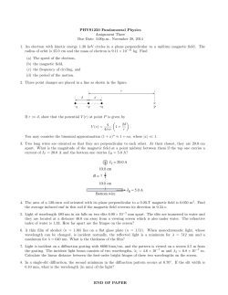

© Copyright 2026