ABC
docz
Explore
Log in
Create new account
Download
Report
science
medicine
surgery
Document 143682
Occupational Therapy – Kids health information Shoulder stability and control Definition Sheet F
Conservative management of a patient with snapping scapula: A case study
Michael J. Sandow
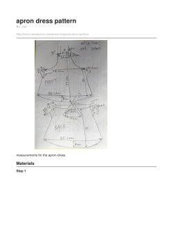
apron dress pattern 1 Materials By: you
R o t a t o r C...
F Frozen Shoulder What Can a Physical Therapist Do
The Hemiparetic Shoulder – Handle with care Sara Gawned Senior Physiotherapist
Bursitis is an inflammation or irritation of the bursa, which... What is subacromial bursitis?
Your Guide to Shoulder Pain and Its Treatment
Shoulder Impingement INFORMATION FOR YOU SHOULDER IMPINGEMENT SHOULDER & ELBOW
Document 148978
Document 148890
© Copyright 2026
About abcdocz
DMCA / GDPR
Report