
Document 143759
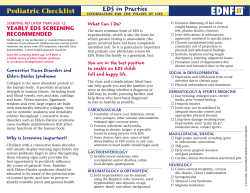
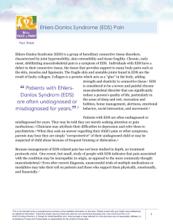
Department of Rehabilitation Services
Occupational Therapy
Standard of Care: Ehlers-Danlos Syndrome
ICD-9 (728)
Case Type / Diagnosis:
Ehlers-Danlos Syndrome (EDS) is the name given to a group of more than 10 different inherited
disorders; all involved a genetic defect in collagen and connective-tissue synthesis and structure.
EDS can affect the skin, joints, and blood vessels. This syndrome is clinically heterogeneous;
the underlying collagen abnormality is different for each type. Clinical recognition of the types
of EDS is important. One type, type IV, is associated with arterial rupture and visceral
perforation, with possible life-threatening consequences. 1
EDS is said to be relatively uncommon with a variety of physical presentations per type and a
prevalence of approximately 1 case in about 400,000 people. The variations in symptomatology,
natural history and mode of inheritance are striking, thus requiring accuracy in diagnosis and
appropriate counseling of the patient. 2
The diagnosis of EDS requires a good physical exam with family history, the presence of
hypermobile joints and skin findings. A key characteristic for diagnosis and differentiation from
general hypermobility syndromes seems to be the addition of skin findings. One of the major
issues remaining is the distinction between the normal range of joint mobility and the presence of
a defined genetic hypermobility syndrome. For example, confusion still occurs regarding the
question of EDS in such patients. Participants at the Berlin conference of 1986 on the Nosology
of Heritable Disorders of Connective Tissues (10) suggested that a diagnosis of EDS should not
be made in the absence of skin findings. Thus, documented generalized joint hypermobility in
the absence of other findings is usually termed benign hypermobility syndrome or familial
articular hypermobility syndrome if autosomal dominant inheritance (ie, parent-to-child
transmission) is demonstrated. 2
To help confirm the diagnosis of certain types, other tests are recommended. They are: Genetic
DNA tests, specific enzyme urine test for the kyphoscoliosis type, skin biopsy for a collagen
abnormality in the vascular type, heart ultrasound to check for mitral valve prolapse and prenatal
diagnostic tests when there is a family history of genetic mutation.
1
Standard of Care: Ehlers-Danlos Syndrome
Copyright © 2009 The Brigham and Women's Hospital, Inc. Department of Rehabilitation
Services. All rights reserved.
Clinical presentation of the various types:
In the past, experts divided Ehlers-Danlos syndrome into 11 subtypes—I through XI. In an
attempt to simplify these classifications, scientists recognized the subtypes according to signs
and symptoms, arranging them into six different groups:
Hypermobility type (formerly type III)
This is the most common form of EDS. It may affect as many as one in 10,000 to 15,000 people.
Signs and symptoms include:
•
•
Loose, unstable joints
Chronic joint pain
Classical type (formerly types I and II)
This type probably affects fewer than one in 20,000 to 40,000 people. Signs and symptoms
include:
•
•
•
•
•
Highly elastic, velvety skin
Fragile skin that bruises or tears easily
Slow and poor wound healing leading to scarring
Noncancerous fibrous growths on pressure areas, such as elbows and knees; fatty
growths on the shins and forearms
Loose joints, which are prone to dislocation and may delay the development of largemotor skills
Vascular type (formerly type IV)
This is one of the most serious forms of EDS. It affects an estimated one in 100,000 to 200,000
people. Signs and symptoms include:
•
•
•
Fragile blood vessels and organs that are prone to tearing (rupture)
Thin, fragile skin that bruises easily
Veins visible beneath the skin
• Distinctive facial features, including protruding eyes, thin nose and lips, sunken
cheeks and small chin
• Loose joints, usually limited to the fingers and toes
Kyphoscoliosis type (formerly called type VI)
This is an uncommon form. Fewer than 60 cases have been reported worldwide. Signs and
symptoms include:
•
•
Progressive curvature of the spine (scoliosis)
Fragile eyes that are easily damaged
2
Standard of Care: Ehlers-Danlos Syndrome
Copyright © 2009 The Brigham and Women's Hospital, Inc. Department of Rehabilitation
Services. All rights reserved.
•
Severe, progressive muscle weakness
Arthrochalasia type (formerly called types VII A and B)
Arthrochalasia is rare. Only about 30 cases have been reported worldwide. Signs and symptoms
include:
•
•
•
•
Very loose joints and dislocations, involving both hips, which may delay the
development of large-motor skills
Stretchy, fragile skin that is prone to bruising
Early-onset arthritis
Increased risk of bone loss and fracture
Dermatosparaxis type (formerly called type VIIC)
This form is also very rare. Only about 10 cases have been reported worldwide. Signs and
symptoms include:
•
•
Extremely fragile and sagging skin
Loose joints, which may delay development of large-motor skills in children. 3, 4
Other types
The remaining subtypes—types V, VIII, IX, X, and XI— are classified as “other”. These are
rare, and some are not well-defined. Some characteristics of these types:
•
Type V has characteristically fragile skin, although bruising and loose joints are
uncommon.
•
Type VIII involves teeth and gums as well as skin and joints (periodontal form).
•
Type IX may cause chronic diarrhea and lightheadedness due to low blood pressure,
in addition to other signs and symptoms.
•
Types X and XI, characterized by loose joints, are very similar and have sometimes
been considered one type rather than two.
•
Type X may lead to problems with blood clotting. 1
It appears to be unclear as to which type of EDS is the most common. A 2000 survey of 205
patients with EDS revealed 53% with hypermobile type, 26% classical type, and 7% vascular
type, and 12% of the respondents’ types were unknown [19]. A recent German study of more
3
Standard of Care: Ehlers-Danlos Syndrome
Copyright © 2009 The Brigham and Women's Hospital, Inc. Department of Rehabilitation
Services. All rights reserved.
than 406 patients with EDS revealed 54% with classical type, 30% hypermobility type, and only
16% vascular type [20]. 4
Genetic influence:
Having a genetic link is one of the strongest risk factors for the disease. The various types of
EDS are either autosomal dominant (AD), autosomal recessive (AR) or X-linked recessive
(XLR). Genes can be dominant or recessive. Dominant genes show their effect even if there is
only one copy of the gene in the pair. For a person to have a recessive disease or characteristic,
the person must have the gene on both chromosomes of the pair. 5
Affected first-degree relatives are identified in as many as 50% of cases. An autosomal
dominant pattern of inheritance is most common, although autosomal recessive and X-linked
transmission also have been documented. 6
Indications for Treatment:
Treatment is aimed at addressing all the areas of impairment. Because of the various types and
the variety of presentations, a wide range of professionals is necessary to care for this population.
The problem list/indications for occupational therapy are as follow:
1.
2.
3.
4.
5.
6.
7.
Fragile skin, easy bruising
Joint hypermobility and excessive dislocations
Poor tissue healing, wide and atrophic scars
Chronic joint pain
Osteopenia and postural changes
Impaired function or performance of activities of daily living (ADLs)
Decreased knowledge of condition and its impact on function
Contraindications / Precautions for Treatment:
Contraindications:
Any activity that would put stress on locked joints or the heart such as weight lifting, strength
testing, carrying heavy weight, playing any wind instruments (requiring forced air) as well as
pushing or pulling.
Precautions:
•
•
•
Avoid skin irritation from treatment modalities
Minimize stress on painful and lax joints
Minimize activities that would be injury producing (i.e. contact sports)
4
Standard of Care: Ehlers-Danlos Syndrome
Copyright © 2009 The Brigham and Women's Hospital, Inc. Department of Rehabilitation
Services. All rights reserved.
•
Avoid drugs which would affect coagulation (vascular issues)
• Surgical sutures removal should be delayed due to skin fragility and impaired healing
Examination
Medical History: Gather all pertinent medical information from other professionals, review
available medical records as well as information in longitudinal medical record (LMR), and
conduct a thorough patient interview regarding past medical history (PMH).
History of Present Illness: Conduct a patient interview regarding the specifics of the present
condition. If an injury, include the mechanism as well as date, symptoms, treatment to date,
previous occupational therapy and list of medications.
Occupational and Personal Profile: Inquire about social history, the layout of home, daily
routine and habits. Gather information about school and or work, use of orthotics, leisure
interests, prior use of durable medical equipment (DME) for ADLs, documentation of patient’s
beliefs, values, and goals for treatment.
Objective tests:
Observation/inspection: Observe patient’s posture (sitting/standing), movement pattern,
functional use of upper extremities with emphasis placed on joint stability. Inspect and
document skin condition, and swelling. Ask questions about school and work sitting positions as
well as bed position at home. Despite the laxity, evaluate for muscle imbalance. Check on splint
wear, fit and position. Document the use of glasses or other aids.
Vital signs: Obtain baseline parameters from medical records and from patient interview.
Document all pertinent information for later comparison.
Pain status: Determine pain intensity with visual analog scale (VAS); check on location, time,
intensifying or limiting factors.
Range of Motion: Identify joints to be measured, document laxity as well as any joint tightness,
and note pattern of imbalance.
Strength and endurance: Evaluate muscle performance through functional activities. No formal
resistive testing should be performed as it is contra-indicated for EDS patients. Inquire about
energy level and coping mechanisms for fatigue.
Function: Check on performance of all ADLs, and IADLs including child care and work.
5
Standard of Care: Ehlers-Danlos Syndrome
Copyright © 2009 The Brigham and Women's Hospital, Inc. Department of Rehabilitation
Services. All rights reserved.
Psychosocial issues: Check on coping skills, self-esteem, interaction with others, leisure and
support system. Coping with a lifelong illness is rarely easy. Depending on the severity of
symptoms the patient may face challenges at home, work and in relationships with others.
Parents of children with EDS have sometimes encountered suspicions of child abuse because of
frequent bruises and cuts. 3
Assessment
Establish Diagnosis and Need for Skilled Services
Problem List: The problem list is derived from the identified problems, dysfunctions and
indication for treatment.
Prognosis and Expected Outcomes: Prognosis depends on the type of EDS involved, other
contributing factors, patient’s knowledge, motivation, age and coping skills. Symptoms vary in
severity, even within one sub-type and the frequency of complications changes on an individual
basis. Some individuals have negligible symptoms while others are severely restricted in their
daily life. Extreme joint instability and scoliosis may limit a person’s mobility. Most
individuals will have a normal lifespan. However, those with blood vessels involvement,
particularly those with EDS vascular type, have an increased risk of fatal complications. 11
Age Specific Considerations: Due to the genetic factors and the wide variety of physical
presentations of this condition, the patient should be provided with information which highlights
their age specific needs. Early in life, counseling and treatment would emphasize physical issues
and contact sports. As young adult, genetic counseling and child bearing would be as important.
Treatment will be scaled to the level of the patient’s ability and needs.
Goals: Aimed at addressing all the impairment areas identified
1. Prevent or minimize skin injury via education about protective strategies
2. Protect unstable joints and decrease rate of dislocation with strong supportive
soft tissues and splints
3. Protect healing tissues and minimize scar adhesion.
4. Decrease pain and increase ability for self-management
5. Decrease incidence of fractures and manage posture and positioning issues
6. Increase functional independence for all ADLs including school, work and
child care
7. Increase knowledge of condition, and ability to avoid complications. Increase
self-monitoring as well as coping skills
6
Standard of Care: Ehlers-Danlos Syndrome
Copyright © 2009 The Brigham and Women's Hospital, Inc. Department of Rehabilitation
Services. All rights reserved.
Treatment Planning / Interventions:
Interventions Intervention/treatment most commonly used for EDS:
•
Splint fabrication for joint stabilization. Patient teaching regarding splint protocol
with emphasis on skin inspection and use of padding. Two of the key factors with
EDS that the therapist should be aware of when splinting are fragile skin and difficulty
healing.
•
A custom made splint may be more appropriate as the therapist would have a chance
to make adjustments per the needs of the patient. Most common types of splints for
this population are protective immobilizing splints or static serial splints if muscle
imbalance is a problem. They may be used to support, protect, or immobilize joints,
allowing healing to occur after bone, tendon, vascular, nerve, joint, or soft tissue injury
or inflammation. Correction of existing deformity represents another commonly
encountered reason for splint application. 7
•
Pain management with support to painful joints. This can be done through splinting as
explained above as well as through gentle exercises with a focus on the muscle
imbalance. Increase awareness of proper positioning for ADLs using less deforming
forces. Provide with adaptive aids to help lessen stress on joints (i.e. use of built-up
equipment and easy spring-loaded type tools). 8 Consider using self-help devices, such
as reachers because stretching puts joints at risk for injury or dislocation. 9
•
Pain management should include psychosocial support for which appropriate referrals
should be made. Chronic pain is a physical and psychosocial experience with a range
of difficult emotions. Amongst these are emotions that manifest themselves in a
variety of anxiety-type symptoms and/or depressed mood. People with Joint
Hypermobility Syndrome JHS who have chronic pain may require help to manage
these types of emotional difficulties when they occur. 10
•
Environmental modifications such as remove or re-arrange furniture as needed to
reduce the risk of injury or falls which may lead to fractures and dislocations. Add
railings, arm supports and relocate bedrooms as needed to decrease need for stair
climbing and potential for falls.
•
Increase functional performance with the use of ergonomic adaptations for home,
school, work and leisure tasks. Use of braces/splints and adaptive aids as documented
in prior sections.
•
Patient and family education regarding available resources, and increase patient’s
knowledge about self-management of their disease/condition. Encourage regular visits
7
Standard of Care: Ehlers-Danlos Syndrome
Copyright © 2009 The Brigham and Women's Hospital, Inc. Department of Rehabilitation
Services. All rights reserved.
with cardiologist, dentist, ophthalmologist, as well as the genetic counselor especially
if the patient is of childbearing age.
Frequency & Duration: Varies based on the examination findings and patient’s needs.
Patient / family education: Preventing injuries, improving self-monitoring skills
as well as increasing knowledge of joint protection should be emphasized.
Suggestions as follow:
•
Avoid injury. Avoid contact sports and other activities that increase risk of injury.
•
Use protective gear. Patients with severe EDS should wear protective clothing to
shield their fragile skin.
•
Reduce the clutter. To prevent falls and injuries at home, keep walkways and
doorways clear of clutter. Avoid loose rugs and electric cords, which can increase
potential for tripping and falling.
•
Use assistive devices. A number of common devices can help decrease stress on
joints, such as jar openers, utensils with wide handles and long-handle equipment.
•
Wear sunscreen. Regular use of sunscreen when exposed to the sun may help reduce
premature aging of skin. 3
Recommendations and referrals to other providers: Depending on the type of EDS
involved and if the patient is not already connected with the pertinent specialists, the
referring MD should be contacted with the evaluation findings and recommendations.
Some of the specialists a patient with EDS may be referred to including the occupational
therapist are: Genetic counselor, dentist, cardiologist, dermatologist, orthopedist,
pediatrician, Orthotist, physical therapist, ophthalmologist, and surgeon.
Re-evaluation
Standard Time Frame: Every 30 days from referral date, or as dictated per patient’s
needs.
Discharge Planning
Patient is discharged when he or she has met his or her goals and is proficient in selfmanagement and independent with any home program.
8
Standard of Care: Ehlers-Danlos Syndrome
Copyright © 2009 The Brigham and Women's Hospital, Inc. Department of Rehabilitation
Services. All rights reserved.
Transfer of Care: Refer back to MD if not improving or has significant change in status.
If the referring MD is not available and the situation is unclear or urgent, the patient
should be sent to the ER. If therapist is not available, plan of care should be available for
covering therapist.
Patient’s discharge instructions: Review home program and self management
information, provide with resources as needed. Talk about maintaining normalcy as much
as possible and review needs for modification. Most people with EDS are able to live a
productive and fulfilling life, even with the limitations imposed by having EDS. 3
Author:
Reviewers:
Marie José Benjamin, OT
Reg B. Wilcox III, PT
5/09
Gayle Lang, OT
9
Standard of Care: Ehlers-Danlos Syndrome
Copyright © 2009 The Brigham and Women's Hospital, Inc. Department of Rehabilitation
Services. All rights reserved.
References
1. Ceccolini E. Ehlers-Danlos Syndrome. Available at:
www.emedicine.com/derm/topic696.htm2008.
2. Raff ML, Byers PH. Joint hypermobility syndromes. Curr Opin Rheumatol.
1996;8:459-466.
3. Ehlers-Danlos Syndrome at: www.mayoclinic.com/print/ehlers-danlossyndrome/DS00706/METHOD=print&D...
4. Schroeder EL, Lavallee ME. Ehlers-danlos syndrome in athletes. Curr Sports
Med Rep. 2006;5:327-334.
5. The Basics on Genes and Genetic Disorders at:
www.kidshealth.org/teenyour_body/health_basics/genes_genetic_disorders.html.
6. Everman DB, Robin NH. Hypermobility syndrome. Pediatr Rev. 1998;19:111117.
7. Fess EE, Gettle K, Philips C, Janson JR. Exercise and splinting for specific
upper extremity problems. In: Hand and Upper Extremtiy Splinting: Principles &
Methods. 3rd ed. St. Louis, Missouri: Elsevier Mosby; 2005:397-436.
8. Cleveland Clinic Heart and Vascular Institute--What is occupational theray at:
http://my.clevelandclinic.org/heart/prevention/exercise/ot.aspx.
10
Standard of Care: Ehlers-Danlos Syndrome
Copyright © 2009 The Brigham and Women's Hospital, Inc. Department of Rehabilitation
Services. All rights reserved.
9. Ehlers-Danlos Syndrome at:
www.orthop.washington.edu/uw/ehlersdanlos/tabID_3376/ItemID_32/PageID_7/...
10. Joint hypermobility syndrome: a complex constellation of symptoms at:
http://bcbsma.medscape.com/viewarticle/537938.
11. The Gale Group. Gale Encyclopedia of Medicine, 3rd ed
11
Standard of Care: Ehlers-Danlos Syndrome
Copyright © 2009 The Brigham and Women's Hospital, Inc. Department of Rehabilitation
Services. All rights reserved.


© Copyright 2026