ABC
docz
Explore
Log in
Create new account
Download
Report
science
medicine
surgery
Management of flail chest Trauma 2001; 3: 235–247
Radiology Frequent MRI/CT Orders - Exams and Indications MRI CT
Hope Cancer Care Health History Form – Important Information
Blunt thoracic trauma: f lail chest, pulmonary contusion, and blast injury
Costochondritis
A Health Matters Kaiser Permanente
Dr Chrissy Gray Sessional GP, GP Support Unit and Ambulatory Care
Operative Repair of Rib Fractures SIGN Conf 2013 Joel Gillard
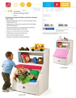
P 883100 885000 Lift & Hide
Clinical assessment tool for children 0-5 years
ATLS initial management Dr. Khalid Abdulwahid CABS, FRCS(England)
© Copyright 2026
About abcdocz
DMCA / GDPR
Report