
Efficacy of low-dose myrrh protocols in the
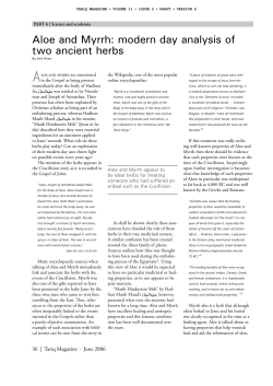
Research and Reports in Tropical Medicine Dovepress open access to scientific and medical research O r i g i n al R e s e ar c h Open Access Full Text Article Efficacy of low-dose myrrh protocols in the treatment of experimental schistosomiasis mansoni: hepatic improvement without parasitologic cure This article was published in the following Dove Press journal: Research and Reports in Tropical Medicine 15 November 2010 Number of times this article has been viewed Rashad Abdul-Ghani 1 Naguiba Loutfy 2 Manal Sheta 3 Azza Hassan 2 1 Department of Medical Parasitology, Faculty of Medicine and Health Sciences, Sana’a University, Sana’a, Yemen; 2Tropical Health Department, High Institute of Public Health, 3 Department of Pathology, Medical Research Institute, Alexandria University, Alexandria, Egypt Abstract: There is a new trend of “back to nature” in searching for antischistosomal drugs, particularly after the concerns raised about the possible emergence of schistosome isolates resistant/tolerant to praziquantel as well as for their relative safety and fewer side effects. Many plant derivatives have been investigated for efficacy against the Egyptian strain of Schistosoma mansoni, but much attention has been paid to myrrh extract, which is a purified sap obtained from Commiphora molmol. This extract has been produced and marketed in Egypt as a pharmaceutical preparation, but with a great discrepancy in its antischistosomal activity in both experimental and clinical studies. Most previous experimental studies used myrrh in the dosing protocol of 500 mg/kg/day for five days. In the present study, we investigated the therapeutic efficacy of three low-dose myrrh protocols against experimental schistosomiasis mansoni. All these protocols showed no significant efficacy in reducing parasite burdens and tissue egg loads or in changing oogram patterns. Nevertheless, there was an amelioration of hepatic lesions, with reductions in mean counts of hepatic granulomas as well as marked healing of these granulomas. Keywords: Schistosoma mansoni, schistosomiasis mansoni, myrrh, Commiphora molmol, antischistosomal chemotherapy Introduction Correspondence: Rashad Abdul-Ghani High Institute of Public Health, Alexandria University, 165 El-Horria Ave, Hadara, Alexandria, Egypt Tel +20 164 621 779 Email [email protected] submit your manuscript | www.dovepress.com Dovepress DOI: 10.2147/RRTM.S14231 Screening natural products found in all sorts of environments is a highly competitive field in which all of the major pharmaceutical companies are involved.1 Although many plant species have been used throughout the world in traditional medicine for the treatment of human helminthes,2 only a few plants have been screened for activity against adult Schistosoma mansoni.3 Experimentally, many plant derivatives have been shown to display antischistosomal activities against the Egyptian strain of S. mansoni, including the blackseed Nigella sativa,4–6 curcumin Curcuma longa,7,8 garlic extract Allium sativum,6,9,10 Cleome droserifolia,11 mandarin orange Citrus reticulata,12 and ginger Zingiber officinale.13 Myrrh is an oleo-gum resin obtained from the stem of Commiphora molmol (also called C. myrrha), and probably other species of the family Bursearacae, growing in north-east Africa and Arabia.14 Myrrh has been recognized to possess marked antiseptic, anesthetic, and antitumor properties.15 It consists of water-soluble gum, alcohol-soluble resins, and volatile oil.16 The oleo-gum contains polysaccharides and proteins, while the volatile oil is composed of steroids, sterols, and terpenes.16 One of the advantages Research and Reports in Tropical Medicine 2010:1 65–71 65 © 2010 Abdul-Ghani et al, publisher and licensee Dove Medical Press Ltd. This is an Open Access article which permits unrestricted noncommercial use, provided the original work is properly cited. Dovepress Abdul-Ghani et al of myrrh is that, like many herbal and botanic products, it is safe in humans,17 so that this natural, flavoring substance has been approved by the US Food and Drug Administration.18 Upon comparison with praziquantel, Omar et al concluded that natural myrrh extract had no hepatotoxic, genotoxic, or carcinogenic effects on adult albino rats.19 Clinically, almost all studies of myrrh demonstrated its safety in humans with few, if any, side effects as has been reviewed by Abdul-Ghani et al.17 In Egypt, myrrh has been investigated, both experimentally and clinically, against human trematode infections, particularly schistosomiasis and fascioliasis with stories of success and disagreement regarding its efficacy against schistosomes. 17 The antischistosomal activity of myrrh extract was first reported in hamsters infected with the Egyptian strain of S. mansoni, with reduction in worm burden reaching 100% two weeks posttreatment, significant oogram changes, and reductions in liver and intestinal tissue egg loads.20 Mirazid®, a pharmaceutical myrrh preparation, is produced and marketed in Egypt as gelatin capsules containing 300 mg of purified C. molmol extract for the treatment of schistosomiasis and fascioliasis.21 The results of studies published on the activity of myrrh in the treatment of S. mansoni-infected mice have yielded conflicting results,17 both experimentally22,23 and clinically.24–28 Overall, in view of these controversial results, myrrh may be considered for those who have undergone treatment with praziquantel and were not cured for some reason, including intolerance, disease relapse, or reinfection.24,29 The present study was carried out to evaluate the antischistosomal efficacy of three low-dose myrrh protocols in Swiss albino mice experimentally infected with the Egyptian strain of S. mansoni. Materials and methods Eight-week-old female Swiss albino mice (CD-1 strain), weighing 20 ± 2 g, were obtained from the Schistosome Biologic Supply Center, Theodore Bilharz Research Institute, Cairo, Egypt. These mice were bred in polypropylene cages under environmentally controlled conditions, and fed with a standard pellet diet and water ad libitum. The parasite used was laboratory-maintained cercariae of Egyptian S. mansoni (CD strain). Purified C. molmol extract (Mirazid ®, Pharco Pharmaceuticals, Alexandria, Egypt), was used as a freshly prepared suspension in 2% Cremophor EL ® (Sigma, St Louis, MO) before administration. 66 submit your manuscript | www.dovepress.com Dovepress Three dosing protocols were applied intragastrically using a stomach tube to the mice treatment groups from day 46 post-infection onwards, when adult S. mansoni stages are harbored in mice. A total of 40 mice infected with S. mansoni were randomly allocated to three experimental groups (I, II, and III) and to a control, with 10 mice each. The infected untreated control received the vehicle solution used for suspending the drug for three days. The dosing protocols comprised: a two-day protocol (group I), 500 mg/kg/day (recommended dose) for two days; a four-day protocol (group II), 250 mg/kg/day (half the recommended dose) for four days; and a six-day protocol (group III), 125 mg/kg/day (quarter the recommended dose) for six days. Parasitologic investigations Mice were infected in batches by subcutaneous injection of each mouse with (60 ± 10) cercariae of laboratory-maintained S. mansoni (Egyptian CD strain) suspended in 0.1 mL solution.30 Worms were recovered from the hepatic and portomesenteric veins using the Smithers and Terry perfusion technique for mice.31 This was performed using a perfusion apparatus (Filamatic®; National Instrument Co., Baltimore, MD). For estimation of tissue egg loads, weighed fragments from the tissues of the liver and small intestine were removed and processed separately by the KOH digestion technique for counting S. mansoni eggs in tissues.32 An oogram study was performed by studying alteration in the percentages of the various stages of viable eggs as well as of the increase in the percentage of dead eggs in the mucosa of the small intestine.33 Histopathologic investigations A small piece of liver was removed from the central part of the left lateral lobe from each mouse after perfusion, and preserved in a suitable quantity of 10% formalin. Liver sections were prepared and subsequently stained with hematoxilyn and eosin in the Pathology Department of Medical Research Institute, Alexandria University. Reductions in mean counts and diameters of hepatic granulomas, as well as their healing in the treated groups, were then determined and compared to those in the infected untreated control. The granulomas were identified as either active granulomas surrounded with immune cells or healed granulomas surrounded with fibrotic tissue. Statistical analysis The data were analyzed using SPSS (version 11.5; SPSS Inc., Chicago, IL) in the Biostatistics Department, High Institute Research and Reports in Tropical Medicine 2010:1 Figure 1 An active hepatic schistosomal granuloma around a central ovum, with reflactile egg shell, surrounded by a giant cell (multinucleated macrophage) and inflammatory cellular infiltrate formed of eosinophils, lymphocytes and few histiocytes (hematoxylin and eosin, ×400). of Public Health, Alexandria University. One-way analysis of variance, followed by Tukey’s post hoc test, was used to test the difference in means between each experimental group and the control. Results Total and female worm reductions Total worm reductions in the treated groups were nonsignificant in the range of 11.5%–13.2% compared to the control. On the other hand, statistically nonsignificant (P . 0.05) reductions ranging between 6.6% and 13.0% were observed in female worm burdens in the treated groups compared to the control (Table 1). Figure 2 A healed schistosomal hepatic granumola with a central ovum, showing distorted mirscidium and egg shell, and surrounded by fibro-collagen bundles entangling fibroblasts and few inflammatory cells (hematoxylin and eosin, ×400). Research and Reports in Tropical Medicine 2010:1 7.71 ± 0.84 6.71 ± 0.29 7.00 ± 0.49 7.20 ± 0.73 NA 13.0 9.2 6.6 16.51 ± 1.69 14.18 ± 1.43 13.08 ± 1.72 14.30 ± 0.80 NA 14.1 20.8 13.4 18.57 ± 1.94 14.28 ± 1.17 16.63 ± 1.70 17.83 ± 1.22 NA 23.1 10.4 4.0 Reduction (%) 66.14 ± 3.55 59.28 ± 2.51 65.60 ± 1.93 62.60 ± 3.24 28.71 ± 4.01 33.43 ± 2.55 29.80 ± 1.02 31.80 ± 3.47 Mature 5.15 ± 0.74 7.29 ± 0.81 4.60 ± 0.51 5.60 ± 0.74 Dead Notes: Groups I, II, and III were tested versus control using one-way analysis of variance followed by Tukey’s post hoc test; P . 0.05, no statistically significant differences. Surviving mice/group is given in parentheses (n). Values are expressed as mean ± SE. Abbreviations: NA, not applicable; TWR, total worm reduction; FWR, female worm reduction; SE, standard error of the mean. NA 11.8 13.2 11.5 20.57 ± 1.21 18.14 ± 0.77 17.86 ± 0.96 18.20 ± 0.58 Intestine × 103 Untreated 500 × 2 250 × 4 125 × 6 Reduction (%) Control (7) I (7) II (5) III (7) FWR (%) Total immature Total females Liver × 103 TWR (%) Dosing protocols (mg/kg/day × days) Group (n) Total worms Oogram changes Tissue egg load reductions Worm reductions Table 1 Effects of different low-dose myrrh protocols on worm burdens, tissue egg loads, and oogram patterns in experimentally infected mice harboring adult S. mansoni (Egyptian CD strain) Dovepress Low-dose myrrh for Schistosoma mansoni submit your manuscript | www.dovepress.com Dovepress 67 Dovepress Abdul-Ghani et al Tissue egg load Nonsignificant (P , 0.05) reductions of 20.8% followed by 14.1% and 13.4% in liver tissue eggs per gram were observed in groups II, I, and III, respectively. In addition, nonsignificant (P . 0.05) reductions of 23.1% and 10.4% in eggs per gram tissue of small intestine were observed in groups I and II, followed by a negligible, nonsignificant (P . 0.05) reduction of only 4.0% in group III (Table 1). Oogram pattern There were no statistically significant (P . 0.05) differences in percentages of total immature stages in groups I, II, and III, ranging from 59.28% to 65.60% compared to a percentage of 66.14% in the control (Table 1). In addition, percentages of mature eggs in the treated groups were comparable ranging from 29.80% to 33.43% (P . 0.05) compared to a percentage of 28.71% in the control. Likewise, there were no statistically significant (P . 0.05) changes in the percentages of dead eggs, which ranged from 4.60% to 7.29% in all treated groups, compared to a percentage of 5.15% in the control (Table 1). Hepatic granuloma measurements Table 2 shows the effects of the different myrrh dosing protocols in reducing the mean granuloma counts/low power field and their diameters in treated groups compared to those in the control. Only groups III and II showed statistically significant (P , 0.05) reductions in the mean count of hepatic granulomas of 39.1% and 34.8%, respectively, whereas a statistically nonsignificant reduction of only 13.0% (P . 0.05) was observed in group I. In contrast, nonsignificant reductions of 17.6%, 13.3%, and 11.6% were observed in the mean diameters of hepatic granulomas in groups I, III, and II, respectively (Table 2). Study of the cellularity of granulomas revealed both active (Figure 1) and healed (Figure 2) granulomas. While the number of healed granulomas represented only about one half of the active granulomas (a healing ratio of 1:2) in the control, all myrrh dosing protocols induced relative increases in the number of healed granulomas, with healing ratios of 3:1, 4:1, and 5:1 in groups I, II, and III, respectively (Table 2). Discussion In the present study, myrrh largely failed to induce significant reductions in total worm burdens in all treated groups with the dosing protocols employed. The total worm burdens were nonsignificantly reduced in the range of 11.5%–13.2%. Female worm reductions were also minimal and statistically nonsignificant; the highest reduction was only 13.0% when myrrh was administered at 500 mg/kg/day for two days. Meanwhile, the effects of the different myrrh dosing protocols on tissue egg loads were negligible. Myrrh caused only nonsignificant reductions in the range of 13.4%–20.0% in liver egg loads. Similarly, myrrh exerted only slight effects on egg loads in the wall of the small intestine when administered at 500 mg/kg/day for two days that showed the highest but nonsignificant lowering effect of 23.1%. On the other hand, none of the myrrh dosing protocols induced any alteration in the oogram pattern of eggs in the mucosa of the small intestine. All immature and mature developmental egg stages were present in percentages approximately equal to those of the control. Changes in the number and character of eggs (oogram) provide a simple, sensitive, and reliable criterion for the screening of drugs active against S. mansoni that assesses the effects of a drug on oviposition, as well as on the maturation and survival of eggs trapped in the intestinal mucosa.33 Even if hepatic shift of schistosomes is not evident, alterations in the oogram can still be detected.34 This indicates the failure of myrrh to interrupt the processes of oviposition and egg development. Moreover, myrrh did not exhibit any ovicidal activity on eggs already laid in the intestinal wall where no increase in the dead eggs was provoked Table 2 Effects of different low-dose myrrh protocols on granuloma characteristics in experimentally infected mice harboring adult S. mansoni (Egyptian CD strain) Group (n) Dosing protocols (mg/kg/day × days) Granuloma count/LPF (mean ± SE) Reduction (%) Granuloma diameter (μm) (mean ± SE) Reduction (%) Healing ratio (healed:active) Control (7) 1 (7) II (5) III (7) Untreated 23.00 ± 1.29 20.00 ± 1.31 15.00 ± 1.84a 14.00 ± 2.39a NA 13.0 34.8 39.1 270.73 ± 17.56 239.22 ± 20.32 223.00 ± 16.64 234.76 ± 16.93 NA 11.6 17.6 13.3 1:2 3:1 4:1 5:1 500 × 2 250 × 4 125 × 6 Notes: aP , 0.05; values are expressed as mean ± SE; surviving mice/group are given in parentheses (n). Groups I, II, and III were tested versus control using one-way analysis of variance followed by Tukey’s post hoc test. Abbreviations: LPF, low-power field; NA, not applicable; SE, standard error of the mean. 68 submit your manuscript | www.dovepress.com Dovepress Research and Reports in Tropical Medicine 2010:1 Dovepress by the drug. This finding is contradictory to the suggestion of Massoud who attributed the efficacy of myrrh extract in the reduction of tissue egg load in hamsters experimentally infected with S. mansoni to a possible toxic effect of the drug on the reproductive organs of the parasite.35 The results of the present study are in agreement with those of a multicenter study carried out in Egypt, Italy, the US, and Brazil.23 In this study, nonsignificant reductions in total worm burdens of S. mansoni (Egyptian CD strain) were recorded, with the highest reductions being only 19% and 27%. These percentages were produced with the lower dose of 250 mg/kg for five days whereas the higher one of 500 mg/kg for five days did not enhance worm reduction. Moreover, there was agreement as regards the nonsignificant reduction in the liver egg load as well as the unaltered oogram pattern where all egg developmental stages were present and comparable with those of the control.23 On the other hand, various extracts and doses of myrrh, including Mirazid®, did not show any demonstrable antischistosomal activity in mice and hamsters infected with different strains of S. mansoni. In the same study, the Egyptian CD strain was shown to be less susceptible to myrrh than the Puerto Rican (Mill Hill) strain of S. mansoni whereas Mirazid® achieved a slightly higher (36%) worm reduction when administered at a lower dose of 100 mg/kg for three days.23 El-lafft reported a total worm reduction of 36.5% with no significant change in antibodies to the parasite in mice experimentally infected with the Egyptian strain of S. mansoni and treated with myrrh.36 However, this efficacy is still low and supports the results of myrrh obtained in the present study. However, the results of the present study are inconsistent with those of Badria et al who reported that oral administration of myrrh twice a day for three consecutive days at doses of 250 and 500 mg/kg produced significant total worm reductions of 76% and 75%, respectively.22 Badria et al also attributed their finding of a progressive reduction in oviposition, as evidenced by oogram changes to a possible effect on female reproductive organs.22 Furthermore, the results of the present study are contradictory to those of Massoud et al who reported a reduction of 98.5% in total worm burden five weeks after oral Mirazid® administration at a dose of 500 mg/kg/day for five days 45 days postinfection in mice.37 They also recorded a marked reduction of more than 90% in hepatic and intestinal tissue egg loads as well as a significant alteration in oogram pattern.37 The findings of the present study are also different to the findings of Hamed and Hetta who reported significant reductions of 81% in total worm burden and of 73% in liver egg load in mice four weeks after Research and Reports in Tropical Medicine 2010:1 Low-dose myrrh for Schistosoma mansoni oral administration of 600 mg Mirazid® for three consecutive days two months postinfection.12 Nevertheless, it should be taken into consideration that the present study is different from the previous studies with respect to the dosing protocols, as well as the timing of mice sacrifice. Botros et al attributed the difference in results of studies on the antischistosomal efficacy of myrrh to the possibility of Mirazid® not being a well defined chemical entity, but simply a plant extract that could vary greatly in its active ingredients among different batches of preparation.28 In fact, all published studies that claimed antischistosomal activity for myrrh did not refer to a specific chemical constituent responsible for such activity. Moreover, the mechanism of action of myrrh on schistosomes is not fully understood.17 It should be stressed that Commiphora comprises more than 200 species,16 but the resins obtained from species other than C. molmol contain many adulterants that complicate the characterization of myrrh.38 In the present study, myrrh induced a moderate reduction in the mean count of hepatic granulomas compared to the infected untreated control. Lower doses over longer periods provoked a higher reduction in mean counts of hepatic granulomas. The highest reduction of 39.1% was achieved in mice receiving myrrh at 125 mg/kg/day for six days followed by 34.8% in those receiving myrrh at 250 mg/kg/day for four days. In contrast, 500 mg/kg/day for two days caused a nonsignificant reduction of only 13.0% in hepatic granulomas compared to the control. Although myrrh did not cause significant reductions in mean granuloma diameters, it appeared to be highly efficacious in inducing healing of hepatic granulomas compared to those in the control. It is worth mentioning that although praziquantel is reported to be highly curative, the patient may still suffer the consequences of progressive disease pathology.39 The results of the present study are in agreement with those of Massoud et al who reported the efficacy of Mirazid® in markedly reducing granulomas in the portal areas and ameliorating intercellular fibrosis in mice experimentally infected with S. mansoni (Egyptian CD strain).37 In addition, Massoud et al reported a significant decrease in number and size of granulomas, with significant reduction in the collagen content deposition in portal tracts and around central veins in S. mansoni-infected mice treated with Mirazid®.40 It should be noted that our study differs in its design from the latter two studies with respect to the dosing protocols and the timing of mice sacrifice. In these studies, myrrh extract was given as 500 mg/kg/day for five consecutive days, and mice were sacrificed 12 weeks postinfection.37,40 submit your manuscript | www.dovepress.com Dovepress 69 Abdul-Ghani et al One possible explanation of the antihistopathologic effect of myrrh is that provided by Badria and Attia who showed that Mirazid® has an inhibitory effect on hepatic plateletactivating factor (PAF) in rats with experimentally induced hepatic fibrosis, and that might account for its antifibrotic activity.41 Liver granulomas in schistosomiasis typically contain a high percentage of activated eosinophils that release potentially toxic inflammatory mediators, including PAF, that not only damage the parasite and surrounding tissues, but also play a role in the perpetuation of the local immune response.42 The inhibition of PAF may partly explain the moderate enhancement of histopathologic lesions in spite of the nonsignificant parasitologic results obtained with myrrh extract in the present study. This creates new opportunities to investigate the possibility of combining myrrh with antischistosomal drugs to relieve the manifestations of hepatic schistosomiasis mansoni. In conclusion, myrrh proved to be an agent of low efficacy against S. mansoni when administered intragastrically in the aforementioned dosing protocols to experimentally infected mice. However, even using myrrh at low dosing protocols proved efficacious in ameliorating the hepatic pathology due to S. mansoni. The present study recommends the re-evaluation of myrrh as an antischistosomal drug experimentally and clinically and carrying out experiments to investigate its possible advantages as an antipathologic adjuvant to antischistosomal drugs. Acknowledgments The authors thank the staff of the Schistosome Biologic Supply Unit, Theodore Bilharz Research Institute, Cairo, Egypt, who assisted in this study. Our thanks are due to Prof. Dr. Amel El Sahn, Tropical Health Department, HIPH, Alexandria University and Prof. Dr. Nibal Hammouda, Department of Medical Parasitology, Faculty of Medicine, Alexandria University for their evaluations and suggestions. We also thank Mr. Hany Hussein, Biostatistics Department, HIPH, Alexandria University, for assistance with the statistical analysis of data. Disclosure The authors report no conflicts of interest in this work. References 1. Kayser O, Kiderlen AF, Croft SL. Natural products as antiparasitic drugs. Parasitol Res. 2003;90 Suppl 2:S55–S62. 2. McGaw LJ, Jäger AK, van Staden J. Antibacterial, anthelmintic and antiamoebic activity in South African medicinal plants. J Ethnopharmacol. 2000;72(1–2):247–263. 70 submit your manuscript | www.dovepress.com Dovepress Dovepress 3. Sanderson L, Bartlett A, Whitfield PJ. In vitro and in vivo studies on the bioactivity of a ginger (Zingiber officinale) extract towards adult schistosomes and their egg production. J Helminthol. 2002;76(3): 241–247. 4. Mahmoud MR, El-Abhar HS, Saleh S. The effect of Nigella sativa oil against the liver damage induced by Schistosoma mansoni infection in mice. J Ethnopharmacol. 2002;79(1):1–11. 5. Mohamed AM, Metwally NM, Mahmoud SS. Sativa seeds against Schistosoma mansoni different stages. Mem Inst Oswaldo Cruz. 2002; 100(2):205–211. 6. El-Shenawy NS, Soliman MF, Reyad SI. The effect of antioxidant p roperties of aqueous garlic extract and Nigella sativa as antischistosomiasis agents in mice. Rev Inst Med Trop Sao Paulo. 2008; 50(1):29–36. 7. El-Ansary AK, Ahmed SA, Aly SA. Antischistosomal and liver protective effects of Curcuma longa extract in Schistosoma mansoni infected mice. Indian J Exp Biol. 2007;45(9):791–801. 8. Allam G. Immunomodulatory effects of curcumin treatment on murine schistosomiasis mansoni. Immunobiology. 2009;214(8):712–727. 9. Riad NHA, Fares NH, Mostafa OMS, Mahmoud Y. The effect of garlic on some parasitological parameters and on hepatic tissue reactions in experimental schistosomiasis mansoni. J Appl Sci Res. 2007;3(10):949–960. 10. Nahed HA, Hoda AT, Yomna IM. Effects of garlic on albino mice experimentally infected with Schistosoma mansoni: A parasitological and ultrastructural study. Trop Biomed. 2009;26(1):40–50. 11. El-Shenawy NS, Soliman MF, Abdel-Nabi IM. Does Cleome droserifolia have anti-schistosomiasis mansoni activity? Rev Inst Med Trop Sao Paulo. 2006;48(4):223–228. 12. Hamed MA, Hetta MH. Efficacy of Citrus reticulata and Mirazid® in treatment of Schistosoma mansoni. Mem Inst Oswaldo Cruz. 2005; 100(7):771–778. 13. Al-Sharkawi IM, El-Shaikh KA, Tabl GA, Ali JA. The effect of ginger on Schistosoma mansoni-infected mice. Delta J Sci. 2007;31:1–10. 14. Greene DA. Gold, frankincense, myrrh, and medicine. N C Med J. 1993; 54(12):620–622. 15. Nomicos EYH. Myrrh: Medical marvel or myth of the Magi? Holist Nurs Pract. 2007;21(6):308–323. 16. Hanus LO, Rezanka T, Dembitsky VM, Moussaieff A. Myrrh – Commiphora chemistry. Biomed Pap Med Fac Univ Palacky Olomouc Czech Repub. 2005;149(1):3–27. 17. Abdul-Ghani R, Loutfy N, Hassan A. Myrrh and trematodoses in Egypt: An overview of safety, efficacy and effectiveness profiles. Parasitol Int. 2009;58(3):210–214. 18. Ford RA, Api MA, Letizia CS. Monographs on fragrance raw materials. Food Chem Toxicol. 1992;30 Suppl:S1–S138. 19. Omar A, Elmesallamy Gel-S, Eassa S. Comparative study of the hepatotoxic, genotoxic and carcinogenic effects of praziquantel distocide and the natural myrrh extract Mirazid® on adult male albino rats. J Egypt Soc Parasitol. 2005;35(1):313–329. 20. Massoud AM. Drug for treatment of bilharziasis (schistosomiasis). United States Patent US 6077513A. 2000 June 1. 21. Fenwick A, Savioli L, Engels D, Robert BN, Todd MH. Drugs for the control of parasitic diseases: Current status and development in schistosomiasis. Trends Parasitol. 2003;19(11):509–515. 22. Badria F, Abou Mohamed G, El-Mowafy A, Massoud A, Salama O. Mirazid®: A new schistosomicidal drug. Pharmaceut Biol. 2001;39(1): 127–131. 23. Botros S, William S, Ebeid F, et al. Lack of evidence for an antischistosomal activity of myrrh in experimental animals. Am J Trop Med Hyg. 2004;71(2):206–210. 24. Sheir Z, Nasr AA, Massoud A, et al. A safe, effective, herbal antischistosomal therapy derived from myrrh. Am J Trop Med Hyg. 2001;65(6): 700–704. 25. Gaballah M, El-Gilany A, El-Shazly A, Motawea SS. Control of schistosomiasis in a rural area using a new safe effective herbal treatment. JOESE. 2001;21:63–84. Research and Reports in Tropical Medicine 2010:1 Dovepress 26. Abo-Madyan AA, Morsy TA, Motawea SM. Efficacy of myrrh in the treatment of schistosomiasis (haematobium and mansoni). J Egypt Soc Parasitol. 2004;34(2):423–446. 27. Barakat R, Elmorshedy H, Fenwick A. Efficacy of myrrh in the treatment of human schistosomiasis mansoni. Am J Trop Med Hyg. 2005; 73(2):365–367. 28. Botros S, Sayed H, El-Dusoki H, et al. Efficacy of mirazid® in comparison with praziquantel in Egyptian Schistosoma mansoni-infected school children and households. Am J Trop Med Hyg. 2005;72(2):119–123. 29. Ribeiro-dos-Santos G, Verjovski-Almeida S, Leite LC. Schistosomiasis – a century searching for chemotherapeutic drugs. Parasitol Res. 2006; 99(5):505–521. 30. Peters AP, Warren KS. A rapid method of infecting mice and other laboratory animals with Schistosoma mansoni: Subcutaneous injection. J Parasitol. 1969;55(3):558–563. 31. Smithers SR, Terry RJ. The infection of laboratory hosts with cercariae of Schistosoma mansoni and the recovery of adult worms. Parasitology. 1965;55(4):695–700. 32. Cheever AW. Conditions affecting the accuracy of potassium hydroxide digestion techniques for counting Schistosoma mansoni eggs in tissues. Bull World Health Organ. 1968;39(2):328–331. 33. Pellegrino J, Oliveira CA, Faria J, Cunah AS. New approach to the screening of drugs in experimental schistosomiasis mansoni in mice. Am J Trop Med Hyg. 1962;11(2):201–215. 34. Pellegrino J, Lima-Costa FF, Carlos ML, Mello RT. Experimental chemotherapy of schistosomiasis mansoni. Parasitol Res. 1977;52(2): 151–168. Low-dose myrrh for Schistosoma mansoni 35. Massoud A. Efficacy of myrrh as a new schistosomicide. An experimental study. Ain Shams Med J. 1999;50:1287–1298. 36. El-lafft RH. Antibilharzial activity and physiologic effect of artesunate and myrrh in mice [Dissertation]. Mansoura: Mansoura University; 2006. 37. Massoud AM, el Ebiary FH, Abd el Salam NF. Effect of myrrh extract on the liver of normal and bilharzially infected mice. An ultrastructural study. J Egypt Soc Parasitol. 2004;34(1):1–21. 38. Dekebo A, Dagne E, Sterner O. Furanosesquiterpenes from Commiphora sphaerocarpa and related adulterants of true myrrh. Fitoterapia. 2002;73(1):48–55. 39. El-Bassiouni EA, Helmy MH, Saad EI, El-Nabi KMA, AbdelMeguid E, Hussein HS. Modulation of the antioxidant defence in different developmental stages of Schistosoma mansoni by praziquantel and artemether. Br J Biomed Sci. 2007;64(4): 168–174. 40. Massoud AM, el Ebiary FH, Ibrahim SH. Light microscopic study of the effect of new antischistosmal drug (myrrh extract) on the liver of mice. J Egypt Soc Parasitol. 2005;35(3):971–988. 41. Badria F, Attia H. Effect of selected natural products, thioproline and Pegasys® on hepatic platelet activating factor (PAF) in CCL4-induced hepatic fibrosis in rats. Saudi Pharm J. 2007;15(2):96–104. 42. Klion AD, Nutman TB. The role of eosinophils in host defense against helminth parasites. J Allergy Clin Immunol. 2004;113(1):30–37. Dovepress Research and Reports in Tropical Medicine Publish your work in this journal Research and Reports in Tropical Medicine is an international, peerreviewed, open access journal publishing original research, case reports, editorials, reviews and commentaries on all areas of tropical medicine, including: Diseases and medicine in tropical regions; Entomology; Epidemiology; Health economics issues; Infectious disease; Laboratory science and new technology in tropical medicine; Parasitology; Public health medicine/health care policy in tropical regions; and Microbiology. The manuscript management system is completely online and includes a very quick and fair peer-review system. Visit http://www.dovepress. com/testimonials.php to read real quotes from published authors. Submit your manuscript here: http://www.dovepress.com/research-and-reports-in-tropical-medicine-journal Research and Reports in Tropical Medicine 2010:1 submit your manuscript | www.dovepress.com Dovepress 71


© Copyright 2026