
clinic e th Cellulitis and
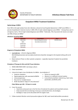
in the clinic
®
in the clinic
Cellulitis and
Soft-Tissue
Infections
Physician Writers
Lawrence J. Eron, MD
Section Editors
Christine Laine, MD, MPH
David R. Goldmann, MD
Harold C. Sox, MD
Prevention
page ITC1-2
Diagnosis
page ITC1-4
Treatment
page ITC1-7
Practice Improvement
page ITC1-14
CME Questions
page ITC1-16
The content of In the Clinic is drawn from the clinical information and
education resources of the American College of Physicians (ACP), including
PIER (Physicians’ Information and Education Resource) and MKSAP (Medical
Knowledge and Self-Assessment Program). Annals of Internal Medicine
editors develop In the Clinic from these primary sources in collaboration with
the ACP’s Medical Education and Publishing Division and with the assistance
of science writers and physician writers. Editorial consultants from PIER and
MKSAP provide expert review of the content. Readers who are interested in these
primary resources for more detail can consult http://pier.acponline.org and other
resources referenced in each issue of In the Clinic.
The information contained herein should never be used as a substitute for clinical
judgment.
© 2008 American College of Physicians
ellulitis is an infection of the skin and underlying tissues. It may follow a break in the skin or a surgical wound but may also occur without an obvious inciting event. The microorganisms most frequently
involved include group A streptococci (Streptococcus pyogenes); groups B, C,
and G β-hemolytic streptococci; and Staphylococcus aureus. Over recent
decades, cellulitis has challenged clinicians in several ways. First, physician
visits for cellulitis and soft-tissue infections have increased from 32 to 48
visits per 1000 population from 1997 to 2005 (1). Second, necrotizing fasciitis due to group A streptococci is now endemic in the United States. Third,
S. aureus, the predominant cause of cellulitis accompanied by abscesses or
wound drainage, has become increasingly resistant to methicillin, requiring
vancomycin and other, newer antimicrobial agents. A particular communityacquired methicillin-resistant S. aureus (CA-MRSA) strain, USA 300, is
replacing nosocomial strains of MRSA in hospitals (2).
C
Prevention
1. Hersh AL, Chambers
HF, Maselli JH, et al.
National trends in
ambulatory visits and
antibiotic prescribing
for skin and soft-tissue infections. Arch
Intern Med.
2008;168:1585-91.
[PMID: 18663172]
2. King MD, Humphrey
BJ, Wang YF, et al.
Emergence of community-acquired
methicillin-resistant
Staphylococcus aureus
USA 300 clone as the
predominant cause
of skin and soft-tissue
infections. Ann Intern
Med. 2006;144:30917. [PMID: 16520471]
3. Semel JD, Goldin H.
Association of athlete’s foot with cellulitis of the lower
extremities: diagnostic value of bacterial
cultures of ipsilateral
interdigital space
samples. Clin Infect
Dis. 1996;23:1162-4.
[PMID: 8922818]
4. Parish LC, Jorizzo JL,
Breton JJ, et al. Topical retapamulin ointment (1%, wt/wt)
twice daily for 5 days
versus oral
cephalexin twice
daily for 10 days in
the treatment of secondarily infected dermatitis: results of a
randomized controlled trial. J Am
Acad Dermatol.
2006;55:1003-13.
[PMID: 17097398]
© 2008 American College of Physicians
What factors increase the risk for
cellulitis and soft-tissue
infections?
Any injury to the skin may lead to
cellulitis or soft-tissue infections.
Human or animal bites have an
especially high risk for subsequent
infection. Crush injuries and open
fractures, particularly those with
vascular injury or gross contamination with soil, are likely to become
infected. Patients with comorbid
conditions that predispose them to
infection (see Box) are also at
increased risk for cellulitis.
A past episode of streptococcal cellulitis puts patients at risk for recurrences, especially if tinea pedis is
present (3). Recurrent cellulitis is
also common in patients with
chronic venous insufficiency, radical
mastectomy with axillary lymph
node dissection, or a coronary artery
bypass graft with saphenous vein
harvesting.
How can patients decrease their
chances of developing cellulitis
and soft-tissue infections?
Wounds due to minor traumatic
injuries should be meticulously
cleaned with antibacterial soap.
Application of neomycin (or
mupirocin ointment when MRSA
is suspected) may be helpful in
patients with comorbid conditions
that predispose them to infection.
ITC1-2
In the Clinic
Although it is not an approved
indication, retapamulin ointment is
an option when mupirocin-resistant
MRSA is suspected or proven (4).
Major traumatic wounds, including
crush injuries and open fractures,
should be cleansed with copious
saline irrigation and, if necessary,
surgical debridement with repair of
injured vessels. Wounds should be
left open to heal by secondary
intention. Antimicrobial prophylaxis should be administered
according to recommendations for
the type of injury (5).
Patients with recurrent lowerextremity cellulitis should be
inspected for tinea pedis, which
should be treated if present.
Risk Factors for Cellulitis and
Soft-Tissue Infection
• Trauma (lacerations, burns, abrasions,
crush injuries, open fractures)
• Intravenous drug use
• Human or animal bites
• Conditions that predispose to infection (diabetes, arterial insufficiency,
chronic venous insufficiency, lymphedema, chronic renal disease, cirrhosis, neutropenia, and hypogammaglobulinemia)
• Past streptococcal cellulitis (especially
if tinea pedis is present)
• Radical mastectomy with axillary
dissection
• Saphenous vein harvesting
Annals of Internal Medicine
6 January 2009
Which strategies reduce the risk
for cellulitis and soft-tissue
infections in patients with
diabetes?
Patients with diabetes should
receive education about proper foot
care and should regularly inspect
their feet to promptly recognize
problem areas. Treatment of superficial plantar ulcers with aggressive
cleansing and off-weighting of the
ulcerated area may prevent subsequent infection. Plantar calluses
should be shaved down. Multidisciplinary foot-care teams may be
helpful (6).
Which strategies decrease
cellulitis and soft-tissue
infections after mammalian bites?
Bite wounds should be thoroughly
cleansed. Clinicians should administer amoxicillin and clavulanate for
prophylaxis of closed-fist injuries
and cat bites and should consider
doing so for other bites when there
is extensive injury to soft tissue or
when the bite site is near a joint or
bone. For patients allergic to penicillin, use moxifloxacin plus clindamycin. Doxycycline may also be
used for prophylaxis of cat bites.
A 2001 review of 8 randomized, controlled
trials compared antibiotics with placebo
or no treatment to prevent infection following mammalian bites. Overall, the use
of antibiotics was associated with a statistically significant reduction in infection
following human bites but not cat or dog
bites. In addition, in analyses of any mammalian bite to the hand, prophylactic
antibiotics reduced the rate of infection
(odds ratio, 0.20 [95% CI, 0.01 to 0.86]) (7).
Avoid suturing bite wounds closed
except if the wounds are on the
face. Wound edges may be approximated with sterile adhesive strips
or closed by delayed primary closure. If sutures are necessary, then
antimicrobial prophylaxis and close
follow-up are indicated.
6 January 2009
Annals of Internal Medicine
Which interventions decrease the
risk for cellulitis and soft-tissue
infections associated with surgical
wounds?
Surgical site infections develop in
2% to 5% of all surgical procedures. To prevent them, health care
providers must practice good hand
hygiene, proper preparation of the
patient’s skin, and good surgical
technique (see Box).
Proper perioperative antimicrobial
prophylaxis, administered within 1
hour before surgical incision, also
prevents surgical wound infection.
Recommendations are for singledose administration or multiple
doses to end within 24 hours following the end of surgery (see Box) (8).
What is the role of surveillance to
identify patients with MRSA
colonization or infection?
Although MRSA can occur in
anyone, athletes, men who have sex
with men, incarcerated individuals,
and people living in public housing
are at greater risk for MRSA infection than are other populations (9).
In the United States, 126 000 hospitalized patients are infected by
MRSA each year, and 5000 die as
a result. To reduce these staggering
numbers, it would seem that clinicians should identify and isolate
patients who are colonized or
infected with MRSA. However,
the connection between colonization and infection and the effectiveness of decolonization in reducing infection are unclear. The
cost-efficacy of active surveillance
cultures to identify MRSA carriers
is also controversial (10).
A study of 812 U.S. soldiers evaluated
changes over time in communityacquired MRSA colonization by collecting
nasal swabs for S. aureus cultures at baseline and 8 to 10 weeks later while observing for soft-tissue infections. At baseline,
24 (3%) participants were colonized with
CA-MRSA and 229 (28%) with methicillinsensitive S. aureus (MSSA). Of those with
CA-MRSA, 9 (38%) developed soft-tissue
infection compared with 8 (3%) with
MSSA (relative risk, 10.7 [CI, 4.6 to 25.2]). At
In the Clinic
ITC1-3
Surgical Techniques that
Reduce the Postoperative
Infection Rate
• Proper skin preparation
• Gentle traction during
surgery
• Effective hemostasis
• Removal of devitalized
tissues
• Obliteration of dead space
• Irrigation of tissues
• Fine, nonabsorbable
monofilament suture
material
• Closed-suction drains
• Wound closure without
tension
Antimicrobial Prophylaxis
in Surgery
• Cefazolin or cefuroxime for
clean surgical procedures
(Clindamycin or vancomycin for patients with
penicillin allergy)
• Cefoxitin, ampicillin/
sulbactam, or metronidazole/cefazolin for gastrointestinal surgery
(Clindamycin or vancomycin plus aztreonam or
ciprofloxacin for patients
with penicillin allergy)
5. Holtom PD. Antibiotic
prophylaxis: current
recommendations. J
Am Acad Orthop
Surg. 2006;14:S98S100.
[PMID: 17003220]
6. Lipsky BA, Berendt
AR, Deery HG, et al.
Infectious Diseases
Society of America.
Diagnosis and treatment of diabetic foot
infections. Clin Infect
Dis. 2004;39:885-910.
[PMID: 15472838]
7. Medeiros I, Saconato
H. Antibiotic prophylaxis for mammalian
bites. Cochrane Database Syst Rev.
2001:CD001738.
[PMID: 11406003]
8. Scottish Intercollegiate Guidelines Network. Antibiotic prophylaxis in surgery.
Guideline No 104;
2008. Accessed at
www.sign.ac.uk/guid
elines/fulltext/104/in
dex.html on 11
November 2008.
© 2008 American College of Physicians
follow-up, CA-MRSA colonization had
decreased to 1.6% without eradication
efforts (11).
9. Diep BA, Chambers HF,
Graber CJ, et al. Emergence of multidrugresistant, communityassociated,
methicillin-resistant
Staphylococcus aureus
clone USA300 in men
who have sex with
men. Ann Intern Med.
2008;148:249-57.
[PMID: 18283202]
10. Webster J, Osborne S.
Preoperative bathing
or showering with
skin antiseptics to
prevent surgical site
infection. Cochrane
Database Syst Rev.
2007:CD004985.
[PMID: 17443562]
11. Ellis MW, Hospenthal
DR, Dooley DP, et al.
Natural history of
community-acquired
methicillin-resistant
Staphylococcus aureus
colonization and
infection in soldiers.
Clin Infect Dis.
2004;39:971-9.
[PMID: 15472848]
12. Harbarth S,
Fankhauser C, Schrenzel J, et al. Universal
screening for methicillin-resistant Staphylococcus aureus at
hospital admission
and nosocomial infection in surgical
patients. JAMA.
2008;299:1149-57.
[PMID: 18334690]
13. Karchmer TB, Durbin
LJ, Simonton BM, et al.
Cost-effectiveness of
active surveillance
cultures and contact/droplet precautions for ` control of
methicillin-resistant
Staphylococcus aureus.
J Hosp Infect.
2002;51:126-32.
[PMID: 12090800]
14. Simor AE, Phillips E,
McGeer A, et al. Randomized controlled
trial of chlorhexidine
gluconate for washing, intranasal
mupirocin, and
rifampin and doxycycline versus no treatment for the eradication of
methicillin-resistant
Staphylococcus aureus
colonization. Clin
Infect Dis.
2007;44:178-85.
[PMID: 17173213]
In a prospective crossover study in which
patients were assigned to either polymerase chain reaction testing of specimens from their nares or no polymerase
chain reaction testing, the incidence of
nosocomial MRSA infection was 1.11 per
1000 patient-days compared with 1.20 in
control participants, a nonsignificant difference (12).
A study examining the cost-effectiveness
of active surveillance cultures and barrier
precautions for controlling MRSA in
neonatal intensive care units of 2 hospitals
found that identifying colonized patients
and implementing preventive measures
was cost-effective (13).
Although there is evidence that
MRSA carriers can be decolonized,
the net benefit of decolonization is
uncertain and decolonization
through the use of topical antimicrobials applied to the nares is not
always successful (14). A review
addressing eradication strategies for
MRSA found insufficient evidence
to support the use of topical or systemic antimicrobial therapy or
combinations of these agents for
eradicating nasal or extranasal
MRSA and identified the potential
for serious adverse events and the
development of antimicrobial
resistance (15).
Prevention... Prevention of cellulitis and soft-tissue infection is best accomplished
by thorough cleansing of wounds and, when indicated, antimicrobial prophylaxis.
In patients with diabetes, examination of the plantar surface of the foot and
proper early management of lesions prevents the development of infection. Central principles in the prevention of surgical site infections are good hand hygiene,
good surgical technique, and appropriate use of perioperative antibiotic prophylaxis. The jury is still out on the net benefits of active surveillance of MRSA carriers with identification and decolonization.
CLINICAL BOTTOM LINE
Diagnosis
© 2008 American College of Physicians
What is the role of the history
and physical examination in the
diagnosis of cellulitis and softtissue infections?
A careful history is important in
determining the potential cause of
the infection. See Box for microorganisms associated with specific
clinical situations.
Skin infection has many causes, and
other conditions may mimic infection (Table 1). The physical examination helps to differentiate cellulitis from other diagnoses and to
determine the potential causes if
cellulitis is present. The absence of
drainage or abscess formation
makes streptococci a likely cause, as
do lymphangitis, a raised indurated
border, and a peau d’orange appearance of the skin. S. aureus is more
likely when abscess, draining
ITC1-4
In the Clinic
wounds, or penetrating trauma are
present (16). Crepitus suggests
Pathogens Associated With
Clinical Scenarios
• Diabetes: Staphylococcus aureus,
group B streptococci, anaerobes,
gram-negative bacilli
• Cirrhosis: Campylobacter fetus,
coliforms, Vibrio vulnificus,
Capnocytophaga canimorsus
• Neutropenia: Pseudomonas
aeruginosa
• Human bite: Eikenella corrodens
• Cat bite: Pasteurella multocida
• Dog bite: P. multocida,
C. canimorsus
• Rat bite: Streptobacillus
moniliformis
• Hot tub exposure: P. aeruginosa
• Fresh water laceration:
Aeromonas hydrophila
• Fish tank exposure:
Mycobacterium marinum
• IV drug use: MRSA, P. aeruginosa
Annals of Internal Medicine
6 January 2009
infection by gas-producing
organisms, such as an anaerobic
bacteria or a gram-negative bacillus. A foul odor may indicate infection by anaerobic microorganisms,
whereas a sweet odor may indicate
the presence of Pseudomonas or
clostridial species.
Impetigo is characterized by blisters and sores on the face or
extremities, is due to streptococci
or staphylococci (S. aureus has
become predominant in recent
years), and usually occurs in children. Necrotizing fasciitis involves
deeper tissue planes (fascia and
muscle), whereas less-severe forms
involve subcutaneous tissue (cellulitis) or epidermis and dermis
(erysipelas) (Figure).
Clinicians must differentiate limbthreatening infection, such as
necrotizing fasciitis, from
non–limb-threatening superficial
cellulitis, because the former
requires hospitalization whereas
outpatient treatment is usually sufficient for the latter (17). See Box
Table 1. Other Conditions to Consider in the Differential Diagnosis of Cellulitis
and Soft-Tissue Infection
Disease
Characteristics
Myositis due to viruses or
parasites
Mild-to-moderate fever. Severe, diffuse pain and little to
no localized pain. Mild systemic toxicity. No gas in tissues. No obvious portal of entry.
Deep venous thrombophlebitis
Deep pain, usually in the calf. Not usually associated with
fever, chills, or reddened skin. Hemodynamic instability
with pulmonary embolism.
Septic arthritis
Fever. Exquisitely painful range of motion.
Olecranon bursa infection
Redness, tenderness, fluctuance, nonpainful range of
motion.
Toxic epidermal necrolysis
Mucous membrane and diffuse cutaneous involvement.
Bullae and sloughing of skin. History of drug exposure.
Hypersensitivity reaction
Usually localized. Insect bite might be present.
Contact dermatitis
Look for unusual patterns of distribution. There is no
fever, but pruritis is present. Exposure to irritant.
Pyoderma gangrenosum
Usually on the anterior shin. Frequently acute bluishblack discoloration. Ragged ulcers with undermined
edges. Often underlying inflammatory bowel disease.
Herpetic whitlow
Vesicles on digits with associated pain, redness, and
swelling; frequently accompanied by lymphangitic streaking. Sometimes occupational (for example, dental hygienist, anesthesiologist).
Erythema migrans (Lyme disease) Resembles erysipelas, but is not painful and progresses
slowly, and fever is less marked.
Early herpes zoster (before
Pain and erythematous papules precede vesicle formation.
vesicles appear)
Hair follicle
Crust
Sebaceous
gland
Bullae
Vesicle
Stratum corneum
Erysipelas
Stratum
germinativum
Dermal papillae
Cellulitis
Post capillary venule
Subcutaneous fat
Deep fascia
Lymphatic channel
Vein
Necrotizing fasciitis
Artery
Myositis
Bone
Muscle
Figure. Anatomy of Skin and Soft Tissues and Different Types of Skin and Soft-Tissue Infections.
6 January 2009
Annals of Internal Medicine
In the Clinic
ITC1-5
15. Loeb M, Main C,
Walker-Dilks C, et al.
Antimicrobial drugs
for treating methicillin-resistant
Staphylococcus
aureus colonization.
Cochrane Database
Syst Rev.
2003:CD003340.
[PMID: 14583969]
16. Stevens DL, Bisno
AL, Chambers HF, et
al. Infectious Diseases Society of
America. Practice
guidelines for the
diagnosis and management of skin and
soft-tissue infections.
Clin Infect Dis.
2005;41:1373-406.
[PMID: 16231249]
17. Bisno AL, Cockerill
FR 3rd, Bermudez
CT. The initial outpatient-physician
encounter in group
A streptococcal
necrotizing fasciitis.
Clin Infect Dis.
2000;31:607-8.
[PMID: 10987730]
18. Anaya DA, Dellinger
EP. Necrotizing softtissue infection:
diagnosis and management. Clin Infect
Dis. 2007;44:705-10.
[PMID: 17278065]
© 2008 American College of Physicians
Physical Examination Findings
that Suggest Necrotizing
Fasciitis
• Rapid increase in size of the
infected area
• Evolution of violaceous bullae
• A reddish-purple discoloration of
the skin
• Woody induration of the infected
area
• A pale appearance of the infected
area rather than erythema
• Pain or severe tenderness out of
proportion to the appearance of
the cellulitis
• Severe systemic toxicity
• Sepsis syndrome
Table 2. Laboratory and Other Studies for Evaluating Cellulitis and Soft-Tissue
Infection
Test
Notes
CBC, differential, and
platelet count
Elevated leukocyte count with marked left shift suggests
deep-seated or systemic infection. Decreased platelet count suggests
bacteremia, the toxic shock syndrome, or gas gangrene. Leukemoid
reaction (>50 000) and hemoconcentration (rising hematocrit, frequently
>60) suggests Clostridium sordellii infection. Low hematocrit, increased
LDH, and intravascular hemolysis suggest C. perfringens infection.
Elevated creatinine concentration suggests group A streptococcal or
clostridial myonecrosis or the toxic shock syndrome.
Elevated glucose level suggests underlying diabetes mellitus.
Elevated CPK concentration suggests rhabdomyolysis, clostridial or
streptococcal myonecrosis, or necrotizing fasciitis.
Low serum bicarbonate concentration suggests metabolic acidosis and
septic shock. Alternatively, in a patient with diabetes, metabolic acidosis
associated with any soft-tissue infection suggests an aggressive process.
A low or decreasing albumin level suggests a diffuse capillary leak
syndrome. Subsequent soft-tissue swelling, third spacing, and
pulmonary edema may result.
A low serum calcium level suggests staphylococcal or streptococcal
toxic shock syndrome or necrotizing fasciitis.
Useful to detect gas in tissue and may also show underlying fracture,
osteomyelitis, or foreign body.
May be useful to localize the site, discern the extent of disease, and
provide for early diagnosis of necrotizing infections.
With necrotizing fasciitis caused by group A streptococcus, distortion
or thickening of the fascia with fluid accumulation can occur in
children. In adults, CT is better than ultrasonography at defining the
extent of disease.
The definitive test for identification of the cause of infection.
Serum creatinine
Serum glucose
Serum CPK
Serum bicarbonate
Serum albumin
Serum calcium
Radiography
CT or MRI
Ultrasonography
Culture and sensitivity
testing
CBC = complete blood count; CPK = creatine phosphokinase; CT = computed tomography; LDH = lactic
dehydrogenase; MRI = magnetic resonance imaging.
for physical examination clues to
necrotizing fasciitis.
19. Arslan A, PierreJerome C, Borthne
A. Necrotizing fasciitis: unreliable MRI
findings in the preoperative diagnosis.
Eur J Radiol.
2000;36:139-43.
[PMID: 11091013]
20. Eron LJ, Lipsky BA.
Use of cultures in
cellulitis: when, how,
and why? [Editorial].
Eur J Clin Microbiol
Infect Dis.
2006;25:615-7.
[PMID: 16896821]
21. Zahar JR, Goveia J,
Lesprit P, et al.
Severe soft tissue
infections of the
extremities in
patients admitted to
an intensive care
unit. Clin Microbiol
Infect. 2005;11:79-82.
[PMID: 15649312]
© 2008 American College of Physicians
What is the role of laboratory
testing in the diagnosis of cellulitis
and soft-tissue infections?
Laboratory testing is useful to judge
the severity of infection and to
guide therapy (Table 2). Appropriate laboratory tests include blood
cultures; complete blood count with
differential; and chemistries, including a creatinine, blood urea nitrogen, creatine phosphokinase, glucose, electrolytes (especially the
bicarbonate), and calcium. Patients
with lower extremity cellulitis
should also be evaluated for
tinea pedis.
tissue. Magnetic resonance imaging
is more sensitive than CT for
detecting edema but may be less
useful in differentiating between cellulitis and necrotizing fasciitis
because of their low specificity
(18,19).
Results of wound and blood cultures may guide antibiotic choices
but are positive in less than 5% of
cases (16, 20). Aspiration or punch
biopsies are reported to yield positive cultures in 2% to 40% of cases
(21), with the lower figure being
closer to most clinical experience.
However, among patients with streptococcal toxic shock syndrome, 60%
have positive blood cultures (21).
Computed tomography (CT) is
more sensitive and specific than plain
radiographs in identifying gas in
Although bullae and vesicles due to
streptococcal toxic shock syndrome
may yield positive cultures, those
ITC1-6
Annals of Internal Medicine
In the Clinic
6 January 2009
Types of Necrotizing Fasciitis
due to nonnecrotizing infection by
streptococci are sterile. Streptococcal
infection, although highly inflammatory, is paucibacterial. In the
future, polymerase chain reaction
technology may permit rapid identification of pathogens that are difficult to culture.
When should clinicians consult an
interventional radiologist or
surgeon during the evaluation of
cellulitis and soft-tissue infection?
Patients with necrotizing fasciitis
require early surgical inspection of
the deep tissues through a small
incision that allows evaluation of
the condition of the fascia and the
viability of underlying musculature.
Diagnostic incisions can be
extended to perform radical
debridement, should necrotizing
infection be visible. Other signs
suggesting the need for surgical
intervention include drainage,
thrombosed vessels, and lack of
resistance to finger dissection in
normally adherent tissues (18).
Interventional radiologists can use
diagnostic imaging to locate, biopsy,
and aspirate deep collections or
masses for diagnostic purposes as
well as for therapeutic drainage but
cannot inspect wounds macroscopically or debride devitalized tissue.
• Necrotizing fasciitis type 1: Mildto-moderate fever and systemic
toxicity. Little to no diffuse pain
and mild-to-moderate localized
pain. Mild-to-moderate gas in tissues. Portal of entry usually obvious. Higher risk with diabetes (S.
aureus, group B streptococci,
anaerobes, and gram-negative
bacilli).
• Necrotizing fasciitis type 2
(“flesh-eating strep”): Moderateto-severe fever. Little to no diffuse
pain and severe localized pain.
Severe systemic toxicity. No gas in
tissues. Portal of entry not obvious
in 50% of cases (commonly streptococci).
• Necrotizing fasciitis type 3 (“gas
gangrene”): Moderate fever. Little
to no diffuse pain and severe localized pain. Severe systemic toxicity.
Severe gas in tissues. Portal of
entry usually obvious (clostridial
species).
Diagnosis... Clinicians must use the medical history, physical examination, and
laboratory testing to identify probable pathogens. Culture identification of the
pathogen should lead to changes from initial broad-spectrum antimicrobial agents
to more narrow-spectrum regimens to avoid the emergence of resistance. Unfortunately, positive cultures are infrequent in patients with nonpurulent cellulitis.
Rapid expansion of the infected area, violaceous bullae or reddish-purple discoloration of the skin, severe pain, or systemic toxicity suggest necrotizing fasciitis, a
condition that requires urgent surgical evaluation.
CLINICAL BOTTOM LINE
Treatment
Which adjuvant measures are
helpful in the treatment of
patients with cellulitis and softtissue infection?
Compression dressings in patients
with chronic venous insufficiency
or pedal edema promote resolution
of cellulitis. Negative pressure
dressings for large, exudative
wounds reduce proteolytic enzymes
and other substances that retard
wound healing. Remediation of
lymphedema or compromised
vascular supply may improve delivery of oxygen and antimicrobials
to the site of infection. There is
insufficient evidence about the
benefits of hyperbaric oxygen for
infected diabetic foot ulcers and gas
gangrene (23).
6 January 2009
Annals of Internal Medicine
When is topical antimicrobial
therapy appropriate?
Topical mupirocin is an option for
impetigo with limited lesions, but it
is expensive and some staphylococci
are resistant. Oral therapy with
penicillinase-resistant penicillins or
first-generation cephalosporins is
preferred (16).
How should clinicians determine
whether to prescribe oral or
parenteral antimicrobials?
High bioavailability makes parenteral administration of antimicrobials largely unnecessary for mild,
uncomplicated cellulitis. Parenteral
therapy is usually reserved for moderate-to-severe infections that may
require hospitalization. However,
In the Clinic
ITC1-7
22. Stevens DL, Bryant
AE, Adams K, et al.
Evaluation of therapy with hyperbaric
oxygen for experimental infection
with Clostridium perfringens. Clin Infect
Dis. 1993;17:231-7.
[PMID: 8399871]
23. Eron LJ, Lipsky BA,
Low DE, et al. Expert
panel on managing
skin and soft tissue
infections. Managing
skin and soft tissue
infections: expert
panel recommendations on key decision points. J Antimicrob Chemother.
2003;52 Suppl 1:i317. [PMID: 14662806]
© 2008 American College of Physicians
Oral Antimicrobials for Mild
Infections Caused by
Streptococci, MSSA, and MRSA
Streptococci only
• Phenoxymethyl penicillin
• Amoxicillin
Streptococci or MSSA
• Amoxicillin–clavulanate
• Cloxacillin, dicloxacillin,
cephalexin
• Clindamycin or a macrolide (if
allergic to penicillins; and if sensitive)
MSSA or MRSA
• Clindamycin (if sensitive)
• Doxycycline or minocycline
• Trimethoprim–sulfamethoxazole
• Linezolid (very expensive)
• Advanced fluoroquinolones–
moxifloxacin and levofloxacin
24. Bergkvist PI, Sjöbeck
K. Antibiotic and
prednisolone therapy of erysipelas: a
randomized, double
blind, placebo-controlled study. Scand
J Infect Dis.
1997;29:377-82.
[PMID: 9360253]
25. Bergkvist PI, Sjöbeck
K. Relapse of
erysipelas following
treatment with
prednisolone or
placebo in addition
to antibiotics: a 1year follow-up.
Scand J Infect Dis.
1998;30:206-7.
[PMID: 9730318]
26. Dall L, Peterson S,
Simmons T, et al.
Rapid resolution of
cellulitis in patients
managed with combination antibiotic
and anti-inflammatory therapy. Cutis.
2005;75:177-80.
[PMID: 15839362]
27. Lee MC, Rios AM,
Aten MF, et al. Management and outcome of children
with skin and soft
tissue abscesses
caused by community-acquired methicillin-resistant
Staphylococcus
aureus. Pediatr Infect
Dis J. 2004;23:123-7.
[PMID: 14872177]
28. Ruhe JJ, Menon A.
Tetracyclines as an
oral treatment
option for patients
with community
onset skin and soft
tissue infections
caused by methicillin-resistant
Staphylococcus
aureus. Antimicrob
Agents Chemother.
2007;51:3298-303.
[PMID: 17576834]
© 2008 American College of Physicians
parenteral agents may also be
administered to outpatients if the
patient is stable and has a social situation that enables out-patient
therapy. Classification schemes
have been proposed to assist in
these treatment decisions (16, 23).
In some patients, cutaneous inflammation worsens during the initial
24 hours of antimicrobial treatment. This is not necessarily a sign
of treatment failure and may merely
represent an inflammatory response
to release of bacterial antigens.
Anti-inflammatory agents may
accelerate the resolution of the
clinical signs of inflammation (2426). When patients do not improve
when receiving what should be
adequate antimicrobials, it could
also represent antimicrobial resistance; a deeper, undrained collection; or complicating comorbid
conditions.
One study suggests that abscesses
less than 5 cm in diameter may be
effectively treated by incision and
drainage without systemic antimicrobial therapy (27), but other
studies show no correlation
between the size of abscesses and
the need for antibiotic therapy (28).
treatment of MSSA include
cloxacillin; dicloxacillin; cephalexin;
and, for penicillin-allergic patients,
clindamycin or a macrolide (erythromycin, clarithromycin, or
azithromycin). However, patients
treated with cephalexin should be
observed for treatment failure, as
some studies have reported high
failure rates in adults (29), possibly
due to poor absorption (30).
Appropriate orally administered
choices for the treatment of MRSA
include trimethoprim–sulfamethoxazole, doxycycline, minocycline, or linezolid. Linezolid is one
of the few orally administered
antimicrobials adequately tested in
comparative trials with parenteral
vancomycin. Clindamycin may be a
very good alternative, if the
S. aureus (MRSA or MSSA) is
susceptible. However, resistance is
becoming increasingly prevalent
among S. aureus. Advanced fluoroquinolones, such as moxifloxacin
and levofloxacin, may also be used,
but only if patients cannot tolerate
any other choices.
Purulence is more often present in
staphylococcal cellulitis. Specimens
from wounds, carbuncles, or furuncles may reveal gram-positive
cocci in clusters, consistent with
S. aureus. Clinicians must judge
whether MRSA or MSSA is likely.
Appropriate choices for the oral
Trimethoprim–sulfamethoxazole is
a reasonable initial choice for the
treatment of mild, uncomplicated
cellulitis. However, some experts
feel that trimethoprim–sulfamethoxazole should not be used for
cellulitis without purulence because
these drugs have relatively poor
activity against streptococci (31). In
one comparative trial, cures were
obtained in 37 of 42 patients
treated with orally administered
trimethoprim–sulfamethoxazole
and 57 of 58 patients treated with
vancomycin, suggesting equivalence
between these 2 drugs in the treatment of mild cellulitis (32). A study
comparing trimethoprim–sulfamethoxazole with doxycycline suggested no statistically significant
difference between these 2 antimicrobials in the treatment of cellulitis and soft-tissue infections caused
by MRSA (33). The use of clindamycin and tetracyclines to treat
ITC1-8
Annals of Internal Medicine
In patients for whom oral therapy
is appropriate, how should
clinicians choose a specific
antimicrobial agent?
Generally, streptococci usually
cause cellulitis without purulence.
Phenoxymethyl penicillin, amoxicillin, or (for penicillin-allergic
patients) clindamycin are all appropriate antibiotic choices (Box).
However, a small number (<10%)
of group A streptococci are resistant to clindamycin.
In the Clinic
6 January 2009
MRSA is supported only by observational studies (34, 35).
In patients for whom parenteral
therapy is appropriate, how should
clinicians choose a specific
antimicrobial regimen?
The choices for parenteral antibiotics for cellulitis probably caused
by streptococci, as well as soft-tissue infection due to MSSA, include
semisynthetic penicillins (nafcillin
and oxacillin), cephalosporins (cefazolin, cefuroxime, or ceftriaxone),
clindamycin (for penicillin-allergic
patients and if the pathogen is
shown to be sensitive). If MRSA is
suspected, vancomycin, daptomycin, tigecycline, and linezolid
are appropriate choices for moderate-to-severe infections. The newer
agents have been shown to be noninferior to vancomycin in comparative trials. Some studies reported
more rapid resolution of clinical
signs of inflammation in patients
receiving daptomycin compared
with a matched cohort of patients
receiving vancomycin (36, 37).
For outpatient parenteral treatment
of MRSA, daptomycin has certain
advantages when compared with
vancomycin. Daptomycin pharmacokinetics allow once-daily administration in patients with healthy renal
function. Rapid administration does
not cause “red-man syndrome” as
Antimicrobials Commonly Used
for Outpatient Parenteral
Antibiotic Therapy
• Ceftriaxone, cefazolin, and
cefuroxime (for streptococci and
MSSA infections)
• Ertapenem, cefepime, ceftazidime
(for gram-negative bacillary
infection)
• Daptomycin and vancomycin (for
MRSA infections)
• Clindamycin (for streptococci,
MSSA, and MRSA infection, if
sensitive)
• Aminoglycosides (for resistant
gram-negative bacillary infection)
6 January 2009
Annals of Internal Medicine
happens with vancomycin, and it is
not necessary to monitor daptomycin levels.
A new antimicrobial agent, dalbavancin, is awaiting approval by the
U.S. Food and Drug Administration. Its pharmacokinetics allow
once-weekly administration, making it attractive for outpatient parenteral treatment. Dalbavancin has
been compared with linezolid and
shows similar efficacy (38) (Table
3). Other parenteral agents active
against MRSA and pending release
include telavancin, ceftobiprole, ceftaroline, and iclaprim.
What is appropriate antimicrobial
therapy for infections associated
with human or animal bites?
See Box for suggested antimicrobial
treatment following bite wounds.
The microorganisms involved in
human bites are most commonly
Eikenella corrodens, anaerobes, and
viridans streptococci. Animal bites
are commonly caused by a mixture
of bacteria, including Pasteurella
mutocida, Streptococcus intermedius,
anaerobes, and S. aureus. A rare
cause of infection following dog
bites is Capnocytophaga canimorsus,
which causes severe sepsis syndrome with disseminated intravascular coagulation but is susceptible
to penicillins.
Are there special considerations
for antimicrobial selection in
immunocompromised patients with
cellulitis and soft-tissue infection?
The clinical signs of infection in
the immunocompromised patient
may be subtle or absent due to
decreased inflammatory response.
Because of the extremely broad
range of opportunistic pathogens in
this patient population, attempts to
definitively identify pathogens with
cultures, biopsies, antigen tests, or
imaging studies are crucial. Empirical therapy should be quickly initiated and guided by patient-specific
clinical parameters and the most
likely pathogens. For example, the
most likely pathogens during the
In the Clinic
ITC1-9
Treatment of Human and
Animal Bites
Oral treatment
• Human bite: amoxicillin/clavulanate
• Animal bite: amoxicillin/clavulanate
Parenteral treatment
• Human bite: ampicillin/sulbactram
• Animal bite: ampicillin/sulbactam
Penicillin allergy
• Human bite: moxifloxacin plus
clindamycin, trimethoprim/
sulfamethoxazole plus
metronidazole
• Animal bite: doxycycline or moxifloxacin or trimethoprim/sulfamethoxazole with either clindamyciin or metronidazole
29. Madaras-Kelly KJ,
Arbogast R, Jue S.
Increased therapeutic failure for
cephalexin versus
comparator antibiotics in the treatment of uncomplicated outpatient
cellulitis. Pharmacotherapy.
2000;20:199-205.
[PMID: 10678298]
30. Madaras-Kelly KJ,
Remington RE,
Oliphant CM, et al.
Efficacy of oral betalactam versus nonbeta-lactam treatment of
uncomplicated cellulitis. Am J Med.
2008;121:419-25.
[PMID: 18456038]
31. Daum RS. Clinical
practice. Skin and
soft-tissue infections
caused by methicillin-resistant
Staphylococcus
aureus. N Engl J
Med. 2007;357:38090. [PMID: 17652653]
32. Markowitz N, Quinn
EL, Saravolatz LD.
Trimethoprim-sulfamethoxazole compared with vancomycin for the
treatment of Staphylococcus aureus
infection. Ann Intern
Med. 1992;117:3908. [PMID: 1503330]
33. Cenizal MJ, Skiest D,
Luber S, et al.
Prospective randomized trial of empiric
therapy with
trimethoprim-sulfamethoxazole or
doxycycline for outpatient skin and soft
tissue infections in
an area of high
prevalence of methicillin-resistant
Staphylococcus
aureus. Antimicrob
Agents Chemother.
2007;51:2628-30.
[PMID: 17502411]
© 2008 American College of Physicians
Table 3. Drug Treatment for Cellulitis and Soft-Tissue Infections
Agent
Dosage
Side Effects
Notes
Low: 0.6–1.2 million IU/d,
IM (benzathine); High: >20
million IU/d, IV (crystalline)
Hypersensitivity
Nafcillin
1.0–2.0 g, q4h IV or IM
Oxacillin
1.0–2.0 g, q4h IV or IM
Dicloxacillin
0.125–0.5 g, q6h PO
Hypersensitivity; reversible neutropenia;
interstitial nephritis; peripheral IV irritating
Hypersensitivity; hepatic dysfunction with
>12 g/d; interstitial nephritis
Hypersensitivity
Amoxicillin
0.5 g, q8h PO; 0.875 g, q12h
Hypersensitivity
Ampicillin
0.25–0.5 g, q6h PO; 150–200
mg/kg•per day, IV
1.0–3.0 g, q6–8h IV
Hypersensitivity
Benzathine useful only for impetigo due
to Staphylococcus pyogenes or syphilis.
Crystalline useful for mild-to-moderate
streptococcal and clostridial infections and
bite wounds
Useful for staphylococcal infections
except MRSA
Useful for staphylococcal infections
except MRSA
Useful for minor infections with Staphylococcus except MRSA and Streptococcus
Useful for Pasteurella and minor streptococcal infections
Useful for Pasteurella and minor
streptococcal infections
Improved spectrum to Staphylococcus
aureus and Bacteroides fragilis
Excellent gram-negative spectrum, including
Pseudomonas species
ß-lactams
Penicillin G
Ampicillin–
sulbactam
Piperacillin–
tazobactam
Macrolides
Erythromycin
Hypersensitivity
2.25–4.5 g, q6h
Hypersensitivity, nausea, vomiting,
diarrhea; phlebitis when given IV;
cholestatic hepatitis (erythromycin estolate)
0.5 g, q12h PO
GI intolerance (4%); reversible dose-related
hearing loss
0.5 g PO on day 1, then 0.25 GI intolerance (4%); reversible dose-related
g/d PO on days 2–5; 0.5 g/d IV hearing loss
Erythromycin resistance in S. aureus and
group A streptococcus
Cephalosporins
Cefazolin
1.0–2.0 g, q8h IV or IM
Hypersensitivity
Cefuroxime
0.75–1.5 g, q6h IV or IM
Hypersensitivity
Cefoxitin
Ceftriaxone
Hypersensitivity
Hypersensitivity; cholelithiasis
Cefpodoxime
Ceftobiprole
1.0 g, q8h; 2.0 g, q4h IV
0.5 g, q12h; 2.0 g, qd (total
<4 g/d) IV or IM
100–200 mg, q12h PO
500 mg, q8h IV
Useful for both staphylococcal and streptococcal infections
More stable than cefazolin against Staphylococcus‚ ß-lactamase. Useful for staphylococcal and streptococcal infections.
Reasonable activity against anaerobes
Useful for outpatient prescription due to
once-a-day dosing
Ceftaroline
600 mg, q12h IV
Clarithromycin
Azithromycin
Fluoroquinolones
Ciprofloxacin
0.25–0.5 g, q6h PO; 15–20
mg/kg per day, IV
500–750 mg, bid PO;
200–400 mg, q12h IV
Levofloxacin
500–750 mg, q24h IV or PO
Moxifloxacin
400 mg, q24h PO
Gatifloxacin
200–400 mg, q24h IV or PO
Hypersensitivity
Active against MRSA, VISA, VRSA, gramnegative bacilli
Active against MRSA, VISA, VRSA, gramnegative bacilli
CNS and GI disturbances; avoid caffeine;
not for use during pregnancy and if age
<18 y; Achilles tendon rupture
CNS and GI disturbances; avoid caffeine;
not for use during pregnancy and if age
<18 y; Achilles tendon rupture
CNS and GI disturbances; avoid caffeine;
not for use during pregnancy and if age
<18 y; Achilles tendon rupture
Hypoglycemia; CNS and GI disturbances;
avoid caffeine; not for use during pregnancy
and if age <18 y; Achilles tendon rupture;
rarely used due to toxicity risk
Aminoglycosides (rarely used due to toxicity risk)
Gentamicin
2 mg/kg load, then 1.5 mg/kg, Ototoxicity and nephrotoxicity
q8h; 5.0 mg/kg (if critically ill,
7.0 mg/kg), q24h load
© 2008 American College of Physicians
ITC1-10
In the Clinic
Excellent activity against gram-negative
bacteria, including Pseudomonas
Improved gram-positive spectrum against
Staphylococcus aureus and Streptococcus
species
Improved gram-positive spectrum against
S. aureus and Streptococcus species
Improved gram-positive spectrum against
Staphylococcus aureus and Streptococcus
species. Contraindicated in diabetes. Use
cautiously in the elderly and if renal insuffi
ciency is present
Good spectrum of activity against resistant
gram-negative bacteria. Follow levels to
guide dosing
Annals of Internal Medicine
6 January 2009
Table 3. Drug Treatment for Cellulitis and Soft-Tissue Infections (continued)
Agent
Tobramycin
Amikacin
Glycopeptides
Vancomycin
Dalbavancin
Lincosamides
Clindamycin
Tetracyclines
Doxycycline
Minocycline
Tigecycline
Dosage
Side Effects
Notes
2 mg/kg load, then 1.5 mg/kg, Ototoxicity and nephrotoxicity
q8h; 5.0 mg/kg (if critically
ill, 7.0 mg/kg), q24h load
15 mg/kg per day
Good spectrum of activity against resistant
gram-negative bacteria. Follow levels to
guide dosing
15 mg/kg, q12h IV. Follow
serum levels
1 g, IV followed 1 week later
by 500 mg, IV
Can cause nephrotoxicity and the “red-man” Used primarily against MRSA
syndrome with rapid infusion.
Active against MRSA, VRE (Van B and Van C
strains only). Use for outpatient therapy
once weekly
0.15–0.45 g, q6h PO; 1.2 g,
IV q12h
Pseudomembranous colitis
Useful for severe group A streptococcal
infections and gas gangrene caused by
Clostridium species; inhibits bacterial toxin
production; use for oral or dental infections
100 mg, bid
Liver toxicity; photosensitivity; tooth
discoloration in children
100 mg, qd or bid
Dizziness
100 mg, IV load, 50 mg, q12h Nausea and vomiting
IV thereafter
Penems
Imipenem–cilastatin 0.5–1.0 g, q6h IV
Option for penicillin-allergic patients
Active against MRSA, streptococci, anaerobic
organisms, and gram-negative bacilli except
Pseudomonas species
Meropenem
Doripenem
1–2 g, q6h IV
500 mg, q8h IV
Superinfection; allergic reactions; phlebitis;
hepatotoxicity; seizures; renal failure
Diarrhea (5%); nausea, headache
Headaches
Broadly active against aerobes, anaerobes,
gram-positives, and gram-negatives
Ertapenem
Oxazolidinone
Linezolid
1 g, q24h
Nausea, diarrhea, rash
2- to 4-fold lower MIC for Pseudomonas
aeruginosa
Use for outpatient therapy once daily
600 mg, q12h IV or PO
Nausea, diarrhea, thrombocytopenia with
prolonged treatment (>2 wk), peripheral
neuropathy, optic neuritis
Active against MRSA and VRE, but also
against other gram-positive bacteria; should
not be used when alternatives are available
Lipopeptides
Daptomycin
4–6 mg/kg per day
Telavancin
7.5 mg/kg, q24h IV
CPK elevation; rhabdomyolysis at high dosage Use for outpatient therapy once daily, use
for bacteremia due to MRSA, as it is rapidly
bactericidal
Taste disturbance, headache
Rapidly bactericidal with a long postantibiotic effect. Active against MRSA, GISA,
VRSA, VRE (including Van A strains), may
have a reduced potential for development of
resistance
bid = twice daily; CNS = central nervous system; DNA = deoxyribonucleic acid; GI = gastrointestinal; GISA = glycopeptide intermediate-resistant
Staphylococcus aureus; IM = intramuscular; IV = intravenous; PO = oral; qd = once daily; MIC = minimal inhibitory concentration; MRSA = methicillinresistant Staphylococcus aureus; VISA = vancomycin intermediate-resistant Staphylococcus aureus; VRE = vancomycin-resistant enterococci;
VRSA = vancomycin-resistant Staphylococcus aureus.
first week of postchemotherapy neutropenia are gram-positive organisms, such as staphylococci, viridans
streptococci, enterococci, and grampositive bacilli, such as Corynebacterium, Clostridia, and Bacillus
species. During the second week of
neutropenia, antibiotic-resistant
bacteria and fungi (Aspergillus,
Candida, and Rhizopus and Mucor
species) become prominent. Initial
6 January 2009
Annals of Internal Medicine
antimicrobial therapy for cellulitis in
immunocompromised patients
should include coverage
of both gram-positive and
gram-negative microorganisms
(ceftazidime, cefepime, piperacillin–
tazobactam, meropenem, doripenem, or imipenem). Penicillinallergic patients should receive
fluoroquinolones (such as
ciprofloxacin) or aztreonam.
In the Clinic
ITC1-11
34. Martínez-Aguilar G,
Hammerman WA,
Mason EO Jr, et al.
Clindamycin treatment of invasive
infections caused by
communityacquired, methicillinresistant and methicillin-susceptible
Staphylococcus
aureus in children.
Pediatr Infect Dis J.
2003;22:593-8.
[PMID: 12867833]
© 2008 American College of Physicians
Risk Factors for MRSA
• Recent antibiotic use
• Recent hospitalization
• Recurrent needle sticks
(IV drug use, hemodialysis, or insulinrequiring diabetic
patients)
• Homelessness,
incarceration
• Contact sports (football,
wrestling)
• Previous MRSA infection
or colonization
Indications for
Hospitalization of
Patients with Cellulitis
and Soft Tissue
Infections
• Necrotizing fasciitis
• Toxic shock syndrome or
multiorgan failure
• Limb-threatening
infection
• Need for surgical debridement or drainage
• Gas in tissue
• Inadequate home situation or patient at risk for
nonadherence
35. Ruhe JJ, Monson T,
Bradsher RW, et al.
Use of long-acting
tetracyclines for
methicillin-resistant
Staphylococcus
aureus infections:
case series and
review of the literature. Clin Infect Dis.
2005;40:1429-34.
[PMID: 15844065]
36. Davis SL, McKinnon
PS, Hall LM, et al.
Daptomycin versus
vancomycin for
complicated skin
and skin structure
infections: clinical
and economic outcomes. Pharmacotherapy.
2007;27:1611-8.
[PMID: 18041881]
37. Krige JE, Lindfield K,
Friedrich L, et al.
Effectiveness and
duration of daptomycin therapy in
resolving clinical
symptoms in the
treatment of complicated skin and skin
structure infections.
Curr Med Res Opin.
2007;23:2147-56.
[PMID: 17669231]
© 2008 American College of Physicians
Monotherapy of Pseudomonas infections seems at least equal to if not
superior to dual therapy, in view of
the toxicity associated with the
addition of an aminoglycoside
(39). Reasons for initiating aminoglycosides for empirical treatment
include a high level of antimicrobial resistance within an
institution.
If patients remain febrile and cultures remain negative, the empirical addition of vancomycin or
daptomycin for MRSA coverage
and addition of amphotericin or an
echinocandin (caspofungin, micafungin, or anidulafungin) or a triazole (voriconazole) are warranted
for antifungal coverage. Patients
with deficiencies of cell-mediated
immunity (such as those with
lymphoma, or HIV infection or
organ transplant recipients) are
beyond the scope of this article (16).
When should clinicians consider
coverage of MRSA when treating
patients with cellulitis and softtissue infections?
Although MRSA should be considered in all cases of cellulitis or
soft-tissue infection (because of its
widespread prevalence), particular
populations are at higher risk than
others (see Box). MRSA and
MSSA should be suspected in
patients with purulent cellulitis,
although streptococci can also cause
purulent cellulitis.
When should clinicians hospitalize
a patient with cellulitis and softtissue infection?
Mild, uncomplicated cellulitis may
be treated out of the hospital with
oral antibiotics. Although surgical
drainage may be required in some
cases, this can also be done on an
outpatient basis. Moderate-tosevere cellulitis, however, may
require hospitalization for observation, control of comorbid conditions, and administration of parenteral antimicrobial agents.
ITC1-12
In the Clinic
Because necrotizing fasciitis is associated with shock and organ failure,
with a mortality rate ranging from
30% to 70% (16), these patients
require hospitalization for surgical
debridement, prompt antibiotic
treatment, and monitoring. When
the diagnosis is in doubt, in-hospital observation is appropriate until
the clinical course becomes clear.
See Box for conditions that may
require hospitalization.
Generally, hospitalized patients
with cellulitis or soft-tissue infection can be discharged when no
longer febrile, when the cellulitis is
no longer spreading, and when the
leukocyte count is trending toward
normal.
When should clinicians consider
consulting infectious disease
experts?
Involvement of an infectious disease
expert may result in more appropriate and cost-effective use of antimicrobial agents and may be helpful
when patients are immunocompromised, have severe infection, or do
not improve on initial therapy.
In one study, more-appropriate treatment
was ordered initially by a consulting infectious disease specialist (78%) than without
such consultation (54%). Following the
availability of culture results, appropriate
treatment increased to 97% and 89%,
respectively (40).
What should clinicians consider in
managing patients with
necrotizing fasciitis?
Type 1 necrotizing fasciitis is usually a polymicrobial infection due to
S. aureus, group B streptococci,
anaerobic organisms, and gramnegative bacilli. Management
includes broad-spectrum antimicrobials, surgical debridement, vascular
evaluation, off-weighting of the
lower extremity, and normalization
of blood glucose levels.
Type 2 necrotizing fasciitis is known
colloquially as “flesh-eating strep,”
because it is caused most frequently
Annals of Internal Medicine
6 January 2009
by streptococci. Occasionally,
staphylococci and gram-negative
bacilli, such as Vibrio vulnificus, are
isolated from necrotizing soft-tissue infections. For streptococcal
necrotizing fasciitis, the antimicrobial agent of choice is clindamycin
combined with penicillin or other
β-lactam antibiotic. As 5% of
streptococci are resistant to clindamycin, penicillin is added to
clindamycin in case of resistance
(16). Linezolid is an expensive but
appropriate choice for staphylococcal
necrotizing fasciitis in penicillinallergic patients (16). However, in
cases of intolerance to linezolid, daptomycin is an option.
Type 3 necrotizing fasciitis is characterized by severe toxicity and gas in
tissues due to infection with
clostridial species. Penicillin plus
clindamycin with debridement is the
therapy of choice (16).
What are the indications for
surgical debridement of cellulitis
and soft-tissue infection?
Evidence of gangrenous or necrotizing infection requires immediate
and thorough debridement. This is
especially true of streptococcal
necrotizing fasciitis. Expert consensus favors the use of intravenous
immunoglobulin to treat streptococcal necrotizing fasciitis,
although studies are conflicting
(41-43). Friable fascia and dark
muscle that do not bleed or twitch
requires debridement, which should
be continued until viable tissue is
reached. Multiple debridements are
often required for patients with
necrotizing fasciitis or myonecrosis.
Delay of surgical debridement
increases mortality.
How long should patients remain on
antimicrobial therapy for cellulitis
and soft-tissue infection?
When treating moderate-to-severe
cellulitis, it is appropriate to switch
from parenteral to oral antibiotics
when the infection is stabilized and
the patient is able to tolerate oral
therapy. Many antimicrobials exhibit
complete oral bioavailability and
comparative trials have shown the
equivalence between parenteral and
orally administered antimicrobials in
the treatment of soft-tissue infections
(44, 45). There may be no advantage
in administering parenteral antibiotics in patients who have infections
susceptible to oral therapies and no
symptoms, such as nausea and vomiting, that would complicate oral therapy. Patients with cellulitis and softtissue infection often receive
antimicrobial therapy for 10 to 14
days or until inflammation resolves.
This practice assumes that inflammation indicates surviving organisms,
but after several days of antimicrobials, inflammation may be due to
antigens released from dead bacteria.
Clinical trial evidence shows similar
outcomes in patients treated for 5
days and 10 days (46).
Treatment... Treat cellulitis and soft-tissue infections empirically with antimicrobials that target the suspected pathogen. Topical treatment is an option for
mild impetigo. Patients with mild, uncomplicated cellulitis who are not at high
risk for MRSA should receive oral antibiotics active against both staphylococci and
streptococci. Abscesses require drainage. Oral antibiotics are sufficient to treat
mild MRSA infection. Patients unable to tolerate oral antibiotics or those with
severe cellulitis and systemic toxicity who are at risk for MRSA should receive parenteral antibiotics active against MRSA and streptococci. Patients at risk for
MRSA should receive coverage for MRSA and streptococci. Antimicrobials should
be adjusted to focus on culture-identified pathogens. Prompt surgical evaluation
is indicated for patients with evidence of necrotizing fasciitis.
CLINICAL BOTTOM LINE
6 January 2009
Annals of Internal Medicine
In the Clinic
ITC1-13
38. Billeter M, Zervos
MJ, Chen AY, et al.
Dalbavancin: a novel
once-weekly lipoglycopeptide antibiotic.
Clin Infect Dis.
2008;46:577-83.
[PMID: 18199045]
39. Paul M, Silbiger I,
Grozinsky S, et al.
Beta lactam antibiotic monotherapy
versus beta lactamaminoglycoside
antibiotic combination therapy for sepsis. Cochrane Database Syst Rev.
2006:CD003344.
[PMID: 16437452]
40. Byl B, Clevenbergh P,
Jacobs F, et al.
Impact of infectious
diseases specialists
and microbiological
data on the appropriateness of antimicrobial therapy for
bacteremia. Clin
Infect Dis.
1999;29:60-6; discussion 67-8.
[PMID: 10433566]
41. Stevens DL. Dilemmas in the treatment of invasive
Streptococcus pyogenes infections [Editorial]. Clin Infect Dis.
2003;37:341-3.
[PMID: 12884157]
42. Kaul R, McGeer A,
Norrby-Teglund A, et
al. Intravenous
immunoglobulin
therapy for streptococcal toxic shock
syndrome—-a comparative observational study. The
Canadian Streptococcal Study Group.
Clin Infect Dis.
1999;28:800-7.
[PMID: 10825042]
43. Darenberg J,
Ihendyane N, Sjölin
J, et al. StreptIg
Study Group. Intravenous
immunoglobulin G
therapy in streptococcal toxic shock
syndrome: a European randomized,
double-blind,
placebo-controlled
trial. Clin Infect Dis.
2003;37:333-40.
[PMID: 12884156]
44.Gentry LO, RamirezRonda CH,
Rodriguez-Noriega
E, et al. Oral
ciprofloxacin vs parenteral cefotaxime
in the treatment of
difficult skin and
skin structure infections. A multicenter
trial. Arch Intern
Med. 1989;149:257983. [PMID: 2684078]
© 2008 American College of Physicians
45. Weigelt J, Itani K,
Stevens D, et.al. Linezolid versus vancomycin in treatment of
complicated skin
and soft tissue infections. Antimicrob
Agents Chemother.
2005;49:2260-6.
[PMID: 15917519]
46. Hepburn MJ. Dooley
DP, Skidmore PJ, et.
Al Comparison of
short course (5 days)
and standard (10
days) treatment for
uncomplicated cellulitis. Arch Intern
Med. 2004;
164:1669-74. [PMID:
15302637]).
What factors do U.S. stakeholders
use to evaluate the quality of care
for soft-tissue infection and
cellulitis?
The Center for Medicare & Medicaid Services (CMS) has developed measures of quality of care to
use in the Physician Quality
Reporting Initiative. None of these
measures relates to the treatment of
cellulitis and soft-tissue infection.
However, several measures focus on
the antibiotic prophylaxis to prevent surgical wound infections.
These measures focus on the
appropriate timing and selection of
antibiotic prophylaxis (started
within 1 hour [2 hours for fluoroquinolone or vancomycin] before
surgical incision and discontinued
within 24 hours of the end of
surgery).
What do professional
organizations recommend
regarding the care of patients
with cellulitis and soft-tissue
infection?
In 2005, the Infectious Disease
Society of America (IDSA)
released guidelines on the management of cellulitis and soft-tissue
infection (16). This review largely
reflects the IDSA recommendations. See the Toolkit for other
guidelines related to the prevention
and treatment of soft-tissue
infections.
PIER Modules
in the clinic
Tool Kit
Cellulitis and
Soft-Tissue
Infections
www.pier.acponline.org
Access the following PIER module: Cellulitis and Soft Tissue Infections. PIER
modules provide evidence-based, updated information on prevention, diagnosis,
and treatment in an electronic format designed for rapid access at the point of care.
Patient Education Resources
www.annals.org/intheclinic/toolkit-cellulitis.html
Access the Patient Information material that appears on the following page for
duplication and distribution to patients.
www.nlm.nih.gov/medlineplus/cellulitis.html
Medline Plus
www3.niaid.nih.gov/topics/streptococcal/
National Institute of Allergy and Infectious Diseases
www.healthsystem.virginia.edu/uvahealth/adult_derm/cell.cfm
Patient information developed at University of Virginia (available in English
and Spanish).
Guidelines
www.journals.uchicago.edu/doi/pdf/10.1086/497143
Infectious Diseases Society of America: Practice Guidelines for the Diagnosis and
Management of Skin and Soft Tissue Infections
www.cdc.gov/ncidod/dhqp/pdf/ar/mdroGuideline2006.pdf
Centers for Disease Control and Prevention: Guidelines for the Management of
Multi-drug Resistant Organisms in Healthcare Settings
www.shea-online.org/Assets/files/position_papers/SHEA_hand.pdf
Healthcare Infection Control Practices Advisory Committee and the
HICPAC/SHEA/APIC/IDSA Hand Hygiene Task Force: Guideline for Hand
Hygiene in Healthcare Settings
www.shea-online.org/Assets/files/position_papers/SHEA_MRSA_VRE.pdf
Society for Healthcare Epidemiology of America
© 2008 American College of Physicians
ITC1-14
In the Clinic
in the clinic
Practice
Improvement
Annals of Internal Medicine
6 January 2009
In the Clinic
Annals of Internal Medicine
annals.org
THINGS PEOPLE SHOULD
KNOW ABOUT CELLULITIS AND
SOFT-TISSUE INFECTION
What is cellultis?
• Cellulitis is an infection that involves the skin
or the muscles and other body tissues directly
under the skin. Symptoms can include redness,
pain, and fever.
Who gets cellulitis?
• Cellulitis can happen after an injury to the
skin, an animal bite, or a surgical wound, but
sometimes there is no obvious cause.
• Conditions that increase the chances of cellulitis include diabetes, circulatory problems,
past surgery or radiation treatment of the
arms or legs, and chronic athlete’s foot.
What is the treatment for cellulitis?
• Patients with diabetes should talk to their
doctors about proper foot care to prevent
infection.
• Keep skin moisturized to prevent cracks.
• Treatment usually involves cleaning the injury
or wound, if present, and antibiotics.
• If you have athlete’s foot, treat it.
• If the infection is severe, then hospitalization
for intravenous antibiotics may be necessary.
Cellulitis can be an emergency. See a doctor
if you notice:
• Sometimes surgery is needed to clean and
drain the infected area.
• a very large area of red, inflamed skin
• Look out for early signs of infection.
• Clean any skin injuries very well.
• See a doctor if you have an animal bite.
• affected area of skin is numb, tingling, or in
severe pain
• skin seems black, purple, or has blisters
• redness or swelling around the eye(s) or
behind the ear(s).
For More Information
Medline Plus
www.nlm.nih.gov/medlineplus/cellulitis.html
National Institute of Allergy
and Infectious Diseases
www3.niaid.nih.gov/topics/streptococcal/
University of Virginia
(information in English and Spanish)
www.healthsystem.virginia.edu/uvahealth/adult_derm/cell.cfm
Patient Information
Can you prevent cellulitis?
• fever
CME Questions
1. A 20-year-old college wrestler has a
3. An 83-year-old woman is brought to the
emergency department physician prescribed warm packs to the area and a
painful lesion on his upper back. He first
emergency department with redness and
course of dicloxacillin. The patient
noted a small painful area 7 days ago,
swelling of her right lower leg of several
returns to the emergency department 2
and the lesion has since enlarged and
days’ duration and a 12-hour history of
days later. The patch is larger and more
became more red. Other members of his
nausea, vomiting, and diarrhea. The
tender but is still not fluctuant. He is
wrestling team have developed similar
patient has type 2 diabetes mellitus and
slightly ill but does not seem toxic. The
lesions. His history is otherwise
coronary artery disease. Medications
emergency department physician
uneventful. Examination of the upper
include glyburide, an angiotensin-conchanges the antibiotic to cephalexin,
back reveals a 1 × 1 cm2 red, raised pusverting enzyme inhibitor; a β-blocker; a
but the patient continues to become
statin; and low-dose aspirin.
tule that is tender to palpation, with a 4
somewhat worse over the next 2 days.
× 4 cm2 area of surrounding erythema.
On physical examination, temperature is
The remainder of the physical examinaWhich of the following is the most
38.9°C (102°F), pulse rate is 102/min,
tion is normal. The lesion is incised,
likely cause of this patient’s clinical
respiration rate is 20/min, and blood
drained, and cultured.
deterioration?
pressure is 92/64 mm Hg. Profuse crackles are heard at both lung bases. Cardiac
Which of the following is the most
A. Lyme disease
examination discloses a regular rhythm
appropriate empiric treatment?
B. An abscess
and no audible murmurs; there is a
A. Levofloxacin
C. Fasciitis
prominent S3. The right leg is more
B. Doxycycline
D. A β-lactam–resistant organism
swollen than the left and is erythemaC. Dicloxacillin
tous with tenderness to the knee. There 5. A 53-year-old man underwent open
D. Cephalexin
are no open lesions, and no inguinal
reduction and internal fixation of a
lymphadenopathy is noted. Hemoglobin
2. A 32-year-old man has a 1-week history
fractured tibia. The patient has diabetes
level is 11.4 g/dL (114 g/L); hematocrit is
mellitus and end-stage renal disease
of worsening erythema and pruritus
34%; leukocyte count is 19.8 × 10⁹/L
and requires hemodialysis by means of
of both axillae. He is otherwise
with 80% neutrophils, 15% lymphoan arteriovenous graft in the left-upper
asymptomatic.
cytes, and 5% monocytes; platelet count
extremity. Three weeks postoperatively,
On physical examination, temperature is
is 281 × 109/L; blood urea nitrogen is 34
his surgical incision became inflamed,
37.1ϒC (98.8ϒF), pulse rate is 72/min,
mg/dL (12.14 mmol/L); serum creatinine
with an open section and drainage of
respiration rate is 16/min, and blood
level is 2.2 mg/dL (194.52 µmol/L); and
cloudy yellow fluid. Culture of the dispressure level is 128/62 mm Hg. Both
serum electrolytes and liver chemisty
charge grew methicillin-resistant
axillae show marked erythema, minimal
studies are normal.
Staphylococcus aureus (MRSA) that was
tenderness, and several small nonpusturesistant to erythromycin, clindamycin,
The patient is hospitalized. Blood cullar vesicles. There is no erythema adjaand tetracycline but was sensitive to
tures
obtained
on
admission
show
no
cent to the axillae and no palpable lymvancomycin and trimethoprim–
growth
at
2
days.
phadenopathy. Hemoglobin level is 15
sulfamethoxazole. The patient was
g/dL (150 g/L); hematocrit is 47%;
Which of the following organisms is
treated intermittently with vancomycin,
leukocyte count is 5.3 ↔ 109/L with
most likely causing this patient’s current
500 mg intravenously. Two months later,
72% neutrophils, 18% lymphocytes, 2%
findings?
the surgical incision is unchanged. Culmonocytes, 8% eosinophils; and platelet
A. Escherichia coli
ture of the discharge now grows Enterocount is 310 × 109/L. Cultures of the
B. Clostridium tetani
coccus faecalis in addition to MRSA.
axillae grow several coagulase-negative
C. Staphylococcus aureus
Both pathogens are resistant to vanstaphylococci, Propionibacterium acnes,
D. Bacillus cereus
comycin. Polymerase chain reaction
and rare Escherichia coli.
shows that the MRSA is vanA ligase–
4. An 18-year-old male basketball player
Which diagnosis is most likely?
positive.
came to the emergency department in
A. Streptococcal cellulitis
Which of the following is the most
February because of a red patch on his
B. Staphylococcal cellulitis
appropriate antibiotic agent?
left forearm. He had been well the day
C. Pasteurella multocida cellulitis
before,
but
woke
up
with
a
painful
area
A. Linezolid
D. Contact dermatitis
2
measuring
about
6
↔
9
cm
on
the
B. Trimethoprim–sulfamethoxazole
E. Hidradenitis suppurativa
volar surface of the forearm. The area
C. Clindamycin
was tender to touch, erythematous,
D. Imipenem
and raised but was not fluctuant. The
E. Quinupristin–dalfopristin
Questions are largely from the ACP’s Medical Knowledge Self-Assessment Program (MKSAP). Go to www.annals.org/intheclinic/
to obtain up to 1.5 CME credits, to view explanations for correct answers, or to purchase the complete MKSAP program.
© 2008 American College of Physicians
ITC1-16
In the Clinic
Annals of Internal Medicine
6 January 2009


© Copyright 2026