
Acute Pain Management of Patients with Multiple Fractured Ribs
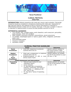
REVIEW ARTICLE The Journal of TRAUMA威 Injury, Infection, and Critical Care Acute Pain Management of Patients with Multiple Fractured Ribs Manoj K. Karmakar, MD, FRCA, and Anthony M.-H. Ho, MS, MD, FRCPC, FCCP Background: Multiple rib fracture causes severe pain that can seriously compromise respiratory mechanics and exacerbate underlying lung injury and preexisting respiratory disease, predisposing to respiratory failure. The cornerstone of management is early institution of effective pain relief, the subject of this review. Methods: A MEDLINE search was conducted for the years 1966 through and up to December 2002 for human studies written in English using the keywords “rib fractures”, “analgesia”, “blunt chest trauma”, “thoracic injury”, and “nerve block”. The reference list of key articles was also searched for relevant articles. The various analgesic techniques used in patients with multiple fractured ribs were summarized. Results: Analgesia could be provided using systemic opioids, transcutaneous electrical nerve stimulation or non steroidal anti-inflammatory drugs. Alternatively, regional analgesic techniques such as intercostal nerve block, epidural analgesia, intrathecal opioids, interpleural analgesia and thoracic paravertebral block have been used effectively. Although invasive, in general, regional blocks tend to be more effective than systemic opioids, and produce less systemic side effects. Conclusion: Based on current evidence it is difficult to recommend a single method that can be safely and effectively used for analgesia in all circumstances in patients with multiple fractured ribs. By understanding the strengths and weaknesses of each analgesic technique, the clinician can weigh the risks and benefits and individualize pain management based on the clinical setting and the extent of trauma. Key Words: Analgesia, Blunt chest trauma, Epidural block, Intercostal nerve block, Interpleural block, Intrathecal opioids, Regional anesthesia, Rib fracture, Thoracic paravertebral block. J Trauma. 2003;54:615–625. C hest wall trauma is most commonly seen after motor vehicle collisions1–3 and accounts for 8% of all trauma admissions.1 It is both a marker of severe injury3 and contributes significantly to the morbidity and mortality of injured patients,1,3 with the elderly1–3 and patients with poor respiratory reserve being most vulnerable. Rib fractures are the commonest of all chest injuries and are identified in 10% of patients after trauma.3 The overall incidence is probably higher because not all rib fractures are seen on chest radiographs or otherwise detected.3 Multiple fractured ribs (MFRs) cause severe pain, which may be more debilitating and harmful than the injury itself.1 Pain limits one’s ability to cough and breath deeply, resulting in sputum retention, atelectasis, and a reduction in functional residual capacity (FRC). These factors in turn result in decreased lung compliance, ventilation-perfusion mismatch, hypoxemia, and respiratory distress. Failure to control pain, compounded by the presence of pulmonary contusion, flail segment, and other insults, can result in serious respiratory complications. Submitted for publication October 22, 2001. Accepted for publication December 5, 2002. Copyright © 2003 by Lippincott Williams & Wilkins, Inc. From the Department of Anesthesia and Intensive Care, The Chinese University of Hong Kong, Prince of Wales Hospital, Shatin, NT, Hong Kong SAR, People’s Republic of China. Address for correspondence: Manoj K. Karmakar, MD, FRCA, Department of Anesthesia and Intensive Care, The Chinese University of Hong Kong, Prince of Wales Hospital, Shatin, NT, Hong Kong SAR, People’s Republic of China; email: [email protected]. DOI: 10.1097/01.TA.0000053197.40145.62 Volume 54 • Number 3 BACKGROUND Hippocrates described hemoptysis secondary to fractured ribs and observed the association of pleurisy and empyema, especially when the chest wall was traumatized.4 Dressing the chest wall with linen was the mainstay of treatment of fractured ribs for centuries.4 The management of blunt chest trauma has evolved through a number of stages since World War II.5 Although external stabilization of the chest was common during the 1930s, the use of various mechanical devices (wires, hooks, screws, or pins) became more common during the next 20 years.6 This was based on belief at that time that the major determinant of morbidity was instability of the chest wall. Avery et al. in 1956 introduced the concept of “internal pneumatic stabilization” and “alkalotic apnea,”7 which necessitated tracheal intubation and mechanical ventilation. This initially lowered the mortality associated with flail chest, but with the widespread use of mechanical ventilation, morbidity related to complications such as ventilatorassociated pneumonia became evident. Trinkle et al.8 in 1975 challenged the routine use of mechanical ventilation in flail chest injury. They compared two groups of patients who were managed either with early tracheal intubation and mechanical ventilation, or with fluid restriction, diuretics, methylprednisolone, albumin, vigorous pulmonary toilet, and intercostal nerve blocks, ignoring the paradox and treating only the underlying lung, and concluded that most patients with flail chest could be safely managed without mechanical ventilation if the underlying lung was treated appropriately.8 More recently, a better understanding of the pathophysiologic effects of blunt chest injury has led to a trend toward conser615 The Journal of TRAUMA威 Injury, Infection, and Critical Care Table 1 Readily Available Options for Controlling Rib Fracture Pain Technique Advantages Disadvantages/Side Effects Contraindications* Systemic opioids Intercostal LA Simplicity, no need for positioning, utility as a supplement Highly effective for 8–24 h with each injection, no CNS depression CNS and respiratory depression, nausea, cough suppression CNS depression, hypotension Risk of pneumothorax, difficulty with first to seventh ribs, not suitable for posterior rib fractures, multiple injections, patient discomfort, high blood LA levels with potential for LA toxicity§ Intercostal LA (extrapleural with catheter) Epidural LA No CNS depression, single placement for multiple injections Risk of pneumothorax, limited dermatomal spread, high blood LA levels with potential for LA toxicity§ Superior analgesia, no CNS depression, opioid sparing, bilateral analgesia, high success rate Hypotension, risk of dural puncture and spinal cord injury, motor blockade, urinary retention, may mask signs of intraabdominal injury Epidural opioids Low dosage requirement, bilateral analgesia, hemodynamic stability, Pruritus, urinary retention, nausea, risk of delayed respiratory intact sensory and motor functions depression, breakthrough pain Epidural LA plus opioids Interpleural LA Improved analgesia with fewer side effects, bilateral analgesia No CNS depression, no need for multiple and repeated injections Paravertebral LA Oral analgesics and NSAIDs Technically simple, safer and easier to perform than thoracic epidural, palpation of rib not necessary and scapula does not interfere with needle placement, uninterrupted chest tube drainage, no CNS depression, maintains hemodynamic stability, preserves bladder sensation and lower limb motor power, no additional nursing surveillance required Simplicity, lack of CNS or cardiovascular side effects, utility as a supplement TENS Simplicity, safety, superior to NSAIDs As for epidural LA and epidural opioids Reduced efficacy in the presence of pleural fluids and adhesions, interruption of chest tube drainage required, high blood LA levels with potential for LA toxicity§ Risk of pneumothorax, dermatomal spread not as predictable as epidural anesthesia, high blood LA levels with potential for LA toxicity§ Risk of peptic ulcerations, platelet dysfunction, risk of renal damage, weak analgesic effect Aortic or mitral stenosis, 1 ICP, previous back surgery, spinal injury, hypovolemia, bleeding disorders 1 ICP, previous back surgery, spinal injury, hemostatic defects Same as epidural LA Peptic ulcer disease, hemostatic defects, renal dysfunction, and hypoperfusion Limited experience and published data, inadequate analgesic effect ICP, intracranial pressure; LA, local anesthetic; CNS, central nervous system; NSAIDs, nonsteroidal anti-inflammatory drugs; TENS, transcutaneous electrical nerve stimulation. * Other contraindications: lack of consent, intolerance to medications used, lack of expertise or supervision, lack of resuscitation personnel and equipment, sepsis (for any instrumentation), infection at the site of needle placement. § Symptoms and signs of LA toxicity (with increasing blood levels of the local anesthetic drug): numbness of tongue, lightheadedness, visual and auditory disturbance, muscular twitching, confusion, convulsion, coma, respiratory depression, cardiovascular depression. vative and nonventilatory regimens.5,9,10 The paradoxical movement of a flail segment is no longer considered the major clinical problem.10 Although it increases the work of breathing, the main cause of hypoxemia is the underlying lung contusion,10 and such patients require assistance with oxygenation rather than ventilation.10 Ventilatory therapy has a role in management of patients who have associated head injury, significant pulmonary contusion causing hypoxemia, after major surgery, or worsening respiratory failure despite adequate analgesia. The latter may be defined as a PaO2 ⬍ 8 kPa breathing room air, PaCO2 ⬎ 6 kPa, respiratory rate ⬎ 30 breaths/min, and vital capacity ⬍ 12 to 15 mL/kg. Selective use of ventilatory therapy has led to reduced morbidity and mortality.5,9,10 Today, the cornerstone of conservative and nonventilatory management is early and effective pain control, aggressive respiratory therapy, measures to avoid fluid overload, and early mobilization. TECHNIQUES USED TO MANAGE PAIN IN PATIENTS WITH MULTIPLE FRACTURED RIBS An ideal method of managing pain in patients with MFRs is one that is safe and simple, provides complete and prolonged analgesia, permits deep breathing and clearance of 616 secretions, and allows cooperation during chest physiotherapy. The method used should also improve respiratory dynamics, have minimal central nervous and systemic side effects, and permit early mobilization. Many different methods have been used to manage pain in patients with MFRs (Table 1) including several regional anesthetic techniques (Fig. 1), but no single method can fulfill all these criteria. Systemic Opioids Systemic opioids are commonly used and are often the first-line management for relieving pain resulting from MFRs. They are used as intermittent on-demand injections,11 continuous intravenous infusion,12 or intravenous patientcontrolled analgesia (IVPCA).13,14 Mackersie et al.12 demonstrated that fentanyl administered as a continuous intravenous infusion improves visual analog pain scores and vital capacity but also results in respiratory depression and hypoxemia. The cause of hypoxemia after opiate administration is multifactorial and may be caused by obstructive apnea, paradoxical breathing, or a possible diminution in the number of sigh breaths.12 Opioids also cause sedation, respiratory depression, and cough suppression. Nonetheless, alfentanil, a potent and short-acting intravenous opioid agent, has been successMarch 2003 Pain Management of Patients with Multiple Fractured Ribs Fig. 1. Regional anesthetic techniques available for pain control in patients with multiple fractured ribs. fully used (intermittent intravenous bolus 100 g at 2- to 3-minute intervals to a total dose of 800 g) for analgesia before chest physiotherapy in a patient with sputum retention after MFRs, thus averting intubation and mechanical ventilation.15 The risk of respiratory depression with this method would restrict its use to locations where resuscitative skills and equipment are readily available. Various researchers have also found systemic opioids to be inadequate in controlling pain caused by MFRs, necessitating a regional anesthetic technique for optimal pain control.11,13 Considering this variable efficacy and potential for side effects associated with the use of opioids in patients with MFRs, it may be preferable to resort to a regional anesthetic technique from the very outset, whenever feasible. Opioids may still be required in conjunction with a regional block in the context of multiple trauma to control pain resulting from associated injuries that is not relieved by a peripheral regional block solely used to control pain caused by the MFRs. Intercostal Nerve Block Intercostal nerve block (ICNB) is a simple and timetested method of managing pain in patients with MFRs. Drugs and dosage commonly used for ICNB in patients with Volume 54 • Number 3 MFRs are outlined in Table 2.6,11–14,16 – 40 Success depends on blocking the intercostal nerve proximal to the fracture site. An ICNB posterior to the midaxillary line will ensure blockade of the lateral cutaneous and anterior branch of the intercostal nerve.41 However, because of overlapping innervation from the segments above and below,42 it is necessary to block the intercostal nerves above and below the fracture site.20 Accurate injection during ICNB requires rib palpation, which may be difficult in patients with MFRs because of pain. MFRs would necessitate multiple intercostal injections, which are not only painful but also time consuming. It may also predispose to local anesthetic toxicity because of the higher doses of local anesthetic used and the fact that local anesthetics are rapidly absorbed from the intercostal space. Adding epinephrine 1:200,000 to bupivacaine 0.5% during ICNB results in lower peak blood bupivacaine levels43 and may reduce the potential for local anesthetic toxicity. Multiple intercostal nerve blocks for MFRs also predispose to a higher incidence of pneumothorax.44 The reported incidence of pneumothorax after ICNB in patients with MFRs is 1.4% for every individual ICNB and 5.6% when multiple ICNBs are simultaneously performed.44 To overcome discomfort associated with rib palpation and the multiple intercostal injec617 The Journal of TRAUMA威 Injury, Infection, and Critical Care Table 2 Reported Drugs and Dosage for the Various Regional Analgesic Techniques Used in Patients with Multiple Fractured Ribs* Method of Analgesia Drugs Intercostal nerve Bupivacaine 0.25–0.5%11,13,16,17,18,19 block Bupivacaine 0.25–0.5% with epinephrine 1:200,00013,18,86 Dosage Schedule Bolus: Multiple level injection: 2–4 mL/segment18,23 Single level injection: 20 mL11,17,18 Regular dosing via catheter: Bupivacaine 0.5%: 10–20 mL every 6–8 h11,17 Infusion: Bupivacaine 0.25%: 3 mL/h18 Lidocaine 1%21 Bolus: Bupivacaine 0.25–0.5%22,23 10–20 mL36,46,47,49 Bupivacaine 0.5% with epinephrine Regular dosing via catheter: 1:200,00024 Bupivacaine 0.5% with epinephrine 1:200,000: 15–20 mL Bupivacaine 0.25% plus 1% lidocaine every 4–6 h (400–450 mg/day)24 25 with 1:400,000 epinephrine Bupivacaine 0.5% 20 mL every 8 h22 Bupivacaine 0.25% plus 1% lidocaine with 1:400,000 epinephrine25: 20 mL every 6 h25 Infusion: Bupivacaine 0.5% with 1:200,000 epinephrine at 5 mL/h26 Thoracic Bupivacaine 0.5%27,28,29 Bolus: paravertebral Bupivacaine 0.5% with 1:200,000 Bupivacaine 0.5%: 20–25 mL67,68,70,72 or 0.3 mL/kg31 block epinephrine30,31 Regular dosing via catheter: Bupivacaine 0.5%: 10–25 mL every 6–8 h27,29 Infusion: Bupivacaine 0.25%: 0.1–0.2 mL/kg/h31 Epidural opioid Lumbar route Bolus: Fentanyl12 Fentanyl (5 g/mL): 1 g/kg12 Morphine33 Morphine: 5 mg diluted to 10 mL with NS33 Buprenorphine6 Buprenorphine: 0.1–0.3 mg diluted in 10–20 mL NS6 Regular dosing via catheter: Morphine 5 mg in 10 mL with NS every 6 h33 Infusion: Fentanyl: 1.03 ⫾ 0.32 g/kg/h,12 50 g/h (5 g/mL)34 Thoracic route Bolus: Morphine9,35 Morphine 2 mg diluted to 10 mL with NS35 Fentanyl plus Morphine14 Morphine 2 mg in 4 mL NS9 Fentanyl 100 g plus duramorph Fentanyl 50 g plus morphine 3 mg14 5 mg36 Morphine 3 mg diluted with NS ⬃ 3–10 mL37 Infusion Morphine 70 g/mL at 8–10 mL/h36 Epidural local Thoracic route Bolus: anesthetic Bupivacaine 0.25–0.5%9,38 Bupivacaine 0.5%; 5.54 ⫾ 1.7 mL38 Infusion: Bupivacaine 0.125% at 8–10 mL/h33 Epidural local Thoracic route Bolus: anesthetic plus Bupivacaine plus morphine37 Morphine 3 mg diluted with NS ⬃ 3–10 mL37 opioid Bupivacaine plus fentanyl (PWH) Infusion: Bupivacaine 0.25% plus morphine (0.005%) at 4–6 mL/h14 Bupivacaine 0.125% plus fentanyl (2.5 g/mL) at 0.1–0.2 mL/kg/h (PWH) Intrathecal opioid Lumbar route Bolus: Morphine39,40 Morphine 1 mg diluted with 4 mL NS,39 morphine 1–2 mg40 Interpleural analgesia Average Duration of Analgesia after Bolus Injection 8–12 h16,17 4.7 ⫾ 0.5–31.3 ⫾ 1.7 h19; Chung et al found that the duration of analgesia was progressively more prolonged after serial injections19 3–3.5 h46,49 9.9 h32 DNA Morphine 2 mg: 6.6 (3–50) h35 40 DNA DNA 30 (9–72) h40 DNA, data not available in patients with MFRs; LA, local anesthetic; NS, normal saline; PWH, regimen used at our hospital (Prince of Wales Hospital, Hong Kong). * Opioids used are preservative free. 618 March 2003 Pain Management of Patients with Multiple Fractured Ribs tions during ICNB, the use of a Doppler ultrasound stethoscope45 and a jet injector device46 have been described. Although both these methods have hypothetical advantages over the conventional method of ICNB, current literature suggests that they are seldom used. Rauck describes the successful use of ICNB in patients with fractured ribs who are discharged home from the emergency department with written instructions of the risk of pneumothorax and who to contact in the event they develop dyspnea.41 Patients selected for this treatment are those in whom pain is not expected to be adequately relieved by narcotics and/or patients who return to the local physician or emergency department during the early posttraumatic period with intractable pain not relieved with oral analgesics.41 In this select group of patients, a single set of ICNB makes subsequent pain management with oral analgesics or opiates much easier.41 ICNB as multiple and repeated injections is also effective in relieving pain in patients admitted to the hospital with MFRs.16 It results in improved flowmeter measurement of forced expiration, but the effects wane after 6 hours.20 Intercostal catheterization at one level for the injection of a large bolus of local anesthetic followed by an infusion for continuous ICNB obviates some of the problems relating to multiple and repeated intercostal injections11 and has been successfully used to control pain in patients with MFRs.13,17 However, the technique of intercostal catheterization lacks a definite end point, and the “give” that has been described as the needle pierces the posterior intercostal membrane to enter the intercostal space47 is not a reliable sign,48,49 resulting in misplaced catheters.48 –50 The exact incidence of misplaced catheters after intercostal catheterization is not known, but Mowbray et al.,50 when performing intercostal catheterization in patients who were about to undergo thoracotomy or median sternotomy, noted at operation that only 12 of the 22 (54.5%) catheters inserted were placed correctly.50 Very easy passage of catheters beyond 3 cm resulted in interpleural catheter placement.50 ICNB or intercostal catheterization for fractures of the upper ribs (second to seventh ribs) is technically difficult because the scapula interferes with needle insertion. Moreover, the exact mechanism of multiple ICNBs after a single intercostal injection of a large volume of local anesthetic is not clear. Spread may be limited and confined to the intercostal space injected;51 may track cephalad and caudad subpleurally;49,50,52 or may enter the paravertebral space,50,53 the epidural space,53 or a combination of the above.53 Mowbray et al.50 followed the spread of an intercostal injection of 20 mL of solution containing bupivacaine and methylene blue through a catheter at thoracotomy and concluded that the major component of segmental block during intercostal catheterization may be secondary to paravertebral spread.50 Mowbray et al. therefore described intercostal catheterization as an alternate approach to the thoracic paravertebral space.50 Volume 54 • Number 3 Epidural Analgesia Epidural analgesia (EA) via the lumbar6,12,34 or the thoracic9,14,22,35,38,54 –56 route using local anesthetic agents,9,22,38,54,55 opioids,6,9,12,34,35 or a combination of both14,56 has been successfully used to manage pain in patients with MFRs. Drugs and dosage commonly used for EA in patients with MFRs are outlined in Table 2. The use of EA in patients with thoracic trauma who are older than 60 years of age is an independent predictor of both decreased mortality and decreased incidence of pulmonary complications.57 Used in patients with chest wall trauma, EA produces pain relief that is dramatic38,54 and superior to that produced by systemic opioids12,14 and interpleural analgesia (IPA).22 It results in an increase in FRC, dynamic lung compliance, and vital capacity; a decrease in airway resistance; and a significant increase in PaO2.55 Shallow breathing changes to near normal and paradoxical chest wall movement is reduced.54 Epidural analgesia also modifies the immune response in patients with chest trauma, as evidenced by a reduction in the plasma levels of interleukin (IL)-8, a proinflammatory chemoattractant implicated in acute lung injury.14 Patients on an EA regimen are alert,38 able to cough adequately and comply with chest physiotherapy,38,54 and develop fewer complications than those treated by intubation and mechanical ventilation.6,9,10 This results in shorter intensive care unit and hospital stay,6 with reduced costs.10 A conservative management regimen that includes EA is more likely to be successful if the vital capacity remains greater than 13 mL/kg and the PaO2 at room air is more than 60 mm Hg,54 with a critical period described between 48 and 72 hours after trauma.54 However, EA is technically demanding, especially in patients distressed with pain. In patients with multiple injuries, it can mask intraabdominal injuries,58 is associated with hypotension38 during the early phase of treatment, and can result in cardiovascular collapse and cardiac arrest in the inadequately resuscitated patient.38 Another serious complication of note is epidural infection.38,56 Although the hemodynamic effects may be minimized with lower doses of local anesthetic agents and by the addition of opioids,35 their use is known to produce undesirable side effects including nausea, vomiting, urinary retention,12,54 respiratory depression,56 and pruritus. Moreover, one must also consider the possibility of inadvertent dural puncture, epidural hematoma and, very rarely, spinal cord trauma after EA.59 Intrathecal Opioids Intrathecal morphine via the lumbar route has been used for analgesia in MFRs.39,40 Drugs and dosage commonly used for intrathecal analgesia in patients with MFRs are outlined in Table 2. Although Kennedy39 found this method to be effective, Dickson and Sutcliffe40 reported that analgesia was unsatisfactory in 27% of patients despite adequate doses of intrathecal morphine, and extradural bupivacaine produced superior analgesia. Dickson and Sutcliffe also ob619 The Journal of TRAUMA威 Injury, Infection, and Critical Care served that the mean duration between doses in their series was 30 hours (range, 9 –27 hours),40 necessitating multiple intrathecal injections.39,40 Intrathecal morphine also produced a high incidence of complications, including excessive drowsiness (26.6%), respiratory depression (13.3%), postspinal headache (6.6%), nausea (20%), and urinary retention.40 This may account for the lack of data in the literature on the use of this method in patients with MFRs. Interpleural Analgesia Kvalheim and Reiestad in 1984 described IPA,60 in which the local anesthetic is injected into the interpleural space via a catheter placed percutaneously. This technique produces multiple unilateral intercostal nerve blockade by gravity-dependent retrograde diffusion of the local anesthetic to reach the intercostal nerve. Rocco et al.61 were the first to describe the use of this method in patients with MFRs, and various other investigators have also successfully used this method to control pain in patients with blunt chest trauma.18,21,23,26 Drugs and dosage commonly used for IPA in patients with MFRs are outlined in Table 2. When comparing IPA to EA for pain relief in chest wall trauma, Shinohara et al.21 found IPA to be comparable to EA, whereas Luchette et al.22 concluded that EA is superior. More recently, in a well-controlled study, Short et al.25 found IPA to be comparable to conventional opioids in controlling pain in patients with blunt chest trauma. This variable efficacy of IPA in patients with blunt chest trauma may be because the success of IPA can be affected by a number of factors, including catheter position, patient position, presence of hemothorax, location of fractured ribs, characteristics of the local anesthetic agent, and the use of epinephrine.25 Moreover, significant amounts of local anesthetic agent can be lost through the intercostal drain.62,63 To improve analgesic efficacy after interpleural injection of local anesthetic, patients are often nursed in the supine position for 20 minutes to facilitate diffusion of local anesthetic through the parietal pleura into the intercostal nerves.64 Nursing a blunt chest trauma patient with decreased FRC and often-compromised respiration in the supine position is not optimal. Although an upright position may be considered advantageous, this may result in a gravity-dependent accumulation of local anesthetic in relation to the diaphragm. Because the diaphragm takes up bupivacaine after interpleural administration,65 this may adversely affect diaphragmatic function.66 With a thoracostomy tube in situ, clamping it for 20 to 30 minutes to prevent siphoning away of the local anesthetic agent is often recommended. This maneuver has raised concerns67 because it can result in a dangerous situation of tension pneumothorax in the event that a significant air leak is present. Interpleural catheter placement can be technically difficult68 and can result in symptomatic pneumothorax,69,70 intrapulmonary catheter placement,70 misplacement into the chest wall,70 or an extrapleural plane. Local anesthetic agents are rapidly absorbed from the interpleural space, resulting in high plasma 620 concentration,66,68 with potential for systemic toxicity.68 Interpleural instillation of local anesthetic can also cause phrenic nerve paralysis71 and Horner syndrome72 and may aggravate bronchospasm.73 Loss of negative interpleural pressure in a patient who is being ventilated makes identification of the interpleural space difficult and can result in catheter misplacement, tension pneumothorax, and intrapulmonary catheter placement.70 It is for this reason that we do not perform interpleural catheter placement in ventilated patients. Thoracic Paravertebral Block Thoracic paravertebral block (TPVB) is the technique of injecting local anesthetic alongside the thoracic vertebrae. This produces multidermatomal ipsilateral somatic and sympathetic nerve blockade74 –76 in contiguous thoracic dermatomes. Drugs and dosages commonly used for TPVB in patients with MFRs are listed in Table 2. Eason and Wyatt in 1979 were the first to describe the use of this method in a patient with MFRs.77 Despite it being more than two decades since this report, there are only a few other publications describing the use of this method in patients with MFRs,27–30,78 and there is a lack of comparative data. This may be because intercostal,11,13,16,17,20 IPA,18,21,23,25,26,61 and EA9,10,12,22,34,35,38,54 –56 have overshadowed the use of TPVB in patients with MFRs. Recently, there has been renewed interest in the use of TPVB to control pain in a variety of conditions involving the chest and abdomen.75,76 Thoracic paravertebral injection of bupivacaine as repeated injections,27 regular dosing via an indwelling catheter,29,77 or continuous infusion28,31 is effective in relieving pain in patients with MFRs, resulting in improved respiratory parameters27,31 and arterial blood gases.27,31 Used to manage pain caused by MFRs in a patient with associated head injury, it avoids the need for sedation and ventilation and allows continuous neurologic assessment.29,78 In patients with concomitant lumbar spinal injury, the unilateral segmental thoracic nerve blockade spares the lumbar and sacral nerve roots, allowing regular neurologic assessment for spinal cord compression.78 Unlike ICNB, TPVB does not require palpation of the ribs and can be easily performed in patients with fractures of the upper ribs. As a regional anesthetic technique, it is simple to perform27,75,77 and technically easier than a thoracic epidural,75,77 especially in a patient distressed with pain. It is associated with a low incidence of complications,79 including urinary retention,76 requires no additional nursing surveillance,76 and has very few absolute contraindications.75,76 Thoracic paravertebral block can be safely performed in patients who are anesthetized or sedated and mechanically ventilated,75,76 unlike thoracic epidural anesthesia, where there may be a greater risk of spinal cord injury;59 or IPA, where there is a greater risk of pleural or pulmonary parenchymal injury70 because of the loss of the negative interpleural pressure. Thoracic paravertebral block reliably blocks the posterior primary ramus and the ipsiMarch 2003 Pain Management of Patients with Multiple Fractured Ribs lateral sympathetic chain,77 which may be involved in afferent pain transmission after MFRs. The unilateral sympathetic nerve blockade74 may explain the low incidence of hypotension in the adequately resuscitated trauma patient,27 an advantage over thoracic epidural anesthesia, particularly in elderly patients. There are few trials that have evaluated the safety and efficacy of TPVB in patients with MFRs.27,31 Therefore, the true incidence of complications is not known. On the basis of published data on TPVB, the overall complication rate appears to be relatively low, varying from 2.6% to 10%,79 – 81 and comparable to those with alternative techniques (EA, IPA, and ICNB).79 The complications of TPVB include hypotension (4.6%), vascular puncture (3.8%), pleural puncture (1.1%), and pneumothorax (0.5%).79 Inadvertent bilateral symmetric anesthesia (EA) is also a possibility32,75,82 and may be caused by extensive epidural spread,82 inadvertent epidural injection,31 inadvertent intrathecal injection into a dural sleeve,83 injection via a medially directed needle,83,84 or the use of large volumes of injection (⬎ 25 mL).32 The medial approach to the thoracic paravertebral space, which was originally described to avoid pleural puncture,84 has been associated with reports of inadvertent dural puncture,81,85 intrathecal injection,81,85 spinal anesthesia,85 and headache,81,85 which can occur with or without obvious dural puncture.85 Recently, pulmonary hemorrhage has been reported after percutaneous TPVB in a patient who had altered paravertebral anatomy because of previous thoracic surgery.86 Despite some of these rare complications, TPVB offers much promise as a regional anesthetic technique for pain control in patients with unilateral MFRs and deserves greater attention and investigation in the future. Transcutaneous Electrical Nerve Stimulation Transcutaneous electrical nerve stimulation (TENS) produces pain relief by releasing endorphins in the spinal cord. Sloan et al.87 used it in patients with MFRs and found it provides better subjective pain relief, with improvement in peak expiratory flow rates and arterial blood gases when compared with a group of patients receiving nonsteroidal anti-inflammatory drugs (NSAIDs). The paucity of data on this mode of analgesia in patients with MFRs suggests that it is rarely used in this group of patients. Oral Analgesic Drugs Oral analgesic drugs such as nonsteroidal anti-inflammatory drugs (e.g., diclofenac, indomethacin) and acetaminophen do not depress the central nervous system or the cardiovascular system and are useful for mild to moderate pain. As with transcutaneous electrical nerve stimulation, there is a paucity of data on the use of oral analgesics in patients with MFRs. However, there may be a place for the use of NSAIDs as adjuncts to other methods of pain relief in patients with MFRs. NSAIDs can cause gastrointestinal upset and platelet and renal dysfunction. The latter would contraindicate the use of NSAIDs in patients who are not adequately resuscitated. Volume 54 • Number 3 COMPARATIVE ANALYSIS OF DIFFERENT ANALGESIC TECHNIQUES IN PATIENTS WITH BLUNT CHEST TRAUMA There are relatively few clinical trials comparing the efficacy of the various analgesia techniques in patients with blunt chest trauma. This may in part be because each technique has unique strengths, weaknesses, and contraindications (Table 1), and pain management is individualized on the basis of the clinical condition and extent of injury. This makes randomized, controlled comparisons difficult or hard to justify. Gabram et al.24 prospectively compared intrapleural bupivacaine with systemic narcotics (morphine, meperidine, or hydromorphone) for the management of 48 patients with rib fractures. The patients with the block statistically had more compromised pulmonary function as measured by forced vital capacity (FVC) at admission; however, they tended toward a greater objective improvement of FVC at discharge, although the difference did not reach statistical significance. When analyzing a cohort of severely impaired patients (initial FVC ⬍ 20%), half of the systemic medication patients compared with only 10% of the block group failed and required another mode of therapy. Catheter complications were minor and did not contribute to overall morbidity. Luchette et al.22 prospectively evaluated analgesia for 72 hours in 19 blunt trauma patients with unilateral rib fractures. They found that thoracic epidural bupivacaine 0.125%, 8 to 10 mL/h continuous infusion, as compared with intrapleural bupivacaine 0.5% intermittent boluses of 20 mL every 8 hours, resulted in significantly lower visual analog scale pain scores, less use of “rescue” narcotics, and greater tidal volume and negative inspiratory force. Vital capacity, FIO2, minute ventilation, and respiratory rate were not affected. Mild hypotension was a common complication with epidural blocks only.22 Shinohara et al.21 had more favorable results with interpleural block. They studied 17 patients with unilateral MFRs and hemopneumothorax in a randomized, crossover, before/after trial on the first and second hospital days. An interpleural catheter was inserted along with a chest tube, and an upper thoracic epidural catheter was also established in the same patient. They administered 10 mL of 1% lidocaine for both blocks. The range of thermohypesthesia was unilateral and shorter with the interpleural block, whereas it was bilateral and wider with the epidural block. The effects of pain relief were almost the same. Respiratory rate decreased and PaO2 tended to elevate similarly. Unlike epidural block, the systemic blood pressure with interpleural block changed only minimally. Serum levels of lidocaine were similar and in the safe range.21 Moon et al.14 compared self-administered opioid-IVPCA and thoracic epidural analgesia using a combination of bupivacaine and morphine in 34 patients with thoracic trauma. Twenty-four patients (IVPCA, 11; epidural analgesia, 13) completed the 3-day study. Epidural analgesia provided bet621 The Journal of TRAUMA威 Injury, Infection, and Critical Care ter pain relief and was associated with superior ventilatory dynamics as evidenced by greater tidal volumes and maximal inspiratory force on day 3, and lower plasma levels of IL-8 on days 2 and 3. In the IVPCA group, there was a progressive decline in tidal volume and maximal inspiratory force throughout the 3-day study period. There were no differences in plasma IL-1, IL-2, IL-6, tumor necrosis factor-␣, or urinary catecholamine levels, although there was a trend for lower IL-6 levels in patients receiving epidural analgesia.14 Because the sample size was small and patients were followed up for only 3 days, it is not known whether the modification in immune response by epidural analgesia translates into reduced inflammatory complications or improved outcome. Mackersie et al.12 compared epidural and intravenous fentanyl for pain control and restoration of ventilatory function after multiple rib fractures in a prospective, randomized trial involving 32 patients. Prefentanyl and postfentanyl parameters were compared in both groups. Both methods significantly improved visual analog pain scores. The epidural method produced improvement in both maximum inspiratory pressure and vital capacity, whereas intravenous analgesia produced improvement in only vital capacity. Intravenous fentanyl produced increases in PaCO2 and decreases in PaO2, whereas no significant changes in arterial blood gases were observed with epidural fentanyl administration. Side effects were similar between the groups, with pruritus being more pronounced with epidural fentanyl.12 Sloan et al.87 compared two groups of patients with MFRs who were randomized to receive either TENS or naproxen sodium 250 mg every 8 hours and also a mixture of paracetamol 1 g and dihydrocodeine tartrate 20 mg on an as-required basis. The peak expiratory flow rate change at 24 hours after the commencement of treatment, the PaO2, and the pain relief were all significantly better in the TENS group.87 CHOICE OF ANALGESIC TECHNIQUE IN DIFFERENT SETTINGS The decision regarding institution of pain relief in patients with blunt chest trauma should be made early, and the most appropriate method chosen should be decided on by someone who has a thorough and clear understanding of the safety and efficacy of the various available methods. Therefore, the patient should be referred to the acute pain management team early. Pain relief is individualized on the basis of a thorough history, clinical examination, and review of the various investigations. Patients with fewer than three rib fractures and without associated injuries are often discharged home on oral medications from the emergency department. For a select group of such patients, intercostal nerve block has been found to be very useful.41 Patients are discharged with written instruction regarding the risk of pneumothorax and who to contact in case the patient develops shortness of breath.41 622 Patients who require immediate surgery are best managed in the immediate postoperative period using intravenous opioids such as patient-controlled analgesia. Once the patient’s condition is stabilized, a regional anesthetic technique should be considered to optimize pain relief and prevent development of respiratory complications. Patients who require immediate intubation and mechanical ventilation in the intensive care unit are also managed initially using intravenous opioids, which also aid in sedating the patient while on the ventilator. Once a decision is made to wean the patient from the ventilator, a regional block should be considered if the clinical condition permits to facilitate weaning from the ventilator. One must exercise extreme caution when performing a regional block in a patient who is sedated and being ventilated because of the possibility of spinal cord trauma after EA59 and the reported high incidence of complications after IPA68 as described earlier. In patients with head injury or mental status changes, EA may be contraindicated, and an alternative such as TPVB,29,78 continuous ICNB, or IPA18 may be preferred. Patients who are discharged to the ward from the emergency room should have a regional anesthetic technique (ICNB, EA, IPA, or TPVB) best suited for the patient decided on at an early stage. A WORD OF CAUTION Regional anesthetic techniques are preferable for pain control in patients with multiple fractured ribs. However, one must exercise extreme caution when performing these techniques in traumatized patients. The majority of patients (94%) with chest wall trauma have associated injuries, and more than half (55%) require an immediate operation.3 Because of the close proximity of the fractured ribs to vital organs, there is an increased likelihood of concurrent splenic and hepatic injury.1 Highly effective pain relief can mask subtle signs of delayed splenic rupture.88 The sympathetic blockade can cause bronchospasm73 and may unmask hypovolemia, which can result in cardiovascular collapse, with disastrous consequences38 as described earlier. Cardiovascular stability must be established, abdominal visceral injury must be excluded, pneumothorax or hemothorax must be drained, and any surgical procedure required must be performed before contemplating any regional anesthetic technique for pain control. Even in patients in whom cardiovascular stability is apparent, delayed hemothorax, a well-known entity,89 can occur and may result in death.24 This condition is usually seen in patients with multiple or displaced rib fractures and occurs 18 hours to 6 days after the injury.89 Bleeding can be significant and is usually heralded by a prodrome of pleuritic chest pain and dyspnea,89 which may also be masked by regional anesthetic techniques. Interpleural, extrapleural, epidural, and thoracic paravertebral block with a local anesthetic agent can all cause Horner syndrome, which can interfere with neurologic assessment and mask occult head injury. Clamping of the chest tube March 2003 Pain Management of Patients with Multiple Fractured Ribs during interpleural injection to prevent siphoning away of the local anesthetic is recommended for 20 to 30 minutes. However, with clamping, any air leak or ongoing bleeding into the pleural space that may be present and not decompressed can result in serious consequences. When in doubt, the chest tube should not be clamped and left to underwater seal drainage without suction. Epidural analgesia should be avoided in patients with spinal injury, and hemostatic defects would absolutely contraindicate EA and relatively contraindicate most other regional techniques. Pain in patients with bilateral rib fractures is probably best managed with EA, although bilateral ICNB, bilateral IPA, and bilateral TPVB as methods of pain control have been described in the literature. However, with bilateral blocks, potential complications such as pneumothorax, local anesthetic toxicity, phrenic nerve paralysis, and bilateral sympathetic blockade would tend to negate any advantage that these unilateral peripheral nerve block techniques may have over EA. The potential for local anesthetic toxicity should also be borne in mind when prolonged continuous infusion of local anesthetic is used via the intercostal, thoracic paravertebral, or interpleural routes. Local anesthetic agents are rapidly taken up from these sites, and accumulation of these agents in blood is known to occur with time. A continuous epidural infusion of a low concentration of a local anesthetic agent in conjunction with an opioid offers a distinct advantage in this respect. REFERENCES 1. 2. 3. 4. 5. 6. 7. 8. 9. 10. 11. 12. 13. CONCLUSION Pain after multiple fractured ribs may compound the problem of lung injury in serious blunt chest trauma. Without adequate analgesia, deep breathing, coughing, and chest physiotherapy are compromised and respiratory failure may ensue. Today, tracheal intubation and mechanical ventilation in the intensive care unit is only used selectively in patients with blunt chest trauma. The cornerstone of management is providing effective pain relief. The analgesic techniques for multiple fractured ribs are intercostal, interpleural, thoracic paravertebral, epidural, and intrathecal blocks using local anesthetics with or without opioids, or systemic opioids and oral analgesic agents. Although invasive, regional blocks tend to be more effective and have less depressive effects on the central nervous system and coughing, but they require more expertise to administer, cause patient discomfort during institution of the blocks, and have other potential serious complications. On the basis of currently available data, it is difficult to recommend a single method that can be safely and effectively used in all circumstances in patients with multiple fractured ribs. By understanding the strengths and weaknesses of each technique, the clinician can weigh the risks and benefits and individualize pain management on the basis of the clinical setting and the extent of trauma. Volume 54 • Number 3 14. 15. 16. 17. 18. 19. 20. 21. 22. Mayberry JC, Trunkey DD. The fractured rib in chest wall trauma. Chest Surg Clin N Am. 1997;7:239 –261. Shorr RM, Rodriguez A, Indeck MC, Crittenden MD, Hartunian S, Cowley RA. Blunt chest trauma in the elderly. J Trauma. 1989; 29:234 –237. Ziegler DW, Agarwal NN. The morbidity and mortality of rib fractures. J Trauma. 1994;37:975–979. Wagner RB, Slivko B. Highlights of the history of nonpenetrating chest trauma. Surg Clin North Am. 1989;69:1–14. Richardson JD, Adams L, Flint LM. Selective management of flail chest and pulmonary contusion. Ann Surg. 1982;196:481– 487. Bolliger CT, Van Eeden SF. Treatment of multiple rib fractures: randomized controlled trial comparing ventilatory with nonventilatory management. Chest. 1990;97:943–948. Avery EE, Morch ET, Benson DW. Critically crushed chests: a new method of treatment with continuous mechanical hyperventilation to produce alkalotic apnea and internal pneumatic stabilization. J Thorac Surg. 1956;32:291–311. Trinkle JK, Richardson JD, Franz JL, Grover FL, Arom KV, Holmstrom FM. Management of flail chest without mechanical ventilation. Ann Thorac Surg. 1975;19:355–363. Linton DM, Potgieter PD. Conservative management of blunt chest trauma. S Afr Med J. 1982;61:917–919. Shackford SR, Virgilio RW, Peters RM. Selective use of ventilator therapy in flail chest injury. J Thorac Cardiovasc Surg. 1981; 81:194 –201. O’Kelly E, Garry B. Continuous pain relief for multiple fractured ribs. Br J Anaesth. 1981;53:989 –991. Mackersie RC, Karagianes TG, Hoyt DB, Davis JW. Prospective evaluation of epidural and intravenous administration of fentanyl for pain control and restoration of ventilatory function following multiple rib fractures. J Trauma. 1991;31:443– 449. Haenel JB, Moore FA, Moore EE, Sauaia A, Read RA, Burch JM. Extrapleural bupivacaine for amelioration of multiple rib fracture pain. J Trauma. 1995;38:22–27. Moon MR, Luchette FA, Gibson SW, et al. Prospective, randomized comparison of epidural versus parenteral opioid analgesia in thoracic trauma. Ann Surg. 1999;229:684 – 691. Ravalia A, Suresh D. I.V. alfentanil analgesia for physiotherapy following rib fractures. Br J Anaesth. 1990;64:746 –748. Gibbons J, James O, Quail A. Relief of pain in chest injury. Br J Anaesth. 1973;45:1136 –1138. Murphy DF. Intercostal nerve blockade for fractured ribs and postoperative analgesia: description of a new technique. Reg Anesth. 1983;8:151–153. Graziotti PJ, Smith GB. Multiple rib fractures and head injury: an indication for intercostal catheterisation and infusion of local anaesthetics. Anaesthesia. 1988;43:964 –966. Chung YT, Sun WZ, Huang FY, Cheung YF. Subpleural block in patients with multiple rib fractures [published erratum appears in Ma Tsui Hsueh Tsa Chi. 1991;29:567]. Ma Tsui Hsueh Tsa Chi. 1990; 28:419 – 424. Pedersen VM, Schulze S, Hoier-Madsen K, Halkier E. Air-flow meter assessment of the effect of intercostal nerve blockade on respiratory function in rib fractures. Acta Chir Scand. 1983;149:119 – 120. Shinohara K, Iwama H, Akama Y, Tase C. Interpleural block for patients with multiple rib fractures: comparison with epidural block. J Emerg Med. 1994;12:441– 446. Luchette FA, Radafshar SM, Kaiser R, Flynn W, Hassett JM. Prospective evaluation of epidural versus intrapleural catheters for analgesia in chest wall trauma. J Trauma. 1994;36:865– 869. 623 The Journal of TRAUMA威 Injury, Infection, and Critical Care 23. 24. 25. 26. 27. 28. 29. 30. 31. 32. 33. 34. 35. 36. 37. 38. 39. 40. 41. 42. 43. 44. 45. Knottenbelt JD, James MF, Bloomfield M. Intrapleural bupivacaine analgesia in chest trauma: a randomized double-blind controlled trial. Injury. 1991;22:114 –116. Gabram SG, Schwartz RJ, Jacobs LM, et al. Clinical management of blunt trauma patients with unilateral rib fractures: a randomized trial. World J Surg. 1995;19:388 –393. Short K, Scheeres D, Mlakar J, Dean R. Evaluation of intrapleural analgesia in the management of blunt traumatic chest wall pain: a clinical trial. Am Surg. 1996;62:488 – 493. Hudes ET. Continuous infusion interpleural analgesia for multiple fractured ribs. Can J Anaesth. 1990;37:705. Gilbert J, Hultman J. Thoracic paravertebral block: a method of pain control. Acta Anaesthesiol Scand. 1989;33:142–145. McKnight CK, Marshall M. Monoplatythela and paravertebral block. Anaesthesia. 1984;39:1147. Williamson S, Kumar CM. Paravertebral block in head injured patient with chest trauma. Anaesthesia. 1997;52:284 –285. Karmakar MK, Kwok WH, Kew J. Thoracic paravertebral block: radiological evidence of contralateral spread anterior to the vertebral bodies. Br J Anaesth. 2000;84:263–265. Karmakar MK, Critchley LA, Ho AMH, Gin T, Lee TW, Yim APC. Continuous thoracic paravertebral infusion of bupivacaine for pain management in patients with multiple fractured ribs. Chest. 2002 (in press). Gilbert J, Schuleman S, Sharp T. Inadvertent paravertebral block. Anaesthesia. 1989;44:527–528. Cicala RS, Voeller GR, Fox T, Fabian TC, Kudsk K, Mangiante EC. Epidural analgesia in thoracic trauma: effects of lumbar morphine and thoracic bupivacaine on pulmonary function. Crit Care Med. 1990;18:229 –231. Mackersie RC, Shackford SR, Hoyt DB, Karagianes TG. Continuous epidural fentanyl analgesia: ventilatory function improvement with routine use in treatment of blunt chest injury. J Trauma. 1987; 27:1207–1212. Johnston JR, McCaughey W. Epidural morphine: a method of management of multiple fractured ribs. Anaesthesia. 1980;35:155– 157. Ullman DA, Fortune JB, Greenhouse BB, Wimpy RE, Kennedy TM. The treatment of patients with multiple rib fractures using continuous thoracic epidural narcotic infusion. Reg Anesth. 1989; 14:43– 47. Rankin AP, Comber RE. Management of fifty cases of chest injury with a regimen of epidural bupivacaine and morphine. Anaesth Intensive Care. 1984;12:311–314. Worthley LI. Thoracic epidural in the management of chest trauma: a study of 161 cases. Intensive Care Med. 1985;11:312–315. Kennedy BM. Intrathecal morphine and multiple fractured ribs. Br J Anaesth. 1985;57:1266 –1267. Dickson GR, Sutcliffe AJ. Intrathecal morphine and multiple fractured ribs. Br J Anaesth. 1986;58:1342–1343. Rauck LR. Trauma. In: Raj PP, ed. Pain Medicine: A Comprehensive Review. St. Louis, MO: Mosby; 1996:346 –357. Tobias MD, Ferrante FM. Complications of paravertebral, intercostal, and interpleural nerve blocks. In: Finucane BT, ed. Complications of Regional Anesthesia. New York: Churchill Livingstone; 1999:77–93. Johnson MD, Mickler T, Arthur GR, Rosenburg S, Wilson R. Bupivacaine with and without epinephrine for intercostal nerve block. J Cardiothorac Anesth. 1990;4:200 –203. Shanti CM, Carlin AM, Tyburski JG. Incidence of pneumothorax from intercostal nerve block for analgesia in rib fractures. J Trauma. 2001;51:536 –539. Vaghadia H, Jenkins LC. Use of a Doppler ultrasound stethoscope for intercostal nerve block. Can J Anaesth. 1988;35:86 – 89. 624 46. 47. 48. 49. 50. 51. 52. 53. 54. 55. 56. 57. 58. 59. 60. 61. 62. 63. 64. 65. 66. 67. 68. 69. 70. 71. Seddon SJ, Doran BR. Alternative method of intercostal blockade: a preliminary study of the use of an injector gun for intercostal nerve blockade. Anaesthesia. 1981;36:304 –306. Murphy DF. Continuous intercostal nerve blockade for pain relief following cholecystectomy. Br J Anaesth. 1983;55:521–524. Baxter AD, Flynn JF, Jennings FO. Continuous intercostal nerve blockade. Br J Anaesth. 1984;56:665– 666. Crossley AW, Hosie HE. Radiographic study of intercostal nerve blockade in healthy volunteers. Br J Anaesth. 1987;59:149 –154. Mowbray A, Wong KK, Murray JM. Intercostal catheterization: an alternative approach to the paravertebral space. Anaesthesia. 1987; 42:958 –961. Moore DC. Intercostal nerve block: spread of india ink injected to the rib’s costal groove. Br J Anaesth. 1981;53:325–329. Nunn JF, Slavin G. Posterior intercostal nerve block for pain relief after cholecystectomy: anatomical basis and efficacy. Br J Anaesth. 1980;52:253–260. Middaugh RE, Menk EJ, Reynolds WJ, Bauman JM, Cawthon MA, Hartshorne MF. Epidural block using large volumes of local anesthetic solution for intercostal nerve block. Anesthesiology. 1985; 63:214 –216. Dittmann M, Ferstl A, Wolff G. Epidural analgesia for the treatment of multiple rib fractures. Eur J Intensive Care Med. 1975;1:71–75. Dittmann M, Keller R, Wolff G. A rationale for epidural analgesia in the treatment of multiple rib fractures. Intensive Care Med. 1978; 4:193–197. Rankin AP, Comber RE. Management of fifty cases of chest injury with a regimen of epidural bupivacaine and morphine. Anaesth Intensive Care. 1984;12:311–314. Wisner DH. A stepwise logistic regression analysis of factors affecting morbidity and mortality after thoracic trauma: effect of epidural analgesia. J Trauma. 1990;30:799 – 804. Ward AJ, Gillatt DA. Delayed diagnosis of traumatic rupture of the spleen: a warning of the use of thoracic epidural analgesia in chest trauma. Injury. 1989;20:178 –179. Mayall MF, Calder I. Spinal cord injury following an attempted thoracic epidural. Anaesthesia. 1999;54:990 –994. Kvalheim L, Reiestad F. Interpleural catheter in the management of postoperative pain. Anesthesiology. 1984;61:A231. Rocco A, Reiestad F, Gudman J, McKay W. Intrapleural administration of local anaesthetics for pain relief in patients with multiple rib fractures: preliminary report. Reg Anesth. 1987;12:10–14. Richardson J, Sabanathan S, Mearns AJ, Shah RD, Goulden C. A prospective, randomized comparison of interpleural and paravertebral analgesia in thoracic surgery. Br J Anaesth. 1995;75:405– 408. Ferrante FM, Chan VW, Arthur GR, Rocco AG. Interpleural analgesia after thoracotomy. Anesth Analg. 1991;72:105–109. Stromskag KE, Hauge O, Steen PA. Distribution of local anesthetics injected into the interpleural space, studied by computerized tomography. Acta Anaesthesiol Scand. 1990;34:323–326. Stromskag KE, Minor BG, Post C. Distribution of bupivacaine after interpleural injection in rats. Reg Anesth. 1991;16:43– 47. Seltzer JL, Larijani GE, Goldberg ME, Marr AT. Intrapleural bupivacaine: a kinetic and dynamic evaluation. Anesthesiology. 1987; 67:798 – 800. Squier RC, Roman R, Morrow JS. Interpleural analgesia: caution in trauma patients. Crit Care Med. 1990;18:246. el-Baz N, Faber LP, Ivankovich AD. Intrapleural infusion of local anesthetic: a word of caution. Anesthesiology. 1988;68:809 – 810. Murphy DF. Interpleural analgesia. Br J Anaesth. 1993;71:426 – 434. Gomez MN, Symreng T, Johnson B, Rossi NP, Chiang CK. Intrapleural bupivacaine for intraoperative analgesia: a dangerous technique. Anesth Analg. 1988;67:S78. Lauder GR. Interpleural analgesia and phrenic nerve paralysis. Anaesthesia. 1993;48:315–316. March 2003 Pain Management of Patients with Multiple Fractured Ribs 72. 73. 74. 75. 76. 77. 78. 79. 80. Parkinson SK, Mueller JB, Rich TJ, Little WL. Unilateral Horner’s syndrome associated with interpleural catheter injection of local anesthetic. Anesth Analg. 1989;68:61– 62. Shantha TR. Unilateral bronchospasm after interpleural analgesia. Anesth Analg. 1992;74:291–293. Cheema SP, Ilsley D, Richardson J, Sabanathan S. A thermographic study of paravertebral analgesia. Anaesthesia. 1995;50:118 –121. Karmakar MK. Thoracic paravertebral block. Anesthesiology. 2001; 95:771–780. Richardson J, Lonnqvist PA. Thoracic paravertebral block. Br J Anaesth. 1998;81:230 –238. Eason MJ, Wyatt R. Paravertebral thoracic block: a reappraisal. Anaesthesia. 1979;34:638 – 642. Karmakar MK, Chui PT, Joynt GM, Ho AM. Thoracic paravertebral block for management of pain associated with multiple fractured ribs in patients with concomitant lumbar spinal trauma. Reg Anesth Pain Med. 2001;26:169 –173. Lonnqvist PA, MacKenzie J, Soni AK, Conacher ID. Paravertebral blockade: failure rate and complications. Anaesthesia. 1995;50:813– 815. Coveney E, Weltz CR, Greengrass R, et al. Use of paravertebral block anesthesia in the surgical management of breast cancer: experience in 156 cases. Ann Surg. 1998;227:496 –501. Volume 54 • Number 3 81. 82. 83. 84. 85. 86. 87. 88. 89. Tenicela R, Pollan SB. Paravertebral-peridural block technique: a unilateral thoracic block. Clin J Pain. 1990;6:227–234. Purcell JG, Pither CE, Justins DM. Paravertebral somatic nerve block: a clinical, radiographic, and computed tomographic study in chronic pain patients. Anesth Analg. 1989;68:32–39. Evans PJ, Lloyd JW, Wood GJ. Accidental intrathecal injection of bupivacaine and dextran. Anaesthesia. 1981;36:685– 687. Shaw WM, Hollis NY. Medial approach for paravertebral somatic nerve block. JAMA. 1952;148:742–744. Sharrock NE. Postural headache following thoracic somatic paravertebral nerve block. Anesthesiology. 1980;52:360 –362. Thomas PW, Sanders DJ, Berrisford RG. Pulmonary haemorrhage after percutaneous paravertebral block. Br J Anaesth. 1999;83:668– 669. Sloan JP, Muwanga CL, Waters EA, Dove AF, Dave SH. Multiple rib fractures: transcutaneous nerve stimulation versus conventional analgesia. J Trauma. 1986;26:1120 –1122. Pond WW, Somerville GM, Thong SH, Ranochak JA, Weaver GA. Pain of delayed traumatic splenic rupture masked by intrapleural lidocaine. Anesthesiology. 1989;70:154 –155. Simon BJ, Chu Q, Emhoff TA, Fiallo VM, Lee KF. Delayed hemothorax after blunt thoracic trauma: an uncommon entity with significant morbidity. J Trauma. 1998;45:673– 676. 625


© Copyright 2026