
Trauma
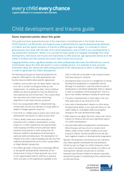
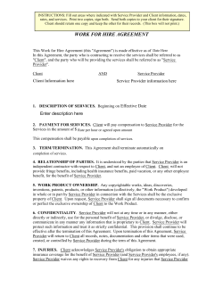
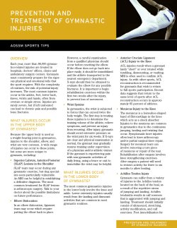
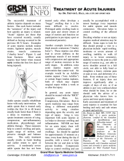
Trauma
• Abdominal trauma
• Pain management
• Burns
• Pelvic injury
• Chest injuries
• Post submersion • Crush injury
• Pre-hospital trauma by-pass
• Electric shock
• Spinal injury
• Eye injury
• Taser ® incidents
• Fluid injection injury
• Trauma in pregnancy
• Hypovolaemic shock
• Traumatic brain injury ( TBI )
• Limb injury
Clinical Practice Guidelines – Trauma
Clinical practice guidelines
Abdominal trauma
Page 1 of 2
Version 1.0 – September 2011
Penetrating and blunt trauma to the abdomen can produce significant
and life-threatening injuries. Many abdominal injuries may appear
insignificant, making it extremely difficult to predict severity. The close
proximity of organs within the torso makes distinguishing between
abdomen, chest and pelvic injuries difficult. Associated injuries outside
that cavity should be considered in all patients.[1]
Blunt abdominal trauma
Blunt trauma results in compression and shearing forces injuries.
Compression forces are those that result in abdominal organs and blood
vessels being crushed between solid objects. Shearing forces cause
tearing and rupture of solid organs and blood vessels at multiple sites.
Penetrating trauma
The extent of vessel and organ damage, including haemmorhage, due
to penetrating trauma is dependent on the mechanism (e.g. stab wound
vs gunshot wound). Many of these patients will require formal surgical
exploration and repair. Apparently small entry wounds may mask
significant internal injury.[2]
Regardless of the mechanism, catastrophic deterioration can develop
quickly and unexpectedly. All penetrating injuries despite the assessed
level of penetration, or actual size of the wound, should be treated as
serious and potentially life threatening.
Children
Due to their physique, children are particularly susceptible to abdominal
trauma. In comparison to adults, their relatively large abdomen is poorly
protected by lower ribs and pelvis and children may present with few
external signs of trauma.[3]
Clinical features
Features may be obvious but in some presentations
unexplained shock may be the only sign of severe
abdominal trauma.[2] Signs and symptoms include:
• ALOC
• Dyspnoea
• Abdominal pain/discomfort, guarding and tenderness on palpation
• Hypovolaemic shock
• Abdominal bruising (Cullen’s sign and Grey Turner
sign), distension (can be a late sign and difficult to determine)
• Shoulder tip pain (Kehr’s sign)
Risk assessment
• Significant abdominal injuries may present with little external evidence of trauma or a trivial
pattern of injury and or mechanism.[1]
• Fluid resuscitation – minimal fluid therapy to achieve a systolic BP 90 mmHg or perfusion of vital organs [4]
• Refer to specific CPG for abdominal trauma in head-injured or pregnant patient.
Additional information
• Pattern bruising such as Cullen’s and Grey Turner’s sign may take hours or days to develop.
• Trauma that presents with eviscerated bowel should be covered
with moist sterile pads or cling wrap.
Standard Cares
Pregnant patient:
Shocked?
Manage as per CPG:
• Trauma in pregnancy
Shocked with TBI?
Consider:
• IV access
• IV fluid (SBP 100 – 120 mmHg )
• Pelvic binder
• FAST
• Blood Consider:
• IV access
• Analgesia
• Antiemetic
• FAST
• Maintain normothermia
• Manage any other
injuries concurrently
Consider:
• IV access
• IV fluid (maintain a radial pulse)
• Pelvic binder
• FAST
• Blood
Transport to hospital
Pre-notify as appropriate
Abdominal trauma – Page 2 of 2
Clinical practice guidelines
Burns
Page 1 of 4
Version 1.0 – September 2011
Most burn injuries are a result of flame burns or scalds, with
electrical and chemical burns less common.[1] Concurrent blast injuries can accompany explosions and need to be considered when assessing a patient with major burns.
Risk assessment
Ensure safety for self and bystanders.
Burns can cause wide range of injuries. In the acute setting, airway
burns and inhalational injury can lead to respiratory compromise.
Over a number of hours, fluid and electrolyte abnormalities develop
in major burns and can lead to shock.[2]
Life threats
Fires in enclosed spaces pose further danger from the production of potentially lethal toxic gases (e.g. carbon monoxide and cyanide).[3]
Consider the possibility of an airway or inhalational burn if there is the presence of:
Respiratory compromise can manifest quickly in airway
and inhalational burns. Early endotracheal intubation is required to ensure airway patency.[5]
•
•
•
•
Clinical features
Depth of burn
Accurate burn-depth assessment can be difficult, as
most burns usually have a mixture of different depths.[4]
Burn depth assessment has implications in guiding
treatment, but lengthy assessment in the pre-hospital setting is not required.
Facial/oral burns
Singed nasal hair
Carbonaceous sputum
Tachypnoea, stridor, hoarseness
Hypovolaemia does not manifest from burns acutely but develops over many hours. The presence of
circulatory shock in the early stages of a burn implies an associated injury (e.g. blast injury).
Depth
Appearance
Sensation
Superficial
Erythema, brisk cap refill
Painful
Features of carbon monoxide and cyanide toxicity
should be sought if the patient was trapped in an
enclosed fire with the potential for significant smoke
inhalation.[3]
Superficial dermal
Moist, reddened with
blisters, brisk cap refill
Painful
Circumferential burns to the torso may restrict
ventilation, requiring urgent surgical intervention.[5]
Deep dermal
White slough,
reddened and
mottled, sluggish or
absent cap refill
Painful
Limb threats
Dry, charred, whitish
Absent cap refill
No Pain
Full thickness
Deep dermal and full thickness burns cause inelastic
dead tissue, referred to as eschar. Circumferential
burns may compromise limb vascular supply leading to ischaemia if untreated. Limbs with circumferential
burns are at risk of vascular compromise.[4] Estimation of surface area affected
The total area burnt can be estimated with the rule of nines, or the
more complicated, but more accurate, Lund Browder burn chart.[6]
Do not include skin with just isolated erythema. The area over the
patient’s palm can also be used to approximate 1% body surface area.
Rule of nines and Lund Browder burn chart estimation charts are below:
A
A
1
3
2
9%
1.5
3
2
2
1.5
2
1.5
1.5
Back 18%
1.5
1.5
1
9%
9%
9%
9%
18%
Back 18%
9%
9%
9%
1%
9%
9%
1.5
2.5
2.5
B
B
B
B
C
C
C
C
1.75
1.75
1.75
1.75
1.5
9%
AREA
14% 14%
9%
Figure 1 – Rule of nines
9%
AGE 0
AGE 1
AGE 5
AGE 10
AGE 15
ADULT
A: half of head
9.5
8.5
6.5
5.5
4.5
3.5
B: half of one thigh
2.75
3.25
4
4.5
4.5
4.75
C: half of one leg
2.5
2.5
2.75
3
3.23
3.5
Figure 2 – Lund Browder chart [6]
Burns – Page 2 of 4
Clinical practice guidelines
Version 1.0 – September 2011
Additional information
• Burns requiring management in dedicated burns unit [7]
-- Partial thickness burns > 20% all ages or
> 10% in < 10 year old and > 50 year old
-- Full thickness burns > 5%
-- Burns involving face, eyes, ears, hands, feet, genitalia,
perineum or overlying a major joint
-- All inhalational burns
-- All significant electrical burns
-- Burns in people with significant co-morbidities (e.g. heart failure)
• IV fluids should not be administered to patients with
significant facial, neck or upper chest burns with high
potential for airway or ventilation comprimise before the
airway is formally secured at hospital. Large amounts of
fluids increase the risk of interstitial oedema and tissue
swelling, potentially increasing the difficulty of endotracheal
intubation.[5]
• Hypothermia must be avoided in major burn injury. [5]
• In the paediatric population, consider non-accidental injury as a mechanism for burn injuries. [8]
• Escharotomies are surgical incisions through burnt eschar
to release tissue pressure in circumferential limb or thoracic
burns. They are best performed in hospital by electrocautery
as the wounds tend to bleed. They may be necessary in
the pre-hospital environment in situations where there is
imminent limb or ventilatory compromise. [4]
Burns
Page 3 of 4
Standard Cares
Evidence of airway burn, inhalation
injury or circumferential thoracic?
Haemodynamically unstable?
Pain?
• Protect against hypothermia
Consider:
• Active cooling ( first 20 min only )
• Cling wrap
• Burnaid dressing only if < 10% BSA kids, < 20% BSA adult
Consider:
• Oxygen
• IPPV
• Early airway assessment
Consider:
• Other injury (e.g. blast, cyanide toxicity)
• IV fluid
• Continue to reassess airway
Consider:
• Active cooling ( first 20 min only )
• Analgesia
• Midazolam
• Ketamine
Transport to hospital
Pre-notify as appropriate
Note: Officers are only to perform
procedures for which they have
received specific training and
authorisation by the QAS.
Burns – Page 4 of 4
Clinical practice guidelines
Chest injuries
Page 1 of 2
Version 1.0 – September 2011
Half of all trauma deaths have some form of chest injury. Although most thoracic trauma in Australia results from blunt forces,[1]
penetrating injuries are on the increase.[2]
Life threatening injuries may not be initially apparent and the
mechanism of injury is important in guiding further investigation (e.g. rib fractures suggest significant force with possible underlying
organ damage). Lack of obvious fractures doesn’t exclude injury
especially in a paediatric patient.
Clinical features
• Depends on mechanism, injuries sustained
• Penetrating trauma:
-- entry and exit wound
-- external bleeding may be evident
• Blunt trauma:
-- contusion/abrasion
-- haemotoma
-- obvious rib fracture
Signs suggesting life-threatening conditions:
•
•
•
•
•
•
•
•
•
•
•
•
Unequal air entry and/or crackles
Asymmetrical or paradoxical chest wall movement
Surgical emphysema
Chest hypomobility
Bubbling or sucking wounds
Extreme tachypnoea
Tracheal shift
Hypotension
Altered conscious state
Jugular venous distension
Muffled heart sounds
Cardiac dysrrhythmias.
Risk assessment
• Over-zealous IPPV may precipitate a tension
pneumothorax, especially in an intubated patient.[3]
• Chest pain in trauma can be due to ischaemia,
but blunt trauma to the heart can precipitate ECG
changes as seen in myocardial contusion.[4]
• Consider the possibility of cardiac arrest after trauma.
• Penetrating trauma to thorax may appear minor, but
life-threatening injury can be sustained (e.g. aortic or ventricular laceration, pneumo or haemothorax).
All wounds are treated as life threatening no matter
the size or perceived depth.
Additional information
Common features:
• pleuritic pain, shallow respirations and postural splinting
• reduced or absent breath sounds (pneumothorax), crepitus/subcutaneous emphysema
• hypoxia, tachypnoea
Standard Cares
Signs of tension
pneumothorax?
Thoracic decompression:
• Needle
• Pneumocath
• Thoracostomy
Shock?
• Stabilise mechanical injuries
Manage as per CPG:
• Shock
Consider:
• Oxygen
• IV access
• Analgesia
• Antiemetic
• IV fluid
• Stabilise mechanical injuries
• FAST
Transport to hospital
Pre-notify as appropriate
Chest injuries – Page 2 of 2
Clinical practice guidelines
Crush injury
Page 1 of 2
Version 1.0 – September 2011
Crush injuries include simple crush injury, compartment syndrome
and crush syndrome. There are many causes ranging from isolated
limb injuries, multisystem trauma, envonmation, drug and toxin
exposure, heat stroke, burns and some bacterial/viral infections.[1]
Crush injury – localised tissue injury that occurs when a compressive
force is applied.[2]
Compartment syndrome – compromised perfusion to tissues within
an anatomical compartment due to increased pressure within
that compartment. Left untreated, this can lead to tissue necrosis,
permanent impairment and crush syndrome.[3]
Crush syndrome – is a systemic condition that results from injuries
sustained by compressive forces sufficient in duration and pressure
to cause widespread ischemia and necrosis to soft tissue. [1] Ischaemia of the muscle leads to increased permeability of cell
membranes and the release of potassium, enzymes, and myoglobin
into the systemic circulation. Crush syndrome is characterised by
rhabdomyolsis, lactic acidosis, hyperkalaemia, renal failure, shock,
dysrrhythmias and death.[4]
The development of crush syndrome is TIME and PRESSURE
dependent. Crush syndrome can develop over a short time period
where the compressive force is large and, conversely, over long
periods where compressive forces are relatively small.[1]
Clinical features
Common presentations of compartment and crush
syndrome:
• Fracture (especially tibial),[5] severe soft-tissue
injury, prolonged limb compression
• Fluid infusion, arterial puncture and haemorrhage
• Envenomation
• Electric shock/burns
• Surgical repair of muscle hernia
• Constriction by casts, circumferential dressings,
clothing
Co-morbidities associated with increased risk:
• Diabetes
• Hypothyroidism
• Bleeding disorders/anticoagulation
Compartment syndrome is characterised by: [6]
• Palpable tension or swelling of an anatomical
compartment
• Pain disproportionate to the injury
• Pain on passive stretching of muscles within the anatomical compartment
• Paraesthesia of skin and paresis of muscles
supplied by nerves traversing the compartment
• Pallor of skin over the compartment
• Pulses diminishing as the condition develops is common, but normal peripheral pulses and
capillary filling is not uncommon.
Clinical features (continued)
Crush syndrome is characterised by: [1]
• Compartment syndrome
• Haemodynamic instability
• Reperfusion injuries leading to:
-- lactic acidosis and hyperkalaemia
-- dysrrhythmias
-- myoglobinaemia leading to renal failure
• Shock
• Hypothermia
Standard Cares
Evidence of hyperkalaemia on ECG?
Compressive force in situ?
Risk assessment
• Compressive force removal [2]
-- Anticipate the development of crush syndrome following removal of compressive force.
-- Anticipate hypovolaemic shock post removal of compressive force.
-- Chest involvement requires immediate
release of compressive force.
• Hypothermia is a potential risk for patients
suffering crush injuries.[1]
Additional information
• Compartment syndrome is a surgical emergency. If not diagnosed quickly and treated appropriately it is associated with a high morbidity. Management includes surgical
decompression of the affected muscle compartment by
fasciotomy and, in the case of circumferential burns,
escharotomy.[5]
Manage as per CPG:
• Hyperkalaemia
Consider:
• Analgesia
• IV fluids
• Elevate limbs
• Control compressive force release
• Anticipate reperfusion injuries
-- hyperkalaemia
-- dysrrhythmias
-- shock
• Consider in trapped patients:
-- torniquet
• Consider:
-- analgesia
-- IV fluid (20 ml/kg prior to release)
Transport to hospital
Pre-notify as appropriate
Note: Officers are only to
perform procedures for which
they have received specific
training and authorisation by the QAS.
Crush injury – Page 2 of 2
Clinical practice guidelines
Electric shock
Page 1 of 2
Version 1.0 – September 2011
All electric shocks (including lightning strike) should be managed as per this CPG.
The extent of injury following electric shock depends on (i) the
amount of current that passes through the body, (ii) the duration of the current, and (iii) the tissues traversed by the current.[1]
Visible injury is not an indicator of severity. There may be serious
internal injury to nerves and vessels as they offer little resistance to electrical energy.[2]
Clinical features
Electric shock can result in the following: [3]
• Neurological injury:
- ALOC
- seizures
- amnesia
- dysphasia
- motor dysfunction
- spinal cord damage
• Respiratory arrest or dysfunction
• Cardiac arrest or dysfunction:
- arrhythmias
- palpitations
- myocardial damage
• Pain (including chest pain or tightness)
• Vascular damage
• Renal failure
Clinical features (continued)
• Trauma:
- burns
- fractures
- entry and exit wounds
- secondary injuries due to falls
- compartment syndrome
Risk assessment
• Safety is paramount.[1]
• The patient must be not be approached until the scene is declared safe by appropriate agency/organisation or personnel.
Additional information
• Lightning strikes may cause respiratory and cardiac arrest
(usually asystole) with fixed dilated pupils. Despite this,
resuscitation should be initiated, as it is often successful
(mortality 30%.) [4]
Standard Cares
Ensure scene safety
Consider
• C-spine injuries
Cardiac arrest?
Unconscious?
Consider:
• C-spine injuries
• IV access
• Analgesia
• 12-Lead ECG
• IV fluid
• Dysrrhythmia treatments
• Burns treatment
Manage as per CPG:
• Resuscitation
Manage as per CPG:
• ALOC
Transport to hospital
Pre-notify as appropriate
Electric shock – Page 2 of 2
Clinical practice guidelines
Eye injuries
Page 1 of 2
Version 1.0 – September 2011
Eye injuries are common and may be serious despite a benign appearance.
All patients with suspected eye trauma and patients who have an ALOC should have their eyes assessed and basic eye protection
precautions implemented.
General management principles include:
• Irrigation with water or saline for chemical or biological fluid exposure, foreign body or thermal burns
• Protect eye with shield (cardboard cone or styrofoam cup)
• Antiemetic
• Position patient head up
Clinical features
• Significant eye injury may be present, despite normal
vision and minimal symptoms.[1]
• If eyelid oedema makes opening of the lids difficult – attempt gentle assessment and document findings.
• General symptoms:
-- pain or sensation of ‘grittiness’ in the eye
-- redness
-- copious tears
-- spasm of the eyelid
-- impaired or double vision
-- photophobia
-- haemorrhage
-- fluid loss from the eye
• Chemical exposure: [2]
-- sensation of foreign body within the eye
-- pain
-- blurred vision, tears
-- redness
Clinical features (continued)
• Penetrating eye injury: [3]
-- abnormally shaped or collapsed globe
-- obvious laceration or presence of prolapsed tissue
-- hyphema
• Blunt eye injury: [3]
-- orbital injury
-- traumatic mydriasis
-- hyphema
-- occasionally retinal detachment
• Retinal detachment: [1]
-- can occur spontaneously or months after an injury
-- history of light flashes
-- presence of floating black specks
-- curtain-like narrowing of peripheral vision
• Flash burns: [2]
-- history of unprotected exposure to welding flash or sun lamp
-- pain develops several hours following exposure
-- foreign body sensation within the eyes
-- redness and photophobia
Risk assessment
• Nil in this setting
Additional information
• With most eye injuries the priority is initial stabilisation of the
patient, protection of the eye and transport to an appropriate
facility (preferably one with an ophthalmologist).
• If possible, patients with eye injuries should have a visual
acuity test completed: [3]
Standard Cares
Blunt or penetrating?
-- Test one eye at a time.
-- Initially test the patient’s ability to count fingers (question patient on clarity of vision).
-- Should the patient be unable to complete this, test for hand motion, or light perception.
-- Do not delay initial treatment to perform visual acuity test.
Remove contact lens
• Administration of an antiemetic following penetrating or blunt
eye injury is highly recommended. Vomiting significantly
increases intraocular pressure and should be avoided.[4]
-- It is recommended that ondansetron is used in these
circumstances especially if opioid pain relief is given.
Chemical exposure?
-- It is highly recommended that medications such as
maxolon are avoided, due to the risk of dystonic reactions
occurring and perpetuating the injury
• Routine padding of eyes is no longer recommended. If padding is used, it must not place pressure on the globe.
Do not pad an eye with a penetrating injury.
• Reducing time for irrigation following chemical exposure is beneficial.
• Patients transported by air may have special requirements. Consult with receiving facility or QCC as to flight restrictions.
• When flushing eyes, place injured/damaged eye down and flush from medial aspect.
• Leave penetrating item in place and protect the eye with a raised shield (e.g. cardboard cone)
• Do not pad the eye
• Irrigate eye with H2O or sodium chloride 0.9% for < 15 minutes
• If foreign body present, attempt removal with a moist cotton bud
• Irrigate the eye and both
lids with H2O or sodium
chloride 0.9% for ≥ 30
minutes
• If capsicum spray,
continue irrigation until
pain subsides
Transport to hospital
Pre-notify as appropriate
• Eye injuries associated with capsicum spray should be
irrigated until pain subsides.
• Preferred positioning for patients with eye injuries is supine
with head elevated.
Eye injuries – Page 2 of 2
Clinical practice guidelines
Fluid injection injury
Page 1 of 2
Version 1.0 – September 2011
The high pressure injection of a fluid such as hydraulic oil, grease, or paint constitutes a medical and surgical emergency.[1] This requires
rapid access to appropriate specialist surgical review. The injury is
frequently worse than it will initially appear and paramedics must
have a high level of suspicion.
After the initial injection, the fluid travels in a stream until resistance
(e.g. from muscle or bone) is encountered.[2] The fluid then rapidly
disperses in all directions along tissue planes, potentially causing
traumatic dissections and compressing neurovascular bundles,
leading to vascular spasm, ischaemia and thrombosis.[1] Furthermore,
the presence of the fluid and subsequent tissue oedema, can cause
a pressure build up, reducing perfusion and resulting in a form of
compartment syndrome.[1]
Two additional issues are the chemical composition of the fluid,
which can have cytolytic properties, and infection, which can occur during the injection, or subsequent to the tissue damage and ischaemia.[1]
Due to the initially benign symptoms, these injuries are often
complicated by a delay to seek medical assistance, often several
hours. Without adequate treatment there is a high rate of
amputation.[1]
Clinical features
• The point of entry may look very small and may not bleed.
• The area of injury will usually be on the working
surface of the hand, however it may be located on any body area.[3]
• Initially, the patient may not complain of pain but may have a feeling of numbness, or increased
pressure within the affected part.
Clinical features (continued)
• Damage in the early stage is normally related to the physical injury as well as damage from the chemicals in the injected material.
• The affected body part will progressively become
increasingly irritated, with the patient complaining
of throbbing pain.
Risk assessment
• If the fluid is a smaller hydrocarbon compound
such as white spirit or kerosene, local anaesthetics
should be avoided as they will potentiate the effects and therefore must NOT be administered.[4]
• With larger sized compounds suck as those typically
used as hydraulic mineral oils, the higher viscosity
usually results in less penetration but is often more
difficult to remove.[4]
Additional information
• Intact skin can be penetrated by pressures of 7 bar (≈ 1000 psi ),
but this requires direct contact. Much higher pressures exist
within industrial machinery, such as paint guns and hydraulic
lines, where infiltration of subcutaneous tissues can occur when the liquid is fired from a distance.[1]
• Dependent upon the entry pressure, injected fluid can travel a significant distance from the initial site of entry, resulting in more widespread tissue damage.
Standard Cares
Provide ongoing appropriate
patient assessment
• Clean and cover wound
• Elevate and splint the affected limb
Consider:
• Analgesia
• Other injuries (e.g. compartment syndrome)
Note: These patients require urgent transfer
to medical facilities with surgical capability.
There are some situations where, due to the
isolation of the patient, the clinician may wish
to liaise with QCC via the regional ambulance
communication centre for advice about the
transfer.
Transport to hospital
Pre-notify as appropriate
Fluid injection injury – Page 2 of 2
Clinical practice guidelines
Hypovolaemic shock
Page 1 of 2
Version 1.0 – September 2011
This CPG deals with hypovolaemic shock. Other sources of shock are dealt with in the relevant CPGs.
Acute haemorrhage, secondary to trauma, is the major cause of
hypovolaemic shock. However, non-haemorrhagic causes must be
considered, i.e. GI losses, environmental exposure and neglect.
Blood loss can be ‘hidden’ and not immediately apparent i.e. pelvic
injury, ruptured ectopic pregnancy or GI haemorrhage or intracranial
bleeding in children.
Awareness of the clinical features of shock is of paramount importance,
as early recognition of hypovolaemia can be life-saving. Assessment
of volume status extends beyond the vital signs and requires a
comprehensive review of the patient. ‘ Treat the patient, not the vital signs.’
Clinical features [1]
Blood Loss
Signs
15% ( 750 mL in 70 kg )
• Minimal or no tachycardia
response
• Blood pressure changes do not usually occur
15 – 30%
( 750 mL – 1500 mL )
• Tachycardia
• Narrow pulse pressure
• Minimal – moderate drop in blood pressure
> 30% ( > 1500 mL )
•
•
•
•
> 40% ( > 2 L )
Tachycardia
Hypotension
Peripheral hypoperfusion
Altered level of consciousness
• Haemodynamic compensation
at its limit
• Decompensation imminent
Clinical features (continued)
Other clinical features
• CVS:
-- pale, cool peripheries, with or without being clammy
-- tachycardia > 100 bpm or bradycardia < 60 bpm
-- decrease pulses peripherally
-- capillary refill > 3 seconds
-- SBP < 100 mmHg
Note: Elderly may not be tachycardic.[2]
BP pseudonormalis. Fit/young patients may have
normal vital signs and be very volume depleted.
• NEURO:
-- ALOC
-- initially quiet with decreased alertness
-- confusion/agitation
-- obtundation (mental blunting)
Note: Be cautious interpreting ALOC as being due to substance misuse or alcohol.
Hypotensive trauma patients may not be secondary to
haemorrhage – consider other causes (i.e. obstructive
shock (tension pneumothorax tamponade) SCI or toxins – See Trauma CPG).
Risk assessment
• Nil in this setting
Standard Cares
Haemorrhagic/traumatic
Associated traumatic brain injury ?
Non-haemorrhagic
• Oxygen
• IV access
• IV fluid
• Control haemorrhage
• Control haemorrhage
• Oxygen
• Oxygen
• IV access
• IV access
• IV fluid
• IV fluid
(SBP 100 – 120 mmHg
• Maintain normothermia
( SBP ≥ 90 mmHg,
PR < 100 bpm)
(Target: palpable radial pulse)
• Maintain normothermia
Transport to hospital
Pre-notify as appropriate
Hypovolaemic shock – Page 2 of 2
Clinical practice guidelines
Limb injury
Page 1 of 2
Version 1.0 – September 2011
Limb injuries can be very painful and visually distressing for a
patient. As such, they can distract the patient and the clinician
from more serious injuries in a multitrauma situation. Gaining a good history of the event to assess the mechanism of injury, and completing a thorough primary and secondary survey are
always essential.
Clinical features
A fracture should be suspected if one or more of the following are present:
• Pain
• Swelling
• Bruising
• Loss of function
• Deformity
• Bony crepitus
Where communication is difficult (e.g. young children or dementia patients) the reluctance to
move a limb may be the only sign of a fracture.
Note: soft tissue injuries can include all but the latter two presentations.
Suspect neurovascular damage if there is poor distal perfusion, or reduced distal sensation or movement.
Risk assessment
• Appropriate analgesia is very important.
• Procedural sedation (ketamine) may be required
when managing complicated injuries (e.g. grossly displaced compound, compromised
vascular supply).[1]
• Limb immobilisation should generally be in near-anatomical position.
Standard Cares
Note 1: Open wounds/compound
injuries should be washed out with
1–2 litres of normal saline following
adequate analgesia.
Patient shocked ?
Manage as per CPG:
• Shock
Consider:
• Traction and splinting
• Analgesia
Limb poorly perfused ?
Consider:
• IV access
• IV fluid
• Analgesia
• Procedural sedation
• Re-alignment, traction and immobilisation
Note 2: Crush injuries to limbs should be treated as per crush injury CPG.
Consider:
• Analgesia
• Positioning
• Immobilisation
Transport to hospital
Pre-notify as appropriate
Limb injury – Page 2 of 2
Clinical practice guidelines
Pain management
Page 1 of 2
Version 1.0 – September 2011
Pain is an unpleasant sensory and emotional experience that is
potentially associated with tissue damage.[1] It is individual and
subjective and is influenced by factors such as culture, previous
experiences, belief, mood and ability to cope. There are therefore no definitive clinical signs of pain.
Clinical features
•
•
•
•
•
•
•
History of potentially painful condition/injury
Self reported pain
Distress
Pallor, muscle tension/guarding, sweating
Dilated pupils
Nausea/vomiting
Increased heart rate, respiratory rate and blood
pressure
• Signs and symptoms specific to underlying cause.
Effective pain management involves the use of an appropriate
pain assessment tool, especially when managing patients who are
unable to effectively communicate (people with an altered level of
consciousness or language barriers, those who are developmentally
delayed, children or the elderly, etc.) [2]
There are two major categories of pain management:
• Non-pharmacological techniques (should always be considered)
--------
appropriately inform patient
reassurance
distraction
posturing
positioning
heat or cold therapy
splinting
• Pharmacological
-- paracetamol, methoxyflurane, GTN, morphine, fentanyl,
ketamine, lignocaine.
-- Consider giving combination pharmacological therapy.
Pain management will not prevent the diagnosis of conditions or
injuries and, therefore, should be implemented for all patients.
Additional information
Wong-Baker FACES Pain Rating Scale [3]
0
NO HURT
2
HURTS
LITTLE BIT
4
HURTS
LITTLE MORE
6
HURTS
EVEN MORE
8
HURTS
WHOLE LOT
10
HURTS
WORST
Point to each face using the words to describe the pain intensity.
Ask the child to choose a face that best describes their own pain
and record the appropriate number.
From: Hockenberry MJ, Wilson D: Wong’s essentials of pediatric nursing, ed. 8, St. Louis, 2009, Mosby. Used with permission. Copyright Mosby.
Visual Analogue Score [4]
Pain as
bad as it
could be
No pain
0
1
2
3
4
5
6
7
8
9
10
Standard Cares
Consider non-pharmacological pain management
techniques:
•
•
•
•
Splinting
Psychological support
Heat or cold therapy
Consider specific injury/condition management
Consider most appropriate pharmacological
strategy:
•
•
•
•
•
•
Paracetamol
Methoxyflurane
Morphine
Fentanyl
Ketamine
Antiemetic
Transport to hospital
Pre-notify as appropriate
Pain alleviated ?
Pain management – Page 2 of 2
Clinical practice guidelines
Pelvic injury
Page 1 of 2
Version 1.0 – September 2011
Pelvic injuries are potentially life threatening and require early
identification and management.
The pelvis is extremely vascular with many blood vessels situated
close to the pelvic bones. Pelvic fractures may cause disruption of
these blood vessels and subsequent internal haemorrhage, shock
and death.[1]
The paediatric pelvis is more compliant, making it less likely to
fracture, but the force is transmitted to the underlying organs.
In most instances considerable force may be required to fracture the
pelvic bones, therefore associated intra-abdominal and pelvic organ
injuries should always be considered.[1]
The application of circumferential pelvic binders in patients with
suspected pelvic fractures can reduce fractures and stabilise the
pelvic ring which will help to decrease active bleeding.[2]
Pelvic trauma should be suspected in all patients with significant mechanism of injury and, in particular, in patients with haemodynamic instability after trauma.
Clinical features
Common mechanisms of injury resulting in pelvic
fracture include:
• traffic, pedestrian and motorcyclist collisions
• falls from heights
• crush.
Clinical features (continued)
Signs and symptoms of pelvic trauma include:
• pain
• bruising:
-- scrotal or vulval bruising
-- flanks (retroperitoneal)
• bleeding:
-- urethral meatus (urethral/prostate
/bladder injury)
-- vaginal (vagina/uterus/bladder injury)
-- rectal (bowel perforation)
• pelvic asymmetry/shortening of limb
• decrease of lower limb pulses
• reduced or absent sensation or power in lower limbs
• haemodynamic instability and shock
Ultrasound investigation (FAST scan) may reveal free fluid in the pelvis.[3]
Note: Pelvic springing is not to be performed.
Springing of the pelvis may disrupt sacral clots and
cause further haemorrhage. In addition to this, clinical
assessment of the pelvis has a low sensitivity for
diagnosing pelvic fractures.[4]
Risk assessment
• Nil in this setting
Standard Cares
Evidence of shock/haemodynamic compromise?
• Apply pelvic binder
Consider:
• Other injuries
• Analgesia
• FAST
Manage as per CPG:
• Shock
Consider:
• Other injuries
• Analgesia
• FAST
Transport to hospital
Pre-notify as appropriate
Pelvic injury – Page 2 of 2
Clinical practice guidelines
Post submersion
Page 1 of 2
Version 1.0 – September 2011
Post submersion refers to survival post submersion/immersion
where potential or actual respiratory compromise has occurred.[1] Presentations may be asymptomatic, with only a history or
mechanism of submersion to respiratory and cardiovascular
compromise deteriorating to cardiac arrest.
Secondary complications post submersion includes noncardiogenic
pulmonary oedema, aspiration pneumonia and anoxic encephalopathy.
A clear history regarding the circumstances surrounding the patient’s
presentation should be gained, including time frames, mechanism of
trauma or medical conditions such as intoxication, seizure, stroke or
ACS that may have precipitated the submersion.[2]
Clinical features
Neurological:
• ALOC
• confusion
• agitation
Cardiovascular:
• dysrrhythmias (usually associated with
hypothermia, unless patient has an underlying
cardiac condition) [2]
• hypotension
• cardiac arrest
Respiratory:
• dyspnoea
• non-cardiogenic pulmonary oedema
• ARDS
• aspiration pneumonia
• respiratory arrest
Clinical features (continued)
Other clinical features include:
• spinal injury
• vomiting and nausea
• hypothermia.
Risk assessment
• All post submersion patients should be transported
to hospital for assessment, due to the potential
for developing complications such as pulmonary
oedema and aspiration pneumonia. These
conditions may occur in patients with few initial
symptoms or a normal initial vital sign survey.[2]
• Hypothermia can occur as a secondary result of submersion or through evaporative heat loss after rescue.
• Spinal immobilisation in all patients with significant mechanism of injury or GCS < 15.
• Gastric tubes should be considered in all intubated
post-submersion patients especially children (to alleviate diaphragmatic splinting and secondary
gastric distention).
Additional information
• Ensure treatable underlying conditions (e.g. overdose,
hypoglycaemia, seizure and/or trauma) are managed
concurrently.
• If the patient is hypothermic, with no pulse and apnoeic, manage as a hypothermic cardiac arrest.
Standard Cares
Cardio/respiratory arrest?
Consider:
• Oxygen
• IPPV
• PEEP
• IV access
• Gastric tube
• Analgesia
• Antiemetic
• Correct reversible causes
• Maintain normothermia
ALOC and/or
SpO2 ≤ 90%?
Manage as per CPG:
• Resuscitation
Consider:
• Oxygen
• IPPV
• IV access
• Analgesia
• Antiemetic
• Maintain normothermia
Transport patient to hospital
Pre-notify as appropriate
Post submersion – Page 2 of 2
Clinical practice guidelines
Pre-hospital trauma by-pass
Page 1 of 4
Version 1.0 – September 2011
The trauma bypass clinical practice guideline is designed to identify
trauma patients who require transport to a Major Trauma Service. Three elements should be considered for the triage of major trauma
patients in Queensland: [1]
• Vital signs
• Mechanisms of injury
• Patterns of injury
Vital signs
NEONATE
INFANT
CHILD
OLDER CHILD
ADULTS
First 28
days
1 – 12
months
1 – 8
years
9 – 14
years
> 14
years
ALOC
ALOC
ALOC
ALOC
ALOC
Respiratory
rate
< 40 or > 60
< 20 or > 50
< 20 or > 35
< 15 or > 25
< 10 or > 30
SpO2
(room air)
< 95%
< 95%
< 95%
< 95%
< 95%
Heart rate
< 100 or > 170
< 90 or > 170
< 75 or > 130
< 65 or > 120
> 120
Systolic BP
N/A
< 60 mmHg
< 70 mmHg
< 80 mmHg
< 90 mmHg
Conscious
state
Table 1: Abnormal vital signs
Vital signs are determined by one or more of the patient’s vital signs
being abnormal, as outlined in the table above.
Mechanism of injury criteria
Triage by mechanism of injury has limited accuracy, however it may
help detect significant occult injury. High-risk mechanism include:
• Ejected from vehicle
• Fall from height ≥ 3 metres
• Involved in an explosion
• Involved in a high impact RTC with incursion into the occupant’s compartment
• Involved in a vehicle rollover
• Involved in an RTC in which there was a fatality in the same vehicle
• Entrapped for > 30 minutes
Injury pattern criteria
• Injuries to the head, neck, chest, abdomen, pelvis, axilla, or groin that:
- are penetrating
- sustained from blasts
- involve two or more of those regions
• Limb amputation above the wrist or ankle.
• Suspected spinal cord injuries.
• Burns in adults > 20%, or in children > 10%, or other
complicated burn injury including burn injury to the hand, face, genitals, airway, or respiratory tract.
• Serious crush injury.
• Major compound fracture, or open dislocation with vascular compromise.
• Fractured pelvis.
• Fractures involving two or more of the following: femur, tibia, or humerus.
Clinical features
Procedure: [1]
• If any of the vital signs criteria or patterns of
injury criteria are present, the patient should be
transported to a Major Trauma Service if there is one within 45 minutes road transport time.
• If a Major Trauma Service is > 45 minutes road
transport time, the patient should be transported to the highest hospital level Regional Trauma
Service if there is one within 45 minute road
transport time.
• If the nearest Regional Trauma Service is > 45
minutes road transport time the patient is to be taken to the closest local hospital. Under these circumstances the Queensland Clinical
Coordination Centre (QCC) must be notified via
the regional Ambulance communication centre to
ensure consideration for early secondary transport.
• If any of the mechanisms of injury criteria are
present, paramedics are to take into account the potential for occult major trauma and consider transporting the patient as per the above procedure.
• If none of the triage criteria are met, the patient
should be transported to the nearest appropriate
facility.
Clinical features (continued)
Transporting patients directly to specialist
facilities
Patients with the following injuries should be
transported directly to the appropriate facilities offering
specialist services, providing it is within 45 minutes road transport time:
• Adults with > 20% body surface area or complicated burns to the Royal Brisbane and Women’s Hospital.
• Children with > 10% body surface area or
complicated burns to the Royal Children’s Hospital.
• Patients with spinal injuries with neurological
deficit to the Princess Alexandra Hospital.
• Patients with amputations to a major trauma service.
Special note: If a Major Trauma Service is within 45 minutes road transport time, it should be the
preferred destination if the patient fits the criteria as stated above – even if this means bypassing a
Regional Trauma Service which may be closer.
Post submersion – Page 2 of 4
Clinical practice guidelines
Pre-hospital trauma by-pass
Page 3 of 4
Version 1.0 – September 2011
Additional information [1]
Major trauma services are provided at:
•
•
•
•
•
•
The Royal Brisbane and Women’s Hospital
The Royal Children’s Hospital (paediatric patients ≤ 14 years only)
Princess Alexandra Hospital
Mater Children’s Hospital (paediatric patients ≤ 14 years only)
Gold Coast Hospital
The Townsville Hospital
Regional trauma services are provided at:
•
•
•
•
•
•
•
•
•
•
•
•
The Tweed Hospital
Logan Hospital
Ipswich Hospital
Toowoomba Hospital
Caboolture Hospital
Redcliffe Hospital
Nambour Hospital
Hervey Bay Hospital
Bundaberg Hospital
Rockhampton Hospital
Mackay Base Hospital
Cairns Base Hospital
• Mt Isa Hospital
Special Note:
Use of retrieval team [2]
Paramedics should consider the use of a medical retrieval team for patients who are trapped or require interventions beyond the scope of the treating paramedics.
Pre-hospital medical retrieval teams should not unnecessarily delay the transport of the patient to a definitive treatment medical
facility. Refer to local work instructions regarding the availability of pre-hospital medical retrieval teams.
Traumatic cardiac arrest
If the patient has life threatening injuries and suffers a cardiac
arrest immediately before or during transport, the patient should
be transported to a Major Trauma Service if within 15 minutes road
transport time. If transport time to a Major Trauma Service is > 15 minutes, consider transport to the nearest appropriate hospital.
Standard Cares
Is this a major trauma
(identified by the above criteria)?
Transport to
local hospital
Can patient be transported
to a Major Trauma Service
(MTS) within 45 minutes drive time?
Transport to Major Trauma
Service (MTS)
Can patient be transported
to a Regional Trauma Service
(RTS) within 45 minutes drive time?
Transport to Regional
Trauma Service (RTS)
Transport patient to closest
hospital and request regional
communication centre notifies QCC
Post submersion – Page 4 of 4
Clinical practice guidelines
Spinal injury
Page 1 of 4
Version 1.0 – September 2011
Spinal cord injury (SCI ) is injury of the spine with associated motor,
sensory and autonomic deficit. SCI can be caused by hyperflexion,
hyper-extension, rotation, compression, or penetrating injury of the
spinal cord. The leading causes of SCI include RTC’s, falls, acts of
violence, sporting injury and recreational water activity.[1]
The diagnostic pattern for SCI involves:
•
•
•
•
Mechanism of injury
Pain/tenderness over or adjacent to the spinal vertebra
Impaired motor or sensory function
Impaired autonomic function.
The principle of pre-hospital management of SCI is to limit neurological
deficit and prevent secondary injury.[1] This is achieved through:
•
•
•
•
•
Appropriate spinal immobilisation
Maintaining a high index of suspicion of SCI
Identification and reversal of life threats in the primary survey
Cardiovascular and ventilatory support
Ensuring appropriate thermoregulation.
Clinical features
Impaired motor function:
• Diaphragmatic ventilation
• Paralysis
• Flaccidity
• Abnormal posturing
Impaired sensory function:
• Local or generalised paresthesia/anaesthesia
• Loss of proprioception
Impaired autonomic function:
• Hypotension
• Bradycardia
• Thermoderegulation
• Priaprism
Consider neurogenic versus spinal shock.[2]
Risk assessment
• Always consider spinal injury in the unconscious
patient (especially cervical spine).[3]
• The extrication board is to be used as an
immobilisation device only and not a restraint device.
• Patients with SCI are high risk for concomitant trauma particularly TBI and haemorrhagic shock.[1]
• KED/NEIJ devices are inappropriate where rapid
extrication is required for patients with life
threatening injury and/or vital sign alteration.[4]
In such circumstance, use MILS with all appropriate
resources for extrication.
Additional information
Canadian C-spine rule (CCR) [7]
Canadian C-spine rule (CCR) has been developed for the
clinical clearance of the cervical spine, without need for
diagnostic imaging, in alert and stable trauma patients.
CCR was considered superior to NEXUS Low risk criteria with
respect to sensitivity and specificity for cervical-spine injury.[5]
Rule does NOT apply if:
Studies have found that paramedics can apply the CCR
reliably, without missing any important cervical spine injuries.[6]
However CCR does not take into account:
• Painful distracting injury or
• Intoxication (alert could include intoxication).
•
•
•
•
•
•
•
Clinical judgement should still take precedent.
1. Any high mechanism that
mandates radiography?
• Age ≥ 65 or
• Dangerous mechanism * or
• Parasthesia in extremities
Non-trauma cases
GCS < 15
Unstable vital signs
Age < 16
Acute paralysis
Known vertebral disease Previous C-spine injury
2. Any low risk factor which
allows safe range of motion?
• Simple rear end RTC **
• Ambulatory at any time
• Delay onset of neck
pain ***
• Absence of midline C-spine
tenderness
Immobilise and
transport
All patients with dangerous mechanisms and distracting
injuries are to have C-spine immobilised.
* Definition dangerous
mechanism • Fall from > 1 metre/5 stairs • Axial load to head (diving)
• High speed RTC (> 100kph),
rollover, ejection
• Motorised recreational vehicle
• Bicycle struck/collision ** Simple rear RTC excludes:
•
•
•
•
3. Able to actively rotate neck?
• 45° left and right?
Pushed into oncoming traffic
Hit by bus/large truck
Rollover
Hit by high speed vehicle
*** Delayed
No Immobilisation
• Not immediate onset of neck pain Spinal injury – Page 2 of 4
Clinical practice guidelines
Spinal injury
Page 3 of 4
Version 1.0 – September 2011
Standard Cares
Spinal immobilisation
Consider neurogenic shock:
Hypotensive and bradycardic
• FAST
• IV fluid
• Inotropic support
Hypotensive and tachycardiac
Consider:
• IV fluids
• FAST
Consider:
• Analgesia
• Antiemetic
Transport to hospital
Pre-notify as appropriate
Spinal injury – Page 4 of 4
Clinical practice guidelines
Taser ® incidents
Page 1 of 2
Version 1.0 – September 2011
Taser ® is a brand name of one of a number of weapons in the general
category of ‘conducted energy devices’ and is currently the only one
in use by the QPS.[1] They are a ‘less than lethal’ use of force option
that may assist officers resolve incidents involving violent people. The
Taser ® is a hand held, neuro-muscular disruption device capable of
incapacitating a person through the application of an electrical current.[2]
The Taser ® has two main capabilities: [2]
• Propelled wired darts embed in the targeted person, followed by
a short duration high voltage electrical pulse, which affects the
sensory and motor functions of the nervous system (probe mode). • Direct contact of the Taser ® to the body, or clothing of a person
(drive-stun mode).
Clinical features
• Taser s® have the potential to cause strong muscle
contractions and the potential for serious secondary
injury including:
-- fractures
-- spinal injuries
-- head injuries
-- soft tissue injuries
-- hyperthermia.
• Death immediately following Taser ® use has been
reported.[3] Follow standard resuscitation guidelines.
Risk assessment
• Ensure safety precautions are taken at all times.
• Ensure the wires from the Taser ® have been
disconnected or cut from the probes.
Probe removal
To remove the probes, one hand should be used to stabilise the skin
around the probe and the other hand should be used to grasp the
probe firmly and pull straight out in a rapid motion.[4] Do not attempt to
pull the probes out by the wires, as they are very fragile and will easily
break. The process is usually painless due to the electrocautery effect
on the surrounding tissue.
Once removed the probes should be:
• inspected to see they are intact, with the straightened barbs still attached to the probe body.
• separated, or removed from the copper coated wires. (These wires are thin and can be cut by scissors or will break easily if pressure is applied).
• disposed of in a sharps container.
Probes should not be removed if embedded in:
• eyes
• genitals
• face or neck.
Manage as per foreign body/penetrating injury and transport to medical facility.
Transport is indicated in patients where:
• probes cannot be removed
• the patient requires a psychiatric evaluation
• assessment of injuries (other than probe injuries) is required
• the patient is affected by substances other than alcohol.
Tasered patients can be left in the care of QPS if they do not meet the above indications for transport and their 12-Lead ECG and BGL are within normal limits.
Standard Cares
Ensure safety issues are considered
Disconnect probes/wire from device
• 12-Lead ECG
• BGL
Consider:
• EEO
• Removal of probes
• Manage injuries
Transport to hospital
Pre-notify as appropriate
Taser® incidents – Page 2 of 2
Clinical practice guidelines
Trauma in pregnancy
Page 1 of 2
Version 1.0 – September 2011
Trauma affects approximately 7% of pregnancies and can cause
premature labour, placental abruption, feto-maternal haemorrhage
and fetal demise.[1] Even seemingly minor injuries can be dangerous.
Approximately half of the trauma experienced in pregnancy is
secondary to motor vehicle accidents, with falls, assaults and burns
occurring less frequently.[2]
Clinical features
Specific trauma related injuries of pregnancy
• Premature labour
• Placental abruption:
-- Shearing forces from deceleration injuries can separate the placenta from the underlying
uterine wall, causing an abruption. It occurs in 1 – 5% minor trauma in pregnancy and in
20 – 50% major trauma.[1] Importantly it can have a delayed manifestation 24 – 48 hours
after the initial injury.
• Uterine rupture: [2]
-- Uterine rupture is a rare but devastating
traumatic complication. It should be
suspected if there is maternal shock, difficulty defining the uterus on palpation or if there are easily palpable fetal parts.
• Feto-maternal haemorrhage [1]
-- Feto-maternal haemorrhage refers to the
spread of fetal blood into the maternal
circulation which can lead to Rhesus
sensitisation in the mother. This has
implications for further pregnancies.
Risk assessment
Important physiological changes occur in pregnancy
which impact on maternal and fetal risk in trauma.
Maternal risk
• Blood volume increases approximately 45%
approaching term – this relative hypervolaemia
can mask haemorrhagic shock. Up to 35% of
maternal blood volume can be lost before signs of haemorrhagic shock appear.[1]
• The diaphragm rises approximately 4 cm in the later stages of pregnancy, with a reduction in residual lung volume.[1] There is
a relative reduction in respiratory reserve in these patients. • Delayed gastric emptying and the displacement
of intra-abdominal organs by the growing fetus
increases the risk of aspiration.[1]
Fetal risk
• Maternal hypovolaemia at any stage, can cause
fetal harm. Adequate maternal blood volume
must be maintained in the traumatised pregnant
patient.
• In the first trimester the uterus resides within
the bony pelvis, protecting it from direct trauma. Beyond the first trimester, there is increased risk to fetus from direct trauma.
Definitive care
The seriously traumatised pregnant patient requires a
multidisciplinary approach, including an urgent obstetric
opinion and possibly a caesarean section. Pregnant patients suffering minor trauma are typically
monitored in the labour ward for a minimum of four hours to detect occult placental abruption.[1]
Standard Cares
Position to left lateral tilt
Anti-D immunoglobulin is administered to Rhesus-negative
mothers to prevent feto-maternal isoimmunisation.[1]
Additional information
Evidence of shock?
• Maternal heart rate can be expected to increase by 15 – 20 beats per minute in the later stages of pregnancy.[1]
• Maternal BP lowers by 10 – 15 mmHg during the 2nd trimester and tends to normalise at term.[1]
• Position pregnant patients with left lateral tilt to avoid aortio-caval compression.
• Consider mechanism of injury including:
-- direct abdominal trauma
-- improper application of lap belt.
• A focused obstetric history should include:
-- gestational age
-- presence of fetal movements
-- PV loss.
Consider:
• Potiental for shock (pregnancy
may mask signs/symptoms in patient)
• Pelvic binder
• IV Fluid
• FAST
• C-spine immobilisation
• Antiemetic
• Analgesia
Transport to hospital
Pre-notify as appropriate
:
Consider resuscitation
• IV fluid
• Pelvic binder
• Antiemetic
• C-Spine immobilisation
• FAST
• Blood
• Analgesia
Be prepared for:
• Spontaneous delivery
Be mindful of:
• Placenta abruption
• Uterine rupture resulting
from traumatic injury
• Managing associated
traumatic injuries
Note: Officers are only to perform
procedures for which they have
received specific training and
authorisation by the QAS.
Trauma in pregnancy – Page 2 of 2
Clinical practice guidelines
Traumatic brain injury
Page 1 of 2
Version 1.0 – September 2011
Traumatic brain injury (TBI ) is a significant cause of morbidity
and mortality in Australia, with over 22,000 people admitted to
hospital per annum. The most common causes of injuries are falls,
motor vehicle accidents and assault.[1]
The goal of pre-hospital care is to reduce secondary brain injury
due to hypoxia, abnormal carbon dioxide levels or hypotension.
In areas where it is available, pre-hospital rapid sequence
intubation using muscle relaxants may be beneficial.[2] Intubation of patients without muscle relaxants, except in arrest, is harmful to TBI patients, and is not indicated.[3]
Clinical features
• External evidence of head injury:
-- scalp abrasion, laceration, haematoma,
complaining of headache
-- obvious depressed skull fracture/open head
injury
-- blood from ears or nose (suggestive of base of skull injury)
-- brain matter on view is an extremely poor prognostic sign
• ALOC/focal neurology
-- reduced GCS (Patients may be agitated and appear intoxicated.)
-- unilateral weakness, seizure, unequal/
unreactive/dilated pupils
Clinical features (continued)
• Abnormal vital signs
-- Bradycardia and hypertension (Cushing’s reflex)
is a sign of raised ICP and suggests imminent
brain herniation.[4]
-- Hypoxia or hypotension have important
prognostic implications and receiving facilities
should be informed of this.[3]
-- Excluding hypoglycaemia is essential.
Risk assessment
• Any episode of hypoxia or hypotension in the
setting of TBI will significantly increase morbidity
and mortality.[3]
• Hyper or hypoventilation of patients causing
abnormal CO2 levels will also impair brain
perfusion (hyperventilation causes hypocapnoeic
vasoconstriction; hypoventilation causes
hypercapnoeic vasodilatation and raised
intracranial pressure (ICP).[5]
• It is important to avoid raised ICP from impaired venous return, by ensuring constricting tapes, ties or collars are loosened from around the neck and the patient is positioned head-up to 30 degrees if possible.[6]
Management
Pre-hospital
Standard Cares
Consider:
• C-spine support
• Oxygen
• IV access
• Analgesia
• IV fluid
• Support oxygenation and ventilation to prevent hypoxia.
• Use appropriate IV fluid and control of haemorrhage (e.g. fracture splints/pelvic binders, external pressure) to prevent/treat hypotension.
GCS = 15?
Additional information
• Some areas may participate in trials assessing the impact of hypothermia in TBI. Except in these situations, the goal is to achieve normothermia.
• ‘Cold’ intubation in the GCS3 patient is extremely predictive of a poor outcome.
Oxygen
IV access
IV fluid (SBP 100 – 120 mmHg )
Consider:
• Basic airway adjuncts
• C-spine support
• Analgesia
• Midazolam
• Hypertonic saline 7.5%
Transport to hospital
Pre-notify as appropriate
Traumatic brain injury – Page 2 of 2


© Copyright 2026