
CET
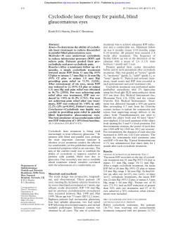
CET
CONTINUING
EDUCATION
& TRAINING
2 FREE CET
POINTS4
4
OT CET content supports Optometry Giving Sight
Approved for Optometrists
Approved for SP Points
Approved for AS Points
4
Medical management
of glaucoma
12/03/10 CET
34
Module 13 Part 3: CLINICAL OPTOMETRY
Course code: C-13472 O/SP/AS
Stephen Bryan MA FRCS FRCOphth
Although glaucoma is largely managed by ophthalmologists in a
hospital setting, there are many optometrists who are intending, or
already are, practising glaucoma management in a shared care setting,
either in their own practice, in a hospital or at a polyclinic. This article
is aimed to provide an overview of the management of glaucoma by
optometrists, to equip them with the necessary skills to perform this
service. Before discussing the actual medical management options,
it has to be emphasised that correct diagnosis based on the patient’s
history and examination is an essential precursor to correct treatment.
Guidelines for the diagnosis and
management of adult chronic open
angle glaucoma and ocular hypertension
from the National Institute for Health
and Clinical Excellence (NICE) aims to
standardise care for glaucoma sufferers
and prevent people from going blind.
This article should therefore be read in
conjunction with this guidance, which
provides greater detail regarding specific
monitoring and treatment pathways. The
guidance is available at www.nice.org.uk.1
Diagnosis – does the patient
really have glaucoma and if so
what kind?
Primary open angle glaucoma (POAG) is
still the most common form of glaucoma
in the UK population, and the majority
of it is suited to medical management
alone. However, the proportion of people
with glaucoma in areas with a significant
ethnic population is actually much more
than the incidence traditionally found
in the UK and American literature.2,3,4
One
example
is
the
increased
incidence of angle closure glaucoma
(ACG) without hypermetropia in the
Chinese population. There is a danger
that diagnosis of POAG is assumed
automatically because it is common and
that the medical treatment of secondary
and complex glaucoma may not control
the condition. Another example is
unilateral glaucoma from previous
trauma, perhaps from a long time ago,
which resulted in secondary glaucoma
from angle recession or peripheral
anterior synechia. These more unusual
diagnoses may be more common than you
think and it is important not to miss them.
The diagnosis of glaucoma is
determined principally by the patient’s
and family history (taking account of
increased risk factors such as diabetes
and hypertension), the presenting
intraocular pressure (IOP) measured
by Goldmann applanation tonometry
(adjusted for central corneal thickness),
gonioscopy, visual field examination and
optic nerve head assessment with dilated
pupils and using stereoscopic indirect
ophthalmoscopy (Figure 1). Optic nerve
and nerve fibre layer imaging are also
useful. The initial diagnosis should be
considered provisional and recorded at
the outset but may need reviewing; for
example POAG that progresses despite
“normal” IOP may need to be changed
to normal tension glaucoma (NTG),
or the drainage angle may be initially
open but become narrower over time.
Deciding to treat: where to set
target IOP
The problem here is trying to decide
where along the continuum between
ocular hypertension and NTG the
individual case lies. The main question
to ask is whether the optic nerves are
already damaged and are not tolerating
the presenting IOP (if so, the patient
has glaucoma) or if the raised IOP is not
causing damage, whether it is so high
that you think the onset of glaucoma is a
certainty, or that there is a risk of retinal
vein occlusion (if so, the patient has
ocular hypertension). If the patient has
So you have decided to treat
and set a target IOP
This now gets more straightforward.
Fortunately, in the last 25 years, there
has been great progress in developing
an arsenal of effective medications. To
give some idea of the change, in the
mid-1980s options were restricted to
pilocarpine and an adrenaline pro-drug,
Propine. Timolol had been introduced
five years earlier but was deemed too
expensive to prescribe. Since then, the
pharmaceutical industry has introduced
topical carbonic anhydrase inhibitors
(Trusopt), alpha-2 agonists (Iopidine)
and, best of all, prostaglandin analogues
(Xalatan), followed by combination drugs
and other rival branded preparations in
these three new categories. There has
been a small retrograde step recently in
the UK with the NICE recommendation
that prostaglandin analogues are not
recommended first line therapy in the
under 65s, on the grounds of cost, but this
may change as Xalatan will shortly go off
patent and so these drugs will fall in price.
As with diagnosis, it is important to
measure IOP with applanation tonometry
and adjust for corneal thickness. As a
very rough rule of thumb, the favoured
first and second line treatments are betablockers or prostaglandin analogues,
unless contraindicated. On their own,
these should achieve up to 30% IOP
reduction and up to 50% together. It then
is a simple matter of arithmetic; a patient
with a presenting IOP of 24mmHg is
probably fine on monotherapy having
set a target IOP of 16mmHg, whilst a
lower target IOP dictates dual therapy.
If you are looking for a reduction
beyond 50%, then surgery may be the
best first line option (it is not unusual
for a few patients with POAG to
present with IOPs of over 40mmHg).
Specific medications
Topical beta-blockers
Topical beta-blockers are a common first
line treatment for existing glaucoma
patients. They are relatively cheap and
have a good track record of efficacy,
with few topical side effects. Betablockers work by reducing aqueous
production and should produce an IOP
reduction of 20% to 30%. Examples of
commonly used beta-blockers include:
• Timolol (Timoptol and TimoptololLA)
• Levobunolol (Betagan)
• Betaxolol (Betoptic)
• Carteolol (Teoptic)
The principle side effects of betablockers are systemic and relate to
pre-existing systemic conditions. Betablockers are contraindicated in patients
with bradycardia and hypotension.
They can worsen peripheral vascular
disease, can cause impotence and some
patients have reported nightmares.
One rare but significant side effect is
that they can mask hypoglycaemia in
diabetics. In addition, a patient may be
35
Figure 1
Glaucomatous cupping
asymptomatic but have a latent tendency
to, for example, reversible airways
disease, which becomes manifest
after you start topical treatment.
Topical beta-blockers are absolutely
contraindicated in heart failure and
some forms of heart block, and in asthma
and chronic obstructive airways disease
(COAD). They should be used with
caution in patients with diabetes and
patients on calcium channel blockers. If
patients are on systemic beta-blockers,
there is a small risk that they may end
up with too much beta-blocker. Also, the
efficacy may be reduced as the patient
is already benefiting from a small IOP
reduction from the systemic beta-blocker.
In some patients, particularly those
with NTG, topical beta-blockers may be
counterproductive at night. They may
exacerbate nocturnal bradycardia, and
cause blood pressure to dip and possibly
impair optic nerve head perfusion.
In these patients, beta-blockers are
prescribed for morning use only.
These systemic interactions may
sound alarming but provided you
exclude the important contraindications,
it is unlikely that a patient under 65
years of age will develop problems. In
older patients, there is a higher risk that
they will have one of these conditions
(COAD has an incidence of 15% and
cardiovascular disease has an incidence
of 25% in the over 65s), and that the betablocker will uncover something they did
not know they had. It is very important
that their GP knows they have been
12/03/10 CET
a “normal” IOP, no history of previous
secondary temporary glaucoma (eg
trauma, vitreoretinal surgery etc.) with
definite glaucomatous damage, the
diagnosis of NTG is straightforward and
you need to set a low target IOP. If the
case is not clear-cut, some factors in the
history may help you to make a decision,
such as family history suggesting NTG,
vasospasm or migraine. Another clue
is the pattern of visual field loss; the
visual field loss of NTG is typically more
central compared with IOP-dependant
POAG. Often, however, it is a case of
“suck it and see”– reviewing target IOPs
as you go. For example, you may have
two patients that present at the same
time with identical moderately raised
IOP and early glaucomatous damage.
Five years later, one patient may have
done fine just with monotherapy
achieving a target IOP of 18mmHg, with
no progression at all. The second patient
may have progressed and needs triple
therapy with a target IOP adjusted to
10mmHg. Sadly, at present, there is no
way of predicting with any certainty
which path an individual patient will
take. Hopefully, ongoing research into
genotyping and phenotyping may
help us to better tailor treatment to the
individual at the point of presentation.
CET
CONTINUING
EDUCATION
& TRAINING
2 FREE CET
POINTS4
4
OT CET content supports Optometry Giving Sight
Approved for Optometrists
Approved for SP Points
Approved for AS Points
4
12/03/10 CET
36
Figure 2
Allergic reaction to glaucoma eye drops
prescribed a beta-blocker and that there
is communication between you both.
Prostaglandin analogues
Examples
of
commonly
used
prostaglandin
analogues
include:
• Latanoprost (Xalatan)
• Travoprost (Travatan)
• Bimatoprost (Lumigan)
Prostaglandin analogues work by
increasing uveoscleral outflow in the
eye. In theory, acting in a physiological
way is an advantage as these agents do
not reduce aqueous production, which
provides essential nutrition to the cornea
and lens. In normal Caucasian eyes, it
was estimated that 10% of outflow is
through the uveoscleral route but this
can be up to 35% in some individuals.
In glaucomatous eyes, which by
definition have impaired trabecular
outflow,
the
uveoscleral
channel
assumes more of the flow. Prostaglandin
analogues are highly effective and
may reduce IOP by up to 35%.
The principle side effects are local
and are i) a tendency to induce a red
eye, particularly upon initial use, ii)
increased iris and eyelash pigmentation,
and in mildly pigmented patients may
cause darkening of the skin around
the eyes, iii) eyelash growth, and iv)
may precipitate or worsen cystoid
macular oedema in aphakic eyes.
Although
initial
theoretical
concerns about a general effect on cell
division have proved unfounded, it
is still contraindicated in pregnancy,
mainly because prostaglandins are
actually used to induce labour.
Topical carbonic anhydrase inhibitors
Examples
of
commonly
used
topical
carbonic
anhydrase
inhibitors
(CAIs)
include:
• Dorzolamide (Trusopt)
• Brinzolamide (Azopt)
Topical CAIs act by reducing aqueous
production and are slightly less effective
than prostaglandin analogues or betablockers. They are thought to reduce
IOP by 18% to 20% but this amount can
be less in some individuals. Generally,
these agents are found to cause more
irritation than the prostaglandin
analogues and can leave a bitter metallic
taste in the mouth. Occasionally, they
can be associated with corneal oedema.
In spite of these issues, topical CAIs are a
very useful second or third line therapy.
Systemic CAIs are rarely used in a
shared care setting outside the hospital.
They are more effective than topical
CAIs and are successful in reducing
very high IOPs quickly, but are a
powerful systemic medication with
significant side effects. Nevertheless,
they have a role in severe chronic
glaucoma that is hard to control
and are safe in selected patients, if
blood electrolytes are monitored.
Trade Name
Active Ingredients
Pilocarpine
Pilocarpine 2% or 4%
Timolol
Timolol 0.25% or 0.5%
Betoptic
Betaxalol Modified Release 0.25%
Betagan
Levobunolol 0.5%
Trusopt
Dorzolamide 2%
Cosopt
Timolol 0.5% & Dorzolamide 2%
Iopidine
Apraclonidine 1%
Saflutan
Tafluprost
Table 1
Preservative-free treatments currently available for glaucoma management
Cholinergic agonists
Examples
of
commonly
used
cholinergic
agonists
include:
• Pilocarpine
• Carbachol
Cholinergic agonists are the original
glaucoma medication of 1856. They are
still used in acute angle closure glaucoma
but they now have a limited role in
chronic glaucoma. They are actually
contraindicated in phacogenic glaucoma
because they cause a forward rotation
of the ciliary body. Cholinergic agonists
work by increasing aqueous outflow by
a mechanical effect on the trabecular
meshwork. This causes dimming of
vision through miosis, blurring through
accommodation in phakic patients, and
red eye. Systemically, these agents are
associated with headache and sometimes
nausea, confusion and even vomiting.
Preservative-free treatment
Remember that each of these topical
medications
can
cause
allergic
reactions to the principle ingredient,
or to the preservative. Preservativefree
treatments currently available
for
glaucoma
management
are
shown in Table 1. Beta-blockers
and Trusopt are available without
preservative and a preservative-free
prostaglandin
analogue,
Saflutan,
has been recently released in the UK.
(a)
(b)
37
Diagnosis made, treatment
started, what next?
Although the patient will come to no
harm for several months if they fail to
respond to treatment, it makes sense to
establish that the patient is achieving
target IOP. Therefore, patients should be
assessed after four weeks unless they are
taking prostaglandin analogues – these
take longer to act and so the response
should be checked in six weeks.
Before the patient leaves the initial
consultation, you need to spend time
explaining the nature of the condition,
especially the fact that glaucoma is silent
and asymptomatic in the earlier stages,
and the importance of treatment and to
emphasise that it is almost certainly for
life. Some patients will have a relation
or will know of someone who has
gone blind from glaucoma, and will be
anxious by the diagnosis. It therefore
helps a great deal to explain that
early-diagnosed glaucoma is treatable.
Patients vary greatly in the amount of
information they want from you, some
will be happy to take your advice and
follow it but others can be reluctant to
accept it. Some have no questions, some
have many, and others will be searching
the Internet as soon as they get home.
Most patients will also need advice
about instilling the drops. Some patients
will find this difficult due to arthritis or
tremor or they may have a psychological
aversion. It is worth finding out if their
partner, friend or a kind neighbour,
could do this for them instead or
whether as a last resort a community
nurse needs to be engaged. The dropper
aids that some companies provide
can sometimes be of value (Figure 3).
Afterwards you need to communicate
with the patient’s GP, as regardless
Figure 3
(a) The Autodrop eye-dropper aid (re-produced with
kind permission of Owen Mumford, Oxfordshire.
UK) (b) The Opticare Arthro eyedrop dispenser
(re-produced with kind permission of the RNIB and
available at rnib.org.uk/shop)
of the system used to prescribe in
your particular service, the repeat
prescription will be the responsibility
of their GP and in theory, they will be
responsible for any adverse reactions.
Also, many patients find that initially
they use up their bottle of drops before
the month is out and need another from
their GP before you see them again.
Monitoring
The first check four or six weeks after
diagnosis is to ensure that patients are
getting the drops in correctly, whether
the drops are causing any problems,
and to check whether the target IOP
has been achieved. The patient may
well have thought of queries since
you last saw them and this is a good
opportunity to make sure they have
all the information they need.
If the target IOP is achieved, it is still
not certain if the process of glaucoma
has been arrested. The initial review
should be at four months with visual
fields assessment and/or optic disc
imaging, then if all remains well further
reviews should be conducted at six
months, and then nine months from
diagnosis. If patients are completely
stable over the first year, then yearly
visits are reasonable from then on.
If at the first visit the target IOP is not
12/03/10 CET
Alpha-2 agonists
Examples
of
commonly
used
alpha-2
agonists
include:
• Apraclonidine (Iopidine)
• Brimonidine (Alphagan)
Alpha-2 agonists are useful in the
hospital service for reducing peak IOPs.
Iopidine is licensed for short-term use
only, whilst brimonidine is licensed for
long-term use as well, although their
chemical structure and mechanism of
action are very similar. These agents
are effective particularly in the short
term with up to 30% reduction in IOP.
Alpha-2 agonists tend to be used as a third
or fourth choice treatment in chronic
glaucoma because of the relatively high
prevalence of allergic conjunctivitis,
which often presents months or even
years after starting (Figure 2). Alpha-2
agonists can cross the blood-brain barrier
and should be used with caution in
elderly people because of risk of lethargy
and confusion. They can also cause
hypotension and have been associated
with respiratory arrest in infants.
CET
CONTINUING
EDUCATION
& TRAINING
2 FREE CET
POINTS4
4
OT CET content supports Optometry Giving Sight
Approved for Optometrists
Approved for SP Points
achieved, compliance should be checked
and then a second line treatment
should be added. The follow up plan
as outlined above should be instigated.
Despite your best efforts the
patient progresses
12/03/10 CET
38
The gold standard for checking
progression is automated perimetry.
The main advantage is that this actually
measures visual function but the
disadvantage is that it is a subjective
test. The Humphrey visual field analyser
is the commonly accepted standard for
monitoring glaucoma in the UK. Patients
with early glaucoma change, however,
may not yet have demonstrable visual
field loss and until now the measure of
progression was traditionally monitoring
optic disc appearance, which is
necessarily subjective. The development
of imaging techniques for the optic disc
and retinal nerve fibre layer has meant
that assessment of possible progression
of glaucoma is now much more objective.
The other group of patients in whom it
is hard to establish whether or not there
is progression are those who find it hard
to perform the visual fields test, either
for mechanical reasons such as tremor,
kyphosis (curvature of the upper spine)
etc. or more commonly because they
lack or are losing the cognitive function
to manage the test properly. Conversely,
it is well known that we get better at
the visual field test with practice, so the
slight deterioration of field exhibited by
some patients may be masked by better
performance over time. The best clue as
to how useful the visual field test result is
lies in the data on fixation, the rate of false
positive and false negative errors, and
the time the test took to complete. If you
feel the visual field test is or has become
“non-contributory”, record the fact so
that the patient doesn’t have to be asked
again to undergo visual field testing.
This group of patients may, in future, be
better monitored with objective imaging.
The most commonly used visual
fields test for glaucoma is the SITA
24-2 algorithm. In advanced glaucoma,
where macular function is most
Approved for AS Points
4
important, the 10-2 algorithm is a more
relevant measure. In end stage glaucoma
(Figure 4), even the 10-2 becomes less
relevant and a simple visual acuity
(VA) is the most useful measure.
One important factor in whether
your treatment plan is really helping is
patient compliance. Sometimes patients
“remember” to take their drops only a
day or two before their appointment and
so although the IOP may appear to be
on target, there may be clear evidence
of progression. Usually, this problem
will be highlighted because there will
be no response to short-term increase in
treatment. Some patients are reluctant
to admit this and the best approach is
to avoid seeming judgmental and to get
the point across that if they can give
an accurate account, it will help you to
treat them and may avoid the need for
an operation. In this situation, if you
ask the patient to ensure compliance
for just a month, you may then find the
IOP is back on target and the patient
has learnt that those drops really do
make a difference. It helps to keep the
drop regime as simple as possible, using
combination preparations if it helps.
Figure 4
End stage glaucoma: cupping of the optic disc
Fixed dose combination treatments
currently
available
for
glaucoma
management are detailed in Table 2.
Some patients, despite full compliance,
genuinely are poor responders to their
first line therapy. In these cases, it would
be more logical to try switching to an
alternative before going to double therapy.
The patient really is progressing
If the patient is progressing then you
need to revise the target IOP downwards.
It is worth re-checking the corneal
thickness at this point, and also to think
again if there are any new findings
such as a narrowing angle or signs of
pigment dispersion, which may not
have been obvious at presentation; these
factors might suggest labile IOPs. At
this point, in a well-supervised shared
care arrangement, it would be useful to
involve the ophthalmologist although,
as is the case with younger patients
with pigmentary glaucoma, you may
want to arrange phasing, in which the
IOP is measured at intervals over the
course of the day. Although shared care
programmes tend not to allow a fee
schedule for this, it need not be too time
IOP is under 12mmHg but is still
progressing
The patient will have NTG in this
scenario. You and the patient will have
to accept that further progression may be
Trade Name
Generic Name
Cosopt
Timolol 0.5% & Dorzolamide 2%
Xalacom
Timolol 0.5% & Latanoprost 0.0005%
Combigan
Timolol 0.5% & Brimonidine 0.2%
Ganfort
Timolol 0.5% & Bimatoprost 0.03%
Duotrav
Timolol 0.5% & Travoprost 0.004%
Azarga
Timolol 0.5% & Brinzolamide 0.001%
Table 2
Fixed-dose combination treatments currently available for glaucoma management
inevitable, as the condition is principally
a vascular neuropathy rather than
primarily IOP-related. Nevertheless, there
is good evidence that keeping the IOP as
low as possible will slow progression
significantly. Despite research at the time
of writing, the only certain therapeutic
lever we have is the lowering of IOP.
Medications that directly help neuroprotection or directly improve optic nerve
head perfusion are still some way off.
Post-glaucoma surgery medical
treatment
You may find that a patient has had
glaucoma surgery with a good result
and is returned to your care but the
IOP has begun to creep up again. Some
patients maintain excellent control
after surgery for years but it is more
common for the IOP to rise again after
a few years, so that you have to reintroduce drops. In this case, the best
first line of treatment is a prostaglandin
analogue as it is helps drainage rather
than reducing aqueous production.
Conclusion
This article has attempted to cover most
of the basic information that might be
helpful to those of you interested or
already working in a shared care glaucoma
setting. Whilst this article is based on the
training and current practice employed by
the author and two optometry colleagues
at the Waltham Forest Community
Glaucoma
Service,
which
has
now been running for nearly five years,
it should be read in conjunction with
the NICE guidelines. The sheer volume
of glaucoma patients and its projected
increase in the UK means that the need
to involve the optometry profession in
glaucoma management will increase
further.
About the author
Mr Bryan is consultant ophthalmologist
at
Whipps
Cross
Hospital,
London, and the Clinical Lead
at
Waltham
Forest
Optometryrun community glaucoma clinic.
References
See www.optometry.co.uk/references
MSc
in
Clinical
Optometry
CITY UNIVERSITY and OT have
joined forces allowing readers to
achieve CET points through to a
full Masters in Clinical Optometry.
The content of this article is part
of
the
forthcoming
Glaucoma
module running July 18 - 20 2010.
Please note that the OT/City exam
will run on May 27 2010 and is based
on the City CET articles published in
2009 – ‘Diabetes’ and ‘Vision in the Aged’
For
further
information
please
contact
Dr
Michelle
L
Hennelly by emailing (m.hennelly@
city.ac.uk) or call 0207 040 8352.
39
12/03/10 CET
consuming and can provide interesting
information. For example, exercise
in pigmentary glaucoma is known
to produce an IOP spike and if your
patient is a regular at the gym or runs/
goes jogging, it is worth asking them to
exercise immediately prior to evaluation.
If you suspect that the patient is
progressing and probably spiking, then
surgery is probably the best option
particularly if the patient is young.
If you feel there is a real chance of
arresting progression with further topical
treatment, it would be quite reasonable
to up the treatment as far as quadruple
therapy, particularly if it is tolerated in
the more elderly patients or those who are
reluctant to have surgery. The important
thing is not to spend time altering therapy
in minor ways if the patient really
needs a substantial IOP reduction. A
frequent error is to make repeated minor
adjustments to treatment with too long a
period between visits, with the result that
a whole year could pass yet the control
was barely improved and the visual fields
have progressed significantly. Clearly,
surgery would have been a better option.
Another situation where it is important
not to delay such treatment is if the
glaucoma is beginning to encroach on the
central 5° of the visual field. If you then
allow progression to begin to threaten
fixation, there is a serious risk that in
this case, there may be decompensation
of the few remaining nerve fibres at the
time of surgery, causing a noticeable
drop in VA and a very unhappy patient.
A final factor to consider is the age of the
patient. The aim of glaucoma treatment
is to maintain good functional vision
until the end of the patient’s lifespan. A
young patient with progression would
need a lower threshold for proceeding
to surgery than an elderly patient whose
functional vision looks like it might last
long enough despite some progression.
CET
CONTINUING
EDUCATION
& TRAINING
2 FREE CET
POINTS4
4
OT CET content supports Optometry Giving Sight
Approved for Optometrists
Approved for SP Points
Approved for AS Points
Module questions 40
Course code- C-13472 O/SP/AS
1. Which one of the following regarding topical beta-blockers is TRUE?
a. They are most effective when given at night
b. They are very expensive
c. They are contraindicated in aphakic eyes
d. They act by reducing aqueous production
8. Azarga is a combination of which two drugs?
a. Travoprost and Timolol
b. Brinzolamide and Timolol
c. Bimatoprost and Betaxolol
d. Dorzolamide and Timolol
2. Which one of the following statements is FALSE? Prostaglandin analogues:
a. Work by increasing uveoscleral outflow
b. Work by reducing aqueous production
c. May produce a red eye
d. Are contraindicated in pregnancy
9. A patient is referred in accordance with NICE guidelines with NCT
IOP readings of 23mmHg in both eyes. Corneal thickness is 595µm and
560µm. Visual fields and optic disc assessments are normal in both eyes.
What is your provisional diagnosis?
a. Primary open angle glaucoma (POAG)
b. Glaucoma suspect
c. Normal
d. Normal tension glaucoma
3. Which one of the following statements regarding pilocarpine is TRUE?
a. It is not available without preservative
b. It significantly alters accommodation in pseudophakic patients
c. It acts by increasing outflow through the trabecular meshwork
d. It causes shortness of breath
12/03/10 CET
4
4. Combigan is a combination of which two drugs?
a. Dorzolamide and Timolol
b. Travoprost and Timolol
c. Brimonidine and Timolol
d. Bimatoprost and Brinzolmide
10. A patient in your optometry practice has an IOP of 19mmHg both
eyes. Corneal thickness is 490μm and 500μm. Visual fields are normal but
the left optic disc is suspicious with a thinning of the inferior
neuro-retinal rim. What is the most appropriate course of action?
a. Review in a year as the IOP is within NICE guidelines
b. Refer as a glaucoma suspect
c. Check visual fields in six months and refer if visual field loss develops
d. Repeat the visual field test
5. A patient has very advanced glaucoma in his left eye and the visual
acuity (VA) has deteriorated from 6/6 to 6/12. There is no other cause for
the drop in VA. What is the most relevant measure for monitoring future
progression?
a. OCT nerve fibre layer
b. Humphrey visual field testing
c. Visual acuity
d. Recording cup:disc ratio
11. A patient of 40 years of age with pigment dispersion glaucoma
appears to progress despite apparent good control of IOP on double
therapy. Phasing shows spikes. What is the most appropriate course of
action?
a. Increase the dose of the current medication
b. Continue to monitor on the same treatment
c. Refer on recommending surgery
d. Advise the patient against prolonged physical exertion
6. Which one of the following is FALSE regarding alpha-2 agonists?
a. Lopidine is licensed for long-term use only
b. Brimonidine is licensed for long-term use
c. A 30% reduction in IOP can be expected
d. There is a high prevalence of allergic conjunctivitis
12. What time frame is usual for the first follow-up appointment if a
patient has been prescribed a prostaglandin analogue?
a. One week
b. Two weeks
c. Four weeks
d. Six weeks
7. Which medical condition is not a risk factor for glaucoma?
a. Diabetes
b. Hypertension
c. Migraine
d. Asthma
PLEASE NOTE There is only one correct answer. All CET is now FREE. Enter online. Please complete online by midnight on April 14 2010 - You will be unable to submit
exams after this date – answers to the module will be published on www.optometry.co.uk
CET
FREE CET 4
Approved for Optometrists
15/01/10 CET
42
CONTINUING
EDUCATION
& TRAINING
Approved for DOs
4
11/09/09 CET
41


© Copyright 2026