
Severe adenovirus infection: an under- recognised disease with limited treatment options Case reports
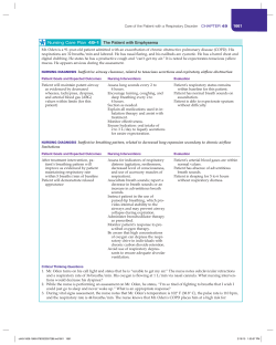
Case reports
© The Intensive Care Society 2012
Severe adenovirus infection: an underrecognised disease with limited treatment
options 3A13
C Houlihan, M Valappil, S Waugh, K Cantlay, DA Price
Severe adenovirus infections can occur in both immunocompromised and immunocompetent patients. New serotypes
are emerging in the UK, and should be considered in patients who present with respiratory tract symptoms and no other
obvious pathogen. The difficulties of diagnosis and treatment of severe adenovirus infection are discussed.
Keywords: Adenoviridae; pneumonia, viral; infection; immunocompromise; lymphopenia
Introduction
Adenovirus (AdV) infections occur worldwide and are
associated with sporadic as well as institutional and
community outbreaks and epidemics. Most infections occur in
children and are mild or asymptomatic. Typically AdV
infections result in self-limiting respiratory, gastrointestinal or
ocular infections,1 however, AdV can cause severe disseminated
disease in immunocompromised patients.2,3 Severe or fatal
infections have been reported in immunocompetent adults4 and
were previously thought to be extremely rare. However,
serotypes with previously limited circulation and geographical
distribution are emerging as significant pathogens,5-7 including
in the UK.8 Several reports now exist of new serotypes,9
variants10 and novel recombinant serotypes. These reports
describe
altered
tissue
specificities
and
clinical
presentations.11- 15 AdV infections may well be easily
overlooked if not clinically suspected.16 We contribute to the
growing literature on these infections by presenting a case of
severe AdV pneumonitis in a patient with no underlying comorbidity, underlining the need to consider and investigate the
possibility of viral pathogens including AdV in patients
admitted to the intensive care unit (ICU) with severe
respiratory infection. Further, we briefly mention two fatal
cases seen in our hospital, where the clinical significance of
AdV detection remains unclear. These cases highlight the
importance of awareness about severe AdV infection and the
challenges of interpretation of AdV tests in the severely ill.
Emergence and spread of recombinant and novel AdV variants
warrants the creation of systems for molecular surveillance of
AdV infections. Finally, in this review we summarise treatment
options for severe AdV infection, highlighting the need for
further research in this area.
history. He presented with a two-week history of fever, myalgia,
dyspnoea with a productive cough and clear sputum. Initial
presentation to his GP led to referral to hospital because of
worsening dyspnoea on exertion. On admission to the
emergency department, he was febrile (39°C), tachycardic
(112 bpm), tachypnoeic (28 breaths/min) and hypoxic (SpO2
90% on room air) but normotensive (119/55 mmHg). His
initial chest radiograph (CXR) is illustrated in Figure 1.
Abnormalities of his initial blood tests included lymphopenia
1.14 × 10⁹/L with a normal total white cell count, a C-reactive
protein (CRP) of 390 mg/L, alanine transaminase (ALT)
1,49U/L and pO2 7.2 kPa on 2 L/min oxygen flow. Urinary
legionella antigen and both serum and urine pneumococcal
antigens were negative, as were HIV antibody, mycoplasma
serology and blood and sputum bacterial cultures. After
48 hours, hypoxaemia persisted and worsened such that he
required non-invasive respiratory support with continuous
Case descriptions
The first patient was a 26-year-old man who had had an
adenoidectomy as a child. He had no other past medical
JICS Volume 13, Number 4, October 2012
Figure 1 CXR with left lower zone infiltrates in patient 1.
337
Case reports
positive airway pressure (CPAP). High dependency care was
required for five days. Broad-spectrum antibiotics (cefuroxime
and clarithromycin) were administered, with the addition of
linezolid on admission to the high dependency unit to cover
resistant staphylococcus and pneumococcus. These were
rationalised after negative serology results and identification of
AdV DNA (serotype 3) from a throat swab. This viral throat
swab was negative for other respiratory viruses including
influenza and parainfluenza viruses. Venous blood was tested
at this time (day 4 of admission), revealing high level of AdV
viraemia (viral load 3.0 × 10⁶ IU/mL). Cidofovir was
considered in light of this result; however at day 4 (day 3
HDU), although the ALT had risen to 584 U/L, the CRP had
dropped to 144 mg/L, the patient’s temperature had normalised
and oxygen dependency decreased. Plasma AdV viral load
was re-tested on day 6 and had dropped to <2 × 10³ IU/mL.
He was discharged 16 days after admission with no respiratory
sequelae.
Two further patients were, firstly, a 60-year-old unemployed
male who was a 20-pack-year smoker with scoliosis and
osteoarthritis but no other medical history. He presented with a
six-day history of dyspnoea and non-productive cough, and on
admission was febrile, tachycardic and hypoxaemic. Admission
blood tests revealed lymphopenia of 1.35 × 10⁹/L, CRP
106 mg/L, with normal liver and renal function. He was
transferred immediately to ICU but had progressive respiratory,
cardiovascular and renal failure in the 24 hours following
admission, requiring escalating ventilatory and inotropic
support and renal replacement therapy. Broncho-alveolar lavage
(BAL) fluid was positive for AdV and serotyping indicated
either type 21 or 50. Cidofovir was commenced on day 3.
Unfortunately, he died from multiple organ failure six days
after admission. The final patient was a 38-year-old HIVpositive male with asthma and Addison’s disease, for which he
used a steroid inhaler and took hydrocortisone. He had a CD4
count of 332 cells/µL (18%) prior to admission and was not on
antiretroviral medication. He presented with a five-day history
of fever, dyspnoea and a non-productive cough, and on
admission was febrile, tachycardic and hypoxaemic. Abnormal
bloods on admission included lymphopenia, 0.41 x 10⁹/L, and
elevated CRP of 334 mg/L. He was admitted directly to ICU
where he was intubated and mechanically ventilated. BAL
specimens were again positive for AdV serotype 3. Cidofovir
was initiated but progressive multi-organ failure ensued and
death occurred three days after admission. Both patients had
normal liver function tests prior to developing elevated
transaminases, both were lymphopenic on admission with
normal total white cell count, and both had negative screening
for alternative pathogens (as with the first patient).
Discussion
Currently, six species of AdV (A-G) with 51 distinct serotypes,
have been identified.17 These serotypes demonstrate organ
tropism; for example, serotypes 8,19 and 37 from species D are
associated with epidemic kerato-conjunctivitis, and serotypes
7,14, and 21 from species B, with acute respiratory disease.2
AdV infection can lead to several other clinical syndromes,
including meningoencephalitis, acute haemorrhagic cystitis,
338
hepatitis, acute febrile pharyngitis and gastroenteritis.
Transmission of the virus is through direct contact with an
infected person, faecal-oral or aerosol transmission, and from
fomites. AdV is usually, but not always, included in the panel
of respiratory viruses tested from throat swabs, nasopharyngeal aspirates and broncheo-alveolar lavage fluid in
hospital laboratories with capabilities to perform polymerasechain reactions (PCR). The development of real-time multiplex
PCR and the investment in laboratory capacity due to
pandemic 2009 influenza A (H1N1) has allowed increased
availability of same-day results. Although there are a variety of
commercial kits available, development of in-house assays is
common in NHS hospitals. In-house validation and
participation in regional and national quality assurance
schemes ensures the standardisation of results, although not
the panel of viruses tested. Recent reports indicate that the
virus genome has altered through recombination11-15 and
epidemics and outbreaks of virulent AdV have been
described.5,6,18 In analysis of AdV positive samples from Ireland
collected between 2009 and 2010, AdV 14 was identified in
nine patients (31%), three of whom died.8 As in the cases
described in this report, severe illness and death from AdV
infection is usually associated with some degree of
immunocompromise and an intact immune system is key to
clearance of the virus.
The first patient reported here had no immunocompromise
at presentation and no factors have been identified during
subsequent clinical follow-up. He was a smoker; however, his
only risk factor for severe AdV infection was being male.19 No
other pathogen was identified to explain his severe
community-acquired pneumonia (CAP). AdV displays the
potential for viral latency/low level persistence; virus has been
detected in the blood of asymptomatic HIV-infected children20
and in tonsillar tissue from children and adults.21,22 The
mechanism through which the virus evades immune clearance
is mediated in part through avoiding recognition by T-cells and
partly by preventing activation of natural killer cells.23
Differentiating latency from active infection can therefore be a
clinical challenge. In the first patient described in this report, a
high level of viraemia with clinical pneumonitis and hepatitis
suggested a disseminated viral illness. Other authors have
highlighted AdV’s unique propensity to cause focal or lobar
changes on chest X-ray.16 Focal changes were seen on X-ray in
the illustrated case, further supporting our conclusion that
AdV was responsible for the patient’s illness. Both this patient,
and the last patient described had extremely high CRPs (390
and 334 mg/L), which has been thought to indicate bacterial
rather than viral pathogens. The association has been
supported in some studies; however, a systematic review of the
diagnostic value of CRP in lower respiratory tract infections in
12 studies concluded that ‘testing for CRP is neither sufficiently
sensitive to rule out nor sufficiently specific to rule in…. bacterial
aetiology of lower respiratory infection.’24 In patients with severe
disseminated viral illness, the use of CRP or even lymphopenia
(which all three patients also had) in differentiating viral and
bacterial illness is not a reliable indicator. This presentation
and clinical findings of the first case highlight the message that
AdV should be tested for in patients with severe respiratory
Volume 13, Number 4, October 2012 JICS
Case reports
illness, with or without competent immune systems.
In the two further cases described, the challenge of
differentiating reactivated latent AdV from disseminated severe
primary AdV infection is clear. Neither patient had peripheral
blood tested for AdV; suspicion was raised by the presence of
AdV in BAL samples. Both patients had an acute severe illness
and in this type of presentation, it is not unusual for there to
be a lack of identification of the responsible pathogen.25
Reactivation of virus in those with pre-existing
immunocompromise, and perhaps an inability to fully clear
less pathogenic viruses, may be an explanation for the
detection of AdV-DNA. Similarly, the presence of viral
remnants after an initial viral infection prior to superadded,
overwhelming unidentified bacterial infection cannot be ruled
out. Legionella and mycoplasma present with similar
symptoms and signs and tests for these lack sensitivity.16 The
challenge in correctly attributing illness to an identified
pathogen should not prevent testing however, especially when
treatments, although limited, are available.
Both ribavirin and cidofovir have been used in
immunocompromised patients with AdV infection; however,
convincing evidence of efficacy in vivo is lacking.26 Ribavirin
is a purine nucleoside analogue with anti-adenoviral activity
in vitro. Although there have been a number of case reports
and case series of its use for AdV infections,27,28 evidence to
date is unconvincing. In the immunocompromised patient, IV
cidofovir, a monophosphate nucleotide analogue, is now
more commonly used for disseminated AdV infection.
Limited evidence, again based on small studies, suggests
cidofovir may be of some value.27,29,30 Despite this, mortality
remains high in this patient group31 and ultimate resolution
of infection often occurs only with the generation of effective
specific T-cell responses.
The use of cidofovir for AdV infection in the
immunocompetent individual has not been studied but it is
doubtful whether it would offer significant advantage in a
patient with an intact immune system. Importantly, side effects
of cidofovir are common and significantly limit use; most
often, these involve renal toxicity. It was for these reasons that
the first patient was not treated, ultimately allowing time for
recovery without antiviral treatment. In the other two patients,
with more severe and ultimately fatal disease, cidofovir
treatment was initiated. Renal failure in these two patients
preceded the initiation of cidofovir, although further
deterioration with its use cannot be ruled out.
Other treatment modalities which have been used include
IV immunoglobulins in combination with antiviral treatments, but there is little evidence to suggest efficacy.32 T-cell
immunotherapy has been successfully used to treat EpsteinBarr virus (EBV) related post-transplant lung disease in
transplant patients. Limited data suggest that this approach
may also be successful for controlling established AdV
disease.33 A new orally active lipid-ester derivative of
cidofovir is currently under investigation, which may prove
less toxic than the IV drug.34 Effective, safe antivirals are
clearly lacking for adenoviral disease. Further research in this
area is urgently needed.
Viral pneumonitis is an important cause of severe respirJICS Volume 13, Number 4, October 2012
atory infection and can easily be overlooked without
appropriate investigation. Viruses account for around 15% of
all patients admitted with pneumonia25 and specific
treatments may be required. It is therefore important that
patients admitted with severe respiratory infections should be
screened for respiratory viruses including AdV by sensitive
techniques as part of their initial investigation. Further,
surveillance for emerging pathogenic AdV sub-types must be
instituted and finally, there is an urgent need for additional
studies to clarify the efficacy of emerging therapies for AdV
infections in various settings.
Conflict of interest
None declared.
References
1. Lenaerts L, De Clercq E, Naesens L. Clinical features and treatment of
adenovirus infections. Rev Med Virol 2008;18:357-74.
2. Echavarria M. Adenoviruses in immunocompromised hosts. Clin
Microbiol Rev 2008;21:704-15.
3. Pham TT, Burchette JL, Jr., Hale LP. Fatal disseminated adenovirus
infections in immunocompromised patients. Am J Clin Pathol
2003;120:575-83.
4. Lewis PF, Schmidt MA, Lu X et al. A community-based outbreak of
severe respiratory illness caused by human adenovirus serotype 14. J
Infect Dis 2009;199:1427-34.
5. Centers for Disease Control and Prevention. Acute respiratory disease
associated with adenovirus serotype 14--four states, 2006-2007. MMWR
Morb Mortal Wkly Rep 2007;56:1181-84.
6. Kajon AE, Lu X, Erdman DD et al. Molecular epidemiology and brief
history of emerging adenovirus 14-associated respiratory disease in the
United States. J Infect Dis 2010;202:93-103.
7. O’Flanagan D, O’Donnell J, Domegan L et al. First reported cases of
human adenovirus serotype 14p1 infection, Ireland, October 2009 to July
2010. Euro Surveill 2011;16:pii=19801.
8. Carr MJ, Kajon AE, Lu X et al. Deaths associated with human
adenovirus-14p1 infections, Europe, 2009-2010. Emerg Infect Dis 2011;17:
1402-08.
9. Jones MS, Harrach B, Ganac RD et al. New adenovirus species found in a
patient presenting with gastroenteritis. J Virol 2007;81:5978-84.
10.Landry ML, Lebeck MG, Capuano AW, McCarthy T, Gray GC.
Adenovirus type 3 outbreak in connecticut associated with a novel
variant. J Med Virol 2009;81:1380-84.
11.Walsh MP, Chintakuntlawar A, Robinson CM et al. Evidence of
molecular evolution driven by recombination events influencing tropism
in a novel human adenovirus that causes epidemic keratoconjunctivitis.
PLoS One 2009;4:e5635.
12.Robinson CM, Rajaiya J, Walsh MP et al. Computational analysis of
human adenovirus type 22 provides evidence for recombination among
species D human adenoviruses in the penton base gene. J Virol
2009;83:8980-85.
13.Kaneko H, Suzutani T, Aoki K et al. Epidemiological and virological
features of epidemic keratoconjunctivitis due to new human adenovirus
type 54 in Japan. Br J Ophthalmol 2010;95:32-36.
14.Rebelo-de-Andrade H, Pereira C, Giria M et al. Outbreak of acute
respiratory infection among infants in Lisbon, Portugal, caused by human
adenovirus serotype 3 and a new 7/3 recombinant strain. J Clin Microbiol
2010;48:1391-96.
15.Yang Z, Zhu Z, Tang L et al. Genomic analyses of recombinant
adenovirus type 11a in China. J Clin Microbiol 2009;47:3082-90.
16.Cunha BA. Severe adenovirus community-acquired pneumonia
mimicking Legionella. Eur J Clin Microbiol Infect Dis 2009;28:313-15.
17.Fields BN, Knipe DM, Howley PM, editors. Fields Virology. 5th ed.
Philadelphia: Lippincott-Raven;2007.
339
Case reports
18.Louie JK, Kajon AE, Holodniy M et al. Severe pneumonia due to
adenovirus serotype 14: a new respiratory threat? Clin Infect Dis
2008;46:421-25.
19.Tate JE, Bunning ML, Lott L et al. Outbreak of severe respiratory disease
associated with emergent human adenovirus serotype 14 at a US air force
training facility in 2007. J Infect Dis 2009;199:1419-26.
20.Ferdman RM, Ross L, Inderlied C, Church JA. Adenovirus viremia in
human immunodeficiency virus-infected children. Pediatr Infect Dis J
1997;16:413-15.
21.Garnett CT, Talekar G, Mahr JA et al. Latent species C adenoviruses in
human tonsil tissues. J Virol 2009;83:2417-28.
22.Neumann R, Genersch E, Eggers HJ. Detection of adenovirus nucleic
acid sequences in human tonsils in the absence of infectious virus. Virus
Res 1987;7:93-97.
23.McSharry BP, Burgert HG, Owen DP et al. Adenovirus E3/19K promotes
evasion of NK cell recognition by intracellular sequestration of the
NKG2D ligands major histocompatibility complex class I chain-related
proteins A and B. J Virol 2008;82:4585-94.
24.van der Meer V, Neven AK, van den Broek PJ, Assendelft WJ. Diagnostic
value of C reactive protein in infections of the lower respiratory tract:
systematic review. BMJ 2005;331:26.
25.Johnstone J, Majumdar SR, Fox JD, Marrie TJ. Viral infection in adults
hospitalized with community-acquired pneumonia: prevalence,
pathogens, and presentation. Chest 2008;134:1141-48.
26.Lenaerts L, Naesens L. Antiviral therapy for adenovirus infections.
Antiviral Res 2006;71:172-80.
27.Bordigoni P, Carret AS, Venard V et al. Treatment of adenovirus
infections in patients undergoing allogeneic hematopoietic stem cell
transplantation. Clin Infect Dis 2001;32:1290-97.
28.Lankester AC, Heemskerk B, Claas EC et al. Effect of ribavirin on the
plasma viral DNA load in patients with disseminating adenovirus
infection. Clin Infect Dis 2004;38:1521-25.
29.Ribaud P, Scieux C, Freymuth F et al. Successful treatment of adenovirus
disease with intravenous in an unrelated stem-cell transplant recipient.
Clin Infect Dis 1999;28:690-91.
30.Legrand F, Berrebi D, Houhou N et al. Early diagnosis of adenovirus
infection and treatment with cidofovir after bone marrow transplantation
in children. Bone Marrow Transplant 2001;27:621-26.
31.Symeonidis N, Jakubowski A, Pierre-Louis S et al. Invasive adenoviral
infections in T-cell-depleted allogeneic hematopoietic stem cell
transplantation: high mortality in the era of cidofovir. Transpl Infect Dis
2007;9:108-13.
32.Emovon OE, Lin A, Howell DN et al. Refractory adenovirus infection
after simultaneous kidney-pancreas transplantation: successful treatment
with intravenous ribavirin and pooled human intravenous
immunoglobulin. Nephrol Dial Transplant 2003;18:2436-38.
33.Feuchtinger T, Matthes-Martin S, Richard C et al. Safe adoptive transfer
of virus-specific T-cell immunity for the treatment of systemic adenovirus
infection after allogeneic stem cell transplantation. Br J Haematol
2006;134:64-76.
34.Paolino K, Sande J, Perez E et al. Eradication of disseminated adenovirus
infection in a pediatric hematopoietic stem cell transplantation recipient
using the novel antiviral agent CMX001. J Clin Virol 2011;50:167-70.
Catherine Houlihan Department of Infectious Disease and
Tropical Medicine, Royal Victoria Hospital, Newcastle upon Tyne
[email protected]
Manoj Valappil Health Protection Agency North East,
Newcastle General Hospital, Newcastle upon Tyne
Sheila Waugh Department of Microbiology, Newcastle upon
Tyne Hospitals NHS Trust
Kaye Cantlay Department of Anaesthetics and Critical Care,
Royal Victoria Hospital, Newcastle upon Tyne
D Ashley Price Department of Infectious Disease and
Tropical Medicine, Royal Victoria Infirmary, University of
Newcastle upon Tyne
Commentary:
A
of each of these infections can occur; they can be difficult to
control and pose a significant infection control challenge.
Less frequent syndromes include meningo-encephalitis,
hepatitis, myocarditis, pancreatitis, genital infections and
haemorrhagic cystitis.2
Most children are infected with at least one respiratory
serotype early on in life. If symptomatic, the usual signs are of
an upper respiratory tract infection and are usually mild and
self-limiting. Lower respiratory infections can be more severe
and may require hospitalisation. Severe pneumonia in children
(those under two years of age are more susceptible) can have a
fatality rate of 16%.3 Serotype 7 (species B) is particularly
associated with serious infection.
AdV was primarily recognised as a respiratory pathogen in
military recruits.4 In the 1950s and ’60s, AdV was one of the
most important causes of illness within the American military
population. Hospitalisation reached 50%5 and fatalities were
reported in previously healthy individuals.6 These numbers
declined with the advent of the oral vaccine, which will be
discussed below.
AdVs are one of the commonest causes of acute
gastroenteritis; long-term sequelae are not seen. Ocular
340
Volume 13, Number 4, October 2012 JICS
denoviruses (AdVs) are large non-enveloped dsDNA
viruses that are found ubiquitously in nature. The
Adenoviridae family is split into four genera: Mastadenovirus,
Aviadenovirus, Atadenovirus and Siadenovirus. Of these, it’s the
Mastadenoviridae that infect humans.1
Infections with AdV are common globally and
predominantly affect children, when they tend to be endemic,
minor and self-limiting. Epidemics and severe disease usually
occurs in neonates and in adulthood; these viruses are
increasingly implicated in high morbidity and mortality in
immunocompromised adults.
Modalities of transmission include direct contact, faecaloral route (particularly in children), aerosols and fomite
contamination.
To date there are six species (A-G) and 53 serotypes
identified. Different species demonstrate organ tropism; for
example species B and C are commonly associated with
respiratory tract infection in children, whereas species F
(especially types 40 and 41, also known as the ‘enteric
adenoviruses’) is linked to gastrointestinal disease.
The major clinical manifestations of AdV infection are
respiratory, gastrointestinal and ocular infections. Outbreaks
Case reports
infections such as pharnygo-conjunctival fever (PCF) and
kerato-conjunctivitis (KC) are seen, again mostly in children,
and outbreaks can occur.7
In most cases, severe AdV disease is seen in
immunocompromised patients. Here, it tends to run a more
prolonged course and can result in a fatal outcome. Mortality
rates approach up to 60% in suppressed patients with
pneumonia, compared with 15% in immunocompetent
people.8 Clinical syndromes seen in this cohort of patients
include pneumonia, hepatitis, colitis, pancreatitis and
disseminated disease. The manifestation depends on the
patient’s age, the serotype of virus and the underlying disease,
with bone marrow transplant and haematopoietic stem-cell
transplantation recipients most at risk of AdV infection.
Recent advancements in diagnostics have greatly aided AdV
recognition. Multiplex polymerase chain reaction (PCR) is
highly sensitive, specific and rapid, with real-time PCR
allowing quantification of the virus. This is of great value in
terms of treatment and follow-up of infected patients.9
With regards to antiviral treatment, there is no specific
therapeutic agent for AdV. Of all the current options, cidofovir
has the most clinical evidence-base, primarily for
immunocompromised patients.10 Ribavirin and ganciclovir
have also been mooted as possible agents.11
The cornerstone of treatment is reducing the level of
immunosuppression. If this is not possible, antiviral
medication should be considered. Other non-antiviral
therapeutic options (less widely used) include donor
lymphocyte infusion12 and intravenous immunoglobulin.
An oral vaccine was developed in the 1960s to prevent acute
respiratory disease in military personnel. The vaccine was
active against serotypes 4 and 7, and was highly successful in
preventing illness. However, manufacturing issues led to the
cessation of the vaccine and a rise in respiratory illness has
occurred in this cohort.13 The vaccine was never considered in
the paediatric populations or for civilians.
AdVs have been studied as vectors for gene therapy to
combat genetic disorders such as cystic fibrosis and myotonic
dystrophy. AdV has also been used as an anti-oncogenetic
agent (with p53 insertion) and as a variety of recombinant
vaccine, with studies investigating possible efficacy against
infectious diseases like HIV and rabies. Although initial
generations of vectors have been unsuccessful, third-generation
vectors are now being investigated; this is a highly active and
exciting area of research.14
JICS Volume 13, Number 4, October 2012
References
1. Benko M, Harrach B, Both GW. Family Adenoviridae. In: Fauquet CM,
Mayo MA, Maniloff J et al, eds. Virus Taxonomy. VIIIth report of the
International Committee on Taxonomy of Viruses. New York: Elsevier;
2005:213-228.
2. Munoz FM, Piedra PA, Demmler GJ. Disseminated adenovirus disease in
immunocompromised and immunocompetent children. Clin Infect Dis
1998;27:1194-200.
3. Murtagh P, Cerqueiro C, Halac A et al. Adenovirus type 7h respiratory
infections: a report of 29 cases of lower respiratory disease. Acta Paediatr
1993;82:557-61.
4. Hilleman MR, Werner J. Recovery of new agent from patients with acute
respiratory illness. Proc Soc Expl Biol Med 1954;85:183-88.
5. Top FH. Control of adenovirus acute respiratory disease in US Army
trainees. Yale J Biol Med 1975;48:185-95.
6. Dudding BA, Wagner SC, Zeller JA et al. Fatal pneumonia associated
with adenovirus type 7 in three military trainees. N Engl J Med
1972;286:1289-92.
7. Turner M, Istre GR, Beauchamp H et al. Community outbreak of
adenovirus type 7a infections associated with a swimming pool. South
Med J 1987;80:712-15.
8. Hierholzer JC. Adenoviruses in the immunocompromised host. Clin
Microbiol Rev 1992;5:262-74.
9. Lankester AC, van Tol M, Claas E et al. Quantification of adenovirus
DNA in plasma for management of infection in stem cell graft recipients.
Clin Infect Dis 2002;34:864-67.
10.Bordigoni P, Carret AS, Venard V et al. Treatment of adenovirus
infections in patients undergoing allogenic haemopoietic stem cell
transplantation. Clin Infect Dis 2001;32:1290-97.
11.Arav-Boger R, Echavarria M, Forman M et al. Clearance of adenoviral
hepatitis with ribavirin therapy in a pediatric liver transplant recipient.
Pediatr Infect Dis J 2000;19:1097-100.
12.Chakrabarti S, Collingham KE, Fegan CD et al. Adenovirus infections
following haemopoietic cell transplantation: is there a role for adoptive
immunotherapy? Bone Marrow Transpl 2000;26:305-07.
13.Kolavic-Gray SA, Binn LN, Sanchez JL et al. Large epidemic of
adenovirus type 4 infection among military trainees; epidemiological,
clinical and laboratory studies. Clin Infect Dis 2002;35:808-18.
14.Russell WC. Update on adenovirus and its vectors. J Gen Virol
2000;81:2573-04.
Rishi H-P Dhillon Specialist Registrar in Microbiology,
Department of Infection and Immunity, Charing Cross Hospital,
Imperial College Healthcare NHS Trust, London
[email protected]
341


© Copyright 2026