
Document 145294
CHAPTER I O
Approaches To Midfoot Degenerative Joint Disease
Stepben J.
Miller, DPM.
INTRODUCTION
The joints of the midfoot are susceptible to degenerative changes as a resuit of a variety of forces,
including biomechanical faults, frank trauma, and
Charcot neuropathy. Such degeneration may be
accelerated by concurrent osteoarthritis, or compen-
sation for
a
functional foot deformity such as
equinus. Diagnosis can be elusive until radiographic
changes ate evident. Conservative treatment is
effective only while symptoms are tolerable, while
surgical intervention becomes the treatment of
choice in order to achieve pain free function.
ANATOMICAL CONSIDERATIONS
The joints of the midfoot include LisFranc's (tarsometatarsal) joint, Chopart's (midtarsal) joint, and
the joints in between. Because of its unique function (which is more in tandem with the subtalar
joint), the midtarsal joint will not be addressed in
this paper.
The tarsometatarsal loints consist of the first,
second, and third metatarsocuneiform joints, as
well as the fourth and fifth metatarsocuboid joints.
This is an intricate nefwork of articulations which
make up the transverse arch of the foot, where the
keystone is the second metatarsal and cuneiform.
The transverse arch is connected by dorsal , plantar,
and interosseus ligaments which bond all
metatarsal-cuneiform/cuboid articulations except
for the first and second metatarsal bases. Although
the bases of these two metatarsals have no ligamentous attachment between them, the second
metatarsal is stabilized by an extremely strong
ligament from the medial cuneiform bone
(LisFranc's ligament). The intertarsal joints consist
of the first, second, and third naviculocuneiform
articulations, as well as the adjacent interfaces
between the first and second cuneiform bones, the
second and third cuneiform bones, and the third
cuneiform-cuboid bones.
Motion is limited to each rzy as well as adjacent afticulations. The shapes of the joints allow
only limited motion, making them pafiicularly susceptible to jamming injuries, which are exaggerated
in the Charcot foot. The midfoot is the interface
between the rearfoot function (subtalar and midtarsal joints) and forefoot function (metatarsais and
digits). In addition, there is a limited amount of
inteftarsal motion.
PATTIOPHYSIOLOGY
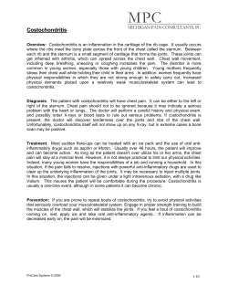
Midfoot degenerative joint disease is the product of
trauma, biomechanical abnormalities, or neuropathy
(Figs. 1A, 1B). Trauma can include blunt or crush
injuries, contusions, or high veloci$z impact. Tarsometatarsal joint dislocations or fracitre/dislocations
can be devastating injuries which rapidly progress to
degenerative joint disease. Injury to this group of
afiiculations can also involve other midfoot bones
and joints, as well as the base of the metatarsals (particularly the second). \fith luauma, there is direct
afiicular damage which causes significant joint
degeneration over time. Underlfng biomechanical
deformrties can fufiher accelerate afihrosis.
Figure 1A. A 67 year-old female with non-traumatic degeneration of
the tarsometatarsal joint.
42
CHAPTER 10
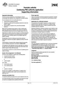
Figure 2A. Lesser tarsus joint destruction aggravated by an equinus
Figure 18. Note the point of sagittal plane collapse on the latency
deformity. Note the point of compensation at the talonavicular 1oint.
racliograph.
The most common biomechanical deformity
that can cause midfoot jamming is pronation.
Once the subtalar joint pronates and unlocks the
midtarsal joint, the distal joints, including those of
the midfoot, tend to lose their inherent functional
stability as a rigid lever. \7ith a loss of stability, the
joints are in malalignment and reach an early functional end range of motion. This causes significant
jamming as the weight of the body continues to
propulse across them. Such forces result in significant degenerative changes where the impact of the
joint is greatest, which is usually dorsally.
In pes ca\.Lrs, there is a significant supination
deformity which again causes midfoot jammlng,
but by a slightly different mechanism. The joints
are inherently stable, but have no available end
range of motion. This results in significant cafiilage
degeneration, pafiicularly at the dorsal margins of
the joints.
Compensation deformities can include forefoot or rearfoot varus, forefoot valgus, and equinus
(Figs. 2A, 2B). Fusion, by way of triple, pantalar, or
even single hindfoot joint arthrodesis, causes
increased stress on the surrounding joints as they
attempt to compensate for the lost motion. This too
can result in joint jamming and destruction.
Perhaps the most dramatic manifestation of
midfoot stresses is in the neuropathic foot. Loss of
shock absorption mechanisms results in rapid joint
destruction, and fractures accelerated by biomechanical deformities such as equinus. Conditions
which predispose patients to degenerative joint
disease of the midfoot include overuse, obesity,
osteoporosis, rheumatologic disorders, biomechanical deformities, and neurologic disorders.
Figure 28. The point of compensation is at the naviculocuneiform
loint.
DIAGNOSIS
Stiffness, midfoot fatigue, and difficulty wearing
shoes, especially if they are tight over the midfoot
area, ate all early symptoms of developing degenerative joint disease. The pain is usually of a dull
aching nature, and is located more dorsally than
plantarly. Pain is due to subchondral osseous damage after there is loss of articular cartilage. There
may also be locaiized synovitis. Pain becomes
more severe during weight bearing, resulting in difficulty with walking. It may present as poststatic
dyskinesia, and the patient may repofi a significant
increase in symptoms when there are temperature
or weather changes.
Usually there is very little swelling, but often
warmth is present at the affected afiiculations. The
patient may often present with an antalgic gait.
Palpation reveals tenderness along the joint lines
dorsally where osteophyes may be evident. There
are seldom significant clicks or crepitus, since there
is insufficient motion in these joints to produce
such signs. Careful and deep palpation of the midfoot from the plantar approach will also often
CHAPTER 10
43
reveal tenderness. One should be able to anatomi-
cally identify the exact ioint(s) affected before
consulting radiographs.
In terms of diagnostic tests, blood studies are
not usually helpful, with the possible exception of
the ESR which is a nonspecific marker for inflammation. Radiographs show a loss of joint space,
often with complete joint obliteration. If the x-ray is
not pointed at the coffect angle, visualization of the
joint may be obscured. Repeat films with adjusted
central ray angles may be necessary. Radiographic
findings include narrowing of the joints, subchondral sclerosis, erosions, and periarlicular osteophyes
(Figs. 3,{, 3B). Subluxation and dislocation may be
evident in the most severe cases, especially in the
Charcot foot. A CT scan is indicated only if joint
damage is extensive, and if the information gained
would influence the treatment.
Flgure 38. Note hor. the change in the angle of
the central radiograph beam "opens" various
midfoot joints.
ing is necessary in order to effect sufficient rest for
relief of symptoms.
Intra-articular corticosteroid injections can
provide a variable duration of relief, but are genera1ly effective in relieving symptoms. A soluble
steroid should be utiiized for the initial test injection. If this is successful, the longer acting
crystalline steroid salts should be utilized. There is
relatively low risk of further joint damage since the
hyaiine cartilage has already been destroyed by the
degenerative process.
SURGICAL TREATMENT
in spite of conservative
care is the primary indication for surgical interwention. However, the patient should not be immediately
Persistent pain and disability
Figure 3A. An example of
changes
C
clegenerative
in the midfoot.
ONSERVAITVE TREATMENT
Early intervention is most beneficial if initiated
prior to radiographic evidence of degenerative
joint disease. Treatment modalities include compression, heat, NSAIDs, and physical therapy. Heat
delivered via ultrasound or a paraffin wax bath is
the most effective treatment. Orthotic support is
also essential. Occasionally, a short period of cast-
scheduled for surgery merely due to marked joint
destruction on the initial radiograph. The nature of
the pathology, prognosis with and without treatment,
and the alternatives for non-operative and operative
care should be clearly delieated to the patient before
surgical intelention is attempted.
Midfoot degenerative joint disease frequently
occurs in the geriatric population. Because it is
often impossible to keep this patient non-weight
bearing during recovery from arthrodesis, alternative
procedures may be considered. One such
procedure is the osteoarthrotomy. This involves
44
CHAPTER 10
resecting one side of a joint (usually the more
mobile side), such as the bases of the metatarsals
at the tarsometatarsal junctions. Complete medial
to lateral resection of the midfoot should not be
attempted, however, one or two joints may be
addressed at a lime. The patient must be made
awate of the fact that a bone graft afihrodesis may
be necessary in the future, if the procedure fails to
produce painfree ambulation.
Silastic implant material can serve as an interface at the osteoafihrotomy site. High density
silicone polymer can be fashioned from a solid
block, and shaped to fit in the appropriate space.
Again, the patient must be prepared for possible failure and the need for further afihrodesis at the site.
There are no long-term studies relative to the effectiveness of silastic fi:raterial in these pafiicular
locations. Postoperative management includes the
use of a closed suction drain and a surgical shoe.
'Weight
bearing is allowed immediately as necessary.
A common and effective procedure for relief of
midfoot degenerative joint disease is arlhrodesis of
either a single joint or a combination of joints,
depending on the degree of pathology. Whether the
bones need to be fused, at their side-to-side interfaces depends again on the pathology presented.
Usually the bones within eachray can be fused, and
it is not imperative to fuse the bones in between the
rays, unless there is severe joint destruction present
(Figs. 44, 4B). The more mobile the joint, the greater
the chance for non-union. Therefore, mobile afiiculations such as the first metatarsocuneiform joint
require greater stabilization.
Figure 4A. Preoperative radiograph of a
54
year-old female with painful degeneration of the
lesser tarsal joints.
Incision Planning
Incision placement requires careful planning due
to the many anatomical structures thatlay between
the scalpel and the affected joints. Each layer
houses neryes and vessels, including the superficial
peroneal nerve branches and the dorsalis
pedis/deep peroneal neurovascular bundle, which
lies on the periosteum of the midfoot. One should
use a skin scribe to carefully draw the target articulations and the vital structures in the area before
making an incision. The incision should be oriented to provide the best exposure with the least
amount of scarring.
Figure 48. Complete relief of painful symptoms
in medial lesser tarsal joints, resolved by fusion.
Postoperative radiograph following arthrodesis
of the first and second tarsometatarsal ,oints.
The patient had complete resolution of pain fo1lowing successful fusion.
CHAPTER 10
45
Joint Resection
Bone Grafting
Bone grafting material, preferably cancellous bone,
should be available for every midfoot arthrodesis
procedure. The order of preference for bone grafting
is autogenous, aliogeneic, or a bone substitute such
as that derived from coral. Bone graft material can be
firmly wedged into the afihrodesis site to add some
measure of compression to insure consolidation.
Fixation
Internal interfragmentary compression fixation is
the ideal goal for immobilization of the afthrodesis
sites. This may be augmented by neutralization
Postoperative Management
'Ihe patient should be placed in a compression cast
until the surgical swelling subsides, usually
between three and five days. A non-weight bearing
below-knee cast is then applied for six to eight
weeks, followed by a walking cast for an additional
two weeks. Rehabilitation to pain-free walking in
shoes is then instituted.
Articular osteophytes will be encountered first, and
these must be removed along with any loose ossicles. The damaged articdar surfaces must then be
resected to expose the underlying cancellous bone.
Overaggressive resection of bone should be
avoided so as to not make consolidation dfficult or
impossible.
plates, such as the one-third tubular plate. A T-plate
is well suited for use along the medial surface of the
first metatarsocuneiform joint, and can be extended
proximally to include the navicular. A smali
dynamic compression plate can be placed over the
tarsal afticulations dorsaily to provide interfragmentary compression and stabrlization. Howeveq there
is a risk of irritation from this plate. Other methods
of fixation include cross k-wires, staples, and
absorbable pins. Staples should be pre-drilled to
prevent fracture into the afihrodesis site.
Special considerations are given to the more
mobile versus the more stable midfoot joints.
Fufihermore, consideration for motion must be
made between the stabilized ray and the adjacent
mobile ray. This is important when deciding to cross
non-damaged joints with internal fixation devices.
Due to the anatomical complexities of the
midfoot, the approach to fixation is variable in
every clinical situation, depending on whether one
or more joints are involved, if transverse fusion is
required, and the age, bone quality, and activiry
level of the patient. Finally, one must address any
concurrent structural or functional deformities.
Controversy exists regarding the crossing of
unaffected joints in the area with screws in order
to enhance fixation and stabilization of the
arthrodesis sites. There is some concern for joint
damage, even if the screws are to remain in place
only temporarily. There is also the possibility that
shear forces will lead to fracture of the screws.
Although some authors have applied these with
impunity, there is insufficient evidence to suggest
that these joints will not break down over time,
once they are damaged by the screws.
SUMMARY
Successful treatment of midfoot degenerative joint
disease can be very gratifying to both the patient
and the surgeon, since it usually resolves a painful
and incapacitating problem. Although normal intertarsal and tarsometatarsal motion is relatively
limited, stabilization can be quite challenging,
especially the application of internal fixation in
order to achieve stable osseous consolidation.
Attention to the principles which have been presented will heip achieve these goals.
BIBLIOGRAPTry
Banks AS, McGlamry ED: Charcot foot. J Am Podicttr lted Assoc 79t2L3235. 7989.
Goosens M, Destoop N: LisFranc's fracture dislocations: etiology, radiology and results of treatment. Clin Orthop 176t 1.54-762, 7983.
Harris JR, Brand P\il: Patterns of disintegration of the tarslls in the
anacsrherit loor. J Bone.loint SurB 18B: I 16. 1966.
Horton GA, Olney B$7: Deformity correction and amhrodesis of the
midfoot with a medial plate. Foot Ankle 14:193-499, 7993.
JohnsonJE, Johnson IiA.: Dowel arthrodesis for degenerative afthritis of
the Tarsometatarsal (LisFranc) joints. Foot Ankle 5:213-253, 1986.
LoGerfo FW, Coffman JD: Vascular and microvascular disease of the
foot in diabetes. N Eng J Med 3111675-18, 1984.
Myerson MS, Fisher RT, Burgess AR, Kenzora JE: Fracture dislocations
of the tarsometatarsal joints: end results correlated with pathology
and treatment. Foot Ankle 6:225-242, 1986.
Sanjeorzan BJ. Veith RG, Hansen ST: Salvage of LisFranc's tarsometararsal joint by arthrodesis. Foot Ankle 1,0:1,93-200, 7990.
Sanjeorzan, BJ, Hansen, ST: Early and late posttraumatic foot reconstruction. Clin Orthop 243:86-91, 1989.


© Copyright 2026