
Foot Injuries Arches
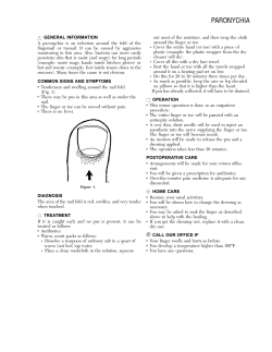
Arches
Foot Injuries
• Three arches in the foot:
1) Lateral longitudinal arch
2) Medial longitudinal arch
3) Transverse arch
• These arches are maintained and supported
by the wedging of the interlocking tarsal and
metatarsal bones, tightening of the
ligaments of the plantar aspect of the foot,
and the extrinsic muscles of the foot and
their tendons.1
1. Magee. Orthopedic Physical Assessment. 2002.
Medial Longitudinal Arch
Lateral Longitudinal Arch
• Bones: Calcaneal tuberosity; Talus;
Navicular; three Cuneiforms; first, second,
and third Metatarsals1
• Supported by the tibialis anterior, tibialis
posterior, flexor digitorum longus, flexor
hallucis longus, abductor hallucis, and
flexor digitorum brevis muscles, along with
the plantar fascia and the plantar
clacaneonavicular ligament.1
• Bones: Calcaneus; Cuboid; fourth and
fifth Metatarsals
• Supported by the peroneus longus,
peroneus brevis, peroneus tertius,
abductor digit minimi, and flexor
digitorum muscles, along with the
plantar fascia and the long and short
plantar ligaments.1
1. Magee. Orthopedic Physical Assessment. 2002.
1. Magee. Orthopedic Physical Assessment. 2002.
Transverse Arch
• Bones: Navicular, Cuneiforms,
Cuboid, Metatarsal bones1
• Supported by the tibialis posterior,
tibilais anterior, and peroneus
longus muscles, along with the
plantar fascia.1
1. Magee. Orthopedic Physical Assessment. 2002.
Arch Strains
• An arch strain could be caused from
an excessive jerk, twist, bend,
plantarflexion, or dorsiflexion to any of
the muscles or ligaments supporting
the arches.1
• Treatment: Rest. Ice. Arch support
system/brace or arch tape for extra
support. Time off of sport related
activity depends on the severity of the
strain.1
1. Magee. Orthopedic Physical Assessment. 2002.
1
Plantar Fascia
• Thick white band of fibrous tissue
originating from the medial
tuberosity of the calcaneus and
ending at the proximal heads of the
metatarsals.2
2. Prentice. Arnheim’s Principles of Athletic Training: A
Competency-Based Approach. 2003.
Plantar Fasciitis
• Inflammation of the Plantar Fascia. Usually
caused by overuse and irritation.1
• Normal movements and ROM with pain
(localized or radiating) in the anterior
medial heel. Sometimesparesthesia in the
sole of the foot will appear. Pain worsens
after long periods of standing, walking, or
running, or if foot is forced into
dorsiflexion.1
1. Magee. Orthopedic Physical Assessment. 2002.
Plantar Faciitis
• Treatment: the best treatment is
orthotic therapy. A night splint may
also be used to help maintain a static
stretch. Athlete should maintain a
stretching routine to add additional
stretching. Anti-inflammatory
medications are recommended.2
2. Prentice. Arnheim’s Principles of Athletic Training: A
Competency-Based Approach. 2003.
2
Heel Bruise (Contusion)
Heel Bruise (Contusion)
• Thick fat pad covering calcaneus
bruises from sport activities that
demand a lot of stop-and-go or sudden
change from horizontal to vertical (e.g.
basketball, volleyball, track and field,
etc…)2
• Symptoms: unable to apply pressure to,
or weight-bear on, the heel. Sometimes
you may notice redness or the heel may
feel warm.2
• Treatment: athlete should not bear
weight for 24 hours. RICE should be
taken into affect. Anti-inflammatory
medication are administered to help
bring down the swelling. A heel cup or
protective doughnut should be applied.2
• 2. Prentice. Arnheim’s Principles of Athletic Training: A
Competency-Based Approach. 2003.
2. Prentice. Arnheim’s Principles of Athletic Training: A CompetencyBased Approach. 2003.
Sprained Digit
Sprained Digit
• Pathology
• Treatment
– Tearing of the ligaments in the toes
– RICE
– casting/splinting
– Buddy taping
– Weight bearing and activity as tolerated1
• Mechanism
– Excessive flexion, extension, or twisting of
the toes
– Kicking an unyielding surface1
1 Prentice, William E. Arnheim’s Principles of Athletic Training, 2003
1 Prentice, William E. Arnheim’s Principles of Athletic Training, 2003
Exostosis
Exostosis
• Pathology
– a.k.a bone spur
– An abnormal bony
outgrowth extending from
the surface of the bone
– An increase in the bone
mass at the site of an
irritative lesion in response
to overuse, trauma, or
excessive pressure1
1 Magee, David J. Orthopedic Physical Assessment. 2002
2 www.podiatryinfo.com/ vphtml/heel.html
• Mechanism
2
– Repeated strain placed on a bone or the bony
insertion of a tendon1
• Treatment
– RICE
– Donut padding
– Surgical intervention2
1 Magee, David J. Orthopedic Physical Assessment. 2002
2 Prentice, William E. Arnheim’s Principles of Athletic Training, 2003
3
Tarsal Tunnel Syndrome
Tarsal Tunnel Syndrome
2
• Pathology
– Entrapment of the
posterior tibial nerve
or one of its medial
or lateral branches
as it passes through
the tarsal tunnel1
• Mechanism
– Compression
– Fracture, dislocation, or inflammation of the
tarsals or local ganglion
• Treatment
– Orthotic
– Surgical intervention1
1 Starkey, Chad. Ryan, Jeff. Evaluation of Orthopedic and Athletic Injuries, 2002
2 www.footcaredirect.com/ tarsal_tunnel.html
1 Starkey, Chad. Ryan, Jeff. Evaluation of Orthopedic and Athletic Injuries, 2002
Ingrown Toe Nail
Bunions
• Pathology:
• Pathology:
– Inflammation of the bursa over
the 1st metatarsophalangeal
joint causes the hallux to
become deformed
– The toe nail grows into
skin, cutting the flesh.
• MOI:
• MOI:
– Trimming nail too short
– Small shoes
http://www.cgh.com.sg/health_public/pamphl
et/general/nail/nail_content2.html
• Treatment:
– Narrow shoes
– Pronated foot
– Depressed or flattened trans
arch
http://www.americasfootdoctor.com/yourfeet_bunions.shtml
• Treatment:
– Remove excess nail
– Place sterile object,
i.e. cotton to push nail
away from skin1
–
–
–
–
Wear wide box shoes
Orthotics
Pad or splint w/ wedge
Surgery1
www.footandheel.com/topics/bunions.htm
1 http://www.cgh.com.sg/health_public/pamphlet/general/nail/nail
_content2.html
http://www.podiatryclinician.com/prod01.htm
1 Prentice. Arnheim’s Principles of Athletic Training.
Blisters
Callous and Corns
• Pathology:
• Pathology:
– Fluid filled sacs covered with a thin
layer of almost transparent skin
– Thickening of skin as a
result of friction
• MOI:
• Callous = bottom of foot
• Corns = Top of foot
– Improper footwear
– Foot structure or gait patterns
– Tape
• MOI:
– Rubbing against shoes
http://images.search.yahoo.com
• Treatment:
• Treatment
– Clean blister and try not to pop it
– If popped…keep area clean
– Wear different shoes
– Cover with moleskin e.g.
– Pads1
• Cover blisters and apply disinfectants
http://www.family-foot.com/foot_ailments/corns_calluses.
1 http://www.family-foot.com/foot_ailments/corns_calluses.
1. “Caring for your Feet.” Our Health Network. Retrieved August 31, 2005, from
http://www.ourfootdoctor.com/yourfeet_injuries_feet_b.shtml
4
Neuroma
Plantar Flexed First Ray
• Pathology:
– Swelling of a nerve
– The most common nerve affected
connects the 3rd and 4th toes
• Pathology:
– Structural deformity occurs when
the big toe lies lower then the
other four metatarsal bones
• MOI:
– Over Pronation
– The pulling of the ligaments under
the foot irritates the nerve
– High heels
• MOI:
http://images.search.yahoo.com
– Poor biomechanics
• Pes cavus
– Genetics
• Treatment:
• Treatment:
– Orthotics
– Dorsal Osteotomy
– Orthotics
– Inflammatory injections
– In severe cases, surgery may be necessary
• Surgical repositioning of the
bone
2. “Neuroma.” Dr. Foot. Retreived August 31, 2005, from http://www.drfoot.co.uk/neuroma.htm.
3. Magee (2002). Orthopedic Physical Assessment. Saunders, Inc. Pg. 787.
Mallet Toe
•
•
•
Claw Toe
Pathology: caused by a flexion contracture at the DIP joint involving the flexor
digitorum longus tendon.
–
Mallet toe usually becomes a fixed deformity, causing the
callus dorsally over the DIP joint or on the tip of the toe.
development of a
Mechanism of injury: wearing shoes that are too small over ayalenewhavenhealth.org/
long period of time.
library/healthguide/en...
Treatment:
–
Conservative treatment
–
Surgery
•
•
•
•
•
http://images.search.yahoo.com
Focusing on relieving pressure by wearing loose footwear
Padding can help with irritation
Shave the callus
Necessary when deformity becomes fixed
Involves straightening the toe using a K-wire, inserted through phalanges into metatarsals
• Pathology: caused by flexion contracture at the
DIP joint but also involves hyperextension at MP
joint.
http://yalenewhavenhealth.org/library/healthguide/en-us/support/topic.asp?hwid=hw143095
– Callus develops over PIP joint and under the
metatarsal head. ¹
• Mechanism of injury: wearing shoes that are too
small over a long period of time.
– Can be associated with Pes Cavus
• Treatment:
– Same as Mallet Toe
www.federatie-pas.nl/ kompas/hfdst10-1.html
Arnheim Principles of Athletic Training, Prentice, 2003
Arnheim Principles of Athletic Training, Prentice, 2003
¹ Physical Examination of Spine and Extremities, Hoppenfeld, 1976
Turf Toe
Sprained Hallux (Great Toe)
www.athleticadvisor.com/.../ turf-toe_images.htm
• Pathology: stretching or tearing of the ligaments or joint
capsule in the first phalange
• Mechanism of injury: force applied to extend joint beyond
normal range, jamming it or twisting it
– Example: kicking a stable object
• Treatment:
– RICE- reduce swelling
– Buddy tape to adjacent toes to immobilize
• Toes are too small to splint or cast!!
• Most common example is turf toe.
Arnheim Principles of Athletic Training, Prentice, 2003
• Pathology: hyperextension of the MP joint of the great
toe resulting in a sprain.
• Mechanism of injury: occur on synthetic turf because
shoes designed for this field are more flexible and allow
dorsiflexion of first toe.
• Treatment:
–
–
–
–
Ice
Ultrasound
Rest, until able to play without pain
Shoe companies have added steel to forefoot areas to decrease
this injury or you can add Orthoplast under the shoe insole
www.amazon.com/gp/browse. html?_encoding=UTF8&...
Arnheim Principles of Athletic Training, Prentice, 2003
5
EQUINUS DEFORMITY
• Pathology – occurs when
there is less than 100 of
dorsiflexion in the ankle1
• MOI – results from
contracture of the Achilles
tendon or gastrocnemius
or soleus muscles. Can
also result from structural
bone deformity1
1. Magee, David. Orthopedic Physical Assessment 4th ed. Pg. 781
http://www.worldortho.com/inventions/cripple/c111.html (picture)
• Treatments – heel
lifts, foot orthodics,
shoe modification,
physical therapy or
surgery2
• All treatments are
based on severity of
deformity.
2. Prescription Custom Foot Orthoses Practice Guidelines, The
American College of Foot and Ankle Orthopedics and Medicine.
December 2004, pg 17
http://www.footmaxx.com/clinicians/five.html#equinus (picture)
Pes Cavus
• Pathology – the
longitudinal arches of the
foot are accentuated all
the way to the toes3
• Treatments – orthodics, lateral wedge,
stretching of the Achilles tendon and
plantar fascia3
– Can cause claw foot3
– Results from excessive
supination3
• MOI – cause of deformity
is caused by abnormal
orthopedic or
neurological condition3
3. Prentice, William, Principles of Athletic Training 11th ed. Pg 510
3. Prentice, William, Principles of Athletic Training 11th ed. 2003
www.ordesignslv.com/ cmt.html (picutre)
http://www.global-technologies.net/ShopSite/Extremities.html (picture)
Pes Planus
• Pathology – the
medial longitudinal
arch is flat3
– Excessive pronation of
the feet3
– “flat feet”
• Treatments – if there is no pain with the
athlete, then nothing should be done to
correct the problem3
– If athlete experiences pain, a properly
constructed orthodics should be used3
– A medial wedge should be used to correct
excessive pronation3
• MOI – overweight3
– Wearing of tight shoes
– Trauma that weakens
supportive structures3
3. Prentice, William, Principles of Athletic Training 11th ed. 2003
http://www.global-technologies.net/ShopSite/Extremities.html (picture)
3. Prentice, William, Principles of Athletic Training 11th ed. 2003
www.supportyourfeet.com/ over_pronation.htm (picture)
6
REARFOOT VARUS
• Pathology – inverted calcaneus relative to
the long axis of the tibia4
• Treatments – depending on severity of the
problem, orthodics would be used to treat
the deformity4
– Subtalar joint is in neutral poistion
• MOI - heel spur syndrome, Tailor’s
bunion, leg fatuige4
4. http://www.footmaxx.com/clinicians/five.html
REARFOOT VALGUS
4. http://www.footmaxx.com/clinicians/five.html
• Treatments – orthodics with a control in
the rear foot5
• Pathology – the calcaneus is inverted relative to
the long axis5
– Rarely observed
– Valgus tibial alignment
• MOI – the rear foot becomes hyper mobile5
– Increased pronation
– Hammer toes
– Lateral ankle pain
5. Starkey, Chad. Evaluation of Orthopedic and Athletic Injuries 2nd ed.
2002
Plantar Fasciitis
• Pathology1
Plantar Fasciitis
• Mechanism of Injury1
– Inflammation of the
plantar fascia
– Inflammation of the
interface between the
fascia and the first
layer of intrinsic
muscles
– Acute
• Forced dorsiflexion of
ankle with toe extension
– Insidious
http://tricitynewbalance.com/images/health/plantar_fascia.jpg
1: Starky, Evaluation of Orthopedic and Athletic
Injuries, 2002
• Additional distance
running
• increased activity levels
• Changing surfaces
• New or different shoes
• Treatment2
– Soft orthotics w/ deep
heel cup built in
– Heel cord stretching
– Plantar fascia
stretching
– NSAIDs
1: Starky, Evaluation of Orthopedic and Athletic Injuries, 2002, 2nd
edition
2: Arnheim, Athletic Training: A Competency-Based Approach, 2003,
11th edition
7
Stress Fracture (Metatarsals)
• Pathology
• Treatment2
– Fracture of
metatarsal(s)
• Mechanism of
Stress Fracture (MT)
Injury1
– Insidious/complex
• Fatigued/ weak toe
extensors
• Diabetes mellitus
• Hypermobility increases
stress on forefoot
• Hypomobility increases
stress on midfoot
– Use a bone scan to detect the fracture
– 3-4 days limited weight bearing
– 2 weeks rest
– Return to running should be very gradual
2: Arnheim, Athletic Training: A Competency-Based Approach, 2003, 11th edition
http://www.footpain.org/Resources/stressfx.gif
1: Starky, Evaluation of Orthopedic and Athletic
Injuries, 2002, 2nd edition
Rear Foot Valgus/ Varus
• Pathology1
• Mechanism of Injury1
– Valgus
– Valgus
• Pronation of subtalar
joint (rarely observed)
• Eversion of the
calcaneus
– Varus
– Varus
• Supination of subtalar
joint
1: Starky, Evaluation of Orthopedic and Athletic
Injuries, 2002
Rear Foot Valgus/ Varus
• Inversion of calcaneus
• Calcaneus does not
completely derotate
during fetal
development
http://www.footdoc-il.com/images/ss1.jpg
Sinus Tarsi Syndrome
Caused by hemorrhage or
inflammation (synovitis) in the sinus
tarsi
Frequently misdiagnosed as chronic
ankle sprain and, if improperly treated,
will result in chronic pain and disability.
Symptoms include pain and instability
when walking on uneven surfaces, and
pain when the affected foot is at rest.
• Treatment1
– Valgus
• Orthotics that wedge
below medial aspect of
calcaneus
– Varus
• Orthotics that wedge
below the lateral aspect
of calcaneus
1: Starky, Evaluation of Orthopedic and Athletic Injuries, 2002
INGROWN TOENAIL
• caused by a toenail that grows or is
pressed into the skin. Most often with the
great toe.
• Treatments include trimming the nail or
surgery or simply wider shoes
Mosby’s medical dictionary, 2002
Oloff , LM, Subtalar joint arthroscopy for sinus tarsi syndrome, 2001
8
Bunion
• An enlargement of the joint at the base of the
great toe (mtp joint) due to hallux valgus has
three phases of formation
• 1. callus 2.thickened bursa 3.exostosis
• treatment can require surgery
Apophysitis (Sever’s Disease)
• Ryan Machuca
• ATPE 396
• 8/1/05
Magee, orthopedic physical assessment, 2002
Etiology
• A traction injury at the apophysis of
the calcaneus.
• A bony outgrowth on the posterior
aspect of the calcaneus where the
Achilles tendon inserts.
• Comparable to Osgood-Schlatter’s
disease at the tibial tubercle of the
knee.
• Effects mostly young physically
active populations.
Management
• RICE
• Stretching
– Slant board
Signs and Symptoms
• Pain on the posterior aspect of the
calcaneus.
• Continued pain with activity.
• Bony growth
Dislocation and Subluxation
• Ryan Machuca
• ATPE 396
• 8/1/05
• Antiinflammatory
medications
(NSAIDS)
• Heel lift/donut
– Helps relieves stress
9
Etiology
• Any displacement of the bones within the
foot due sudden movement such as…
– Hyper-flexion
– Hyper-extension
– Lateral rotation
– Medial rotation
Etiology
•
•
•
•
Stubbing toes
Kicking an object
Being stepped on
Usually dislocations and subluxations are
accompanied with fractures.
• or a combination of more that one.
Signs and Symptoms
• Swelling
• Crepitus
Management
• Immediate reduction by a physician for all
dislocations.
• Buddy taping
– Pop or grind
•
•
•
•
Ecchymosis
Deformity/Displacement
Severe pain
Loss of function
– phalanges
• Orthodics
– Cuboid subluxation
• Depending on the severity of the injury it is not
uncommon for the athlete to return to play
almost immediately.
Forefoot Varus and Valgus
• Ryan Machuca
• ATPE 396
• 8/1/05
Etiology/Signs and Symptoms
•
•
F.F. Varus- excessive pronation
F.F. Valgus- excessive supination
Due to…
1. Poor footwear
2. Poor gait
3. Hereditary
10
Management
• Orthodics
– F.F. Varus
• Medial wedge applied
under the head of the
1st metatarsal
– F.F. Valgus
• Later wedge applied
under the head of the
5th metatarsal
11


© Copyright 2026