
Acute Intermittent Porphyria – Diagnostic and Treatment Traps
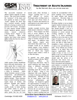
CASE REPORT
Acute Intermittent Porphyria – Diagnostic and Treatment Traps
INIMIOARA MIHAELA COJOCARU1, 2, VIOLETA SAPIRA2, 3, GABRIELA SOCOLIUC2, CRISTINA HERTEA2,
M. BALEA4, CARMEN URSACHE5, M. COJOCARU6
1
“Carol Davila” University of Medicine and Pharmacy, Bucharest, Romania
Department of Neurology, “Colentina” Clinical Hospital, Bucharest, Romania
3
“Ovidius” University, Faculty of Dental Medicine and Faculty of General Medicine, Constanţa, Romania
4
Department of Hematology, “Colentina” Clinical Hospital, Bucharest, Romania
5
Department of Anesthesiology and Intensive Care, “Colentina” Clinical Hospital, Bucharest, Romania
6
“Titu Maiorescu” University, Faculty of Medicine, Department of Physiology, Bucharest, Romania
2
Acute intermittent porphyria (AIP) is a rare metabolic disease defined by mutations coding the
deaminaze enzyme of porphobilinogen (PBGD). Porphyrias are somewhat misdiagnosed as a
consequence of light symptoms in patients. Acute forms of porphyria can be life-threatening, so a
correct diagnosis and an accurate treatment are highly important. The authors presented the case of a
38-years-old patient admitted for persistent abdominal pain that previously presented two generalized
convulsive seizures. The diagnosis of AIP was established by the raised concentration of urinary
porphyrins. Despite treatment with carbohydrates and hemines, the clinical picture of the patient
worsened, with tetraplegia and severe respiratory failure. The patient died seven weeks after the initial
presentation of the disease.
Key words: acute intermittent porphyria (AIP), neurologic complications.
Porphyrins were named for the Greek root for
“purple” (porphyria) [1][2]. The name porphyria is
commonly credited to Schultz, who was a German
medical student in 1874. B.J. Stokvis, MD, made
the first clinical description of acute porphyria in
1889. In 1930, Hans Fischer, a Nobel laureate,
described heme as the crypt that makes blood red
and grass green. In 1937, Waldenström in Sweden
published his findings on one specific type of
porphyria, acute intermittent porphyria (AIP). By
the 1960s, all known types of porphyria had been
identified and environmental factors were shown to
affect the disease course. Research in the 1980s
and 1990s led to the identification of the molecular
defects in each type of porphyria [3]. Currently,
scientists have focused on gene therapy as a treatment for porphyria [2].
Porphyrias are a group of 8 inherited disorders
of heme biosynthesis. A deficiency of any of the
8 enzymes in the biosynthetic pathway can lead to
a variety of clinical symptoms. The disorders are
classified as neurovisceral, cutaneous, or mixed,
depending on the clinical manifestations [1][4][5].
Acute intermittent porphyria has been mistaken
for so many diverse syndromes that, in the words
of Watson “it well deserves the sobriquet of little
simulator” [7] or “little imitator” by Waldenström
[8][9].
ROM. J. INTERN. MED., 2012, 50, 1, 33–41
Misdiagnosis is relatively common because
the symptoms are vague. Acute attacks were the
most feared manifestation, and are mainly a consequence of central, peripheral and autonomic nervous
damage [10]. The most common signs and symptoms
of AIP are abdominal pain (80%), constipation
(50%), nausea and vomiting (50%), tachycardia
(40%), hypertension 31%), urine discoloration
(25%), fever (16%), seizures (10–16%), respiratory
paralysis (9–14%) [2][11–13].
Many factors can precipitate acute attacks in
patients with porphyria, including hormones, medications, nutritional status, tobacco use, infection,
liver damage, surgery, and stress [15–20]. Numerous
medications or “second hits” trigger or unmask
symptoms [20]. The most common include sulfonamides, barbiturates, and ethanol [21]. Drugs may
precipitate symptoms by inducing cytochrome
P-450s and increasing the demand for heme
synthesis [22]. Starvation and illness activate heme
oxygenase, which depletes the hepatic heme pool
leading to aminolevulinic acid synthase (ALAS)
induction. Tobacco contains polycyclic aromatic
hydrocarbons, which also induce cytochromes and
increase heme synthesis [8].
However, acute forms of porphyria can be
life-threatening, so accurate diagnosis and initiation
of proper medical management is important [22].
34
Inimioara Mihaela Cojocaru et al.
2
CASE PRESENTATION
Subsequent hospital course
History and initial evaluation
The patient was transferred in the Department
of Hematology, at that moment she presented with
peripheral facial diplegia, predominant proximal
teraplegia, all deep tendon reflexes were abolished,
severe bradypsychia, bradylalia; the laboratory tests
evidenced severe hyponatremia (103 mmol/L),
severe hypokaliemia (1.4 mmol/L), hypochloremia
(60 mmol/L), leukocytosis with neutrophilia.
Severe hydroelectrolytic disturbances and
acute respiratory failure, secondary to respiratory
muscles palsy imposed the transfer to the Intensive
Care Unit and the orotracheal intubation with
assisted mechanical ventilation.
The lack of improvement in symptomatology
after the therapy with glucose infusions and the
progressive course of motor deficit represented an
indication for hematine therapy-4 mg/kgw/24 hrs, a
total of 15 hematine doses were administered.
The persistence of fever was an indicator for
spute examination that pointed an infection with
Enterobacter which enforced antibiotic treatment
allowed in AIP, depending on bacterial sensitivity
(that was modified during hospitalization) and in
permanent adjustment of doses function of creatinine
clearance.
Also, the patient presented with major hemodynamic instability, with blood pressure variations
from 60 to 220 mm Hg, that imposed inotrop-vasopressor support constantly and in raising doses,
especially in the end stage.
Echography of the heart and of brachial veins
designated the presence of pericardic fluid in
medium quantity and permeable brachial veins.
The neurophysiologic examination (the
measuring of motor and sensitive conduction velocities of peripheral nerves) and the neuroimagistic
evaluation (cerebral MRI) could not be performed
for logistic reasons and in the presence of severe
respiratory failure that needed constant mechanical
ventilation.
The clinical course was constantly worsening,
the patient died secondary to toxico-septic shock
with multiple organ and system failure (Table III).
The patient, a 38-years-old woman, was hospitalized in the Department of Surgery of Colentina
Clinical Hospital for diffuse severe abdominal pain;
the diagnosis established was acute pancreatitis. The
abdominal pain started two weeks before the
admission, she attended emergency room consultation,
but while she had menstruation, modifications of
urine pigmentation could not be identified. Then
she presented at home two generalized seizures.
The patient was a non-smoker, she was not an
alcohol drinker or consumer of forbidden substances,
she did not present a history of psychiatric disorders
or skin sensitivity to sunlight and her family history
was not significant.
At admission, the physical examination revealed
paleness of the skin, cyanosis of the inferior limbs,
spontaneous and palpatory sensitive abdomen,
predominantly in the epigastric and right hypochondrial areas, hyperchromic urine (“pot-wine
urine”), diminished deep tendon reflexes, bradylalia
and bradypsychia.
At this moment, the paraclinic evaluation
showed hyponatremia, hypochloremia and raised
hepatic cytolyse enzymes; 3 days later the patient
presented with a severe electrolytical imbalance
(Table I). The other biochemical markers were in
normal ranges.
Symptomatic therapy was administered, but
the persistent symptomatology imposed an interdisciplinary evaluation: psychiatric, that diagnosed
an acute psychotic episode, neurological, that
highlighted moderate decrease in muscular force
upmost present at the proximal level of inferior
limbs, global diminished deep tendon reflexes,
bradylalia, bradypsychia, and hematological evaluation. Unexplained hyponatremia, the recurrence
of abdominal pain, the acute psychotic episode and
changes of pigmentation in urine raised the
suspicion of AIP; an elevated urine porphyrin
concentration confirmed the diagnosis (Table II).
Hydroelectrolytical infusions, thiamine and
glucose infusions with a total intake of 400 grams
of carbohydrates were administered.
Table I
Biochemical changes in the first days of admission
Kalium (mmol/L)
Natrium (mmol/L)
Chloride (mmol/L)
ALT (IU/L)
AST (IU/L)
At admission
3.75
125
85
140.3
82.2
3 days after admission
2.27
102
61
57.7
58.7
Normal values
3.6–5.2
135–148
98–110
Oct 31st
Oct 31st
3
Acute intermittent porphyria – diagnostic and treatment traps
35
Table II
Biochemical changes of porphyirin in the first days of admission
Urinary metabolites
Lead
ALA/24 hrs
Porphobilinogen/24 hrs
Total porphyrins
Uroporphyrin
Coproporphyrin
Urinary volume/24 hrs
Values at the
diagnosis moment
0.066
111
81
4723
3319
1404
3100
Normal values
In normal ranges
0–7 mg/24 hrs
0–4 mg/hrs
<220 µg/24 hrs
15–50 µg/24 hrs
35–150 µg/24 hrs
1500–2500 mL/24 hrs
Table III
Biochemical parameters in the end stage
Ionogram
Kalium (mEq/L)
Natrium (mEq/L)
Chloride (mEq/L)
Liver function tests
Direct bilirubins (mg/dL)
Total bilirubins (mg/dL)
ALT (U/L)
AST (U/L)
Renal tests
Seric creatinine (mgdL)
Coagulation tests
APTTr
INR
Nonspecific inflammatory tests Fibrinogen (mg/dL)
d-Dimers
CRP (mg/dL)
ESR (mm/h)
DISCUSSION
Pathogenesis of acute intermittent porphyria
AIP, also known as pyrroloporphyria, is the
most common acute porphyria and is caused by
deficiency of hepatic porphobilinogen (PBG) deaminase activity. Affected persons have a 50%
reduction in PBG deaminase activity in their
erythrocytes [23].
Porphyrins are intermediate products in the
heme biosynthetic pathway. Heme is a necessary
component of both cellular hemoproteins produced
primarily in the bone marrow, which makes
hemoglobin and, in the liver which makes the
metabolizing enzymes, cytochrome P-450s. Of the
250 mg of heme synthesized per day, 200 mg is for
hemoglobin. Normally, only 4 to 5 mg of the intermediates in the pathway accumulate, presumably
owing to their toxic nature. Heme that is not used
immediately in a protein complex, is metabolized
to bile pigment [2][24].
The first step is the formation of δ-aminolevulinic acid (ALA) from glycine and succinyl
coenzyme A. Subsequent steps involve the following:
Values in the end
stage
7.25
126
83
0.97
1.5
82
44
3
1.38
1.53
643
3.7
236
55
1) synthesis of a substituted pyrrole compound,
porphobilinogen; 2) condensation of 4 porphobilinogen molecules to yield porphyrinogen;
3) modification of the side chain and ring; and
4) introduction of iron to form heme. In most
tissues, heme, the end product, is required to
suppress further production of heme. If no heme is
present to inhibit the pathway, production of heme
continues unopposed. Any drug, chemical, or hormone
that induces heme synthesis also produces an
increase in heme precursors [2][8][22][24] [26–28].
Only protoporphyrin is used in heme synthesis.
The other porphyrins have no physiologic function
and must be excreted [12]: proto-and coproporphyrins in feces; uro- and coproporphyrins in
the urine and plasma [25][29]. Their fluorescent
properties account for the diagnostic appearance of
urine in some patients.
Diagnosis of acute intermittent porphyria
Diagnosis of porphyrias is usually made by
clinical history in association with increased amounts
of porphyrins or porphyrin precursors in the urine,
feces and blood [16][17][30–32].
36
Inimioara Mihaela Cojocaru et al.
Assays of urinary ALA and porphobilinogen
(PBG) assist in this differential diagnosis. The
urinary excretion of these porphyrin precursors is
markedly increased during acute attacks and the
urine may appear reddish brown because of the
nonenzymatic formation of porphyrins and other
pigments from PBG. Normal levels of ALA in
particular, and of PBG, indicate that the patient is
not suffering from an acute attack, but do not
exclude an underlying porphyria. Markedly raised
excretion of ALA and PBG, which improves following
4
an infusion of the specific treatment heme arginate,
confirms the diagnosis of acute porphyria. Intermediate values may cause some diagnostic difficulty,
but a therapeutic trial of heme arginate usually
proves of value. Differentiation of the four types of
acute or mixed porphyria requires specialized
biochemical analysis of plasma, red cells, urine and
feces, usually undertaken at a specialized centre
(Fig. 1) [2]. Rapid screening tests are useful in the
initial evaluation. The Watson-Schwartz and Hoesch
tests detect PBG in the urine by its reaction with
Ehrlich’s reagent to produce a red color.
Figure 1. Algorithm for the laboratory workup of neurovisceral porphyrias.–normal; +elevated; ADP, δ-aminolevulinic acid
dehydratase porphyria; AIP, acute intermittent porphyria; ALA, δ-aminolevulinic acid; HCP, hereditary coproporphyria; HPLC,
high-performance liquid chromatography; PBG, porphobilinogen; VP, variegate porphyria [2].
Although the enzyme defects underlying the
porphyrias have been identified, molecular genetics
is not yet suitable for primary diagnostic purposes.
Analysis of gene mutations is largely used for
family studies, allowing pre-symptomatic diagnosis,
when biochemical investigations are equivocal.
False-positive results can be seen in certain
clinical situations [1][33]. Patients with hepatobiliary disease or lead toxicity may have elevated
urine porphyrin levels. Factors such as a diet rich in
meat or gastrointestinal bleeding can increase fecal
protoporphyrins [2]. In iron deficiency anemia or
lead toxicity, red blood cell porphyrins can be
elevated; however, more than 90% are zinc protoporphyrins, which will not leak into the plasma [1].
Patients with chronic renal failure do not excrete
porphyrins and can have pseudoporphyria and
elevated uroporphyrin levels in the urine [34].
Several diseases can masquerade as porphyria, such
as intoxication (ethanol, chemicals-hexachlorobenzene, polyhalogenated biphenyls, dioxins, vinyl
chloride, carbon tetrachloride, benzene, chloroform,
lead, arsenic, mercury), liver disease, infectious
disease (mononucleosis), hematologic disease aplastic
anemia, thalassemia, leukemia, pernicious anemia,
iron deficiency anemia), cancer (hepatocellular,
metastatic, pancreatic), pregnancy, hereditary hyper-
bilirubinemia, carbohydrate fasting, and hereditary
tyrosinemia. It is important to differentiate between
porphyrias and other more common diseases
[17][22].
Particularity of the case
The patient presented previous to admission
two generalized convulsive seizures that can be
secondary to both toxic effects of ALA and PBG at
the hypothalamic level (acting as direct neurotoxins)
[12], and the syndrome of inappropriate secretion
of antidiuretic hormone and consecutive hyponatremia (identified at admission).
Abdominal pain, peripheral neuropathy, and
changes in mental status are the classic triad of an
acute attack [5][50], this triad raising also in our
case the suspicion of AIP. Severe abdominal pain is
the most commonly reported presenting symptom
during acute attack [6][13][21][28][35][37][38]. A
clinical presentation without abdominal pain is
unusual in acute porphyria, and some authors
suggest that initial screening tests should be limited
to patients with this symptom [5][13][39][40].
Among patients with acute porphyria, 20–
58% have neuropsychiatric symptoms during acute
5
Acute intermittent porphyria – diagnostic and treatment traps
exacerbations [5][20][38][41]. The neuropsychiatric
symptoms (anxiety, depression) were present in our
patient too, imposing psychiatric evaluation from
admission; the diagnosis of acute psychotic episode
was established. The pathogenesis of the neuropsychiatric manifestations remains unclear and
poorly understood, but multiple hypotheses have been
proposed, including CNS metabolic abnormalities,
ischemia, demyelination, oxidative stress, free
radical damage, PBG or ALA neurotoxicity, ALA
promoting the generation of reactive oxygen
species (ROS) which may result in oxidative
damage to membrane structures within the central
nervous system [5][42][43], ALA inducing lipid
peroxidation in the cerebellum and hippocampus
too [5][42], nervous tissue heme deficiency which
may be deleterious for the synthesis of important
hemoproteins such as cytochrome P450 or nitric
oxide synthase, inhibition of gamma-aminobutyric
acid (GABA) release at central synapses by ALA,
decreased activity of hepatic tryptophan pyrrolase,
a heme-dependent enzyme, leading to increased
levels of brain tryptophan and increased turnover of
5-hydroxytryptamine, a neurotransmitter, decreased
level of melatonin levels, which enhanced ALAmediated lipid peroxidation [5][9][11][28][30][44].
Peripheral neuropathy, which is predominantly a motor neuropathy, initially affecting
mainly the proximal rather than distal muscles, is
next in order of frequency. Patients typically
present with predominantly motor involvement,
characterized by weakness and areflexia, which
may mimic Guillain Barré syndrome, as in our
patient. Sensory loss over the trunk is also common,
although it is often overlooked. Cranial nerves can
also be impaired in cases of AIP, with the facial
and vague nerves being involved more than the
others [12]. Porphyrinic neuropathy typically presents
as a motor neuropathy of the axonal type [48][49].
The pathophysiology underlying axonal dysfunction
in AIP remains unknown. Heme is an essential
component of the mitochondrial electron transport
chain and critical to aerobic metabolism and ATP
production. Fast axonal transport is highly energy
dependent, and diminished ATP availability would
disrupt this process. The accumulation of neurotoxic
heme precursors may lead to impaired function of
the axonal Na+/K+ pump with consequent alteration
in membrane potential to cause neuronal cell death
and axonal degeneration [48].
The most common sign in acute porphyrinic
attacks is tachycardia, which occurs in as many as
80% of patients with acute attacks. Tachycardia
37
results, in part, from the release of catecholamine
during an acute attack and has been implicated in
the sudden death attributed to cardiac arrhythmias
[12].
Our patient presented with hyponatremia
which was constant during hospitalization. Marked
hyponatremia frequently complicates acute porphyrias
[5][11][21][28][35][38][52]. Its mechanism is not
fully understood, but the syndrome of inappropriate
secretion of antidiuretic hormone (SIADH) [5][11]
[21][35][38][53], with resultant dehydration [21]
[54], nephrotoxicity [5][21], excess renal sodium
excretion, and damage to the supraoptic nuclei of
the hypothalamus, has been implicated in its
pathogenesis [5][12][21].
The sub-fever (37o–38oC) and the leukocytosis were initially attributed to the main disease
while repeated hemocultures, done during the fever
attack, were constantly negative. Then, during the
final state the prolonged fever syndrome (39oC)
and the leukemoid reaction (leukocytosis with
neutrophilia – 70000 leukocytes/mm3) were attributed to
the bacterial infection with Acynetobacter (confirmed
at the last hemoculture), sensitive only to Colystin,
antibiotic avoided in persons diagnosed with AIP.
Acute renal failure may be a complication of
an acute AIP attack. Vomiting or diarrhea as a
result of an AIP attack can lead to hypovolemia,
resulting in prerenal azotemia. Urinary retention
may also cause a post obstructive uropathy. Excessive
dosing of hemine may cause also reversible acute
renal tubular damage [55–57].
Management
It is important to understand the pathogenesis, diagnosis, and management of porphyrias,
because they often are misdiagnosed and therefore,
mismanaged in treatable patients.
Prevention is the key. AIP patients should be
advised to use as few drugs as possible. Drugs that
are used should be “safe” based on experimental
and clinical experience. Authoritative websites should
be consulted to avoid exposure to harmful drugs in
treating intercurrent illnesses or symptoms. Oral
carbohydrate intake should always be adequate (a
well-balanced diet somewhat high in carbohydrates –
60 to 70 percent of total calories) and starvation
and fat diets should be avoided. Infections should
be treated promptly. Stresses should be avoided if
possible. Smoking cessation should be recommended. Relatives who are at risk for AIP should
be screened for the disease, probably best done at
38
Inimioara Mihaela Cojocaru et al.
centers with special expertise in the diagnosis and
management of porphyria [31].
Frequently recurring attacks confined to the
luteal phase of the menstrual cycle can be
prevented with GnRH analogue [15][17][59]. If
treatment is effective after several months, lowdose estradiol, preferably by transdermal route, or a
biphosphonate may be added to prevent bone loss
and other side effects, or treatment changed to a
low-dose oral contraceptive.
During pregnancy, hormonal changes, prolonged
fasting due to hyperemesis, and several drugs can
initiate an acute porphirinic attack. Mother’s
mortality rates range from 27 to 42.5%. Symptomatic exacerbations are generally due to exposing
the patients to certain drugs that can modify the
course of pregnancy, resulting in abortion, preterm
births, and other pregnancy complications. Up to
60% of pregnancy complications happen at the
beginning, in early gestational ages. Nearly 15% of
the complications occur during the second trimester
and are generally severe. Although during the last
weeks of pregnancy, women present high ALA and
PBG levels in urine, in porphyrinic pregnant
women the levels tend to be higher [60][61].
Hemin prophylactically administered once or
twice weekly can prevent frequent, non-cyclic
attacks of porphyria in some patients [15].
Treatment of porphyrias depends on the
patients symptomatology [2][3][58][62]. After securing the patients airway, breathing, and circulation,
the disease initially is managed by stopping
exacerbating medications, treating the underlying
infections or illnesses and gaining pain control with
nonporphyrinogenic opiates [2][21][63–67].
Key principles of management are summarized in Table IV [18][31].
Table IV
Therapy of acute attacks of porphyria
9 Remove inciting factors: alcohol, drugs, toxins, chemicals,
smoking
9 Nutritional supplementation: at least 300 grams glucose/
day
9 Frequent checks of neurologic status: especially watch
for development of paresis of muscles of respiration
9 Monitor for hypoventilation (rising arterial pCO2),
hyponatremia or hypomagnesemia and treat vigorously
if found
9 Intravenous heme: 3–4 mg/kg/day for 3–5 days
9 Parenteral meperidine or morphine for pain
9 Phenothiazines for nausea, vomiting, agitation, etc.
Treatment of AIP consists of 3 elements:
eliminating all precipitating factors by discontinuing
6
drugs or treating infections, starting a high-carbohydrate diet or glucose infusion, and starting a
hemin infusion [21][58][70][71].
Mild attacks may be treated with general
supportive measures. A minimum daily intake of
300 g of carbohydrates is also required during
attacks. If oral intake is inadequate, carbohydrates
should be administered intravenously. Glucose or
another readily metabolized carbohydrate should be
used (300–500 grams). Replace fluids, particularly if
patients have poor oral intake or have been
vomiting. Monitor patients for potential hyponatremia or hypomagnesemia and modify administration
of electrolytes and water appropriately.
Tachycardia and/or systemic arterial hypertension can be treated cautiously with betaadrenergic blocking agents such as propranolol or
nadolol. Typical daily doses are 40–240 mg of
propranolol or 60–120 mg of nadolol. Caution must
be exercised when using beta blockers because
hypotension and serious bradycardia have been
reported even after low doses [18] [69]. The tachycardia and hypertension can be quite unsteady.
Seizures are treated by careful correction of
hyponatremia. Almost all anticonvulsivant drugs
have at least some potential for exacerbating acute
porphyrias. Clonazepam may be less harmful than
phenitoin, barbiturates or valproic acid. Bromides,
gabapentin, and vigabatin are safe [65–67].
Intravenous heme therapy is also effective in
managing acute attacks; it can be administered as
hematin. The usual dose consists of 3 to 5 mg
hematin/kg body weight once a day for 3 to 5 days,
beginning as soon as possible after onset of the
attack [12][31][72–75]. These patients should be
monitored for complications of coagulopathy,
thrombophlebitis and hemolysis.
Cimetidine is a cost-effective alternative to
hemin for treating acute attacks (uncontrolled
observations). Cimetidine inhibits hepatic CYPs
and can prevent experimental forms of porphyria
induced by chemicals that are activated by CYPs.
However, chemicals activated by CYPs are rarely,
if ever, implicated in human AIP. Therefore, cimetidine cannot be recommended as an alternative to
hemin [12][76] [77].
Liver transplantation has been highly effective
in patients disabled by recurrent attacks of AIP,
this may be an option for severely affected patients
without advanced motor weakness [78][79].
Long-term monitoring is necessary in patients
with AIP while they are at risk for developing
chronic renal failure and hepatocellular carcinoma.
7
Acute intermittent porphyria – diagnostic and treatment traps
If present, hypertension should be treated, nephrotoxic drugs avoided, and renal function monitored.
Current recommendations are that patients over 50
with acute porphyrias, and especially those with
continued elevations of ALA and PBG, should be
screened by hepatic imaging at least annually for
early detection of hepatocellular carcinoma [78]
[79].
39
CONCLUSION
AIP is a rare but fatal disease and the
physician must consider it in the differential
diagnosis of any acute abdominal pain. Future
research is needed to investigate new treatment
options and to define clinical parameters to monitor
treatment effects and outcome in patients with AIP.
Porfiria acută intermitent (PAI) este o boală metabolică rară caracterizată
prin mutaţii ale genei deaminazei porfobilinogenului (PBGD). Porfiriile sunt
frecvent nediagnosticate deoarece pacienţii prezintă simptome uşoare. Formele
acute de porfirie pot fi ameninţătoare ale vieţii, astfel este important să se
stabilească un diagnostic corect şi să se iniţieze un tratament medical adecvat.
Autorii prezintă cazul unei paciente de 38 ani care a fost internată pentru dureri
abdominal persistente, anterior prezentând două crize convulsive generalizate.
Diagnosticul de PAI a fost confirmat de concentraţia urinară crescută de porfirine.
Evoluţia clinică a fost constant ingravescentă, cu tetraplegie şi insuficienţă
respiratorie, tratamentul cu perfuzii cu glucoză şi hematină neameliorând tabloul
clinic. Pacienta a decedat la şapte săptămâni de la prima manifestare a bolii.
Corresponding author: Senior lecturer Inimioara Mihaela Cojocaru MD, PhD
Department of Neurology, “Colentina” Clinical Hospital
19–21 Şos. Ştefan cel Mare, 020125 Bucharest, Romania
E-mail: [email protected]
REFERENCES
1.
2.
3.
4.
5.
6.
7.
8.
9.
10.
11.
12.
13.
14.
15.
RATNAIKE S., BLAKE D., The diagnosis and follow-up of porphyria. Pathology 1995, 27: 142–153.
FORAN C.E., ABEL G., Guide to the porphyrias. A historical and clinical perspective. Am. J. Clin. Pathol. 2003, 119 (Suppl
1): S86–S93.
McDONAGH A.F., BISSELL D.M., Porphyria and porphyrinology: the past fifteen years. Semin Liver Dis. 1998, 18: 3–15.
VALEAN S., CUCUIANU M., Porfiriile. Biochimie, etiopatogeneza, forme clinice, tratament. Editura medicală universitară
“Iuliu Haţieganu”, Cluj-Napoca, 2003.
GONZÁLEZ-ARRIAZA H.L., BOSTWICK J.M., Acute Porphyrias: A Case Report and Review. Am J Psychiatry 2003, 160:
450–458.
WINKLER A.S., PETERS T.J., ELWES R.D. et al., Neuropsychiatric porphyria in patients with refractory epilepsy: Report of
three cases. J Neurol Neurosurg Psychiatry 2005, 76: 380–383.
WATSON C.J., Porphyria. In Dock W., and Snapper I., eds., Advances in Internal Medicine. Vol. VI. Chicago, Year Book
Publishers, 1954, 235–299.
ABEL G., PALMER-TOY D., Heme synthesis and catabolism. In: McClatchey KD, ed. Clinical Laboratory Medicine. 2nd ed.
Philadelphia, PA: Lippincott Williams & Wilkins; 2002, 407–17.
CRIMLISK H.L., The little imitator-porphyria: a neuropsychiatric disorder. J Neurol Neurosurg Psychiatry 1997, 62: 319–28.
PALMIERI C.,VIGUSHIN D.M., PETERS T.J., Managing malignant disease in patients with porphyria. Q J Med 2004, 97:
115–26.
SUAREZ J.I., COHEN M.L., LARKIN J. et al., Acute intermittent porphyria: clinicopathologic correlation. Report of a case
and review of the literature. Neurology 1997, 48: 1678–84.
SHAH M.A., REMOROZA R., AZIZ K., Acute intermittent porphyria. Hospital Physician, 2002, 38: 67–71.
LITHNER F., Could attacks of abdominal pain in cases of acute intermittent porphyria be due to intestinal angina? J Intern
Med 2000, 247: 407–9.
LOZANO A., TOVAR O.J., ORTIZ C.A., Porfiria aguta: reporte de caso i revision de tema. Revista Med de la Facultad de
Medicina 2008, 16: 106–14.
DE BLOCK C.E., LEEUW I.H., GAAL L.F., Premenstrual attacks of acute intermittent porphyria: hormonal and metabolic
aspects-a case report. Eur J Endocrinol 1999, 141: 50–4.
40
Inimioara Mihaela Cojocaru et al.
8
16. BONKOVSKY H.L., Porphyrin and heme metabolism and the porphyrias. In: Zakim D., Boyer T., eds. Hepatology: a Textbook
of Liver Disease. Philadelphia Saunders, 1982, 351–93.
17. BONKOVSKY H.L., BARNARD G.F., Diagnosis of porphyric syndromes: a practical approach in the era of molecular
biology. Semin Liver Dis. 1998, 18: 57–65.
18. BONKOVSKY H.L., Neurovisceral Porphyrias: What a Hematologist Needs to Know, Hematology, 2005, 2005: 24–30.
19. BONKOVSKY H.L.,SIAO P., ROIG Z. et al., Case records of the Massachusetts General Hospital, Case 20–2008, A 57-year-old
woman with abdominal pain and weakness after gastric bypass surgery. N Engl J Med 2008, 358: 2813–25.
20. REGAN L., GONSALVES L., TESAR G., Acute intermittent porphyria. Psychosomatics 1999, 40: 521–3.
21. THADANI H., DEACON A., PETERS T., Diagnosis and management of porphyria. BMJ. 2000, 320: 1647–51.
22. NUTTALL K.L., Porphyrins and disorders of porphyrin metabolism. In: Burtis C.A., Ashwood E.R., eds. Tietz Textbook of
Clinical Chemistry. 3rd ed. Philadelphia, PA: Saunders, 1999, 1711–32.
23. MEYER U.A., STRAND L.J., DOSS M., REES A.C., MARVER H.S., Intermittent acute porphyria – demonstration of a
genetic defect in porphobilinogen metabolism. N Engl J Med 1972, 286: 1277–82.
24. SAMUELS M.A., Case records of Massachusetts General Hospital. N Engl J Med. 1984, 311: 839–847.
25. KAUPPINEN R., FRAUNBERG M., Molecular and Biochemical Studies of Acute Intermittent Porphyria in 196 Patients and
Their Families, Clinical Chemistry, 2002, 48: 1891–900.
26. FLODERUS Y., SARDH E., MOLLER C. et al., Variations in porphobilinogen and δ-aminolevulinic acid concentrations and
plasma and urine from asymptomatic carriers of the acute intermittent porphyria gene with increased porphirin precursor
excretion. Clin Chem 2006, 52: 701–7.
27. GROSS U., HOFFMANN G.F., DOSS M.O., Erythropoietic and hepatic porphyrias. J Inherit Metab Dis 200, 23: 641–61.
28. SASSA S., KAPPAS A., Molecular aspects of the inherited porphyrias. J Intern Med 2000, 247: 169–78.
29. MOORE M.R., MCCOLL K.E.L., RIMINGTON C. et al., Disorders of porphyrin metabolism. New York: Plenum Publishing
Corporation, 1987, 46–61, 227–55.
30. BLOOMER J.R., BONKOVSKY H.L., The porphyrias. Dis Mon 1989, 35: 1–54.
31. CHEMMANUR A.T., BONKOVSKY H.L., Hepatic porphyrias: diagnosis and management, Clin Liver Dis, 2004, 8: 807–38.
32. PERIASAMY V., SHUBAILI A., GIRSH Y., Diagnostic dilemmas in acute intermittent porphyria. A case report. Med Princ
Pract 2002, 11: 108–11.
33. DEACON A.C., ELDER G.H., ACP Best Practice No. 165: front line tests for the investigation of suspected porphyria. J Clin
Pathol. 2001, 54: 500–7.
34. HINDMARSH J.T., OLIVERAS L., GREENWAY D.C., Plasma porphyrins in the porphyrias. Clin Chem. 1999, 45: 1070–6.
35. SCARLETT Y.V., BRENNER D.A., Porphyrias. J Clin Gastroenterol 1998, 27: 192–8.
36. GAZZANIGA V., Uroporphyria: some notes on its ancient historical background. Am J Nephrol 1999, 19: 159–62.
37. GRANDCHAMP B., Acute intermittent porphyria. Semin Liver Dis, 1998, 18: 17–24.
38. BURGOVNE K., SWARTZ R., ANANTH J., Porphyria: reexamination of psychiatric implications. Psychother Psychosom
1995, 64: 121–30.
39. TAN C.H., YEOW Y.K., Acute intermittent porphyria (AIP) – an unusual cause of acute confusional state: a case report. Ann
Acad Med Singapore 1988, 17: 451–3.
40. BYLESJO I., ANDERSON C., LITHNER F., Clinical aspects of acute intermittent porphyria in northern Sweden:
a population-based study. Scand J Clin Lab Invest 2009, 69: 612–8.
41. SANTOSH P.J., MALHOTRA S., Varied psychiatric manifestations of acute intermittent porphyria. Biol Psychiatry 1994, 36:
744–7.
42. HOLROYD S., SEWARD R.L., Psychotropic drugs in acute intermittent porphyria. Clin Pharmacol Ther 1999, 66: 323–5.
43. SZE G., Cortical brain lesions in acute intermittent porphyria. Ann Int Med 1996, 125: 422–3.
44. HAMNER M.B., Obsessive-compulsive symptoms associated with acute intermittent porphyria. Psychosomatics 1992, 33: 329–1.
45. GHOSH S., CHAUDHURY P.K.R., GOSWAMI H.K., An analysis of six cases of acute intermittent porphyria. Indian
J Psychiatry 2006, 48: 189–92.
46. SATOH Y., IWADATE R., WATANABE Y. et al., Manifestation of psychiatric behaviors in a mouse model of griseofulvininduced hepatic porphyria. J Toxicological Sciences 2008, 33: 599–608.
47. WINKLER A.S., PETERS T.J., ELWES R.D. et al., Neuropsychiatric porphyria in patients with refractory epilepsy: Report of
three cases. J Neurol Neurosurg Psychiatry 2005, 76: 380–3.
48. ALBERS J.W, FINK J.K., Porphyric neuropathy. Muscle Nerve 2004, 30: 410–22.
49. WILKBERG A., ANDERSON C., LITHNER F., Signs of neuropathy in the lower legs and feet of patients with acute
intermittent porphyria. J Int Med 2000, 248: 27.
50. REGAN L., GONSALVES L., TESAR G., Acute intermittent porphyria. Case report. Psychosomatics 199, 40: 521–23.
51. KOCHAR D.K., PAL M., KOCHAR S.K. et al., Acute intermittent porphyria presenting with neurological emergency: Review
of six cases. Neurol India 2007, 55: 413–5.
52. HIFT R.J., MEISSNER P.N., KIRSCH R.E., The clinical diagnosis, prevention and management of the hepatic porphyrias.
Trop Gastroenterol 1997, 18: 41–4.
53. HELLMAN E.S., TSCHUDY D.P., BARTTER P.C., Abnormal electrolyte and water metabolism in acute intermittent
porphyria. The transient inappropriate secretion of antidiuretic hormone. Am J Med 1962, 32: 734–46.
54. JENSEN N.F., FIDDLER D.S., STRIEPE V., Anesthetic considerations in porphyrias. Anesth Analg 1995, 80: 591–9.
55. SARDHE ANDERSON D.E., HENRICHSON A. et al., Porphyrin precursors and porphyrins in three patients with acute
intermittent porphyria and end-stage renal disease under different therapy regimes. Cell Mol Biol (Nosy-le-grand) 2009, 55: 66.
56. ANDERSON C., WIKBERG A., STEGMAYR B., Renal symptomatology in patients with acute intermittent porphyria.
A population-based study. J Int Med 2000, 248: 319.
9
Acute intermittent porphyria – diagnostic and treatment traps
41
57. MARDSEN J.T., CHOWDHURY P., WANG J. et al., Acute intermittent porphyria and chronic renal failure. Clin Nephrol
2008, 69: 339.
58. ANDERSON K.E., BLOOMER J.R., BONKOVSKY H.L. et al., Recommendations for the diagnosis and treatment of the acute
porphyrias. Ann Intern Med. 2005, 142: 439–50.
59. ANDERSON K.E., SPITZ I.M., BARDIN C.W. et al., A gonadotropin releasing hormone analogue prevents cyclical attacks of
porphyria. Arch Intern Med 1990, 150: 1469.
60. MARTINEZ N., NIETO H., GUZMAN M. et al., Porphyria and pregnancy. Case report. Colombia Medica 2011, 42: 107–10.
61. GONZALES C.J., LOPEZ R.E., HERRERA A.B.P. et al., Porfiria aguta intermitente en el embarazo. Am Med Assoc Med
Hosp ABC 2006, 51: 134–7.
62. HARPER P., WAHLIN S., Treatment options in acute porphyria, porphyria cutanea tarda, and erythropoietic protoporphyria.
Curr Treat Options Gastroenterol 2007, 10: 444–55.
63. MEHTA M., RATH G.P., PADHY U.P. et al., Intensive care management of patients with acute intermittent porphyria. Clinical
report of four cases and review of literature. Indian J Crit Care Med 2010, 14: 88–91.
64. OOMMAN A., GURTOO A., Acute intermittent porphyria as a cause of acute respiratory failure. J Indian Med Assoc 2002,
100: 44–6.
65. HARRISON J.C., McAULEY F.T., Propofol for sedation in intensive care in a patient with an acute porphyric attack.
Anesthesia 1992, 47: 335–6.
66. PANDEY C.K., SINGH N., BOSE N. et al., Gabapentin and propofol for treatment of status epilepticus in acute intermittent
porphyria. J Postgrad Med 2003, 49: 285–5.
67. ZADRA M., GRANDI R., ERLI L.C. et al., Treatment of seizures in acute intermittent porphyria: safety and efficacy of
gabapentin. Seizure1998, 7: 415–6.
68. ARORA A., MAHAJAN V., Gabapentin in seizures due to acute intermittent porphyria. Neurol India 100, 48: 194.
69. BONKOWSKY H.L., TSCHUDY D.P., Letter: Hazard of propranolol in treatment of acute prophyria. Br Med J. 1974, 4: 47–8.
70. HERRICK A.L., MCCOLL K.E., Acute intermittent porphyria. Best Pract Res Clin Gastroenterol. 2005, 19: 235–49.
71. ASSELBERGS F.W., KREMER HOVINGA T.K., BOUWSMA C. et al., Acute Intermittent Porphyria as a Cause of
Respiratory Failure: Case report. Am J Crit Care 2009, 18: 180–178.
72. ANDERSON K.E., BONKOVSKY H.L., BLOOMER J.R., Reconstitution of hematin for intravenous infusion. Ann Intern Med
2006, 144: 537–8.
73. DAIMON M., SUSA S., IGARASHI M. et al., Administration of heme arginate, but not hematin, caused anaphylactic shock.
Am. J. Med 2001, 110: 240.
74. www.porphyriafoundation.com.
75. www.porphyria-europe.com.
76. CHEREM J.H., MALAGON J., NELLEN H. et al., Cimetidine and acute intermittent porphyria. Ann Intern Med 2005, 143:
694–5.
77. ROGERS P.D., Cimetidine and acute intermittent porphyria. Ann Pharmacother 1987, 31: 365–7.
78. SOONAWALLA Z.F., ORUG T., BADMINTON M.N. et al., Liver transplantation as a cure for acute intermittent porphyria.
Lancet 2004, 363: 705–6.
79. SETH A.K., BADMINTON M.N., MIRZA D. et al., Liver transplantation for porphyria:who,when,and how? Liver Transpl
2007, 13: 1219–27.
Received January 11, 2012


© Copyright 2026