
companion Faecal tenesmus and dyschezia
companion
JANUARY 2013
The essential publication for BSAVA members
Faecal
tenesmus
and dyschezia
Imaginative
imaging
CT and MRI
P4
How To…
Recognise cutaneous
markers of internal
disease P12
Congress Science
Some highlights
for April
P22
3
4–7
8–11
What’s in
BSAVA News
Latest from your Association
JSAP
Imaginative imaging in practice
The benefits of CT and MRI
Clinical Conundrum
A case of faecal tenesmus and
dyschezia
this month?
12–20 How To…
Recognise cutaneous markers of
internal disease
22–23 Congress Management
Programme
A story of thriving success to inspire
24–25 Congress Pain Lecture
Discover the secret signs of pain
26
By the profession for the
profession
Here are just a few of the
topics that will feature in
your January issue:
Clinical efficacy of a
water-soluble micellar paclitaxel
in canine mastocytomas
Dentoalveolar injuries in
patients with maxillofacial
fractures
Statistics: dealing with
categorical data
New Congress Manager highlights
her plans
27
Publications plans
New Manuals out in time for
Congress
28–29 Hypertension encephalopathy
Simon Platt looks over a case
30–31 PetSavers
Latest fundraising and funding news
32–33 WSAVA News
The World Small Animal
Veterinary Association
34–35 The companion Interview
Kimberly Palgrave
37
Focus On…
The new West Midlands Region
38–39 CPD Diary
What’s on in your area
The findings of this study show the
value of fully assessing the oral cavity in
patients with maxillofacial fractures, as
dentoalveolar injuries are common and
can be predicted by age and mechanism
of trauma.
Radiographic kidney
measurements in pet ferrets
The results of this radiographic study
may allow practitioners to have a more
objective clinical radiographic evaluation
of kidney size of pet ferrets based on
individual traits.
Proteinuria in dogs with
lymphoma
Additional stock photography:
www.dreamstime.com
© Erik Lam; © Hupeng; © Igorr; © Jorge Salcedo;
© Pieter Snijder
companion is published monthly by the British
Small Animal Veterinary Association, Woodrow
House, 1 Telford Way, Waterwells Business Park,
Quedgeley, Gloucester GL2 2AB. This magazine
is a member-only benefit. Veterinary schools
interested in receiving
companion should
email companion@
bsava.com. We welcome
all comments and ideas
for future articles.
Tel: 01452 726700
Email: companion@
bsava.com
Mild proteinuria is common in canine
lymphoma. This paper concludes
that the impact of the proteinuria is
probably low.
2
| companion
This is the fifth of a series of JSAP
articles on statistics in veterinary
medicine and addresses questions
that are widely applicable to clinical
research papers.
This issue of JSAP also includes the
minutes of the BSAVA’s Annual
General Meeting.
Log on to www.bsava.com to access
the JSAP archive online. ■
SPECIAL ISSUE
OF EJCAP NOW
AVAILABLE
Don’t forget that
as a BSAVA member
you are entitled to
free online access
to EJCAP – register
at www.fecava.org/EJCAP to access the
latest issue.
Editorial Board
Editor – Mark Goodfellow MA VetMB CertVR DSAM
DipECVIM-CA MRCVS
CPD Editor – Simon Tappin MA VetMB CertSAM
DipECVIM-CA MRCVS
Past President – Andrew Ash BVetMed CertSAM MBA
MRCVS
■
CPD Editorial Team
Patricia Ibarrola DVM DSAM DipECVIM-CA MRCVS
Tony Ryan MVB CertSAS DipECVS MRCVS
Lucy McMahon BVetMed (Hons) DipACVIM MRCVS
Dan Batchelor BVSc PhD DSAM DipECVIM-CA MRCVS
Eleanor Raffan BVM&S CertSAM DipECVIM-CA MRCVS
■
Features Editorial Team
Andrew Fullerton BVSc (Hons) MRCVS
Mathew Hennessey BVSc MRCVS
■
Design and Production
BSAVA Headquarters, Woodrow House
Web: www.bsava.com
ISSN: 2041-2487
The authors conclude that this
formulation appears to be clinically safe
and effective treatment for canine mast
cell tumours.
No part of this publication may be reproduced
in any form without written permission of the
publisher. Views expressed within this
publication do not necessarily represent those
of the Editor or the British Small Animal
Veterinary Association.
For future issues, unsolicited features,
particularly Clinical Conundrums, are
welcomed and guidelines for authors are
available on request; while the publishers will
take every care of material received no
responsibility can be accepted for any loss or
damage incurred.
BSAVA is committed to reducing the
environmental impact of its publications
wherever possible and companion is printed
on paper made from sustainable resources
and can be recycled. When you have finished
with this edition please recycle it in your
kerbside collection or local recycling point.
Members can access the online archive of
companion at www.bsava.com .
BSAVA on your behalf
Volunteers and staff from your
Association attend selected
meetings throughout the year
in order to ensure we are well
informed and able to represent
the interest of small animal
professionals. Here’s some
news from recent meetings…
B
SAVA Scientific Policy Officer, Sally Everitt,
and Penny Watson, chair of Scientific
Committee, attended the Open Meeting held
by the Veterinary Medicines Directorate and
Veterinary Products Committee in November. As well
as updating the audience on the work of these
organisations over the last year, there was discussion
about the review of the Veterinary Medicines
Regulations due to take place in 2013. Please look out
for details of the consultation which will appear on the
BSAVA website.
There was also a rather gruesome presentation
discussing needle-stick injuries in veterinary practice
(with pictures) and we will be undertaking a member
consultation in the Spring to assess the extent of the
problem in small animal practice.
Dog breeding
Sally also attended the meeting of the Advisory
Council on Welfare issues of Dog Breeding, held in
public in November. The Advisory Council have
produced a report and recommendations on their first
eight welfare priorities (ocular problems related to
head conformation, breathing difficulty related to head
conformation, Syringomyelia and Chiari-like
malformation, idiopathic epilepsy, heart disease
with a known or suspected basis, breed-related and
inherited skin conditions, limb defects and
separation-related behaviour).
The Council has also developed a website to
provide advice for prospective owners thinking of
buying a puppy – www.dogadvisorycouncil.com/
puppy and a health check form to complement the
BVA-AWF / RSPCA Puppy Contract http://
puppycontract.rspca.org.uk/home. If you are
presented with any of this paperwork let us know how
you get on by emailing [email protected]. ■
Today Congress 2013
Early Bird Discount
THURSDAY
January
The Early Bird rate for Congress ends on
31 January – so still time to get the best price
for four days of quality CPD.
Once you have registered, remember to
download the free Congress App (for both
iPhone and Android) to help you make the
most of your time at the event. ■
Membership
reminder
Make the most of
your membership
9876
Expires 31.12.2013
Full Member
Mr Paul Smith
987654
Anyone paying by Direct Debit
should have received their 2013 membership cards;
all others will be sent their cards on receipt of
payment. Winter membership renewals must be paid
in January in order for you to maintain your benefits
and ongoing loyalty privileges. If you have any
questions about your membership plan, then email
[email protected] and a member of our
team will be happy to help you. ■
Regional Contact: [email protected]
How to reward loyal members
The member loyalty benefit for 2013 is a selection of
‘How to’ articles from companion. This compendium
will cover a wide variety of procedures, decisions,
techniques and species; from microchipping
chelonians to the approach to the anorexic rabbit; from
performing a successful joint tap to selecting the right
urinary catheter. With useful nuggets of knowledge
written by experts in their field, this book provides
readers with a diverse wealth of information that
can be applied when making decisions in
practice in the future. Eligible members
(those renewing with no break in their
subscription) will be able to collect their
copy at Congress on the BSAVA
Balcony in the NIA. The rest will be sent
out during May and June. ■
companion
|3
Imaginative
imaging
in practice
Advanced imaging techniques are becoming more widely available
in general practice and offer considerable advantages in increased
diagnostic efficiency. But they are still much more costly than traditional
radiography – and won’t necessarily always be the best tools for the job.
John Bonner asks two leading veterinary radiologists for advice on the right times
to choose computed tomography (CT) or magnetic resonance imaging (MRI)
4
| companion
M
RI and CT scanners are the Rolls Royce
options in diagnostic technology; they are
big shiny expensive machines that will get
you where need to go in comfort and style.
However, sometimes when making the journey
towards a definitive diagnosis in veterinary practice,
the older established technologies of conventional
radiography or ultrasonography will give clinicians all
the information that they need. In other words, it would
be simpler and easier to hop on the bus.
When these high-tech scanners were still the
playthings of veterinary surgeons working in university
hospitals and other specialist centres, there was little
incentive for general practitioners to learn all there is to
know about their capabilities. Now there are many
more machines being used at private referral centres
and larger general practices all around the country.
The costs of the scans are going down and small
animal practitioners need to recognise which of their
patients would benefit from a CT or MRI scan – and to
understand which technology is best for investigating
each specific clinical condition.
Practitioners attending BSAVA Congress in
Birmingham in April can find out which patients they
should be sending for an MRI or a CT examination and
what additional information they can expect to see in
the radiologist’s report. Allison Zwingenberger from the
University of California, Davis and Cambridge-based
consultant Victoria Johnson will deliver two linked
presentations surveying both the potential applications
and limitations of these two imaging modalities.
Dog about to go into a CT scanner
mobile scanners to the veterinary market in 1993. There
is no equivalent to the company’s truck-based service
in continental Europe and in countries like Germany
and France, it is CT technology that has become much
more widely used, Victoria notes.
There is an argument that it is pet insurance that
has really driven the growth in advanced imaging in
the UK, although there has been the same rise in
demand for the new technologies from general
practitioners and their clients in countries where pet
insurance is much less common. “The insurance
industry in the US is small compared to the UK and so
Cost-effective solutions
Both professionals maintain that rather than being
looked on as an expensive luxury, advanced imaging
should be regarded as the most cost-effective option
for many situations. “There is a significant cost to CT
scans but I believe they are in line with other hospital
procedures in terms of personnel time and
equipment maintenance. If CT is the best imaging
modality to obtain a diagnosis, then it is worth the
cost,” Allison suggests.
The same holds for MRI, which was the first of the
new technologies to make an impression on general
practice in the UK. This was mainly due to the efforts of
Burgess Diagnostics, the company which introduced
Allison putting a tortoise into a CT scanner
companion
|5
Imaginative imaging in practice
the increased use of CT is simply a product of its value
in the clinical workup of cases,” Allison says.
In the case of MRI, it is especially valuable for
investigations of the brain and spinal cord and there is
a growing appreciation among practitioners of its
range of applications in the diagnosis of
musculoskeletal disease, said Victoria. But beyond
those areas, there is still some confusion about which
body systems and conditions are suitable for this
approach, and some colleagues will use the
technology inappropriately.
Making the right choice
Part of the problem is that the scanners became
widely available before there were sufficient numbers
of clinicians who were comfortable with using them as
one of their main diagnostic tools. In the past decade,
advanced imaging has become a key part of the
veterinary undergraduate curriculum but the proportion
of technology-savvy vets out working in practice is still
relatively small. As such, there is an ongoing need for
the sort of educational opportunities that will be
offered at Congress.
Practitioners will usually refer a case for an MRI or
CT scan after carrying out a full diagnostic work up
including conventional radiology and/or
ultrasonography. One of the key lessons for general
practitioners is to recognise which cases should be
sent immediately for a scan rather than carrying out
initial radiographic examinations. “Patients with
middle ear disease would be one obvious example.
Sagital STIR of a
myxosarcoma in a
dog’s stifle
6
| companion
In the past we may have taken several different
screening radiographs of these patients, but even
with multiple views conventional radiographs are not
very informative. So sending the patient for a scan
when the clinical examination suggests that this is the
problem will save time and money for the client and
means that the animal does not undergo an
unnecessary additional general anaesthetic.”
Relevant treatment to the patient
As Victoria will explain, the attending vets should try to
identify which imaging modality offers the optimal
results for a particular case. The best choice will be
the technology or combination of methods that
provides all relevant data in way that is the least
invasive, most cost efficient and best overall for the
individual patient, she says.
So it is equally important for practitioners to
recognise which cases are not suitable for advanced
imaging techniques and where more traditional
methods will provide the information needed with less
risk to the patient. For example, any patient in an
unstable state would be a poor candidate for an MRI
scan in view of the time needed to carry out the
procedure. That will vary from about 40 minutes to well
over an hour, depending on the part of the body
concerned and the type of scanner being used.
Any patient with a pacemaker or some other
metallic implant would be at serious risk in the strong
magnetic field generated by an MRI scanner. Patient
safety is also a major issue for those performing CT
Transverse T1W normal nose Dorsal T2W normal nose
Image of a dog with a tumour in a rib
scans on human patients because of the risks from the
radiation used. Victoria says she is surprised never to
have been asked about radiation hazards by the
owners of pets undergoing a CT scan. But even those
clients that understand the principles behind CT
technology seem to take a pragmatic view of the costs
and benefits in their own animals. “Our patients have
shorter lifetimes and so the long term effects of
medical radiation are unlikely to develop in that period.
But radiation safety for patients and personnel is
always taken into consideration during the scanning
procedures,” Allison notes.
utilisation of multi-slice CT machines in veterinary
clinics, offering ultrafast scanning times and
improved spatial resolution in the images. “There is a
learning curve for the software that manipulates these
images but it can be very rewarding. CT can also be
used to gain functional information about tissues such
as perfusion in tumours and glomerular filtration rate.
It’s a very versatile modality that is very exciting to
work with.” ■
Gain more knowledge
As Allison and Victoria will explain, there is a range of
other conditions for which CT or MRI are not generally
used but where either could be considered at an early
stage of the investigation. Evaluating a migrating
foreign body such as a grass seed would be an
obvious example. But there are others where one
modality has clear advantages over the other, such as
investigations of lung disease, where MRI has limited
value because of its lack of sensitivity in looking at
air-filled structures which CT is particularly useful for.
They will also describe clinical scenarios in which
advanced imaging may not be the obvious first line
imaging modality but where it can provide vital
additional information to help guide and improve the
patient’s treatment. Using MRI or CT in oncology
patients to evaluate the tumour margins and presence
of any regional metastases will provide significant
advantages when planning surgical treatment, they
point out.
Even those practitioners that feel that they have a
good understanding of all the potential applications of
these imaging modalities in small animal practice may
benefit from attending the imaging sessions at
Congress. That’s because the technology is changing
and improving with every passing year and so it is
wise to keep an eye out for any developments that may
be appearing over the horizon.
So Victoria will be giving a taste of what’s to come
in small animal practice as techniques such as MRI
angiography with established applications in human
medicine start to filter through to the veterinary area.
Meanwhile, Allison sees great opportunities in the
MRI is good for
showing foreign
bodies – this is a stick
(arrowed) in the
pharynx of a dog
ALLISON AND VICTORIA AT CONGRESS
Friday 5 April – Hall 8 ICC
■
■
10.55–11.40
Allison Zwingenberger – Nurse Imaging
– Radiographic contrast studies
11.45–12.30
Victoria Johnson – Nurse Imaging
– What is the difference between CT and MRI? (Advanced)
Saturday 6 April – Hall 4 ICC
■
■
■
■
■
08.30–09.15
Allison Zwingenberger – Interactive Film Reading –
Why is this cat dyspnoeic? (Electric Voting)
11.05–11.50
Allison Zwingenberger – Interactive Film Reading –
Why is this abdomen painful? (Electric Voting)
14.05–14.50
Allison Zwingenberger – Interactive Imaging – How to
get more out of my X-rays (Electric Voting)
16.50–17.35
Allison Zwingenberger – Interactive Imaging –
When should I consider CT? (Electric Voting)
17.45–18.30
Victoria Johnson – Interactive Imaging –
When is MRI a good option? (Electric Voting)
Victoria
Johnson
Allison
Zwingenberger
companion
|7
Clinical conundrum
Andy Tomlinson, an Intern in Small Animal Studies
at the University of Liverpool, Small Animal Teaching
Hospital, invites companion readers to consider
faecal tenesmus and dyschezia in a 7-year-old
Border Collie
Case presentation
A 7-year-old female neutered
Border Collie presented with a
2-week history of constipation,
dyschezia and faecal tenesmus.
The case was initially managed with
stool softeners, which resulted in
the passage of small volumes of
diarrhoea or loose ribbon-like
faeces; however, dyschezia and
faecal tenesmus persisted.
Routine haematology and
biochemistry had been performed
by a colleague on the day prior to
examination (Table 1).
On presentation the dog was
bright and alert. Abdominal
palpation identified a large mass in
the caudodorsal abdomen. Digital
rectal examination identified a
narrowed rectal lumen with a firm
density compressing the dorsal
rectal wall. Anal sacs were palpably
normal and no perineal hernia was
detected. Rectal temperature was
39.9°C. The remainder of the
clinical examination was normal.
Parameter
Value
Normal
range
Albumin
33 g/l
25–44
ALP
94 IU/l
20–150
ALT
42 IU/l
10–118
Amylase
593 IU/l
200–1200
Total bilirubin
6 µmol/l
2–10
Calcium
2.80 mmol/l
2.15–2.95
Creatinine
110 µmol/l
27–124
Urea
5.4 mmol/l
2.5–8.9
Globulin
41 g/l
23–52
Glucose
2.9 mmol/l
3.3–6.1
Inorganic
phosphate
1.55 mmol/l
0.94–2.13
Na+
148 mmol/l
138–160
K+
4.4 mmol/l
3.7–5.8
TP
74 g/l
54–82
WBC
10.20
6–18 x109/l
RBC
6.55
5.4–8.0
x1012/l
Hb
15.4
12–18 g/dl
HCT
0.458
0.35–0.55
MCV
70.0
65–75 fl
MCH
23.4
19.5–24.5 pg
MCHC
33.5
32–37 g/dl
Platelets
215
150–400
x109/l
Neutrophils
9.20
3–12 x109/l
Lymphocytes
0.14
1.2–3.8
x109/l
Monocytes
0.71
0–1.2 x10⁹/l
Eosinophils
0.1
0.1–1.3
x109/l
Basophils
0.01
0–0.1 x10⁹/l
Reticulocytes
0.06
X1012/l
Table 1: Haematology and biochemistry results
performed the day prior to presentation
(abnormal results in bold)
8
| companion
Create a problem list
■■
■■
■■
■■
■■
Constipation, dyschezia and faecal
tenesmus
Caudal abdominal mass
Pyrexia
Lymphopenia
Mild hypoglycaemia
Consider the differential
diagnosis for your problems.
Can your differentials be
prioritised based on the
history and physical
examination findings?
Passage of small amounts of diarrhoea or
ribbon-like faeces with faecal tenesmus
and dyschezia is consistent with
intraluminal/intramural or extraluminal
colonic/rectal obstruction. An intraluminal
mass was not palpated on rectal
examination but an intraluminal colonic
mass cannot be excluded. A palpable
abdominal mass and dorsal compression
of the rectal lumen suggests an
extraluminal cause of obstruction.
Differential diagnoses for intraluminal/
intramural and extraluminal obstruction are
listed in Figure 1.
Mild lymphopenia is likely a
reflection of stress. Blood glucose was
re-assessed at presentation and
measured 5.3 mmol/l; the initial result
was most likely an artefact reflecting
delayed separation of serum from red
blood cells, although a paraneoplastic
process could not be excluded.
Pyrexia is indicative of inflammation
secondary to an infectious, immunemediated or neoplastic process.
What initial diagnostic plan
would you consider?
Abdominal radiography is required to
assess the caudal abdominal mass (size
and location), assessment of
Intraluminal/intramural
■■
■■
■■
■■
Intraluminal/intramural neoplasia:
– Adenomatous polyp
– Adenoma/carcinoma in situ
– Leiomyosarcoma/leiomyoma
– Adenocarcinoma
– Lymphoma
Granuloma
Stricture
Foreign body
Extraluminal
■■
■■
■■
■■
■■
■■
*
A
Caudal abdominal neoplasia
Granuloma
Abscess
Pelvic fracture
Sublumbar lymphadenopathy
Organomegaly
Figure 1: Differential diagnoses for
intraluminal/intramural and extraluminal
colonic obstruction
organomegaly, presence of a foreign body
and pelvic conformation. As abdominal
neoplasia is a differential, thoracic
radiographs are indicated to assess for the
presence of metastatic disease.
What is your interpretation of
the thoracic and abdominal
radiographs?
Inflated right and left lateral and
dorsoventral thoracic radiographs were
obtained (Figure 2). There is a rounded
soft tissue opacity dorsal to the second
sternebra on the left and right lateral
views (arrow on Figure 2A and 2B). The
dorsoventral view revealed focal left
sided mediastinal widening between
T3 and T5 ( on Figure 2C). A generalised
bronchial lung parenchymal pattern can
be seen, but the bronchial markings are
largely fine and well defined. The
remainder of the lung parenchyma is
unremarkable.
The lateral and dorsoventral
abdominal radiographs demonstrate a
large rounded homogenous soft tissue
B
C
Figure 2: Inflated thoracic radiographs. (A) Left lateral, (B) right lateral and (C) dorsoventral views
opacity ventral to L4–L7, causing ventral
displacement of the descending
colon (arrow on Figure 3). The mass
extended into the pelvic inlet, causing
significant narrowing of the terminal colon/
cranial rectum.
In light of the radiographic
findings refine your differential
diagnosis list
A bronchial lung pattern can be a
radiographic artefact seen with expiratory
*
A
Figure 3: (A) Right lateral and (B) ventrodorsal radiographic
views of the abdomen
B
companion
|9
Clinical conundrum
exposure, but as these were inflated
thoracic images it is likely that the
bronchial pattern reflects age-related
change and is not significant in this case.
The sternal lymph node is the draining
lymph node of the abdominal cavity whilst
the sublumbar lymph nodes drain the
dorsal half of the abdomen, pelvis and
pelvic limb. Sternal and sublumbar
lymphadenopathy can be secondary to
reactive hyperplasia and primary
haemopoietic or metastatic neoplasia.
Sternal lymphadenopathy is most
commonly associated with a neoplastic
process and, given the lack of
haematological changes combined with
degree of lymphadenopathy, a primary
haemopoietic or metastatic neoplastic
process seems most likely.
Revised differential diagnoses include:
Disseminated neoplastic disease
– Lymphoma
– Histiocytic sarcoma
– Mast cell tumour
■■ Granulomatous disease
■■
What further investigations are
indicated?
Ultrasound-guided fine-needle aspiration
of the affected lymph nodes is required to
assess cell type and morphology.
A
Abdominal ultrasonography will allow
thorough assessment of the abdominal
pathology and facilitate harvesting of
samples required for diagnosis.
What is your interpretation of
the ultrasonographic
examination?
Ultrasonographic examination of the
spleen and liver was unremarkable. The
sternal lymph node was visualised and
measured 37 x 25 mm (Figure 4A).
Enlarged medial iliac lymph nodes (MILN)
were identified, with the largest left MILN
measuring 50 x 33 mm and the largest
right MILN measuring 66 x 25 mm (Figure
4B). This represents multi-centric
lymphadenopathy, and fine-needle
aspiration of the sternal and MILNs was
carried out under ultrasound guidance.
Marked pyelectasis was identified in
both kidneys, with the left renal pelvis
measuring 13 mm and the right renal pelvis
measuring 10 mm in diameter (Figure 4C).
There was dilatation of both ureters
proximally although they were of normal
diameter when entering the bladder.
Pyelectasis can be associated with
pyelonephritis and obstructive ureteral
disease (e.g. ureteral stones, trigonal
neoplasia); mild dilatation can be seen in
patients with polyuria/polydipsia and in
B
those on high rates of intravenous fluid
therapy. In this case pyelectasis and
ureteral dilatation was most likely
secondary to obstruction/compression
from the MILNs, with resolution expected
with treatment of the lymphadenopathy.
What is your interpretation of
the lymph node fine-needle
aspirates (Figure 5)?
There is a monomorphic population of
large lymphoblasts, approximately 2.5
times the diameter of small lymphocytes.
They have a large round nucleus with open
granular chromatin and 2–3 nucleoli and
have sparse basophilic cytoplasm. The
mitotic rate is high and there are
occasional tangible body macrophages
(Figure 5). The predominance of a large
lymphoblast population and clinical
staging information is consistent with stage
IIIa multi-centric high-grade lymphoma.
What is your treatment plan and
expected long-term outcome?
Around 90% of patients with lymphoma
respond to chemotherapy, with many
having good periods of remission.
Various chemotherapy options were
available, starting with steroids alone
utilising a COP protocol or adding an
anthracycline to create a CHOP protocol.
C
Figure 4: Ultrasonographic appearance of the (A) right sternal lymph node, (B) medial iliac lymph nodes and (C) left kidney
10
| companion
Figure 5: Fine-needle aspirate from the medial
iliac lymph node
Each has potential benefits (increasing
intensiveness of treatment should
lead to longer survival times) and
disadvantages (costs, practical aspects
in terms of monitoring and frequency of
interventions). Potential side effects
such as gastrointestinal upset,
myelosuppression and the potential risk of
living with a cytotoxic pet, also need to be
considered. After careful discussion with
the owner, a standard 25-week
discontinuous multi-agent chemotherapy
protocol was commenced (L-CHOP which
contains: crisantaspase (L-asparaginase),
vincristine, epirubicin, cyclophosphamide
and prednisolone).
Herding breeds are known to carry
the MDR-1 gene mutation, which impairs
their ability to exclude cytotoxic drugs,
such as vincristine, from their cells,
increasing their susceptibility to adverse
effects. Blood was submitted for genetic
testing and it was found that the patient
was negative for this mutation.
The use of crisantaspase as part of
standard doxorubicin-based
chemotherapy protocols (L-CHOP) has not
been associated with increased survival
times for dogs with lymphoma when
compared to non-crisantaspasecontaining protocols (CHOP). However,
anecdotally, its use has been associated
with a faster reduction in tumour burden,
which would provide rapid relief of clinical
signs, alleviating the signs of dyschezia
and faecal tenesmus seen in this case.
Additionally, the use of multiple drugs with
different mechanisms of action can
eradicate more neoplastic sub-clones.
Classically doxorubicin is the
anthracyline used in CHOP-based
protocols. Epirubicin is used at our
institution due to its potential
for reduced toxic side effects particularly
with regard to cardiotoxicity. A recent
retrospective study showed that the
epirubicin-CHOP protocol has comparable
survival times to the standard doxorubicinCHOP protocol. Average time to first
relapse and survival time of 216
and 342 days, respectively, can be
expected for patients with multi-centric
lymphoma treated at our hospital with the
L-CHOP protocol.
The staging of our dog did not reveal
evidence of negative prognostic factors.
The patient was well (WHO sub-stage a)
and there was no evidence of
hypercalcaemia or cytological criteria to
suggest a T-cell phenotype.
AVAILABLE FROM BSAVA
BSAVA Manual of
Canine and Feline
Oncology
3rd edition
Edited by: Jane Dobson
and Duncan Lascelles
Building on the success
of previous editions,
this fully updated
Manual encompasses
the important advances made over recent
years, while keeping the text practical and
user-friendly. A wealth of new photographs
has been included to illustrate the clinical,
diagnostic and therapeutic aspects of a range
of tumours. The growing importance of
ethical considerations and palliative care are
also recognized, and exciting developments
and treatment possibilities explored.
“...an excellent publication…sufficiently
rewritten and updated to make it a
worthwhile purchase, even for those
veterinarians already in possession of the
second edition. The content is ‘state of the
art’ as much as possible… remains very
easy to read…a very attractive purchase for
practitioners and students…” JOURNAL OF
SMALL ANIMAL PRACTICE
Member price: £55.00
Price to non-members: £85.00
Outcome
The patient was re-examined one week
after starting the L-CHOP chemotherapy
protocol. The medial iliac
lymphadenopathy had greatly reduced and
was no longer palpable on abdominal or
rectal examination. The clinical signs had
resolved and the dog was once again
passing faeces without any tenesmus or
dychezia. The patient is now entering week
6 of her chemotherapy protocol and has
suffered no toxic side-effects or recurrence
of her clinical signs. ■
CONTRIBUTE A CLINICAL CONUNDRUM
If you have an unusual or interesting case
that you would like to share with your
colleagues, please submit photographs
and brief history, with relevant questions
and a short but comprehensive explanation,
in no more than 1500 words to
[email protected]
All submissions will be peer-reviewed.
companion
| 11
How to recognise
cutaneous markers
of internal disease
Laura Buckley, veterinary dermatologist at
Calder Vets, Dewsbury, and Tim Nuttall of
the University of Liverpool Small Animal
Teaching Hospital help us spot the clues
C
utaneous markers of internal disease are
dermatological lesions that are highly
specific for, or diagnostic of, a particular
systemic disorder. Included in this group are
the cutaneous paraneoplastic syndromes, which are
non-neoplastic skin disorders associated with internal
malignancy. The known veterinary diseases include:
■■
■■
■■
■■
■■
■■
■■
Feline paraneoplastic alopecia
Feline thymoma-associated exfoliative dermatitis
Superficial necrolytic dermatitis
Canine nodular dermatofibrosis
Pancreatic panniculitis
Paraneoplastic pemphigus
Endocrine disease (hypothyroidism,
hyperadrenocorticism, feline acquired skin fragility
syndrome, canine testicular tumour and
feminisation syndrome).
Most of these diseases are uncommon but they are
well described. This article summarises the clinical
presentations of this interesting group of dermatoses
and the steps necessary to confirm the diagnosis.
Pathophysiology
The aetiopathogenesis of the primary disease process
is often known, although the exact pathophysiology of
the cutaneous lesions in most cases is unclear.
Proposed mechanisms include:
Tumour-induced antigen–antibody interactions
involving cross-reactivity between tumour antigens
and self antigens
■■ Abnormal or excessive production of biologically
active substances (hormones, enzymes, growth
factors, cytokines) by tumour cells or by accessory
cells in response to the disease process
■■ Tumour-induced or organ dysfunction-associated
depletion of certain physiological substances
■■
12
| companion
Clinical importance
Prompt recognition of the cutaneous markers of internal
disease permits the early detection and treatment of
the primary underlying disease. The cutaneous
markers also provide a means of monitoring for
recurrence of disease during and after therapy.
Accurate diagnosis and treatment of the cutaneous
disease is very important as many of the cutaneous
syndromes are more debilitating for the animal than the
underlying disease.
Feline paraneoplastic alopecia
This is a cutaneous paraneoplastic syndrome involving
non-pruritic, progressive alopecia associated with an
underlying internal malignancy.
Cutaneous markers:
Alopecia (Figure 1A): Acute-onset, non-pruritic,
progressive symmetrical alopecia, initially affecting
the ventral abdomen and limbs and then becoming
generalised. Hair is easily epilated.
■■ Glistening skin (Figure 1B): Alopecic skin is
inelastic and thin and has a smooth, shiny
appearance.
■■ Footpad lesions (Figure 1C): Some cats have
concentric scale, crusting and painful fissures
affecting the footpads.
■■ Malassezia dermatitis: Secondary Malassezia
dermatitis with brown greasy accumulations
around the eyes, nose and claw beds has been
reported. These lesions may be pruritic.
■■
Other clinical signs: Weight loss, lethargy,
inappetence/anorexia, vomiting and diarrhoea, with or
without clinical signs associated with metastatic
disease to the liver and/or lungs.
Signalment: Older cats of 7–16 years; no sex or breed
predilection.
Aetiopathogenesis: Associated predominantly with
pancreatic carcinoma, but bile duct carcinomas have
also been reported.
Pathophysiology of cutaneous lesions: Unknown.
Histopathologically there is atrophy, miniaturisation
and telogenisation of the hair follicles, hyperplasia of
A
B
C
Figure 1: Feline paraneoplastic alopecia. (A) Symmetrical alopecia affecting the ventral abdomen and limbs in a cat with
pancreatic neoplasia. (B) The same cat as in (A); the ventral abdominal skin is thin and has a shiny appearance; (C) Concentric
scale affecting the footpads in another cat
the epidermis and thinning or absence of the stratum
corneum, and occasionally yeasts in the stratum
corneum. It has been suggested that the
histopathological presence of yeasts in cats with
generalized skin disease is also a marker for internal
malignancy.
Differential diagnoses: Dermatophytosis,
demodicosis, self-induced alopecia (pruritic and/or
behavioural skin disease), endocrinopathies,
superficial necrolytic dermatitis.
Diagnostic tests:
1. Skin scrapings, trichography and examination of
coat brushings to rule out demodicosis and other
parasitic diseases. Trichography shows
telogenisation of hair follicles.
2. Cytology to investigate Malassezia dermatitis.
3. Fungal culture to investigate dermatophytosis.
4. Haematology, biochemistry, urinalysis and total T4
to assess for organ dysfunction and endocrine
disease.
5. Skin biopsy, abdominal ultrasonography and
biopsy of the primary tumour for definitive
diagnosis.
6. Screening for metastatic disease if neoplasia is
suspected or confirmed.
interdigital skin, claw beds and ear canals. In some
cases this is associated with Malassezia dermatitis.
■■ Crusting and ulceration: Some cats develop
crusts and ulcers with advanced disease.
Other clinical signs: In many cases there are no
other clinical signs. In advanced cases the thoracic
mass and/or pleural effusion may cause dyspnoea.
Metastasis of thymomas is uncommon.
Signalment: Older cats, no sex or breed predilection.
Aetiopathogenesis: Most of the reported cases are
associated with thymoma, although some cases can
be idiopathic.
A
Feline thymoma-associated exfoliative
dermatitis
This is a generalised exfoliative dermatitis that in most
cases is associated with thymoma, although some
cases may be idiopathic. It is important to rule out a
cutaneous drug eruption as some of the cutaneous
features are shared with erythema multiforme, which
can occur as a result of drug administration.
B
Cutaneous markers:
Exfoliative dermatitis (Figure 2A,B):
Non-pruritic, diffuse erythema and skin
exfoliation/scaling with associated alopecia.
Lesions begin on the head and pinnae, and then
become generalised.
■■ Keratosebaceous accumulations (Figure 2C):
Brown, keratosebaceous debris affecting the
■■
C
Figure 2: Feline
thymona-associated
exfoliative dermatitis.
(A) Generalised skin
exfoliation and
alopecia. (B) The skin is
mildly erythematous
with diffuse adherent
scale. (C) Thick brown
keratosebaceous debris
adhered to the distal
limbs and claw beds
companion
| 13
How to recognise cutaneous markers
of internal disease
Pathophysiology of cutaneous lesions: Unknown,
but probably associated with self-active T-cell clones.
Histopathologically there is a lymphocytic interface
dermatitis, and epidermal hyperplasia and
hyperkeratosis with focal parakeratosis. Sebaceous
glands are often absent.
Differential diagnoses: Cutaneous drug eruption
(CDE), erythema multiforme, contact dermatitis,
pemphigus foliaceus (PF), systemic lupus
erythematosus (SLE), bacterial pyoderma, Malassezia
dermatitis, dermatophytosis, demodicosis,
endocrinopathies, lymphocytic mural folliculitis.
Diagnostic tests:
1. Skin scrapings, trichography and examination of
coat brushings to rule out demodicosis and other
parasitic diseases.
2. Cytology to investigate bacterial pyoderma and
Malassezia dermatitis.
3. Fungal culture to investigate dermatophytosis.
4. Haematology, biochemistry, urinalysis and total T4
to assess for organ dysfunction and endocrine
disease.
5. Skin biopsy, thoracic radiography or CT and biopsy
of the primary tumour for definitive diagnosis.
Superficial necrolytic dermatitis (SND)
Previously known as necrolytic migratory erythema and
hepatocutaneous syndrome, SND is an uncommon
A
dermatosis that predominantly affects dogs but has
also been reported in cats. SND causes distinctive
lesions of the footpads and mucocutaneous junctions
in association with metabolic hepatic disease or, less
commonly, glucagonoma of the pancreas or intestine.
Cutaneous markers:
Footpad hyperkeratosis (Figure 3A): Erythema,
crusting, hyperkeratosis and fissure formation
affecting the footpads on all four feet. Severe
lesions can cause pain and pruritus.
■■ Crusting dermatitis (Figures 3B): Alopecia,
erosions/ulceration and adherent crusts affecting
pressure points, mucocutaneous junctions and
feet. In cats the lesions may be more subtle, and
alopecia and scaling may be a more prominent
feature. Pruritus and Malassezia dermatitis have
also been reported.
■■
Other clinical signs: In the early stages of disease
the cutaneous lesions are the only clinical signs and
animals are systemically well. Over several weeks to
months animals develop lethargy, inappetence and
further systemic signs in association with hepatic or
pancreatic disease. Some cases develop diabetes
mellitus in association with hepatic disease.
Signalment: Older dogs (around 10 years) and
cats of any breed or sex. Smaller dog breeds are
over-represented.
B
Figure 3: Superficial necrolytic dermatitis. (A) Severe hyperkeratosis, fissuring and ulceration of the footpads in a dog with
SND. (B) Erythema, alopecia and adherent crusts affecting the mucocutaneous skin
Reproduced from the BSAVA Manual of Canine and Feline Dermatology, 3rd edition
14
| companion
Aetiopathogenesis: In dogs most cases are
associated with a vacuolar hepatopathy (cirrhosis,
neoplasia, hepatic lipidosis, chronic active hepatitis
and drug-induced hepatitis (especially associated with
phenobarbital)). A smaller number of cases have been
reported in association with pancreatic glucagonomas,
which may also occur in the proximal small intestine. In
cats SND has been reported in association with
pancreatic carcinoma, hepatopathy, thymic
amyloidosis and intestinal lymphoma.
Pathophysiology of cutaneous lesions: The
exact pathophysiology is unknown but lesions are
thought to arise from a deficiency in cutaneous
amino acids, which results in keratinocyte
degeneration and skin necrosis. This may be
secondary to increased hepatic catabolism of
amino acids. Histopathologically there is a distinctive
‘red, white and blue pattern’ when stained with
haematoxylin and eosin as a result of parakeratosis,
keratinocyte swelling and oedema, and hyperplasia of
basal keratinocytes, respectively.
Differential diagnoses: Erythema multiforme, PF,
SLE, zinc-responsive dermatosis, CDE, generic dog
food dermatosis.
Diagnostic tests:
1. Skin scrapings, trichography and examination of
coat brushings to rule out demodicosis and other
parasitic diseases.
2. Cytology to investigate secondary bacterial
pyoderma and Malassezia dermatitis.
3. Haematology, biochemistry, urinalysis, bile
acid stimulation test and total T4 (plus TSH in
dogs) to assess for organ dysfunction and
endocrine disease.
4. Skin biopsy, abdominal ultrasonography and
biopsy of the liver or pancreas for definitive
diagnosis. Ultrasonographically, the liver has a
unique ‘honeycomb’ or ‘Swiss cheese’ appearance
due to variably-sized, hypoechoic regions with
echogenic borders.
5. Plasma can be submitted to detect increased
glucagon levels and reduced levels of amino acids
but the findings are non-specific.
Figure 4:
Multiple cutaneous
nodules due to
dermatofibrosis in a
Labrador Retriever
crossbreed
Reproduced from the
BSAVA Manual of Small
Animal Dermatology,
2nd edition
Canine nodular dermatofibrosis (CND)
This is a cutaneous paraneoplastic syndrome involving
benign collagenous nodular disease that develops in
association with renal neoplasia and, less commonly,
uterine tumours.
Cutaneous markers:
■■
Cutaneous and subcutaneous nodules (Figure
4): Multiple firm dermal and subcutaneous nodules
of variable size that are mainly located on the
extremities but can be generalised. The overlying
epidermis is usually intact but can be ulcerated.
Other clinical signs: In many cases the cutaneous
lesions are the only clinical signs, as they precede
renal disease by many months to years. In addition to
the usual clinical signs associated with renal
dysfunction, affected dogs may present with
haematuria, pyrexia, abdominal distension and pain
secondary to rupture of renal cysts.
Signalment: Middle-aged to older animals but cases
have been reported in dogs as young as 2 years.
German Shepherd Dogs are most commonly affected
but cases have been seen in Golden Retrievers, Boxers,
German Shorthaired Pointers and mixed breeds.
companion
| 15
How to recognise cutaneous markers
of internal disease
Aetiopathogenesis: Cutaneous lesions are
associated with renal cystadenocarcinoma or
cystadenoma and, less commonly, uterine leiomyoma.
Birt–Hogg–Dube (BHD) syndrome in man shows
similar development of renal neoplasia and benign
cutaneous trichofolliculomas. Pedigree analysis has
revealed an autosomal recessive mode of inheritance,
with gene mapping localising the suspected
causative gene to a region on chromosome 5 that
overlaps with the BHD locus. In addition, one study
showed increased levels of the cytokine TGF-β1
(a potent stimulator of fibrosis) in the hair follicles and
renal tubules of German Shepherd Dogs with CND
and renal cystadenocarcinoma, when compared to
normal dogs.
Pathophysiology of cutaneous lesions: It is
possible that a common genetic defect, partially
mediated by an overproduction or expression of
fibrosis-inducing cytokines, leads to the development
of renal/uterine neoplasia and collagenous nodules.
Differential diagnoses: Infectious nodular disease
(deep bacterial or fungal, mycobacterial and protozoal
infection), immune-mediated disease (sterile nodular
panniculitis, sterile pyogranuloma/granuloma
syndrome, reactive histiocytosis, erythema nodosum,
pyoderma gangrenosum, canine sarcoidosis),
neoplasia (lymphoma, mast cell tumour, malignant
histiocytosis), other nodular skin diseases (calcinosis
circumscripta, foreign body reaction).
Diagnostic tests:
1. Fine-needle aspiration cytology using routine and
special stains to investigate infectious and sterile
nodular disease.
2. Haematology, biochemistry and urinalysis to
investigate renal disease.
3. Skin biopsy, abdominal ultrasonography and
radiography ± contrast nephrography, and biopsy
of the primary tumour for definitive diagnosis.
Pancreatic panniculitis
This is a form of panniculitis specifically associated
with pancreatic disease. It has been reported in both
dogs and cats.
16
| companion
Figure 5: Multiple subcutaneous nodules affecting the trunk
and proximal limbs of a Greyhound with panniculitis
associated with pancreatitis. Where some of the nodules
have ruptured there is dark brown, oily debris adhered to
the hair coat
Cutaneous markers:
1. Panniculitis (Figure 5): Single or multiple large,
deep-seated nodules which can be firm and well
defined or soft and poorly demarcated. The
overlying epidermis can be intact or ulcerated, and
drains a serosanguineous to purulent oily fluid.
Other clinical signs: Systemic signs consistent with
pancreatitis.
Signalment: Any age, sex or breed.
Aetiopathogenesis: There are many causes of
panniculitis, but the pancreatic form has been
associated with pancreatitis, pancreatic necrosis,
nodular pancreatic hyperplasia and pancreatic
neoplasia.
Pathophysiology of cutaneous lesions: Panniculitis
is characterised by inflammation of the subcutaneous
adipose tissue, release of lipid from damaged
adipocytes, and hydrolysis of lipid to glycerol and
pro-inflammatory fatty acids. In pancreatic panniculitis
it is proposed, though not proven, that pancreatic
enzymes released into the bloodstream localise in
adipose tissue, where they cause inflammation and
hydrolysis of fatty tissue.
Differential diagnoses: There are a huge number of
differential diagnoses for panniculitis including rabies
vaccination, depot steroid injection, sterile idiopathic,
infectious (fungal, bacterial, atypical bacterial,
mycobacterial), diet, trauma, CDE and SLE.
Figure 6:
Paraneoplastic
pemphigus.
(A) Erosions and
ulcerations affecting
the anal
mucocutaneous
junction in a Cocker
Spaniel. (B) Erosion,
ulceration and
associated
exudation affecting
the vulva
Diagnostic tests: Although less likely, it is possible for
unrelated panniculitis and pancreatic disease to occur
concurrently and it is therefore important to investigate
other causes of panniculitis, whether or not there are
signs associated with pancreatic disease.
1. Fine-needle aspiration cytology using routine and
special stains to investigate infectious and sterile
nodular disease.
2. Haematology, biochemistry, canine or feline
specific pancreatic lipase and urinalysis.
3. Surgical excision of a nodule for histopathology to
confirm panniculitis. Special staining of tissue
impression smears and histopathological
sections, and tissue culture to rule out infectious
causes of panniculitis.
4. Abdominal ultrasonography and radiography and
pancreatic biopsy for definitive diagnosis.
A
Paraneoplastic pemphigus (PNP)
PNP is a very rare cutaneous paraneoplastic syndrome
characterised by autoimmune-induced ulceration of the
mucosae and mucocutaneous junctions.
B
Cutaneous markers:
■■
Oral and mucocutaneous ulceration (Figures
6A,B): Vesicles that rapidly rupture to cause
severe ulceration of the oral cavity and
mucocutaneous junctions. Lesions are often
bilaterally symmetrical. The claw beds and
pressure points may also be affected.
Other clinical signs: Affected animals are often
depressed and inappetent due to pyrexia and the oral
lesions. Some cases present with clinical signs
associated with the underlying neoplasia. The ulcers
may become secondarily infected.
Signalment: There are too few reported cases to
establish age, sex or breed predispositions.
Aetiopathogenesis: PNP has been reported in
association with lymphoma, thymoma, splenic
sarcoma and a metastatic thymic mass.
Pathophysiology of cutaneous lesions: The disease
is characterised by the development of autoantibodies
(IgG) against components of the desmosomes
(envoplakin, periplakin and desmoglein 3) linking the
basal keratinocytes. The exact pathomechanism is
unknown but is thought to be due to the antibodies
generated against tumour antigens cross-reacting with
self antigens. Histopathological features include
epidermal acantholysis, suprabasilar cleft formation,
keratinocyte apoptosis, and a variable lymphocytic
interface dermatitis.
companion
| 17
How to recognise cutaneous markers
of internal disease
Differential diagnoses: Pemphigus vulgaris,
epidermolysis bullosa acquisita, mucous membrane
pemphigoid, bullous pemphigoid, vesicular cutaneous
lupus erythematosus, SLE, erythema multiforme, toxic
epidermal necrolysis, CDE.
Diagnostic tests:
1. Impression smear cytology to investigate
infectious disease.
2. Typical lesion distribution and histopathological
changes are suggestive.
3. Haematology, biochemistry and urinalysis to
screen for internal disease plus further tests
appropriate to the animal’s presenting
systemic signs.
4. Survey thoracic and abdominal radiography and
abdominal ultrasonography plus fine-needle
aspirate cytology and/or biopsy of the primary
tumour for definitive diagnosis.
5. Indirect immunofluorescence and
immunoprecipitation can be used to detect
autoantibodies in serum, and direct
immunofluorescence can be used to identify
epidermal and basement membrane zone
deposition of IgG and complement on
histopathology samples (although these tests are
not commercially available).
Some cutaneous lesions are more strongly
associated with particular endocrine diseases than
others. For example, the myxoedematous tragic facial
expression seen with some cases of hypothyroidism
(see Figure 7B) and craniodorsal calcinosis cutis in
some cases of hyperadrenocorticism (see Figure 8C)
are seen very rarely in any other disease. Linear
preputial erythema (see Figure 10C) is pathognomonic
for canine testicular tumour and feminisation syndrome
and the identification of this lesion should prompt
assessment of both scrotal and cryptorchid testes.
Feline acquired skin fragility syndrome (see Figure 9)
Figure 7:
Hypothyroidism.
(A) Well demarcated
focal alopecia of the
dorsal muzzle and
cutaneous
hyperpigmentation
in a Cavalier King
Charles Spaniel.
(B) Tragic facial
expression due to
myxoedema in a
Labrador
A
Endocrinopathies
A number of endocrine diseases can present with a
variety of dermatological lesions. These diseases and
associated changes are summarised in Table 1.
Although they are still cutaneous markers of internal
disease, many of the lesions, including bilaterally
symmetrical alopecia, cutaneous hyperpigmentation,
comedones, cutaneous atrophy and secondary
microbial infections are not specific for a particular
internal disease. However, the majority of animals will
present with one or more accompanying systemic
signs, which in combination with cutaneous changes,
should alert the clinician to the probability of
endocrine disease. A thorough general and
dermatological history and careful physical
examination may increase the suspicion of one
endocrine disease over another and this can direct
further investigations.
18
| companion
B
Hypothyroidism
Canine hyperadrenocorticism
Feline acquired skin
fragility syndrome
Canine testicular tumours
and feminisation syndrome
Cutaneous
markers
Focal alopecia of the nose (Figure
7A) and tail, bilaterally symmetrical
truncal alopecia, seborrhoea, poor
coat quality and change in colour,
hyperpigmentation, mucinosis and
myxoedema leading to ‘tragic facial
expression’ (Figure 7B), secondary
microbial infections
Bilaterally symmetrical truncal
alopecia that spares the head and
distal limbs (Figure 8A), thin and
inelastic skin, hyperpigmentation,
comedones (Figure 8B), calcinosis
cutis (Figure 8C), and secondary
microbial infections
Thin, translucent skin that
tears easily with minor
trauma (Figure 9). Scars
often thin and ‘tissuepaper’-like
Bilaterally symmetrical
alopecia affecting ventral
neck, lumbar region (Figure
10A), perineum (Figure
10B) and genital area, linear
preputial erythema (Figure
10C), variable skin thinning,
coat colour change, macular
melanosis
Other
clinical signs
Weight gain, lethargy, mental
dullness, unwillingness to
exercise, intolerance of cold, sinus
bradycardia, anoestrus, testicular
atrophy, neurological abnormalities,
behavioural change
Polydipsia, polyuria, polyphagia,
lethargy, panting, pendulous
abdomen, muscle atrophy and
weakness, hepatomegaly, anoestrus,
signs associated with UTI and
thromboembolic disease, testicular
atrophy, facial nerve paralysis. These
may be variable in atypical cases not
associated with elevated cortisol
Variable and dependent on
the underlying aetiology
Testicular asymmetry or
cryptorchidism, feminisation
syndrome (gynaecomastia,
pendulous prepuce,
attractiveness to male dogs
± signs associated with
myelosuppression)
Signalment
Middle-aged to older dogs of either
sex. Many breeds at increased risk
(e.g. Boxer, Dobermann, Standard
Poodle, Afghan Hound). (Extremely
rare in cats)
Middle-aged to older dogs. No sex
predilection for pituitary-dependent
HAC; females may be at increased
risk for adrenocortical tumours.
Boxers, Boston Terriers, Dachshunds
and Miniature Poodles predisposed
Middle-aged to older cats
of either sex and any breed
Older male dogs. Boxers,
Shetland Sheepdogs,
Pekingese, Weimaraners,
Cairn Terriers and collies
may be predisposed. Less
common in females
Aetiology
Primary HT: 50% lymphocytic
thyroiditis, idiopathic thyroid
gland atrophy, neoplasia or
metastatic infiltration, therapy
with potentiated sulphonamides
(reversible)
Secondary HT (deficiency of TSH):
rare, pituitary malformations and
neoplasia
80–85% Pituitary adenoma
15–20% Adrenal adenoma or
adenocarcinoma
Iatrogenic and naturally
occurring HAC, diabetes
mellitus, treatment with
progestational agents.
Less commonly phenytoin
drug reaction, severe
hepatic disease, feline
dysautonomia, nephrosis,
idiopathic
Functional Sertoli cell
tumour, seminoma or
interstitial cell tumour.
Functional ovarian cyst or
neoplasia in females
Pathophysiology
of cutaneous
lesions
Thyroid hormone deficiency has
multiple effects on the skin and
hair follicle, in particular follicular
growth arrest
Excess cortisol has multiple effects
on the skin and hair follicle, in
particular cutaneous and follicular
atrophy. Rare atypical cases are
associated with a variety of steroid
precursors rather than cortisol
Unknown
Uncertain, thought to be sex
hormone aberration, such
as hyperoestrogenism or an
imbalance of multiple sex
hormones
Differential
diagnoses
Other endocrinopathies, follicular
dysplasia, cyclical flank alopecia,
bacterial pyoderma/folliculitis,
demodicosis
Other endocrinopathies, follicular
dysplasia, cyclical flank alopecia,
bacterial pyoderma/folliculitis,
demodicosis
Trauma
HAC, HT, follicular dysplasia,
cyclical flank alopecia
Diagnostic
tests
BSS, HBU, total T4 TSH, free T4
BSS, HBU, ACTH stimulation test/
LDDST, UCCR, endogenous ACTH
assay, abdominal US
HBU, ACTH stimulation
test/LDDST, UCCR,
endogenous ACTH assay,
abdominal US
Testicular palpation, HBU,
abdominal and testicular US,
orchidectomy/ovariectomy
with histopathology
Table 1: Summary of cutaneous markers associated with endocrine disease in dogs and cats
BSS, basic skin sampling (skin scrapings, trichography and surface cytology); HAC, hyperadrenocorticism; HBU, haematology, biochemistry and urinalysis; HT, hypothyroidism; LDDST, low-dose
dexamethasone suppression test; UCCR, urine cortisol:creatinine ratio; US, ultrasonography; UTI, urinary tract infection
A
B
C
Figure 8: Canine hyperadrenocorticism. (A) Diffuse partial alopecia sparing the head and limbs. Note the pot-bellied appearance and the dorsal crusting
and erythema due to calcinosis cutis. (B) Thinning of the ventral abdominal skin and comedone formation. (C) Same dog as in (A), close-up view of
calcinosis cutis affecting the dorsal neck and shoulders. The erythematous plaques are exudative and crusted and can be severely pruritic
companion
| 19
How to recognise cutaneous markers
of internal disease
A
Figure 9: A partially healed skin wound in a cat with skin
fragility syndrome. The skin tears easily with handling
is a rare condition which is easily diagnosed based on
history and physical examination. The difficulty in
these cases is in the management of the patient whilst
the possible underlying diseases are investigated and
treatment is implicated.
Conclusion
Most cutaneous markers of internal disease are
uncommon to rare cutaneous syndromes with
distinctive and well described presenting signs. The
more common endocrine diseases are usually
accompanied by well known systemic signs that make
them easier to identify. It is important to be aware of
the cutaneous markers and their underlying diseases
in order to achieve prompt diagnosis and treatment of
affected animals.
Many of the reported cases in the literature are
associated with poor outcomes, but where animals are
diagnosed early in the course of disease the prognosis
may be better. For example, resolution of the
cutaneous signs is seen following prompt excision of
pancreatic tumours or thymomas in paraneoplastic
disease and management of the hepatopathy and
amino-acid deficiency in SND. ■
B
REFERENCES
References are available online at the companion area of
the BSAVA website – www.bsava.com/companion
20
| companion
C
Figure 10:
(A) A dog with a
Sertoli cell tumour
and feminisation
syndrome. Alopecia
of the ventral neck,
dorsum and
lumbar region.
(B) Alopecia
affecting the
caudal thighs and
perineum. (C) Linear
preputial erythema:
a well demarcated
linear erythematous
patch extending
from the prepuce to
the scrotum. There
is also a discrepancy
in testicular size due
to atrophy of the
unaffected testicle
Should I give it
steroids?
Problems in small animal
gastroenterology
7 February
Starting with the history and physical
examination, this course will illustrate the
approach to diagnosis and management of
canine and feline GI diseases
SPEAKER
Ed Hall
VENUE
Stonehouse Court, Gloucestershire GL10 3RA
FEES
BSAVA Member: £233.00
Non BSAVA Member: £350.00
A clinical dissection
of brain disease in
dogs and cats
5 March
A day of superb CPD with an engaging
expert – with the option to add
quad biking to your experience
SPEAKER
Pete Smith
VENUE
Wildpark Leisure,
Derbyshire DE6 3BM
FEES
WITH QUAD BIKING
BSAVA Member: £250.00
Non BSAVA Member: £375.00
Practical approach to
the diagnostic and
management issues in
cats with kidney
disease
19 February
Bringing the busy practitioner up-to-date
with the issues in diagnosis and treatment,
focusing on the main disease presentations
SPEAKER
Jonathan Elliott
VENUE
London Hilton Stansted Airport
FEES
BSAVA Member: £233.00
Non BSAVA Member: £350.00
Learn@Lunch
webinars
These regular monthly lunchtime (1–2 pm) webinars are
FREE to BSAVA members – just book your place through
the website in order to attend. The topics will be clinically
relevant, and particularly aimed at vets and nurses in
first opinion practice. There will be separate webinar
programmes for vets and for nurses.
This is a valuable MEMBER BENEFIT.
Coming soon…
■
■
■
■
Vomiting and regurgitation in the dog –
webinar for vets, 16 January
How to tell if your patients are in pain –
webinar for nurses, 23 January
Cruciate disease: which technique when –
webinar for vets, 13 February
Theatre practice – webinar for
nurses, 20 February
COURSE ONLY
BSAVA Member: £200.00
Non BSAVA Member: £300.00
For more information or to book your course
www.bsava.com
All prices are inclusive of VAT.
Congress · 4–7 APRIL 2013
Success secrets
at Congress
Delegates at BSAVA
Congress 2013 can find out
how one independent
veterinary practice is thriving
– developing over five sites
in seven years
A
n energetic, ambitious young veterinary
graduate might dream about making a
success out of running their own
business, but in the current economic
climate, you would forgive anyone for being scared
to take on the financial risks of setting up an
independent practice.
However, it is possible for a brand new
veterinary practice to thrive, even in
today’s harsh business environment.
Peter Heathcote is proof of this
and he will be sharing his
experiences with delegates at
Congress in April.
Peter formerly worked for
an animal welfare charity and
set up Budget Vets in 2005
with his veterinary surgeon
spouse, Katherine. Even
before the start of the current
recession, many would have
been fearful of the huge
capital investment needed
to equip a modern
veterinary clinic, and so
might choose either to
join a conventional
partnership or to
establish a joint
venture with one of the
growing number of
corporate practices.
22
| companion
Peter and Katherine’s business, based in Newport,
South Wales, was originally intended to have a slightly
different focus from other vets in the area, offering just
low cost vaccination and neutering services. But within
a few months, the response from its clients suggested
that the clinic should provide the full range of
veterinary procedures. The business continued to
grow and Peter and Katherine are now running a group
practice with five different sites.
Is it all in a name?
Deciding on a name for the new business was just one
of hundreds of decisions that had to be made in the
run-up to opening the first premises. With hindsight it
may have been one that the partners should have
spent more time over. “Having the word ‘budget’ in our
title is probably our biggest asset but it also causes
some of our biggest challenges. Price is a factor in
deciding where a pet owner chooses to take their
animal but is a pretty small element, it is certainly not
the be-all-and-end-all. Good value is much more
important and if the clients feel they are getting that
then they are satisfied,” Peter points out.
In his presentation in the Practice Management
stream entitled ‘Our cultural journey from zero to hero’,
Peter will talk about the process that he and Katherine
went through when planning their practice. He also
hopes that the session will develop into a discussion
with the audience in which everybody will share their
experiences of what works and what doesn’t in a
modern veterinary practice.
Ask those in the know
Getting sound advice from professionally qualified
advisors is obviously essential for anyone opening up a
new business, yet Peter finds the veterinary profession
is curiously reluctant to seek the opinions of two groups
of people who can offer valuable insights on how the
business functions – its own staff and its clients.
He says it is essential for a business to know where
its clients are coming from, and why their business
was chosen. The business is still picking up
considerable numbers of new clients and asks each
one the reasons why they came. A recommendation
from family and friends is by far the most common
explanation.
Congress · 4–7 APRIL 2013
Peter did a lot of research on what his potential
clients wanted from their veterinary practice before
opening the business and he continues to ask them
how they feel staff can improve the services on offer.
Equally, he thinks it is important to give experienced
and trusted staff the opportunity to contribute to the
way the business is run.
“At veterinary meetings these days, people will
admit that they are struggling to make a reasonable
profit. Times are hard and for most practice principals,
the usual policy is to keep quiet about it. Sometimes
the first that the nurses will know about it is when it
gets so bad they have to be made redundant.”
Learn from others
Even with the best available advice from professional
advisors and senior staff, a new business is bound to
make mistakes and Peter admits to having made
plenty in the early days of running his business. “The
important thing is to recognise that these things
happen and to learn from them, and it will help if we
are more open with each other and can find out how
others have dealt with their problems.”
Peter will be happy to share his views on how to
grow a successful business, and will be talking about
the benefits of developing good business analytical
tools, of achieving Investors in People status and of
receiving RCVS accreditation. He says that the single
biggest factor in ensuring that a veterinary business
4–7 April
continues to prosper is to remember always to
consider the needs of the people that sit in the waiting
room, and to understand why they are there. “You may
know what you want for your business, but in reality
your business is what your clients want it to be. The
more it fits with what they want the more successful
you will be.” ■
PETER AT CONGRESS
■
Friday 5 April, 8.30–9.15,
Olympian Suite
Managing a practice:
our cultural journey from
zero to hero
2013
The Congress for the whole team
Early Bird Deadline 31 January
Today
THURSDAY
January
Book online at www.bsava.com
companion
| 23
Congress · 4–7 APRIL 2013
Looking pain
in the face
White coat syndrome and a stiff upper lip are
not the reserves of the human patient – making
it a challenge to see what level of pain and
discomfort an animal in your care might be
experiencing. However, delegates at BSAVA
Congress 2013 will discover ways to better
identify the condition of a patient
T
he expression on a patient’s face can illustrate
whether that animal is experiencing
postoperative pain – but not all patients are so
expressive. One of the main barriers to better
pain management in small animal practice is the
frustrating fact that patients may try to conceal any
sign of discomfort from the very people who would be
able to relieve it. However, research is beginning to
produce tools for accurately assessing the patient’s
comfort after major surgery and this will help veterinary
surgeons and nurses to providing more effective
postoperative care, BSAVA members will be told at
their annual congress in Birmingham next year.
Sheilah Robertson, an assistant director of the
animal welfare division of the American Veterinary
Medical Association, will be crossing the Atlantic to
24
| companion
talk about understanding and recognising pain in
companion animals. A board-recognised specialist in
welfare science, ethics and law, Sheilah spent many
years studying the diagnosis and control of pain at the
University of Florida before taking up her current post
in March 2012
She says there have been major improvements
over the past few years in the way that most practices
deal with patients following painful abdominal and
orthopaedic surgery. Yet surveys in several countries
have shown that a significant minority of patients are
still receiving inadequate postoperative analgesia. The
numbers vary according to the type of operation and
dogs tend to be better cared for than cats.
Working together to be better
Older male veterinary surgeons are those least
likely to provide appropriate pain relief for their
patients, while young female practitioners provide
better care because they are better at empathising
with their patients. However, the key factor in
determining whether cats and dogs are given
suitable pain relief is the presence of trained
veterinary nurses. “They see the animal when it
arrives for treatment and get to know its normal
behaviour. They are then able to detect subtle
changes after surgery that may indicate that the
patient is uncomfortable and so they can act as its
advocate,” Professor Robertson explains.
Congress · 4–7 APRIL 2013
A 1980 graduate of Glasgow veterinary school,
Sheilah has carried out collaborative research with UK
colleagues at both her alma mater and at Bristol
University. She contributed to the work which has
produced the Glasgow Composite Pain Scale, the first
practical tool for practitioners and nurses for
assessing whether a dog is feeling pain. She is now
working with colleagues in the UK and Brazil to
develop a similar system for assessing the physical
comfort of feline patients based on observations of
their posture, vocalisations, general demeanour,
response to people, etc.
Unless the animal is checked carefully, it is easy to
misinterpret its behaviour and to assume that it is
comfortable because of the way that it responds to
people in the kennel area. “If you have a video camera
recording what happens in the cage when there is
no-one around you will see all sorts of behaviour that
you won’t see when there are people in the room. So
you have a dog showing signs of pain but which
rushes to the front of the cage and wags its tail when
someone comes near – the need for social interaction
overrides everything.”
Taking us forward
Sheilah will explain what vets and VNs should be
looking for as the key indicators of pain in both dogs
and cats. She hopes that the results of the studies on
the cat pain assessment tool will have been validated
and be ready for publication some time in 2013. Once
it is ready, this tool should be applicable in practice by
any suitably trained staff member. The process will
only take about a minute and patients can be
reassessed at regular intervals according to factors
such as the type of surgery and analgesic agents
being used, as well as the practical demands of staff
availability, etc.
As with the canine pain assessment tool, the one
being developed for use in cats is likely to rely solely
on behavioural assessments, as research has shown
that physiological measurements are too unreliable to
have much practical value. “We have tried to include
factors such as heart rate but the problem is that cats,
just as in humans, suffer from white coat syndrome.
Their heart rate goes up when they are in a clinic being
examined by the medical staff. We also find that heart
rate increases in anticipation of events, irrespective of
whether the experience is likely to be a positive or
negative one.”
Examining the animal’s face may offer a much
more reliable indicator of whether or not it is feeling
pain. But the changes in facial expression are subtle
and staff would need careful training in being able to
interpret the information. Research of facial expression
changes in response to pain has been pioneered by
researchers at the University of Newcastle looking at
laboratory animals. But as rabbits, rats and mice are
all prey species which will try to conceal any signs of
weakness, the changes may only be visible when
recorded and watched by observers outside the room.
However the reported observations are consistent
across species which makes this a potentially
powerful tool for researchers looking for new and
better ways of assessing animal welfare. “We have
looked at what cats do when they experience a
noxious stimulus, such as an IV catheter being put in.
They do have a particular facial expression related to
the noxious stimuli. Newborn babies do exactly the
same and so it appears that the face is a primal
exhibitor of pain responses.” ■
SHEILAH AT CONGRESS
Thursday 4 April
■
■
9.30–12.30, Executive Room 1 ICC
Advanced thoracic surgery (Small group session)
14.50–15.35, Hall 8 ICC
Anaesthesia and analgesia: understanding pain mechanisms
(Advanced)
Saturday 6 April
■
■
■
8.30–9.15, Hall 10 ICC
Pain management: understanding and recognising pain
(Advanced)
9.25–10.10, Hall 10 ICC
Pain management: management of chronic pain
15.00–15.45, Hall 10 ICC
Pain management: Using ketamine to improve perioperative analgesia
(Advanced)
Sunday 7 April
■
9.55–10.40, Hall 10 ICC
Controversies in anaesthesia: Pre-medication: just ACP and buprenorphine?
(Advanced with electronic voting)
companion
| 25
Congress · 4–7 APRIL 2013
A Congress by the profession,
for the profession
As Amanda Stranack takes the reigns of
BSAVA Congress, she talks about how the
event relies on the input of the profession
and industry to make sure that the very best
in science and innovation is available to all
What are your first impressions of
BSAVA Congress?
Having started at BSAVA in September
I have yet to experience Congress, so I am
really looking forward to seeing everything
come together in April. It is clearly a
phenomenal event and the scale is
extraordinary – over 300 lectures, more
than 250 exhibitors, and an extensive
social programme. I imagine the biggest
challenge for a delegate is choosing how
best to spend their time. Which is exactly
why this year we aim to be very clear about
who each lecture is designed for and what
they will gain from it.
The main point for me is that this entire
event is pulled together by BSAVA
volunteers, working with a small team at
Woodrow House. The value our volunteers
bring to Congress and the return it delivers
to the profession is huge. This event really
is created by vet professionals for vet
professionals – something that even after
all these years, not everybody realises.
What are the biggest challenges
for Congress at the moment?
The Exhibition is the financial lifeblood of
Congress; the contribution made by
exhibiting companies enables us to
subsidise the extensive science
programme – and to deliver great social
events too. It also provides the Association
with a surplus that we reinvest in new
initiatives to benefit our members and the
profession as a whole.
Our exhibitors really value being part of
BSAVA Congress because it is such an
important element of the small animal
veterinary profession. But of course they
still have to see a financial return, and they
get that through meeting and engaging
with our delegates.
Some exhibitors have concerns that
there has been a drop in number of
delegates visiting the Exhibition over the
26
| companion
last couple of years, despite us having just
had two record-breaking years in terms of
delegate numbers. We think their
experience has been compounded by
some of the external hospitality offered by
commercial companies in the areas around
the ICC and NIA. So we would encourage
all attendees to spend time in the
Exhibition, not only to expand their own
knowledge around some truly impressive
industry expertise, but also to support
Congress and the Association as a whole.
In more recent times commercial
events, such as the London Vet Show, have
changed the face of the exhibition market
in the veterinary sector. For veterinary
professionals I am sure it is useful to have
a choice of exhibitions and CPD at a
different time of year and in a different
location, but in the current climate
exhibitors’ budgets are squeezed and
companies may well have to make choices
as to whether they can exhibit at every
event. We are doing everything we can to
support our colleagues working in the
veterinary industry and need to ensure
our members understand the value they
bring to Congress and the necessity to
support them.
Where do you see opportunities to
keep Congress fresh in the future?
I have lots of ideas but until I see Congress
take place it is difficult to say. I do think we
should make our science more widely
available after the event. It’s great that we
do the podcasts but scientific
presentations with important visual
information through the slides would be a
valuable addition, so we will be exploring
this over the next few months. And I am
looking forward to seeing how the small
group sessions are received this year –
particularly the practical ones, as I think
they will be really valuable; we welcome
delegate feedback.
What are you most looking forward
to when you get to Congress?
I have spent a lot of time with volunteers
and exhibitors over the last three months.
The one group of people I have yet to
speak to is our members and other
delegates from both the UK and overseas.
I am looking forward to talking to people
about their Congress experience and
incorporating their views into our planning.
The great thing about the scale of
Congress is that we can provide something
valuable and engaging for everyone,
regardless of their role or career stage.
I would ask all members to think about
what they need from Congress and to
provide us with feedback. There are so
many channels available, either the
traditional feedback form available after
Congress, or via email to congress@
bsava.com – and at any time throughout
the year we can be found on LinkedIn,
Facebook and Twitter – or if you would
prefer, just send us a letter! n
Congress · 4–7 APRIL 2013
NeW for
Congress
2013
The new titles being
launched at Congress
will mean a very busy
BSAVA Balcony for the
publications team
Foundation Manual series
Written for the student and new graduate,
as well as those returning to practice or just
wanting to brush up on their knowledge,
the Foundation Manual series provides
useful and practical information on the core
elements of a broad subject. The current
titles in the series are:
The BSAVA Manual of Canine and
Feline Surgical Principles – provides
key information on surgical facilities
and equipment, perioperative
considerations for the surgical patient,
and surgical biology and techniques
n■ The BSAVA Manual of Exotic Pets –
provides a solid grounding on the
biology, husbandry, handling,
diagnostic approach, supportive care
and common surgical procedures of
exotic pets, from small animals, through
birds, reptiles, amphibians and
invertebrates
n■
Two new additions to the Foundation
Manual series will be launched at
Congress: the BSAVA Manual of Feline
Practice and the BSAVA Manual of Canine
and Feline Radiography and Radiology.
Feline Practice
With a focus on the feline problems
commonly seen in general practice, this
manual provides an easily accessible
source of practical and easy-to-follow
advice. It is divided into three sections: an
introduction to important general issues in
effective feline practice; a problemoriented section providing step-by-step
guides to investigating and managing
common problems; and a systems-based
section with more detailed information on
the management of disorders. A variety of
Quick Reference Guides, highlighting
practical treatments or techniques, are
provided throughout.
Radiography and Radiology
Designed to replace the classic BSAVA
Manual of Small Animal Diagnostic
Imaging, this manual serves as an
introduction to the subject. The first
section outlines the basic principles of
X-ray beam generation, image formation,
radiographic technique and radiological
interpretation. The second section
provides information on imaging the
various body systems, including the skull,
appendicular skeleton, spine, thorax and
abdomen. Each chapter emphasises the
relevant radiographic anatomy, details the
value of radiographic assessment of
disease, describes a systematic
approach and illustrates how this
approach can be used to diagnose
common diseases.
Pocketbooks
In conjunction with the Membership
Development Committee (MDC), we have
published two new pocketbooks: one for
vets and one for nurses. These provide a
‘quick glance’ reference for the busy
professional and pull together key
information from a range of BSAVA
Manuals and other sources.
The BSAVA Pocketbook for Vets
provides the new graduate (and
possibly the not so new) with a quick
guide to the important drugs, tests and
procedures most commonly
encountered in small animal practice.
First Year Qualified Member are
entitled to a complimentary copy
n■ The BSAVA Pocketbook for Veterinary
Nurses offers veterinary nurses the
information needed in each of the key
areas of nursing: patient assessment;
inpatient care; laboratory; bandaging
and wound care; triage and emergency
care; imaging; anaesthesia and
analgesia; and theatre. All nurse
members in 2013 will be entitled to a
complimentary copy. n
n■
BALCONY BOOKSHOP
The BSAVA Publications Stand forms part of
the BSAVA Balcony in the exhibition hall at
the NIA. Visit us during Congress to purchase
your copies of these or the other new titles
being debuted this year:
n■
n■
The BSAVA Manual of Canine and Feline
Neurology, 4th edition – this latest edition
has been thoroughly revised and updated
in light of developments within the field.
New to this edition is the addition of a
DVD featuring over 100 video clips (see
also page 29)
The BSAVA Manual of Exotic Pet and
Wildlife Nursing – this newest edition
to the Nursing Manual series aims
to provide veterinary nurses with a
greater understanding of the specific
requirements of exotic pet and wildlife
patients, enabling them to modify and
apply their skills
4–7 April
Discounts
Special Offers
Competitions
exhibition
vouchers
The ICC / NIA – Birmingham – UK
www.bsava.com
BSAVA exclusive member
offer: BSAVA members
attending Congress 2013
can save £5 on each title
purchased on production
of a valid BSAVA
membership card.
companion
| 27
The ‘pressure’
to keep up
Hypertension encephalopathy in dogs
and cats is a newly recognised disease.
Simon Platt, co-editor of the new edition of the
popular BSAVA Manual of Canine and Feline
Neurology, explains with a case-study
Case presentation
A 9-year-old male Shih Tzu was brought to the hospital for sudden
onset of blindness and compulsive but aimless pacing around the
house. The dog had not previously been ill and was up to date on
vaccinations and parasite control. The neurological examination
confirmed postural reaction deficits in all four limbs, with an absent
bilateral menace response and inappropriate mentation.
Lesion localization
The lesion localization was compatible with a diffuse
symmetrical disease of the cerebrum. Typically when
considering brain disease in older patients, stroke,
seizures and encephalitis are considered, but often
these conditions result in asymmetrical dysfunction of
the central nervous system (CNS). Symmetrical
disease of the CNS is usually caused by nutritional,
toxic or degenerative diseases. The dog had been on
a standard complete diet and had not been exposed
to toxic substances.
Further investigations
Although there had not been any clinical signs of
systemic illness, it was decided to perform a minimum
database consisting of haematology, serum
biochemistry and urinalysis, along with a blood
pressure screen and thoracic radiography. As definitive
investigations of brain disease require tests such as
magnetic resonance imaging (MRI) and cerebrospinal
fluid (CSF) analysis under general anaesthesia, it was
considered wise to ensure that there were no
underlying systemic illnesses which would increase the
risk of the patient undergoing such procedures.
In this case, repeated mean blood pressure
assessments ranged between 220 and 270 mmHg.
Such significant hypertension can result in brain
disease, so an MRI investigation was pursued.
Bilaterally symmetrical white matter lesions of the
parietal and occipital lobes of the cerebrum were
identified (Figure 1), similar to those described in
humans associated with hypertensive encephalopathy.
Diagnosis and treatment
Based on a normal CSF tap, a presumptive diagnosis
of hypertensive encephalopathy was made and the
dog was treated with amlodipine. Based on further
testing, the underlying cause of the hypertension was
believed to be a protein-losing nephropathy. The dog
started to improve within 3 days and was considered
normal by the owners within 10 days. Sixteen months
later the dog is still on amlodipine and has had no
further CNS dysfunction.
Hypertension in humans
A
B
Figure 1: T2-weighted FLAIR MR images of the cerebrum showing with matter
hyperintensity (arrowed) in the (A) parietal and (B) occipital lobes
28
| companion
Hypertensive encephalopathy is well recognized in
humans. In addition to the observation of systemic
hypertension concurrently with CNS dysfunction, the
best criterion for confirming the diagnosis is resolution
of the clinical signs with appropriate anti-hypertensive
therapy. The clinical signs of hypertensive
encephalopathy are often reversible, with resolution of
the neurological signs occurring within hours to the
first few days following initiation of therapy. Given the
reversibility of the clinical signs and the anatomical
distribution of the pathology in the caudal portion of
the cerebellum, hypertensive encephalopathy in
humans is often referred to as posterior reversible
encephalopathy. It should also be noted that although
underlying hypertension is present in the majority of
cases, 20–30% of affected individuals are
normotensive at the time of diagnosis.
Hypertension in veterinary patients
Hypertensive encephalopathy has become
increasingly recognized in veterinary medicine, with
the more routine use of MRI being primarily
responsible both for ruling out other diseases and for
depicting characteristics compatible with bilateral
vascular compromise. It is associated with either an
acute (>30 mmHg) or a sustained (>180 mmHg)
elevation in systolic arterial blood pressure. The clinical
signs reflect the involvement of the prosencephalon
and include seizures, altered mentation and blindness.
In addition, signs may also relate to dysfunction of the
CNS structures of the caudal fossa and include altered
mentation, vestibular or cerebellar ataxia and
abnormal nystagmus.
Pathogenesis
Although the pathogenesis of hypertensive
encephalopathy is not completely understood, the
most widely held explanation suggests that the lesions
are probably the consequence of vasogenic and
interstitial cerebral oedema, which occurs as a result
of failed autoregulation of the cerebral vasculature.
When the myogenic autoregulatory mechanisms for
cerebral perfusion are compromised, hyperperfusion
ensues. Hyperperfusion results in alteration of the
blood–brain barrier, leading to the development of
vasogenic oedema.
An alternative explanation has been proposed in
humans: lesions may be the consequence of initial
hypoperfusion, secondary to a systemic inflammatory
response and endothelial activation and injury.
Consequently, systemic vasoconstriction ensues to
increase perfusion and reverse brain hypoxaemia.
Autoregulatory vasoconstriction in response to the
initial hypoperfusion may further reduce brain
perfusion and induce ischaemia, which leads to the
development of oedema.
Vasogenic oedema typically results in an increased
signal intensity within the white matter on T2-weighted
MR images. The prognosis can be good, depending
on the underlying cause of the hypertension and the
response to anti-hypertensive therapy. ■
NOW AVAILABLE
Hypertensive encephalopathy is just
one of several new diseases of the
nervous system covered in the new
edition of the BSAVA Manual of Canine
and Feline Neurology. Over the last
8 years, since the publication of the
third edition, the field of neurology has
advanced greatly and this has been
reflected in the new Manual.
■
■
■
Comprehensively updated but retains practice nature
Neurological genetics (reflecting work on the canine
genome)
DVD featuring over 100 video clips covering the
neurological examination and common manifestations
of neurological disease.
Member price: £55
Non-member price: £89
CONGRESS
LECTURES
Simon Platt will be giving a number
of lectures at BSAVA Congress in
April. Some of the highlights include:
Thursday 4 April
■
15.00–15.45, Hall 4 ICC
Head trauma
Friday 5 April
■
■
08.30–09.15, Kingston Theatre, Austin Court
Investigating exercise intolerance
16.40–17.30, Kingston Theatre, Austin Court
Neurological diseases of young dogs
Saturday 6 April
■
09.30–12.30, Executive 2 ICC
Video case-based neurology problems
(Small Group Session)
companion
| 29
Fighting fit for the New Year
Thousands of pounds have been raised by putting
on trainers and running for PetSavers. Here’s how to
get fit and support your veterinary charity
O
ne of the
simplest
ways to raise
funds for
PetSavers is to take
part in a running event.
Running is one of the
most popular past
times in the UK and if
you are a regular
runner, or if you have
made a new year’s
resolution to get fit,
then why not take part
in a running event on
behalf of PetSavers
and raise much
needed funds?
The London 10K
The British 10K London Run is held each
year in July, and is often run by top
athletes and celebrities. PetSavers is lucky
enough to have a limited number of
spaces for 2013 which enables a select
30
| companion
few people to get involved and fundraise
for PetSavers through sponsorship for
taking part in the run.
If you are interested in taking part in
the London 10K on Sunday 13 July please
email [email protected] or call
Gemma on 01452 726 723. Competition
for places in 2013 will be high as numbers
are limited, so be sure to secure your
place now.
How we can help you
As part of the PetSavers London 10K team
you will be expected to raise a minimum of
£250, and it return you will receive:
■
■
■
■
■
■
■
FREE Registration
A guaranteed place
A PetSavers running vest
Chip timing
Help and support from PetSavers with
your fundraising and training
A finishing medal from the 10K
organizers upon completion of the race
A fun day out in London!
Run your own race –
and let us help
In June last year, Charlotte Clough got in
touch to tell us that she was celebrating
her 50th birthday in style by running the
South Downs Marathon to raise money
for PetSavers.
Charlotte, who trained alongside her
Border Collie Ted, commented, “Sadly not
all the patients I see in my work as a vet
can be as active as Ted, whether through
accident or disease. PetSavers does great
work in providing funds for original
research into common problems that affect
our pets every day.”
She completed the 26.2 mile stretch in
a fantastic time of 4 hours and 47 minutes,
finishing 375th out of 555 runners. She
says, “The weather was perfect and the
South Downs looked stunning with views to
the coastline. The training paid off so
I ended the race in good shape. Thank you
very much for all your support.” Charlotte
raised a fantastic £522.29 for PetSavers
which will continue the fight against
disease and illness.
If you have been inspired by
Charlotte, and have an event in mind,
we would love to hear from you. ■
Support Pay PetSavers a visit
Congress
the future at
As you walk along the
of pet health
P
etSavers enables
groundbreaking research by
funding projects and degrees
that don’t use experimental
animals. The first PetSavers award set
the standard when a study of joint
disease in the dog provided vets with
criteria for accurate diagnosis of
certain joint diseases, thus leading to
more appropriate treatment.
PetSavers’ funding of work into canine
parvovirus when this virus first
emerged allowed groundbreaking
work on its diagnosis to
be rapidly undertaken,
which contributed to
the national control of
the disease.
Recognising
your contribution
When PetSavers receives your gift
there may be an opportunity to
recognise your contribution by naming
a research project in your name. For
example, Jane Bailey, who cared
deeply about animals, made a
residuary bequest to PetSavers. We
were able to say thank you by naming
two of our projects after her – the Jane
Bailey Lymphoma Research Projects.
You can help us continue our vital
research by leaving us a gift in your
will. Request a free booklet today by
calling Gemma on 01452 726 723 or
emailing [email protected].
Alternatively, visit the PetSavers
website www.petsavers.org.uk
where you can download the
booklet. ■
BSAVA Balcony, be sure
to stop and talk to the
team on the PetSavers
stand about how you
can help them to raise
funds, but also how
they can help you and
your practice
N
ot only does Petsavers fund vital
research, the charity also offers
some really useful resources
and products, which you can
discover at BSAVA Congress in April.
Practices all over the country are
especially thankful for the Coping with the
Loss of your Pet leaflet, which can provide
invaluable support to your clients in that
most difficult period of bereavement.
There will be copies for you to take away,
and you can request more to be sent to
your practice at the stand.
Then there are the superb products,
such as the heated blankets and cages,
which are not only of the highest quality, but
you can be confident that your purchase is
also funding a really good cause.
What we can do for you
You might be interested in finding out about
PetSavers grants – and not only will the
staff be able to talk you through the
application process and what kind of grants
are available, you can even meet previous
grateful recipients who are keen to give
something back to PetSavers by working
with us at Congress.
Ways to get invovled
Of course, the team on the stand will be
able to give you plenty of money-raising
ideas for your practice team to get involved
with – not least the incredibly popular
London 10K run and other ways to
fundraise and stay fit at the same time.
If all of this isn’t enough to tempt you to
visit the Balcony, there will be a brilliant
prize in the PetSavers Congress
Competition – and there are usually
sweeties on the stand too!
So please do come and visit Gemma
White and her team – or in the meantime,
if you have any questions email her at
[email protected]. ■
WIN WORK OF ART
BSAVA banquet is a sparkling evening of fabulous food and great entertainment. On Friday
evening at Congress the ICC is transformed into a fine dining experience where more than 700
guests put their gladrags on and let their hair down. At the same time there is the chance to
win a beautiful piece of art and raise money for PetSavers.
Our kind friends at the Halcyon Gallery in the ICC donate one of their pictures each year in
order for us to raffle it at the Banquet. This year the limited edition print is called ‘Midsummer
Moon’ by the popular artist Lawrence Coulson. So if
you are coming to Banquet, be sure to bring some
cash for a very good cause.
You can book your Banquet place at the same time
as you register, or add it in afterwards. If you have
any questions about Banquet or Congress email
[email protected] or call 01452 726700.
companion
| 31
Update from the
Acting President
Professor Jolle
Kirpensteijn has stepped
in as Acting WSAVA
President owing to Peter
Ihrke’s ongoing health
problems, and here
provides an update on
recent activity
Left to right: Professor Jolle Kirpensteijn and
Dr Peter Ihrke
32
| companion
A
s many of you know, Professor
Peter Ihrke has unfortunately been
battling serious health problems
for some time. He has recently
had further surgery and is doing well but
has resigned as President of the WSAVA so
that he can concentrate on rebuilding his
health. We are very sad but his health must
come first. I have been asked by the
Executive Board to serve as Acting
President while the Leadership and
Nominations Committee review applications
for a new President and this I am happy to
do. Peter has asked me to pass on his
thanks to all those who have sent well
wishes, cards and emails. He is humbled
by so much attention and support.
On other matters, during last Autumn
I was privileged to undertake a speaking
tour of the Far East and Australasia where
I talked on surgical oncology and wound
management to more than 1,350
veterinarians. The fantastic thing about the
tour was that I promoted the WSAVA during
every lecture, thanks to my slides which
showed off our new ‘house style’. Everyone
loved it! In every country I met enthusiastic
veterinarians, eager to learn and
passionate about their work. I gave
lectures to nurses and veterinarians and
relished responding to their questions
because this is WSAVA CE at its best.
Finally, I’m delighted that our rebranding
work is complete and that our new website
will be launched very soon. I’d like to thank
Siraya and the members of the PR
Committee for their work to deliver them. ■
10,000
cranes
for Peter
O
n learrning of Peter Ihrke’s
illness, delegates at the
Japanese Board of
Veterinary Practitioners
(JBVP) annual conference came
together to fold origami paper cranes
as a symbol of healing and prayer.
This form of message of support is
deeply traditional in Japan because an
ancient legend promises that anyone
who folds a thousand origami cranes
will be granted a wish by a crane.
Dr Takuo Ishida, President of
JBVP, commented: “We are a global
veterinary community and our support
extends to beyond scientific exchange
and development. It is also our
exchange of compassion and caring
for each other beyond borders and
language.”
Peter Ihrke was deeply touched by
this gesture: “I am deeply honoured
by the news that the Japanese
veterinarians are folding 10,000
cranes for me! I hope you can convey
to them my deepest gratitude for this
lovely and
heartfelt gesture.
I have always
loved origami.
Imagine: 10,000
cranes! Again,
I will not let
them down.” ■
New guidance on
pain management
The WSAVA and two of
its affiliated members
are working to produce
guidelines to help vets
around the world
W
e all use non-steroidal
anti-inflammatory drugs
(NSAIDs) for the short-term
control of pain in cats, but to
ensure the control of chronic, painful
conditions in the long term without
side-effects the WSAVA recommends
adherence to a set of guidelines recently
launched by the International Society of
Feline Medicine and the American
Association of Feline Practitioners, both
affiliate members of the WSAVA.
The IFSM and AAFP Consensus
Guidelines for the Long Term Use of
NSAIDS in Cats have been developed by a
panel of experts from both organisations.
The Guidelines cover recognising the
signs and common causes of chronic pain.
They provide an update on the types of
drugs available and give practical advice
on administration doses and techniques.
They signpost adverse effects to look out
for and also make panel recommendations
that are practical and useful to clinicians.
WSAVA Pain Treatise
These guidelines will soon be supported
by a further initiative the WSAVA Global
Pain Council (GPC), which will launch
its own WSAVA Pain Treatise at a
pre-Congress session (‘Assessing and
managing pain in companion animals’) at
WSAVA/FASAVA World Congress 2013 on
5th March. A pragmatic approach to pain
management, the session will be informal
and interactive, covering the assessment
and management of pain in companion
animals. The full agenda includes:
■
The WSAVA Pain Treatise: Using the
Guidelines based on Pharmacology,
Pathophysiology and Principles of pain
management. This lecture will highlight
the ‘have and have not’ of analgesics
and the challenge of managing pain
around the world. The construction of
the guidelines utilising the ‘3 Ps’, will be
explained, showing how they can serve
as a tool for veterinarians around the
world using the drugs and techniques
available to them.
■
■
■
Recognising acute pain: How do we
know they hurt? Video case study
presentations.
Treating peri-operative pain:
developing sound protocols based on
available products. The protocols will
be based on the IFSM and AAFP
Guidelines, presenting various
treatment modalities and techniques
based on the best available products
throughout the world.
Clinical dilemmas: a session covering
common yet potentially problematic
clinical scenarios, including
traumatised, critically ill, paediatric,
pregnant or post-caesarian cats or
those suffering medical pain. These
patients have unique physiological
considerations with respect to
analgesic selection. All scenarios of
drug availability and management will
be presented.
Don’t miss this opportunity to update
your knowledge of the management of
pain in companion animals. The GPC
guidelines are currently in preparation to
be submitted to the Journal of Small
Animal Practice. ■
Other pre-congress workshops
at WSAVA/FASAVA World Congress 2013
If you’re attending WSAVA/FSAVA World Congress 2013, don’t forget that
while it starts on Wednesday 6 March, a full range of pre-Congress workshops
take place the day before. Aside from ‘Assessing and Managing Pain in
Companion Animals’ led by the WSAVA Global Pain Council as mentioned above, these include:
■
■
■
‘Navigating Nutrition’, led by Drs Cave, Takashima and Freeman who head the WSAVA’s Global
Nutrition Committee
‘Simple fracture repair techniques for general practitioners’ – led by Drs Beale and Hulse
A one-day symposium entitled ‘The Art and Science of Feline Practice’ covering key issues in
feline medicine, including feline behaviour and managing stress in multi-cat households. The
symposium will be led by leading feline experts from the UK: Sarah Heath, Andrea Harvey,
Andrew Sparkes and Ross Tiffin.
Full details of all pre-congress sessions are available on the WSAVA/FSAVA 2013 website.
companion
| 33
the companion interview
Kimberly
Palgrave
Q
A
Kimberly Palgrave was born in Athens, Greece,
as her parents were in the US military. From the
age of two she lived in San Antonio, Texas.
Her first degree was a Bachelor of Science (BS)
in Animal Science and Chemistry from Texas
A&M University. Her mother is originally from
Scotland and encouraged Kimberly to apply to
study in Edinburgh. She moved to the city in
2002 to attend the R(D)SVS. She married Chris,
a veterinary pathologist at Bristol University, at
St Margaret’s Chapel in Edinburgh Castle
during their final year. Having worked in equine
referral and small animal practice, as well as
returning to teach at her old vet school,
Kimberly is now working in industry, as the
Clinical Development Manager and in-house
vet for BCF Technology.
34
| companion
What did you do in those first years after
graduation?
My first job was as an intern in a private equine
referral practice in Texas; then I worked as a
mixed vet in rural Pennsylvania for 6 months.
We moved to North Carolina in 2008, where I was in
small animal practice while Chris was a pathology
resident. We returned to Scotland in January 2010,
where I worked for a year as maternity cover in the
Small Animal Practice at the Hospital for Small
Animals, R(D)SVS. It was great to be back at the Dick
Vet, where I was primarily involved in teaching final
year students.
What was it like to return to the US after qualifying
in the UK?
My husband is British and has always wanted to
experience life in another culture, so we took the
opportunity to live and work in the USA. It was
interesting to go back as I feel more at home in the UK,
but it gave me the chance to work in a busy, referral
practice where I was able to immediately put into
practice all of the theory and skills I had learned in vet
school. Coming back to the States was quite a culture
shock as the people and environment were a stark
contrast to that of the UK. For example, many of the
equine clients had vast numbers of broodmares which
were managed in a similar way to cattle.
In terms of veterinary experiences, many of the
diseases which are considered ‘exotic’ or ‘emerging’ in
the UK are more commonplace in the USA. For
example, heartworm disease was regularly diagnosed,
therefore I had to improve my working knowledge of
the diagnostics, treatment and monitoring of patients
with this condition as well as becoming fully versed in
the various methods of prevention available.
I also realised how integral I was to the safety of
the community against diseases such as rabies and
how effective communication between local law
…sometimes negative
experiences can be a
powerful reminder of exactly
how not to be, while positive
interactions can give you
something to strive for…
regular basis, but I work as a locum
when possible to ensure that I keep my
clinical skills up! I also have the
opportunity to continue working with
various species including dogs, cats,
horses, cattle and exotics – most recently
to include taking dental radiographs of a
polar bear!
What is the most important lesson life
has taught you?
enforcement, veterinarians, human
medics and pet owners is essential to
maintaining the health and well-being of
that community.
I was always keen to return to the UK
and we returned once my husband had
finished his residency and was offered a
post back at the Dick Vet in Edinburgh.
I enjoy clinical practice and I also enjoy
teaching, which is why I was thrilled to be
given the opportunity to work in the small
animal first opinion practice at the Dick
Vet, where I was able to treat patients,
interact with clients and train final year
students on rotation.
What fascinates you most about
imaging?
I believe imaging is integral to nearly
every other aspect of veterinary medicine
– from surgery to medical diagnostics –
and it is a generally non-invasive method
for rapidly obtaining information which
can allow you (along with other parts of
your diagnostic ‘toolkit’) to arrive at a
diagnosis, plan medical or surgical
treatment, and monitor response to
treatment and disease progression.
Prior to starting vet school, I worked for
Dr Andra Voges, a Veterinary Radiologist in
the US at a small animal referral practice,
and her boundless enthusiasm for
radiology, ultrasonography and nuclear
scintigraphy was infectious. She took me
under her wing and encouraged me to
always strive to do the best job possible
while never forgetting that the interests and
well-being of not only the pet, but also of
the client/owner, should always remain at
the forefront of our approach to every case.
She has been my professional inspiration.
So I have always had a keen interest in
diagnostic imaging, and I was eager to
obtain more knowledge of both the theory
and practical application of these
modalities (particularly radiography and
ultrasonography as they tend to be more
readily available in the general practice
setting). This is primarily why I enrolled on
the Certificate programme.
That you can learn something from every
person you meet – whether it’s knowledge,
skills, attitude, work ethic, etc. And that
includes the good and bad – sometimes
negative experiences can be a powerful
reminder of exactly how not to be, while
positive interactions can give you
something to strive for.
What have been some of the most
interesting advances in imaging in
recent years?
I had always planned on being an equine
vet, although I also worked for small animal
vets throughout high school and my
undergraduate degree. However, when
looking for my first job following the equine
internship, there were no equine positions
available. I also realised that I lacked some
of the fundamental skills for working in first
opinion small animal practice as my focus
throughout vet school had been relatively
narrow. My theoretical knowledge was
good, but I didn’t have sufficient
experience of how to implement that
knowledge without significant advice and
mentoring from my boss. I was very
blessed that my boss in the mixed practice
job in Pennsylvania, Dr Amy Hinton, was
willing to take the time and effort necessary
to guide me through those first few months
in what was to be a very steep learning
curve! In hindsight, I would have
encouraged myself to gain more practical
exposure and experience in general small
animal practice while at vet school. I would
also encourage new graduates looking for
their first job to discuss mentorship with
potential employers as I believe this has a
significant impact on your integration into
the veterinary profession. ■
Digital imaging and the ability to readily
share images among colleagues and
clients has made a significant impact on
veterinary medicine. For instance, the fact
that digital images can be viewed online
(particularly via email or in the ‘cloud’)
gives us the ability to obtain specialist
opinions from more experienced
colleagues more easily and rapidly. This
can ultimately enable us to treat our
patients more effectively.
The availability of CT and MRI within
the veterinary profession has significantly
increased in the past decade. This has
allowed us to provide greater options to
clients for the investigation of disease.
There are a number of advances in the
human medical field, particularly in regards
to the sophistication of ultrasound
technology with the use of techniques such
as 4D scanning and contrast imaging,
which are available in some practices but
may become more widely utilised in future.
Do you miss being in practice?
I do miss interacting with clients on a
What advice would you like to go back
and give to yourself?
companion
| 35
Missed this
recent edition?
Got this in your
practice library?
Dermatology
Abdominal Imaging
BSAVA Manual of Canine and Feline
3rd edition
This fully-updated Manual presents a problem-based
approach and covers:
■
■
■
Examination and investigation techniques
Common conditions
Major skin diseases caused by bacteria, yeasts,
fungi and parasites
WHAT THEY SAY
BSAVA Manual of Canine and Feline
Provides the reader with a grounding in the imaging modalities
used, with chapters covering the individual body systems.
■
■
■
Radiographic anatomy and variations
Interpretive principles
Diseases and imaging findings
WHAT THEY SAY
“…I recommend that this book be put on the
clinic bookshelves of general practices, close
to the other well known BSAVA manuals…”
EUROPEAN JOURNAL OF COMPANION
ANIMAL PRACTICE
“…extremely useful for the general
veterinary practitioner… a valuable
learning tool for veterinary students…”
AUSTRALIAN VETERINARY JOURNAL
BSAVA Member Price: £55.00
BSAVA Member Price: £49.00
On special offer
companion offer of the month
Price to non-members: £89.00
BSAVA Manual of
Ornamental Fish
2nd edition
An indispensable resource covering:
■
■
■
■
Husbandry and filter systems
Diseases by system and cause
Diagnosis and treatment
Anaesthetic systems and surgery
WHAT THEY SAY
“…a superb manual…a must for all
veterinary practitioners who have, or
might have occasion to deal with fish…”
FISH VETERINARY JOURNAL
BSAVA Member Price:
£57.00 £30.00
Price to non-members: £85.00 £50.00
For more information or to order
www.bsava.com
BSAVA reserves the right to alter prices where necessary without prior notice.
Price to non-members: £75.00
Exclusive offer for companion readers –
call BSAVA on 01452 726700 and
quote ‘companion offer – MSI’
Extra 25% discount off member price
BSAVA Manual of Canine and Feline
Musculoskeletal
Imaging
WHAT THEY SAY
“…a welcome addition to the arsenal of information needed
to address diagnostic challenges…” JOURNAL OF THE
AMERICAN VETERINARY MEDICAL ASSOCIATION
Offer is available to BSAVA members
only. Ends 31 January 2013. Free P&P
on telephone orders for UK and Eire
delivery, online rates of P&P apply for
overseas orders.
companion offer: £44.00 £33.00
Price to non-members: £70.00
BSAVA Publications
COMMUNICATING VETERINARY KNOWLEDGE
Go West
In 2013 the regions are having a re-fresh. This
means the unification of Wales, the merger of Surrey
& Sussex with Kent, and the splitting of Midlands
into East and West. We talk to David Godfrey, Chair
of the new West Midlands committee about what this
means for vets in his area and how BSAVA hopes this
will deliver even more effective CPD at a local level
How will the recent changes impact on BSAVA’s
ability to deliver affordable, accessible CPD in the
regions? What does is mean for your region?
The BSAVA is one of the key providers of CPD in the
UK and we are keen to maintain this privileged
position. Because volunteers run the regions and
because BSAVA is not-for-profit, we can deliver local
CPD in a very effective way. One of our concerns
during the development phase of the regional map
was to make sure that the changes would never
compromise the provision of CPD in any way. I think
we have achieved that – and can be confident that the
BSAVA’s mission to promote excellence in small animal
practice and its vision to be the premier provider of
training and scientific support for small animal vets will
be improved by these small changes. It has simply
allowed us the chance to re-focus our efforts – and for
a few regions, like ours, to bring in some fresh ideas
with new volunteers.
Splitting the old Midlands region into East and
West means twice as many local CPD meetings for
members in our area – so there will always be a
meeting close-by.
So, with regions run by vets and VNs from around
the area, what do you hope to offer your West
Midlands colleagues – and, how can they get
involved?
I know that all the regional committees are very happy
to receive feedback and, most of all, would be
delighted to have the support of more proactive
members. Although most regions are filled with
enthusiastic volunteers, some have had difficulty in
recruiting committee members in the past – and no
region is ever going to turn down help, whether that is
in a formal capacity on the committee, or just with
ideas for speakers, subjects and locations. Whenever
I speak to one of the Officers, or even whoever is the
current president, they will so often say that their first
experience of the BSAVA was in the regions – and they
had the best time doing it. You meet great people, and
can learn as much from them as you do from the
speakers. Plus you get to help select what CPD
happens in your area – and attend it free of charge.
So whether you are attending as a delegate or helping
organise the event, the Regions have a lot to offer
and are a member benefit you really want to take
advantage of.
Do you need any new volunteers on the
West Midlands Committee?
We would love to have a VN member join us to help us
make sure we deliver relevant CPD to nurses in our
region. Anyone who is interested can email me
[email protected] – or call Jennie at Woodrow
House on 01452 726738.
Can I only attend a course in the region I belong to?
All BSAVA members can attend any regional course in
any part of the country – but you also have the option
to select a Secondary Region in your profile to make
sure you hear about their news too. If you want to take
that up you can email [email protected]
and they will sort that out for you at HQ in Gloucester.
Tell us about some of the first courses you have
organised to launch your new region.
We are really keen to offer a more hands-on approach
to some of our CPD, so have two great practical
courses coming up on handling reptiles (5 February
at Solihull College) to be attended by BSAVA
President, Mark Johnston, and handling poultry (2 July
also at Solihull College). Our whole programme is
available online at www.bsava.com and you can
check the diary at the back of companion each month
to find out what is on. I think we have a great
programme for 2013. ■
Left to right:
Simon Godsall,
Joanna Godsall,
David Godfrey,
David Fisher and
Isuru Gajanayake
at the inaugural
committee meeting
at Woodrow House in
November. (Also on
the committee –
Hannah Garnham
and Derek Attride)
companion
| 37
CPD diary
January 2013
Day MEETing
Tuesday 15 January
Feline medicine: feline viral
disease
Speaker: Andy Sparkes
Hilton, Stansted Airport, Essex CM24 1SF
Details from [email protected]
LUnCHTiME WEbinar
Wednesday 16 January
13:00–14:00
vomiting and regurgitation
in the dog
Speaker: Alison Ridyard
Online
Details from [email protected]
EvEning MEETing –
EasT MiDLanDs rEgion
Wednesday 16 January
Large intestine diarrhoea
Speaker: David Murdoch
Yew Tree Lodge Best Western Hotel,
33 Packington Hill, Kegworth, Derby DE74 2DF
Details from [email protected]
EvEning MEETing –
CyMrU/WELsH rEgion
Thursday 17 January
DNA testing: DOs, DON’Ts, and
what it means when you have
Speaker: Cathryn Mellersh
The Unicorn Inn, Llanedeyrn, Cardiff CF3 6YA
Details from [email protected]
Day MEETing –
soUTH WEsT rEgion
Friday 18 January
Canine lower urinary tract disease
Speakers: Ian Battersby and Ronan Doyle
Alveston House Hotel, Alveston,
Bristol BS35 2LA
Details from [email protected]
EvEning MEETing –
soUTHErn rEgion
Wednesday 23 January
a practical approach to
ophthalmological emergencies
in small animals
Speaker: Robert Lowe
Potters Heron Hotel, Romsey, Hampshire
Details from [email protected]
Day MEETing –
norTH EasT rEgion
sunday 27 January
Commonly encountered conditions
of rabbits and small furries
Speaker: Anna Meredith
The Pavilions, Great Yorkshire Showground,
Railway Road, Harrogate,
North Yorkshire HG2 8NZ
Details from [email protected]
Day MEETing
Tuesday 5 February
Feeding back to health: clinical
nutrition in general practice
Speaker: Isuru Gajanayake and Rachel Lumbis
Telford Golf and Spa Hotel
Details from [email protected]
aFTErnoon/EvEning MEETing –
METroPoLiTan rEgion
Tuesday 5 February
approach to backyard poultry
Speaker: Steve Smith
The Oxford Belfry, Milton Common, Thame,
Oxfordshire OX9 2JW
Details from [email protected]
sunday 27 January
gastrointestinal disease:
approach, supportive care and
imaging for vets
Tuesday 5 February
save that last breath for another
day: dealing with a respiratory
emergency
Day MEETing – sCoTTisH rEgion
EvEning MEETing –
norTH EasT rEgion
Speaker: Alison Ridyard
Edinburgh
Details from [email protected]
sunday 27 January
oncology for nurses
Speaker: Katy Calder
Edinburgh
Details from [email protected]
EvEning WEbinar
Monday 28 January
20:00–21:00
new, important and emerging
information on feline viral disease
Speaker: Andy Sparkes
Online
Details from [email protected]
February 2013
Wednesday 23 January
13:00–14:00
How to tell if your patients
are in pain
sunday 3 February
Hot topics in feline medicine:
an interactive day of case-based
lectures
| companion
Speaker: Sarah Pellet
Animal Care Department, Solihull College,
Blossomfield Road, Solihull B91 1SB
Details from [email protected]
EvEning MEETing –
soUTH WEsT rEgion
Day MEETing –
EasT angLia rEgion
38
Tuesday 5 February
reptiles: handling and
husbandry – hands on with
lizards, snakes and chelonians
Day MEETing – sCoTTisH rEgion
LUnCHTiME WEbinar
Speaker: Karen Walsh
Online
Details from [email protected]
EvEning MEETing –
WEsT MiDLanD rEgion
Speaker: TBC
Animal Health Trust, Newmarket, Suffolk
Details from [email protected]
Speaker: Dan Lewis
The Devon Hotel, Matford, Exeter EX2 8XU
Details from [email protected]
Wednesday 6 February
Cat dentals
Speaker: Bob Partridge
IDEXX Laboratories Wetherby,
Grange House, Sandbeck Way, Wetherby,
West Yorkshire LS22 7DN
Details from [email protected]
EvEning MEETing –
sUrrEy anD sUssEx rEgion
Wednesday 6 February
Tips, tricks and pitfalls in rigid and
flexible endoscopy
Speaker: Philip Lhermette
The Holiday Inn, Guildford, Surrey
Details from [email protected]
Day MEETing
Thursday 7 February
should i give it steroids?
Problems in small animal
gastroenterology
Speaker: Ed Hall
Stonehouse Court Hotel,
Gloucestershire GL10 3RA
Details from [email protected]
EvEning WEbinar
LUnCHTiME WEbinar
EvEning WEbinar
Thursday 7 February
20:00–21:00
Case presentations:
systemic disease and the eye
Wednesday 20 February
13:00–14:00
Theatre practice
Wednesday 6 March
20:00–21:00
basic principles of wildlife rescue
and first aid
Speaker: David Gould
Online
Details from [email protected]
Day MEETing –
soUTHErn rEgion
sunday 10 February
How to solve common problems in
small furries, including
anaesthesia and post op care
Speaker: John Chitty
The Potters Heron Hotel, Ampfield, Romsey,
Hampshire SO51 9ZF
Details from [email protected]
EvEning MEETing –
EasT MiDLanDs rEgion
Tuesday 12 February
Exploratory laparotomy:
a guided tour
Speaker: Stephen Baines
Yew Tree Lodge Best Western Hotel, 33
Packington Hill, Kegworth, Derby DE74 2DF
Details from [email protected]
LUnCHTiME WEbinar
Wednesday 13 February
13:00–14:00
Cruciate disease: which
technique when
Speaker: Sorrel Langley-Hobbs
Online
Details from [email protected]
EvEning MEETing –
CyMrU/WELsH rEgion
Wednesday 13 February
sweetness and light: diabetes
explained
Speaker: Grant Petrie
Carmarthen Veterinary Centre SA31 3SA
Details from [email protected]
Day MEETing
Tuesday 19 February
Practical approach to the
diagnostic and management
issues in cats with kidney disease
Speaker: Jonathan Elliott
Hilton, Stansted Airport
Details from [email protected]
EvEning MEETing –
norTH WEsT rEgion
Tuesday 19 February
immunology
Speaker: Nat Whitley
Holiday Inn, Chester
Details from [email protected]
Speaker: Alison Young
Online
Details from [email protected]
EvEning MEETing –
sCoTTisH rEgion
Thursday 21 February
Urinary soft tissue surgery:
surgery of the blocked cat
Speaker: Richard Coe
Holiday Inn, Westhill, Aberdeen
Details from [email protected]
Day MEETing –
soUTH WEsT rEgion
Thursday 21 February
immune-mediated and
haematological disease
Speaker: Nat Whitley
Kendleshire Golf Club, Henfield Road,
Coalpit Heath, Bristol, Avon BS36 2TG
Details from [email protected]
Day MEETing –
soUTH WEsT rEgion
Friday 22 February
immune-mediated and
haematological disease
Speaker: Nat Whitley
Kingsley Village, A30, Penhale, Fraddon,
Cornwall TR9 6NA
Details from [email protected]
Speaker: Liz Mullineaux
Online
Details from [email protected]
Day MEETing –
norTH EasT rEgion
sunday 10 March
smelly ears
Speaker: Sue Patterson
Wetherby Racecourse
Details from [email protected]
LUnCHTiME WEbinar
Wednesday 13 March
13:00–14:00
surgical management of aural
disease
Speaker: Alison Moores
Online
Details from [email protected]
EvEning MEETing –
EasT MiDLanDs rEgion
Wednesday 13 March
Diagnosis and management of
liver disease in cats and dogs
Speaker: Nick Bexfield
Yew Tree Lodge Best Western Hotel,
33 Packington Hill, Kegworth, Derby DE74 2DF
Details from [email protected]
March 2013
EvEning WEbinar
Monday 4 March
20:00–21:00
Practical approach to diagnostic
and management issues in cats
with kidney disease
Speaker: Jonathan Elliott
Online
Details from [email protected]
Day MEETing
Tuesday 5 March
a clinical dissection of brain
disease in dogs and cats
Speaker: Pete Smith
Wildpark Farm, Ashbourne, Derbyshire DE6 3BN
Details from [email protected]
EvEning MEETing –
WEsT MiDLanDs rEgion
Tuesday 5 March
acute pain management/
peri-operative analgesia
Speaker: Matthew Gurney
Wolverhampton Medical Institute, New Cross
Hospital, Wolverhampton WV10 0QP
Details from [email protected]
OthER UpCOMIng BSAVA CpD COURSES
See www.bsava.com for further details
■■
■■
■■
■■
■■
BSAVA Education
Monday 18 March
Case-based clinical approach to
stifle lameness
Metropolitan Region
Tuesday 19 March
Wound Management
BSAVA Education
Wednesday 20 March
Chemotherapy
BSAVA Education
Thursday 21 March
BSAVA Dispensing course
South West Region
Thursday 21 March
Medical and surgical aspects of
gastrointestinal disease
EXCLUSIVE FOR MEMBERS
Extra 10% discount on all BSAVA
publications for members attending any
BSAVA CPD event.
All dates were correct at time of going to print; however, we
would suggest that you contact the organisers for confirmation.
companion
| 39
4–7 April 2013
Party a little,
learn a lot
BSAVA Congress Party
Night is always one of the
highlights of the year and
2013 will be no exception
with a host of comedians
plus musical acts to keep
you dancing through to
the early hours (or as long
as your feet will allow!)
ensuring that it is a night
to remember!
Party Night will feature:
■
■
■
Alan Davies
Chris Ramsey
Rhodri Rhys
Register online now
www.bsava.com/congress
The ICC / NIA – Birmingham – UK
Today
THURSDAY
January
Register before
31 January 2013
to take advantage
of the Early Bird
rates
Be the first
to know
Follow us on Facebook
(www.facebook.com/
theBSAVA) and Twitter
(www.twitter.com/
BSAVACongress)
or visit the website
to be kept up to date
with the latest news
and information.


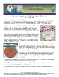


© Copyright 2026