
Oral Health Care for Patients with Epidermolysis Bullosa - Best
DOI: 10.1111/j.1365-263X.2012.01247.x
Oral Health Care for Patients with Epidermolysis Bullosa - Best
Clinical Practice Guidelines
Susanne M. Kra¨mer1,2,3, Marı´a Concepcio´n Serrano4, Gisela Zillmann1,3, Pablo Ga´lvez3,
Ignacio Araya5, Nicola´s Yanine5, Alonso Carrasco-Labra5, Patricio Oliva6,
Romina Brignardello-Petersen5 & Julio Villanueva5
1
Department of Paediatric Dentistry, Facultad de Odontologı´a, Universidad de Chile, Chile, 2Unit Of Oral Medicine and
Special Care Dentistry, UCL Eastman Dental Institute, London, UK, 3DEBRA Chile, 4Private practice, Valencia, Spain,
5
Evidence based Dentistry Unit, Facultad de Odontologı´a, Universidad de Chile, Chile and 6PhD candidate in Public Health
and Biomedical Research Methods, Universitat Auto`noma de Barcelona, Barcelona, Spain
International Journal of Paediatric Dentistry 2012; 22
(Suppl. 1): 1–35
Objective. To provide the users with information
on the current best practices for managing the
oral health care of people living with EB.
Methods. A systematic literature search, in which
the main topic is dental care in patients with
Epidermolysis Bullosa, was performed. Consulted
sources, ranging from 1970 to 2010, included
MEDLINE, EMBASE, CINAHL, The Cochrane
Library, DARE, and the Cochrane controlled trials
register (CENTRAL). In order to formulate the
recommendations of the selected studies the SIGN
system was used. The first draft was analysed and
discussed by clinical experts, methodologists and
patients representatives on a two days consensus
INTRODUCTION
DEBRA International is a worldwide network
of national groups working on behalf of those
affected by the genetic skin blistering condition, epidermolysis bullosa (EB).
Epidermolysis bullosa is a rare disease with
multiple oral manifestations, which requires a
special approach from the dental point of view.
Because of its low prevalence, many dentists
have limited knowledge of the disease. The scientific literature regarding oral health care of
people living with EB is relatively scarce. This
makes it difficult for dentists with no experience in treating people with EB to know how
Correspondence to:
Susanne Marie Kra¨mer, Department of Pediatric Dentistry,
Facultad de Odontologı´a, Universidad de Chile. Sergio
Livingstone 943, Independencia, Santiago, Chile. Email:
[email protected]
meeting. The resulting document went through
an external review process by a panel of experts,
other health care professionals, patient representatives and lay reviewers. The final document was
piloted in three different centres in United Kingdom, Czech Republic and Argentina.
Results. The guideline is composed of 93 recommendations divided into 3 main areas: 1) Oral Care
- access issues, early referral, preventative strategies, management of microstomia, prescriptions
and review appointments-, 2) Dental treatment:
general treatment modifications, radiographs,
restorations, endodontics, oral rehabilitation,
periodontal treatment, oral surgery and orthodontics-, and 3) Anaesthetic management of dental
treatment.
Conclusions. A preventive protocol is today’s dental management approach of choice.
to approach them in a safe manner given all
the special care these patients might need.
As part of their vision for working to ensure
access to the best quality support and medical
care for people living with EB, DEBRA International entrusted the development of Clinical Guidelines to health care professionals
with significant experience in EB around the
world.
It became necessary to gather experts from
different centres around the world to discuss
the different treatment alternatives and to work
towards establishing the best clinical practice
guidelines. These guidelines contain the appropriate precautions that people with EB might
require to receive optimal oral health care.
The present guidelines on dental care for people living with EB have been developed by an
international team of dentists, using a standard
methodology based on a systematic review of
the currently available scientific evidence.
2012 The Authors
International Journal of Paediatric Dentistry 2012 BSPD, IAPD and Blackwell Publishing Ltd
1
2
S. M. Kra¨mer et al.
These guidelines contain a chapter on general information on dental care of patients
with EB, followed by a chapter explaining the
precautions that should be taken into account
when treating patients with each subtype of
EB, as well as recommendations for dental
treatment. The appendix includes a glossary,
general information on EB, and a description
of its oral manifestations.
1.1 AIM
1)
2++
2+
2)
3
4
To provide the users with information on the
current best practices for managing the oral
health care of people living with EB.
1.2 USERS
Specialists in Paediatric Dentistry, Special
Care Dentistry, Orthodontics, Oral and Maxillofacial Surgery, Rehabilitation and General
Dental Practitioners, Dental hygienists, Paediatricians, Dermatologists, Dietitians, parents,
and those living with inherited epidermolysis
bullosa.
GRADES OF RECOMMENDATION
Note: The grade of recommendation relates to
the strength of the evidence on which the recommendation is based. It does not reflect the clinical
importance of the recommendation.
A
B
1.3 TARGET GROUP
These guidelines can be applied to all patients
diagnosed with epidermolysis bullosa. As
such, the guideline considers information for
all four major types of EB: EB simplex, junctional EB, dystrophic EB, and Kindler syndrome.
Meta-analyses, systematic reviews, or RCTs with a high risk
of bias
High-quality systematic reviews of case–control or cohort
studies
High-quality case–control or cohort studies with a very low
risk of confounding or bias and a high probability that the
relationship is causal
Well-conducted case–control or cohort studies with a low
risk of confounding or bias and a moderate probability
that the relationship is causal
Case–control or cohort studies with a high risk of
confounding or bias and a significant risk that the
relationship is not causal
Nonanalytic studies, for example, case reports, case series
Expert opinion
C
D
At least one meta-analysis, systematic review, or RCT
rated as 1++, and directly applicable to the target
population; or
A body of evidence consisting principally of studies
rated as 1+, directly applicable to the target population,
and demonstrating overall consistency of results
A body of evidence including studies rated as 2++,
directly applicable to the target population, and
demonstrating overall consistency of results; or
Extrapolated evidence from studies rated as 1++ or 1+
A body of evidence including studies rated as 2+,
directly applicable to the target population, and
demonstrating overall consistency of results; or
Extrapolated evidence from studies rated as 2++
Evidence level 3 or 4; or
Extrapolated evidence from studies rated as 2+
GOOD PRACTICE POINTS
1.4 Method used for Formulating the
Recommendation
To formulate the recommendations, from the
selected studies, the SIGN Guidelines were
used.
Recommended best practice based on the clinical
experience of the guideline development group
Fiftieth Guideline Developer’s Handbook,
NHS Scottish Intercollegiate Guidelines Network SIGN. Revised Edition January 2008.
LEVELS OF EVIDENCE
1++
1+
High quality meta-analyses, systematic reviews of RCTs, or
RCTs with a very low risk of bias
Well-conducted meta-analyses, systematic reviews, or RCTs
with a low risk of bias
2012 The Authors
International Journal of Paediatric Dentistry 2012 BSPD, IAPD and Blackwell Publishing Ltd
Clinical Guidelines for Oral Care in Epidermolysis Bullosa
3
2 Oral care for patients with inherited Epidermolysis
BullosaA
2.1 Introduction
A preventive protocol is today’s dental
management approach of choice1-3.
The approach to dental treatment for
patients with epidermolysis bullosa (EB), in
particular for those with the more severe
types, has changed dramatically over the last
30 years. Crawford et al.4 considered extraction of all teeth to be the treatment of choice
for patients with RDEB. Two decades later, in
1999, Wright5 declared that it was possible to
manage dental abnormalities successfully
with a combination of anaesthetic and restorative techniques. Recently, Skogedal et al.2
demonstrated that caries can be successfully
prevented in patients with RDEB by continuous follow-up aimed at dietary advice, oral
hygiene habits, frequent professional cleaning, and fluoride therapy.
D
2.2 Importance of oral preventive care and dental
treatmentB
1) To prevent and treat pain and infection.
This is important considering that
patients with oral pain will reduce their
nutritional intake.
2) To improve aesthetics and self-esteem.
3) A healthy dentition allows patients to
improve their nutritional state6, as they
will be able to chew better. Maintaining a functional dentition reduces the
potential for oral and oesophageal soft
tissue damage through more efficient
mastication7,8; however, a nutritional
consultation is important, as advice
must be given on the nutritional content of the diet and the consistency of
the food.
4) Improved phonetics when anterior
teeth are restored, allowing for better
positioning of the tongue.
A
The Oral manifestations of EB can be found in page 22.
This list was ordered according to the preferences of the patients and
their representatives.
B
5) Improved swallowing: doing mouth
opening exercises strengthens the muscles and allows for better access for
routine hygiene.
6) Maintaining a harmonious relationship
between teeth stabilizes the occlusion
for better function and aesthetics and
allows better hygiene.
2.3 Access to dental clinic
The clinic must be of easy access for
patients using wheelchairs and walking
frames.
Allow patients to accommodate on their
own giving them enough time. Do not
try to assist them if you are not aware of
the areas where they have wounds.
If the patient has to travel a long distance to attend the specialist dentist in
the EB unit, a shared care approach can
be arranged with a local dentist, who
can provide more regular preventative
care.
Access to dental care can be a challenge for
some patients. Even though in most developed countries it is guaranteed, it is still a
privilege for many patients around the world.
There is a lack of knowledge about the disease in the dental profession9 and other
healthcare professionals. Dental care can be
complicated by the fears of both the patient
and the dentist10. Allow yourself plenty of
time. Even the most simple procedures, such
as an oral exam, takes longer because of the
limited access, discomfort, or fear of developing blisters secondary to soft tissue manipulation.
2.4 Early referral
Members of the multidisciplinary team should
refer patients to the dentist before oral problems present, as early referral and close follow-up are the key to keeping patients as
healthy as possible from the oral point of
view (Image 1).
2012 The Authors
International Journal of Paediatric Dentistry 2012 BSPD, IAPD and Blackwell Publishing Ltd
4
S. M. Kra¨mer et al.
Image 1 Early diagnosis and education to parents on bullae
management.
Image 3 Early diagnosis of Generalized Enamel Hypoplasia
in JEB.
2.5 Oral bullae and ulcerations
Patients with EB should be referred to
the dentist for the first consultation at
the age of 3–6 months. The first consultation should be aimed at:
(a) Education of the parents and caregivers:
counselling on diet (including sugar-free
medications), oral hygiene routines, fluorides, technical aids, and oral manifestations of EB. This preventative advice
should be provided even before the teeth
erupt (Image 2).
(b) Early diagnosis of enamel abnormalities
such as those seen in junctional EB
(JEB). This is possible as soon as the first
primary tooth erupts (Image 3).
(c) Early diagnosis of tooth crowding, mainly
in recessive dystrophic EB (RDEB).
(d) Early diagnosis of incipient caries lesions.
Patients with EB should be referred to a
dentist as early as possible to identify any feature related to EB that needs special attention, for example, generalized enamel
hypoplasia5,10-13. This enables dentists to start
preventive programmes and reduces the risk
of developing dental diseases14. Many case
reports have shown that patients visit the
dentist only when they already have several
carious lesions or pain7,11,15,16.
Image 2 Early oral hygiene instruction.
Although oral bullae, ulcers, and erosions are
the most common oral feature of EB, there is
only one published study of a therapy for
these oral lesions. Marini et al.17 found that
sucralfate suspension reduced the development and duration of oral mucosal blisters
and ulcers, reduced the associated oral pain,
and improved plaque and gingival inflammation indices17.
2.6 Preventative Strategies
Oral Hygiene. At home – Concern is expressed
by some patients, parents, and dentists
regarding the use of tooth brushes and potential damage to the oral mucosa.
Tooth brushing is possible in all patients
with EB, even in patients with the
severe generalized RDEB subtype. The
following suggestions can help determine the appropriate toothbrush for
each patient:
(a) Small head5,7,8,11,13.
(b) Soft bristle5,8,11,13,18.
(c) Bristles can be further softened by soaking them in warm ⁄ hot water10,13.
(d) In patients with severe microstomia, short
bristles are indicated to access occlusal surfaces of molars. For this purpose, bristles
can be cut. If bristles are cut, one needs to
ensure that they remain soft and do not
harm the tissue.
(e) Special adaptations of the toothbrush
handle can be advantageous for patients
with pseudosyndactyly and manual dexterity problems (Images 4).
(f) Parents or caregivers are advised to assist
children, to improve plaque removal, and
D
2012 The Authors
International Journal of Paediatric Dentistry 2012 BSPD, IAPD and Blackwell Publishing Ltd
Clinical Guidelines for Oral Care in Epidermolysis Bullosa
helping to reduce the risk of tissue damage7.
(g) A manual toothbrush may be preferable
to an electric brush7 (There is no evidence to support this statement. This
reflects an author’s opinion).
(h) Special toothbrushes, for example, Collis
Curve (TM) toothbrush, might be a good
option for patients with RDEB, but more
research on its efficiency is needed
(Images 5).
(i) Cotton buds (cotton swabs), disposable
mini brushes, clean cotton cloth, or gauze
can be used to clean the teeth if a patient
is temporarily not able to brush because
the mouth is very sore.
D
Rinsing with water during the day, particularly after meals10,19, also helps oral
hygiene as it improves removing food
debris or sugar deposits.
Disclosing solution or tablets to help
identify dental plaque are a useful tool
to help patients assess their effectiveness
when brushing their teeth. They can be
used by all patients with EB.
D
Professional Hygiene. Gentle and careful
ultrasonic scaler and polish techniques
can be used in all patients, including
severe RDEB11. Haemorrhagic bulla can
appear because of vibration on the
mucosa. If this happens, they should be
drained.
Adjuvant therapies.
Chlorhexidine
D
Chlorhexidine 0.12% has been widely
advocated for oral disease prevention in
5
Image 5 Collis Curve (TM) toothbrush brushing the palatal
and buccal sides of the tooth simultaneously.
patients with EB5,7,10,11,16,19,20. It has
shown to be effective for candida while
ineffective for caries control.
A variety of application methods have
been used, including mouthwashes, swabs,
sprays, gels, and topical varnish applications.
An example of a preventative treatment
protocols is as follows: a rinse two times a
day for 2 weeks every 3 months.
d
d
D
Alcohol-free formulations are advised in
patients with oral lesions8,10,11.
Fluoride
D
Topical applications of high-dose fluoride
varnish are suggested every 3 months in
patients with high caries risk or at each
dental visit5,7,19.
For children resident in nonfluoridated
communities, the importance of daily
fluoride supplements has been highlighted10.
Fluoride can also be prescribed as a gel
preparation or mouth wash. Gel preparations
can be applied with a toothbrush, in a custom-made plastic tray10 or with cotton rolls.
Mouth wash formulations should be alcohol
free in patients with oral lesions. These
0.05% and 0.2% fluoridated solutions can
also be applied topically with a cotton bud on
all teeth once a day21.
D
Dietary modifications. As indicated previously,
the dietary habits ⁄ requirements of patients
with EB may increase the risk of caries.
D
Image 4 Patient with RDEB and pseudosyndactyly
performing oral hygiene.
A dietary caries-prevention programme
should be instigated at early age16,18.
2012 The Authors
International Journal of Paediatric Dentistry 2012 BSPD, IAPD and Blackwell Publishing Ltd
6
S. M. Kra¨mer et al.
It is essential that dentists and nutritionists collaborate on an appropriate programme for each patient, as opposed to
giving contradictory advice that may
confuse patients and parents ⁄ guardians.
Fissure sealants and other aids.
D
Sealing fissures and fossae has been
recommended, as oral hygiene and
other preventive measures can be difficult to perform10,13,22. Some clinical
experts, however, have apprehensions
regarding this advice, as the technique
is very sensitive and may not be an
option for some patients because of limited cooperation, compromised access,
and difficult long-term follow-up.
consulted literature provides no definitive
solutions. Slight increments in the maximum
oral aperture have been obtained with
mechanical techniques. Four techniques have
been described.
In one patient, resin plugs of progressively
increasing calibre increased maximal mouth
opening from 19 to 23 mm after 10 min of
exercise and to 30 mm at the end of a treatment session22. Unfortunately, this parameter
returned to the initial values on discontinuing
mechanical therapy.
Other suggestions include daily exercises
with wooden spatulas26, mouth trainer, and
threaded acrylic cone.
2.8 Prescriptions
Other remineralization techniques, such
as Recaldent (CPP-ACP), can also be used
for the noninvasive management of early
caries lesions in patients with EB.
When prescribing medications in tablet
form to patients with RDEB, it is important to consider that swallowing them
could be difficult because of oesophageal
stenosis or could cause oesophageal
trauma. Therefore, prescriptions should
be in soluble or liquid form. If sugar-free
preparations are not available, parents
should be advised of the sugar content
and advised ideally to brush or at least
rinse the child’s teeth with water
directly after administration of the medication to reduce the risk of decay.
2.7 Microstomia
Patients with severe generalized RDEB
should perform daily exercises to
improve ⁄ maintain a good mouth opening. This can be performed, for example,
during dressing changes.
Improving mouth opening also favours
phonation and swallowing.
Performing exercises half an hour before
dental treatment helps improving
access22.
Limited mouth opening has been reported
as the greatest clinical difficulty for providing
dental treatment23,24 as well as complicating
intubation (Image 6)25. In this context, the
D
2.9 Review appointments
D
(a)
(b)
(c)
(d)
(e)
Image 6 Microstomia makes the access to the oral cavity
difficult.
D
Frequency of dental review should be
scheduled on an individual basis according to the amount of plaque present and
risk of caries. Every 3–6 months may be
sufficient for some patients, and for others, monthly appointments may be necessary3,5,15,22,27. The review sessions
should be aimed at3,7,15,19,22:
Caries prevention ⁄ early diagnosis.
Professional plaque removal.
Topical fluoride application.
Dietary advice.
Review progress or deterioration of
patient’s oral condition.
As the predisposition to develop intraoral squamous cell carcinoma (OSSC)
2012 The Authors
International Journal of Paediatric Dentistry 2012 BSPD, IAPD and Blackwell Publishing Ltd
Clinical Guidelines for Oral Care in Epidermolysis Bullosa
increases with age, cancer screening
must be considered a very important
aspect of the review appointment in
patients with RDEB from the second
decade on19,28.
Any unusual ulcer or white or red
patches should be biopsied to ensure
that these do not represent pre-cancer or
cancer in the mouth.
Frequent recall visits have shown to be useful to maintain dental health in patients with
EB6,7,15. There are examples of patients who
previously had extensive carious teeth who
7
remained caries free when attending frequent
review appointments6,7. On the other hand,
clinical cases have been reported showing
that patients who failed to attend the review
visits developed several caries within 2 years,
despite a preventive programme being
explained11,16.
As many patients have to commute long
distances, review appointments should be
scheduled together with other health care
appointments. A shared care approach can be
considered.
2012 The Authors
International Journal of Paediatric Dentistry 2012 BSPD, IAPD and Blackwell Publishing Ltd
8
S. M. Kra¨mer et al.
3 Dental treatment
3.1 Treatment modifications and precautions
Even though patients with milder oral
involvement do not require many treatment
modifications, a careful approach benefits
every patient. Patients with the severe generalized RDEB subtype of EB require the most
specific precautions during treatment to minimize soft tissue damage.
Image 8 Five-year-old patient with JEB and no perioral
lesion.
3.1.1 EB simplex (EBS).
D
Most authors agree that routine dental
treatment can be provided5,22,29.
Clinicians should ask about history of mucosal
fragility because manipulation can precipitate
lesions in mildly affected patients5. Although
this has never happened to the members of the
expert consensus; we recognize that EB is very
diverse and that it could happen.
Image 9 Two-year-old patient with JEB and severe perioral
granulation tissue.
3.1.2 Junctional EB (JEB).
D
Dental management does not require
many modifications4; however, a careful
approach is advised as tissue manipulation
can produce oral ulceration (Image 7).
This group requires an aggressive preventive programme and frequent visits to the
dentist as they present with generalized
enamel hypoplasia, leading to an increased
risk of cavities and severe attrition.
Mucosal and skin fragility vary considerably
between subtypes of JEB and patients, and
the avoidance of adhesive contact with the
skin and careful manipulation is always
advised. Following the suggestions listed in
‘Recessive DEB’ can be of help for these
patients as well.
This group of patients will require a special
dental rehabilitation plan, as they present
with generalized enamel hypoplasia (Images 8
and 9).
3.1.3 Dominant DEB (DDEB).
D
Patients with DDEB are able to receive
routine dental treatment with little or no
modifications28. Nevertheless, a careful
approach is still advised as tissue manipulation can produce oral ulceration.
There is a report of a patient who has been
wearing dentures for several years without
difficulties4.
3.1.4 Recessive DEB (RDEB).
Patients with the severe generalized RDEB subtype of EB require several treatment modifications and a careful approach to avoid as much
tissue damage as possible. Management of
these patients ideally requires a well-organized
multidisciplinary team approach27,30 with good
communication involving case discussion.
1. Lubrication
Image 7 Ulcer on the tongue and commissure of a patient
with JEB.
D
Lips should always be lubricated
with Vaseline ⁄ petrolatum or other
2012 The Authors
International Journal of Paediatric Dentistry 2012 BSPD, IAPD and Blackwell Publishing Ltd
Clinical Guidelines for Oral Care in Epidermolysis Bullosa
appropriate lubricants before any procedure is performed to reduce adherence and reduce shearing forces that
lead to tissues separation and lesions
formation1,5,18,27,31.
There have been reports suggesting the
lubrication of the buccal mucosa and instruments as well, but the consensus group
believes this does not benefit the patient and
makes treatment more difficult.
In the operating room, a water-soluble
lubricant should be used instead of petrolatum because it is not flammable.
2. Suction tip
D
Bullae formation or epithelial sloughing
can occur upon contact with the suction
tip1. It is suggested to lean the suction
tip or saliva ejector upon hard tissue,
that is, on occlusal tooth surface or on a
wet cotton roll32 (Image 10).
Avoid use of high vacuum suction as
this could cause sloughing of extensive
areas of tissue.
3. Bullae
D
Blood- or fluid-filled bullae that occur
during treatment should be drained with
a sterile needle or by a cut with scissors
to avoid lesion expansion because of
fluid pressure13,22,23,33.
4. Pressure
D
Extreme care of fragile tissues is important. To handle tissues, a little pressure
(compressive forces) can be applied, but
no sliding movements (lateral traction or
other shear forces) should be used, as
these can cause tissue sloughing5,11,23.
9
5. Air syringe
D
Can be used but should be managed
carefully11. Air-filled bullae can occur. If
this happens, they have to be drained.
6. Instruments
D
Because of limited access, it is easier to
use paediatric size instruments13.
A laryngeal mirror can also be helpful in
patients with severe microstomia. Flat
malleable retractors are useful for separating the cheeks, as they spread force over
larger area and can protect tissue if having
to prep a tooth for restorative treatment.
They come in various widths and are typically available in hospital operating rooms.
7. Isolation
Relative isolation: When using cotton
rolls, it is advised to lubricate them with
Vaseline ⁄ petrolatum or other aqueous
products for intraoral lubrication before
placing them inside the mouth. When
removing them, they must be soaked
with water.
Consider reducing the size of the cotton
rolls so they can fit in limited spaces.
Complete isolation: Rubber dam can be
used with or without clamps, aided with
wooden wedges. Use with caution as the
placement and position of the clamp,
and the rubber dam against the lip and
cheeks can cause blisters.
8. Visual access
In severe microstomia, it is easier to separate the lip using the handle of the mirror instead of the mirror itself, or flat
malleable retractors as explained before.
When possible, consider use of head
light.
9. End of session
Image 10 Suction tip leaned on tooth surface to avoid
mucosal sloughing.
At the end of every clinical session, it is
important to check for fluid-filled blisters
and drain them. It is also important to
check and remove any remnants of dental materials in the sublingual space or
vestibule, as the patients have ankyloglossia and cannot clear the mouth easily. This
can be carried out with a wet cotton roll.
2012 The Authors
International Journal of Paediatric Dentistry 2012 BSPD, IAPD and Blackwell Publishing Ltd
10
S. M. Kra¨mer et al.
3.1.5 Kindler Syndrome:
D A careful approach is advised, as mucosal
sloughing can form following dental
treatment such as scaling34.
The scarce literature available suggests periodontal health as main area of concern for
dental therapy34,35.
isolation, caries risk, and cultural and
economic factors. The use of stainless
steel crowns should be considered.
The atraumatic restorative treatment
(ART) technique can be used in difficult
or special circumstances.
D
Restorations and dentures should be
carefully adapted and highly polished to
lessen the risk of iatrogenic oral mucosal
blisters and ulcers18.
D
Iatrogenic blisters can develop after
treatment even if all precautions are in
place22.
D
Soft tissue lesions resulting from restorative treatment typically heal in one to
two weeks and require no specific treatment32.
3.2 Dental radiographs:
In patients with EBS, JEB, DDEB, and
Kindler syndrome, all diagnostic techniques can be used with no or little
technique modification.
D
In patients with severe generalized
RDEB, periapical technique has been
proved to be extremely difficult ) especially in the posterior area – because of
microstomia, ankyloglossia, and scarring
of the sublingual area. Orthopantomography (panoramic) is the radiograph of
choice32.
Other techniques that can also be helpful and diagnostic are as follows:
Bitewings using small films.
Some digital panoramic radiographs have
extraoral bitewings capabilities making
them a good option for patients with limited access.
As an alternative, an occlusal technique
can also be used for anterior teeth or lateral oblique for mandibular posterior teeth.
d
d
d
D
If periapical radiographs are required in
RDEB, care must be taken not to damage the mucosa11. Lubrication of the
film packet has been advised to avoid
tissue damage36.
3.3 Restorations
Restorative treatment can be difficult in
patients with RDEB because of microstomia,
soft tissue fragility, and complex anaesthetic
management37.
D
There are no contraindications to the
use of conventional dental materials5,38.
The restorative material to be used will
depend on the possibility of achieving
If required, analgesics can be prescribed.
3.4 Endodontics
D
Root canal treatment (endodontic treatment) can be performed in all patients,
unless there is no access because of limited mouth opening32.
In patients with severe microstomia,
access to the pulp chamber might need
to be modified. For example, anterior
teeth might need vestibular access.
For determining root canal working
length in patients with RDEB and severe
microstomia, it is best to use electronic
apex locators or, if unavailable, a panoramic radiograph (as periapical radiographs are difficult to take).
Concern has been raised regarding the
use of hypochlorite when isolation is not
ideal. The experience of the working
group is that there are no major problems using this agent.
3.5 Impression taking
Although there are no reports of any
adverse events (i.e., mucosal damage),
impressions should be taken with special
care in RDEB32,39,40.
2012 The Authors
International Journal of Paediatric Dentistry 2012 BSPD, IAPD and Blackwell Publishing Ltd
Clinical Guidelines for Oral Care in Epidermolysis Bullosa
11
RDEB11,18, improving aesthetics, oral
function, and enhancing patients’ confidence (Image 11)18.
Image 11 Before and after fixed crown restoration.
All type of impression material can be
used.
D
Microstomia can be a real challenge. As
an alternative to stock impression trays,
specially cut topical gel application trays
and custom-made acrylic trays have been
proposed18. Another alternative is to do a
first impression with hard (putty) silicone
and to use this as a tray adding light body
silicone on a second step.
If the cervical margin is subgingival, a
gingivectomy may be needed. For information on this matter, consult the Gingivectomy section.
D
Computer-generated
stereolitographic
template can be a noninvasive harmless
impression solution for surgical and
prosthodontic implant planning and
placement in RDEB24.
3.6 Oral rehabilitation
Oral rehabilitation can be fixed or removable
depending on the health system and financial
possibilities.
Whenever possible, fixed rehabilitation
is advised.
A. Fixed rehabilitation.
D
The use of stainless steel crowns has
been reported as a successful approach
in children with RDEB and JEB4,5,22,41.
D
Successful oral rehabilitation with fixed
bridges has been reported in several
patients
with
severe
generalized
In cases with generalized enamel hypoplasia, restoration of the entire dentition
with full crowns may be necessary.
This treatment has to be planned carefully
and discussed with the parents and the
patient, as it may consist of several stages
until full permanent dentition has been established and restored33,42.
B. Removable dentures.
The tolerance to bear tissue-supported dentures depends on the degree of mucosal fragility of each EB subtype and patient.
D
Reports of patients successfully tolerating
these types of dentures include patients
with EBS, JEB, DDEB, and pre-tibial
RDEB4,22,43,44.
Few patients with RDEB can bear dentures if the buccal margins are adapted
and the retainers are flat.
D
Overdentures have been described as a
practical, economic, nonsurgical treatment option for patients with JEB and
generalized hypoplastic enamel who
present with failure of eruption43. Careful follow-up is needed because of the
high risk of caries.
C. Implant rehabilitation.
C
Fixed prostheses are the rehabilitation
technique of choice31,39.
Short dental arch rehabilitation scheme
is advised39,40.
A variety of implant supported prostheses
can be considered for complete denture rehabilitation, such as fixed bridges or overdentures31,39,40. The level of satisfaction after
implant therapy in one series was slightly
higher in the fixed prosthesis group (n = 3,
mean 9.6) than in the overdenture group
(n = 3, mean 8.8). Oral mucosal ulcerations
were observed in areas in contact with overdentures, whereas in patients with fixed dentures, the tissues appeared healthier31.
Limited mouth opening, limited posterior
D
2012 The Authors
International Journal of Paediatric Dentistry 2012 BSPD, IAPD and Blackwell Publishing Ltd
12
S. M. Kra¨mer et al.
space, and oral hygiene difficulties may make
it necessary to use a short dental arch rehabilitation scheme39,40.
3.8.4 Biopsy.
Biopsies of oral tissues may be required
when a squamous cell carcinoma (SCC)
is suspected.
3.7 Periodontal treatment
D
Periodontal treatment can be performed
in all patients with EB. Special care must
be taken in patients with RDEB, as there
might be substantial bleeding during the
procedure11,32.
3.8 Oral Surgery
3.8.5 Surgical Extractions. Contemporary oral
health care is targeted at prevention of oral
disease, but some patients still require extractions because of severe caries or the need for
orthodontic care that involves severe dental
crowding. Surgical ⁄ difficult extractions should
be performed by an oral surgeon.
D
When planning surgical extractions,
especially if multiple extractions are
needed, it is advisable to consult the
patient’s physician as profound anaemia
could complicate the dental surgery30.
D
For multiple extractions, it has been suggested to extract first the anterior teeth
(i.e., from premolar to premolar) and then
the molars to allow optimal access30.
D
An atraumatic technique should be used,
making firm and safe mucosal incisions
to prevent bullae formation10,23.
3.8.1 Suturing. There has been debate in the
literature about the feasibility of suturing
after oral surgery in patients with
EB7,9,23,27,41,45.
Sutures can be used safely in all patients
with EB, but need careful placement.
3.8.2 Gingivectomy.
Gingivectomy using a laser or electric
scalpel is the technique of choice. In
patients with Kindler syndrome, this
technique may be needed to remove hyperplasic gingival papillae.
3.8.3 Vestibuloplasty. Severe obliteration of
the vestibule can cause difficulty in eating46,
performing oral hygiene46, providing dental
treatment, and reduces food clearance
because of reduced mobility.
Periodontal plastic surgery and vestibuloplasty to deepen the vestibule or to
restore the alveolar ridge height has
been reported in two patients with
dominant dystrophic EB (DDEB)46,47.
The consensus of experts has limited
but positive experience on this kind of
surgery in patients with RDEB. This
surgery is recommended when required,
that is, when the obliteration affects the
patient’s quality of life or oral function.
D
Inserting a soft acrylic stent extending to
the newly established vestibule avoids
fusion of the connective tissue layers
and allows time for epithelium migration
on both surfaces47.
Haemostasis can be achieved with gentle
pressure using gauze packs9,41. These
should be wet to avoid tissue adherence.
Some authors have reported the extraction
of healthy third or even second permanent
molars in patients with severe generalized
RDEB to improve or facilitate oral
hygiene2,48. There is controversy among different authors about this intervention. Severe
tooth crowding12,22,49, reduced alveolar
arches secondary to growth retardation8,50,
and severe microstomia1,7,22,23,31,45,51,52 are
described in patients with severe generalized
RDEB, which would justify preventive extractions. However, nowadays most patients
receive dietetic advice that optimizes nutrition
and growth. They receive orthodontic treatment (serial extractions) and are advised on
exercises to improve microstomia. Therefore,
preventive extractions of permanent molars
need to be assessed very carefully on an individual basis.
D
Perioperative
complications:
Despite
attempts to use as gentle manipulation as
2012 The Authors
International Journal of Paediatric Dentistry 2012 BSPD, IAPD and Blackwell Publishing Ltd
Clinical Guidelines for Oral Care in Epidermolysis Bullosa
possible and all the special precautions,
mucosal sloughing and blister formation have
been reported after almost every surgical
extraction
in
patients
with
severe
RDEB1,9,22,30,41. Blisters can arise at the
angles of the mouth, lips, vestibule, tongue,
and any sites of manipulation (Image 12);
some measuring up to 4 by 3 cm1,30. In some
instances, they might only be noticed by the
patient or carer only on the second post-operative day9.
Image 12 Bullae, ulcers, and mucosal sloughing after
surgical extractions.
Post-operative complications: Despite the
potential for extensive mucosal damage during surgery, post-operative complications are
rare9,30,53. Healing of the oral tissues occurs
gradually after one to 2 weeks16,21,41. Healing
of the alveolar sockets seems to be uneventful6,9. Nevertheless, there is a suggestion that
scarring of the oral commissure can be accentuated after surgery1,9. The use of post-operative antibiotics will depend on each
individual case.
3.8.6 Osseointegrated implants.
D
To avoid destruction of the atrophic
residual alveolar ridges of the maxilla,
an osteotome technique is advised23,31.
D
Surgical management can be complicated by bleeding and bullae23,31,54.
When needed, bone grafts can be
placed simultaneously with implants to
reduce the number of surgical interventions and, therefore, mucosal ⁄ skin damage54.
Successful rehabilitation using dental
implants has been reported in patients with
D
13
generalized RDEB, non-Herlitz JEB, and
RDEB-I5,23,31,55. The most extensive report
encompasses 38 dental implants with a success rate of 97.9% with a follow-up of 1–
9 years (average 5.5). Peri-implant mucosa
remained in good condition in all
patients24,31,54. It has been reported that after
rehabilitation, patients improved their ability
to chew, swallow, and their quality of
life23,31,39,40. Block and particulate allograft
and autografts have been used successfully in
patients with RDEB54. For information on stereolitography, see Impression taking.
These results are encouraging and dental
implants seem a possible solution for edentulous patients with EB and mucosal fragility. It
is important to note, however, that patients
with RDEB and JEB have been shown to
have lower bone mineral density scores56.
There has been clinical evidence of bone
atrophy
during
implant
surgery
as
well23,31,40.
When planning this type of rehabilitation,
advice from the medical team should be
sought, as extensive surgery might need to be
delayed or discouraged because of concomitant pathology as, for example, severe anaemia or poor prognosis SCC.
3.9 Orthodontics
D
Orthodontic treatment typically only
requires minor modifications in patients
with EBS, JEB, and DDEB5. Patients
with EBS Dowling–Meara, however, can
have more mucosal fragility requiring
the precautions indicated below.
For patients with RDEB, we strongly
recommend serial extractions to prevent
dental crowding, as this contributes to
high caries risk and periodontal disease.
a. The aim of orthodontics in RDEB should
be to obtain tooth alignment.
b. Serial extractions should be performed at
the appropriate stage of dental development.
c. A risk-benefit analysis should be performed on an individual basis to avoid the
need for repeated general anaesthetic for
serial extractions. These procedures should
2012 The Authors
International Journal of Paediatric Dentistry 2012 BSPD, IAPD and Blackwell Publishing Ltd
14
S. M. Kra¨mer et al.
ideally be performed with behavioural
management techniques and local anaesthesia.
In patients with RDEB, it is possible to
achieve tooth movement using fixed orthodontics, such as to: (1) correct a one tooth
cross-bite, (2) close diastema, and (3) align
the anterior teeth.
A tooth-borne removable appliance may
also be possible, for example, inclined, anterior bite plane to correct a cross-bite.
D
To prevent lesions on the soft tissues,
orthodontic wax ⁄ relief wax can be
applied on the brackets48.
2012 The Authors
International Journal of Paediatric Dentistry 2012 BSPD, IAPD and Blackwell Publishing Ltd
Clinical Guidelines for Oral Care in Epidermolysis Bullosa
15
4 Anaesthetic management
All kinds of dental treatment for patients with
EB can be provided under local anaesthesia,
conscious sedation, or general anaesthesia.
The decision on which type of anaesthetic
management approach to choose must be
agreed between the patient and the dentist
based on the risks, advantages, and disadvantages of each technique, as well as the availability of specialized services. It is important
to highlight that conscious sedation should
not be performed in-office on patients with
potential for compromised airway or difficult
intubation.
For patients with mild forms of EB and for
small, atraumatic procedures, using local
anaesthesia is the technique of choice. General anaesthesia can be indicated for some
extensive procedures in patients with severe
forms of EB, but the support of an experienced team is crucial.
4.1 Local anaesthesia
Topical anaesthesia in gel form can be
used normally.
D
To avoid blister formation, the anaesthetic solution must be injected deeply
into the tissues and at a slow rate, to
avoid the liquid causing mechanical separation of the tissue5,23,31.
Iatrogenic blisters may develop following
local anaesthesia injection6,11,18,21–23,57.
If this happens, the lesions have to be
drained.
Post-operative instruction must highlight
that the patients should not bite, rub, or
traumatize their lip while under the
effect of local anaesthesia.
The main benefits of local anaesthesia are
that it maintains airway patency and provides
prolonged post-operative pain relief. Examples of successful treatments provided under
local anaesthesia include multiple extractions,
implants, root canal treatment, and restorations6,16,23.
Some authors suggest that less mucosal
damage is produced when patients are treated
under local anaesthesia when compared to
general anaesthesia.
4.2 General anaesthesia
D
When planning a procedure under general anaesthesia, the patient’s MD ⁄ GP
should be consulted13.
The availability of an anaesthetic team
with experience in EB is crucial. If this
is not available, the use of local anaesthesia should be considered.
Treatment under general anaesthesia allows
the provision of extensive reconstructive dental treatment and multiple extractions regardless of the severity of soft tissue fragility and
microstomia present5,7. The fact that the
patient will be asleep, however, does not
mean that the procedure will be easy to perform. Patients with severe fragility will still
develop intra-operative generalized mucosal
sloughing secondary to retraction and minor
trauma of the procedure itself1,7,36. Oral surgery and restorative procedures can be combined with other surgical procedures, as for
example, oesophageal dilatation1. As stated
previously, a water-soluble lubricant should
be used instead of petrolatum in the operating room because it is not flammable.
2012 The Authors
International Journal of Paediatric Dentistry 2012 BSPD, IAPD and Blackwell Publishing Ltd
16
S. M. Kra¨mer et al.
5 Summary of recommendations
D
A preventive protocol is today’s dental
management approach of choice1,2.
Dietary modifications
D
Early referral
A dietary caries-prevention programme
should be instigated at early age16,18.
It is essential that dentists and nutritionists collaborate on an appropriate programme for each patient, as opposed to
giving contradictory advice that may
confuse patients and parents ⁄ guardians.
Patients with EB should be referred to
the dentist for the first consultation at
the age of 3–6 months.
Oral hygiene
D
(a)
(b)
(c)
(d)
(e)
Tooth brushing is possible in all patients
with EB, even in patients with the
severe generalized RDEB subtype. The
following suggestions can help determine the appropriate toothbrush for
each patient:
Small head5,7,8,11,13.
Soft bristle5,8,11,13,18.
Bristles can be further softened by soaking them in warm ⁄ hot water10,13.
In patients with severe microstomia,
short bristles are indicated to access
occlusal surfaces of molars. For this purpose, bristles can be cut.
Parents or caregivers are advised to assist
children, to improve plaque removal, and
helping to reduce the risk of tissue damage7.
Microstomia
Patients with severe generalized RDEB
should perform daily exercises to
improve ⁄ maintain a good mouth opening. This can be performed, for example,
during dressing changes.
Prescriptions.
When prescribing drugs to patients with
RDEB, it is important to consider that
they often present oesophageal stenosis.
Therefore, prescriptions should be in
liquid form, that is, soluble painkillers
(analgesics), ideally a sugar-free form.
Review appointments.
D
Frequency of dental review should be
scheduled according to the risk of caries
every 3 to 6 months5,15,22,27.
D
As the predisposition to develop intraoral carcinoma (SSC) increases with age,
cancer screening must be considered a
very important aspect of the review
appointment in patients with RDEB
from the second decade on19,28.
Professional hygiene
D
Gentle and careful ultrasonic scaler and
polish techniques can be used in all
patients, including severe RDEB11.
Fluoride
D
D
Topical applications of high-dose fluoride
varnish are suggested every 3 months in
patients with high caries risk or at each
dental visit5,7,19.
For children resident in nonfluoridated
communities, the importance of daily
fluoride supplements has been highlighted10.
Treatment modifications – precautions
EB simplex (EBS).
D
Routine dental treatment can be provided5,22,29.
Junctional EB (JEB).
D
Dental management does not require
many modifications4; however, a careful
2012 The Authors
International Journal of Paediatric Dentistry 2012 BSPD, IAPD and Blackwell Publishing Ltd
Clinical Guidelines for Oral Care in Epidermolysis Bullosa
approach is advised as tissue manipulation can produce oral ulceration.
This group of patients requires an
aggressive preventive programme and
frequent visits to the dentist as they
present enamel hypoplasia ⁄ defects, leading to an increased risk for cavities and
severe attrition.
Dominant DEB (DDEB).
D
Patients with DDEB are able to receive
routine dental treatment with little or
no modifications28.
5. End of session
At the end of every clinical session, it is
important to check for fluid-filled blisters and drain them. It is also important
to check whether there are remnants of
dental materials.
Kindler syndrome
D
In patients with severe generalized
RDEB, periapical technique is difficult
in the posterior area because of microstomia, ankyloglossia, and scarring of
the sublingual area. Orthopantomography (panoramic) is the investigation of
choice. Other alternatives are as follows:
small films bitewings, extraoral bitewings capabilities in panoramic radiographs (if equipment is available), and
occlusal or lateral oblique techniques.
D
Bullae formation or epithelium sloughing can occur upon contact with the suction tip1. It is suggested to lean the
suction tip or saliva ejector upon hard
tissue, that is, on the tooth surface. High
vacuum suction should be avoided.
3. Bullae
Blood- or fluid-filled bullae that occur
during treatment have to be drained
with a sterile needle or by a cut with
scissors to avoid lesion expansion
because of fluid pressure13,22,23,33.
4. Pressure
D
D
Extreme care of fragile tissues is important. To handle tissues, a little pressure
(compressive forces) can be applied, but
no sliding movements (lateral traction
or other shear forces) should be used, as
these can cause tissue sloughing5,11,23.
A careful approach is advised, as mucosal sloughing can form following dental
treatment34.
Dental radiographs.
Recessive DEB (RDEB).
Patients with the severe generalized RDEB subtype of EB require several treatment modifications and a careful approach to avoid as much
tissue damage as possible. Management of
these patients ideally requires a well-organized
multidisciplinary team approach27,30 with good
communication involving case discussion.
1. Lubrication
Lips should always be lubricated with
Vaseline ⁄ petrolatum or other appropriate lubricant before any procedure is
performed to reduce adherence and
lesions formation1,5,18,27,31.
2. Suction tip
17
Restorations.
D There are no contraindications to the use
of conventional dental materials5,38.
D
Restorations and dentures should be
carefully adapted and highly polished to
lessen the risk of iatrogenic oral mucosal
blisters and ulcers18.
Endodontics.
D
Root canal treatment (endodontic treatment) can be performed in all patients,
unless there is no access because of limited mouth opening32.
Oral rehabilitation.
Whenever possible, fixed rehabilitation is
advised.
In cases with generalized enamel hypoplasia, restoration of the entire dentition
with full crowns may be necessary.
2012 The Authors
International Journal of Paediatric Dentistry 2012 BSPD, IAPD and Blackwell Publishing Ltd
18
S. M. Kra¨mer et al.
Suturing.
Sutures can be used safely in all patients
with EB, but need careful placement.
Surgical extractions.
D
When planning surgical extractions,
especially if multiple extractions are
needed, it is advisable to consult the
patient’s physician as profound anaemia could complicate the dental surgery30.
conscious sedation, or general anaesthesia.
The decision on which type of analgesia to
choose will have to be agreed between the
patient and the dentist based on the advantages and disadvantages of each technique, as
well as the availability of specialized services.
It is important to highlight that conscious
sedation should not be performed in-office on
patients with potential for compromised airway or difficult intubation.
Local anaesthesia.
D
Osseointegrated implants.
To avoid destruction of the atrophic
residual alveolar ridges of the maxilla,
an osteotome technique is advised23,31.
To avoid blister formation, the anaesthetic solution should be injected deeply into
the tissues and at a slow rate, to avoid the
liquid causing mechanical separation of the
tissue5,23,31.
General anaesthesia.
Orthodontics.
For patients with RDEB, we strongly recommend serial extractions to prevent
dental crowding, as this contributes to
high caries risk and periodontal disease.
Anaesthetic management
D
When planning a procedure under general anaesthesia, the patient’s MD ⁄ GP
should be consulted13.
The availability of an anaesthetic team
with experience in EB is crucial. If this
is not available, the use of local anaesthesia should be considered.
All kinds of dental treatment for patients with
EB can be provided under local anaesthesia,
2012 The Authors
International Journal of Paediatric Dentistry 2012 BSPD, IAPD and Blackwell Publishing Ltd
Clinical Guidelines for Oral Care in Epidermolysis Bullosa
19
6 Development of the guideline
6.1 The guideline development group
6.1.1 Clinical Experts.
Prof. Dr. Susanne Kra¨mer
Dr. Marı´a Concepcio´n Serrano
Prof. Dr. Gisela Zillmann
Dr. Pablo Ga´lvez
Department of Paediatric Dentistry, Facultad de Odontologı´a, Universidad de Chile
Oral Medicine and Special Care Dentistry Unit, UCL Eastman Dental Institute, London, UK
Dentist, DEBRA Chile
Private practice, Valencia, Spain
Department of Paediatric Dentistry, Facultad de Odontologı´a, Universidad de Chile
Dentist, DEBRA Chile
Dentist, DEBRA Chile
6.1.2 Patient Representatives.
Mr. John Dart
Mr. Scott O’Sullivan
Chief Operating Officer. DEBRA International, UK
Patient representative, UK
6.1.3 Methodologists.
Prof. Dr. Julio Villanueva
Dr. Ignacio Araya
Dr. Alonso Carrasco-Labra
Dr. Romina Brignardello-Petersen
Mr. Patricio Oliva
Dr. Nicola´s Yanine
Prof. Dr. Marco Cornejo
Evidence based Dentistry Unit, Facultad de Odontologı´a,
Evidence based Dentistry Unit, Facultad de Odontologı´a,
Evidence based Dentistry Unit, Facultad de Odontologı´a,
Evidence based Dentistry Unit, Facultad de Odontologı´a,
PhD candidate in Public Health and Biomedical Research
Universitat Auto`noma de Barcelona, Barcelona, Spain.
Evidence based Dentistry Unit, Facultad de Odontologı´a,
Evidence based Dentistry Unit, Facultad de Odontologı´a,
6.2 Funding for Guide Development
The guideline was funded by a grant from
DEBRA UK.
6.3 Updating Procedure
The guideline will be updated every two years
after its first version. If new relevant evidence
is detected before the update, the information
will be published on the web site http://
www.debra-international.org/.
The team in charge of this update will be
formed by Dr. Susanne Kra¨mer and Dr. Julio
Villanueva in 2013
6.4 Methodology
6.4.1 Systematic Literature Searching. Literature
Sources
The literature search ranged from 1970 to
November 2010. Consulted sources included
the electronic databases MEDLINE (1970 to
November 2010), EMBASE (1980 to November 2010), CINAHL (1980 to November 2010),
Universidad
Universidad
Universidad
Universidad
Methods,
de
de
de
de
Chile
Chile
Chile
Chile
Universidad de Chile
Universidad de Chile
The Cochrane Library (2010), DARE (2010),
and the Cochrane controlled trials register
(CENTRAL) (2010). In addition, handsearching journals, reviewing conference
proceedings, and other guidelines sources
such as The US National Guideline Clearinghouse and The German Guidelines Clearinghouse were carried out.
Dissertations, conference proceedings, technical reports, and other unpublished documents that meet the selection criteria were
also included. The reference lists of all papers
for relevant citations were reviewed. When
all the relevant studies were identified, they
were sent to the experts to review for completeness.
Selection criteria of the articles – Primary or
secondary articles in which the main topic is
dental care (diagnosis, and ⁄ or treatment
and ⁄ or prognosis) in patients with epidermolysis bullosa, published between 1970 and
2010 in English, Spanish, French, German, or
Italian were considered.
Search strategy – To identify studies for
this review, detailed search strategies were
2012 The Authors
International Journal of Paediatric Dentistry 2012 BSPD, IAPD and Blackwell Publishing Ltd
20
S. M. Kra¨mer et al.
developed for each database. These were
based on the search strategy developed for
MEDLINE, but revised appropriately for each
database.
The search strategy used a combination of
controlled vocabulary and free text terms
based on:
#1 (Epidermolysis Bullosa):ti, ab, kw
#2 MeSH descriptor epidermolysis bullosa
explode all trees
#3 (Dentistry): ti, ab, kw
#4 MeSH descriptor Oral Health explode all
trees
#5 (Mouth Disease MeSH term)
#6 (Mouth Disease): ti, ab, kw
#7 (Mouth Rehabilitation MeSH term)
#8 (#1 AND #3)
#9 (#2 OR #3)
#10 (#1 AND #4)
#11 (#1 AND #5)
#12 (#2 AND (#5 OR #7))
# 13 (#1 AND (#4 OR #6 OR #7))
#14 (#8 AND #6)
With the aim of seeking specifically for randomized controlled trials and epidermolysis
bullosa, the search terms described above
were combined with the following terms:
1) Randomized controlled trial.pt.
2) Controlled clinical trial.pt.
3) Randomized controlled trials.sh.
4) Random allocation.sh.
5) Double-blind method.sh.
6) Single-blind method.sh.
7) or ⁄ 1–6
8) Animal ⁄ not human ⁄
9) 7 not 8
10) Clinical trial.pt.
11) exp clinical trials ⁄
12) (clin$ adj25 trial$).ti,ab.
13) [(singl$ or doubl$ or trebl$ or tripl$)
adj25 (blind$ or mask$)].ti,ab.
6.4.2 Methods Used for Formulating the
Recommendations. To formulate the recommendations of the selected studies, the SIGN
system was used as described on the 50
Guideline Developer’s Handbook, NHS Scottish Intercollegiate Guidelines Network SIGN.
Revised Edition January 2008 (See figure on
page 2 of this guideline).
6.5 External Review
6.5.2 Patient Group. Patients and representatives from the DEBRA association groups of
Australia, Belgium, Canada, Germany, New
Zealand, and the United Kingdom were
invited to review the document in order to
make sure that the degree to which the evidence addresses patients’ concerns is reflected
in the guideline.
6.5.3 Other health care professionals and lay
reviewers.
Anne W
Lucky, MD
Acting Director, Division of Pediatric dermatology
Cincinnati Children’s Hospital. Cincinnati,
Ohio, USA
Professor of Dermatology and Pediatrics
The University of Cincinnati College of Medicine
Cincinnati, Ohio USA
Lesley Haynes
Formerly Principal Paediatric Dietitian
for EB, Great Ormond Street Hospital for
Children, London, UK
Lynne Hubbard Specialist Dietitian, St. Thomas’ Hospital,
London, UK
Christian
Lay reviewer, Chile
Fingerhuth
6.5.1 Specialist Review:
Prof. Dr. Tim Wright
Dr. Marie Callen
Dr. Carol Mason
Prof. Dr. Stephen Porter
Dr. Nina Skogedal
Dr. Kari Storhaug
Dr. Reinhard Schilke
Bawden Distinguished Professor and Chair, The Department of Pediatric Dentistry, The
University of North Carolina, Chapel Hill, NC USA
Private practice, Cincinnati, USA
Consultant in Paediatric Dentistry, Great Ormond Street Hospital for Children NHS Trust, London, UK
Institute Director and Professor of Oral Medicine, UCL Eastman Dental Institute, London, UK
Specialist in Paediatric Dentistry, National Resource Centre for Oral Health in Rare Medical
Conditions (TAKO-centre), Lovisenberg Diakonale Hospital, Oslo, Norway.
Director dr.odont., National Resource Centre for Oral Health in Rare Medical
Conditions (TAKO-centre), Lovisenberg Diakonale Hospital, Oslo, Norway.
Department of Conservative Dentistry, Periodontologie und Preventive Dentistry,
Hannover Medical School, Germany.
2012 The Authors
International Journal of Paediatric Dentistry 2012 BSPD, IAPD and Blackwell Publishing Ltd
Clinical Guidelines for Oral Care in Epidermolysis Bullosa
6.6 Pilot
d
The guideline was piloted in three centres for
a period of three months. At the end of the
pilot period a feedback form was sent to the
authors.
d
d
Dr. Victoria Clark
Dr. Gabriela Scagnet
Dr. Mariana Armada
Dr. Adela Stepanska
Dr. Renata Gaillyova
Dr. Sylvia Stepanska
Consultant in Paediatric Dentistry,
Birmingham Children s Hospital, UK
Dentist, DEBRA Argentina & Universidad
de Buenos Aires and Hospital de
Odontologı´a Infantil Quinquela Martin
Gobierno, Buenos Aires, Argentina
Hospital de Odontologia Infantil
Quinquela Martin Gobierno, Buenos
Aires, Argentina
Dentist, DEBRA Czech Republic
Department of Genetics, University
Hospital, Brno, Czech Republic
Practical dentist, Brno, Czech Republic
Lack of knowledge and training of some
health professionals to implement the recommendations.
Lack of patient’s adherence to the recommendations.
Insufficient provision of health services in
some parts of the world.
6.9 Further areas of research:
d
d
d
d
6.7 Patient involvement
One patient, Scott O’Sullivan from England,
participated during the consensus meeting
held in Santiago, Chile in November 2010
expressing his opinion and experience regarding dental treatment. Patients and representatives from seven DEBRA association groups
were invited to review the document in July
and August 2011.
21
d
d
d
d
A more detailed study on the effect of sucralfate.
To study the relationship between nutritional state (weight) and oral rehabilitation.
To study which is the best toothbrush for
patients with EB.
To study the usefulness and safety of using
chewing gum with polyol sweeteners such
as xylitol to reduce caries in patients with
EB.
Techniques to minimize or improve microstomia.
Techniques to minimize or avoid vestibule
obliteration.
Which is the best technique to treat vestibule obliteration?
Is atraumatic orthopaedic tooth alignment
in patients with RDEB possible?
6.8 Implementation barriers
According to the context of implementation
of this guideline, some barriers to be considered are:
2012 The Authors
International Journal of Paediatric Dentistry 2012 BSPD, IAPD and Blackwell Publishing Ltd
22
S. M. Kra¨mer et al.
7 Appendix
7.1 List of abbreviations
Abbreviations
EB
EBS
JEB
DEB
RDEB
DDEB
RDEB, sev gen
SCC
Epidermolysis bullosa
EB simplex
Junctional EB
Dystrophic EB
Recessive DEB
Dominant DEB
Severe generalized RDEB
Squamous cell carcinoma
7.2 Oral Manifestations of EB
The frequency and severity of the oral manifestations of EB vary with the type of
disease; however, in general, the oral mucosal lesions of EB comprise vesiculobullous
lesions that vary from small, discrete vesicles to large bullae. These lesions can be
distributed on all of the mucosal surfaces.
Differences exist with regard to the prevalence and severity of oral involvement
among the different EB types, patients with
the generalized RDEB being the most
severely affected19,28.
The hard tissues also present different
involvement depending on the form of EB.
Patients with JEB present with generalized
enamel hypoplasia, and individuals with
RDEB and JEB have significantly more caries
when compared with other EB types or unaffected controls, whereas patients with EBS
and DDEB do not have an increased caries
risk19.
early report. A more recent case series
reported greater involvement, although oral
mucosal involvement was not always determined by direct clinical examination but by a
history of oral ulceration28. 40.3% of the
group of 124 patients with EBS had oral
ulcers with 58.6% of those with generalized
and 34.7% with localized EB. Oral mucosal
involvement was reported to be more common during the perinatal period, but in some
patients, it persisted during early childhood or
even later (Image 13)28.
EBS, localized (EBS-loc) (previously
termed EBS Weber-Cockayne)
There is some dispute as to the frequency
of oral mucosal lesions in EBS-loc. Whereas
Sedano59 reported this subtype does not give
rise to oral mucosal lesions, Wright28 reported
that 34.7% (33 ⁄ 95) of the patient with localized EBS had a history of or presence of oral
mucosal blisters at examination.
EBS, Dowling–Meara (EBS-DM)
37% of one case series of patients with
EBS-DM were noted to have intraoral soft
tissue damage5.
EBS, other generalized (EBS, gen-nonDM) (includes patients previously classified
as having EBS-Koebner)
A review suggested that this group of
patients may have occasional intraoral blisters
that are less severe than those of other EB
types59.
EBS with muscular dystrophy (EBS-MD)
Only one report of this uncommon subtype
of EBS included details of oral features. The
patient had lost her teeth, which had enamel
EB simplex
Although the most recent classification58 considers two major subtypes and 12 minor subtypes of EBS, most of the literature on the
oral aspects of EBS precedes this classifications system; hence, the following text will
consider the oral manifestations of EBS as a
group and will only reflect on the subtype
when available.
Oral ulcers. Oral mucosal ulceration was
described in 2% of patients with EBS in an
Image 13 Ulcer on the lateral border of the tongue in a
patient with EBS.
2012 The Authors
International Journal of Paediatric Dentistry 2012 BSPD, IAPD and Blackwell Publishing Ltd
Clinical Guidelines for Oral Care in Epidermolysis Bullosa
defects, by the age of 16 years. The mucous
membranes were normal60.
Junctional EB
Intraoral soft tissue involvement. Major oral
mucosal bullae and areas of granulation tissue
seem infrequent5,28, although a history of
and presence of blisters is high (88.8%)28.
Patients rarely present evidence of severe
intraoral scarring4,19,28.
Perioral tissue involvement. Perioral and perinasal crusted and granular haemorrhagic
lesions, which can involve large areas of the
face and cause occlusion of the nostrils, tend
to develop between the sixth and twelfth
month of life in patients with the Herlitz subtype (Image 14). The lesions were noted in all
patients with Herlitz JEB and tended to
resolve during or after adolescence in patients
who survived (Image 15)28,59. They are
believed to be pathognomonic for JEB-H59.
Microstomia. One case series studied the commissure-to-commissure distance obtaining
39.2 mm in Herlitz JEB, 46.7 mm in nonHerlitz JEB, and 44.7 mm in the healthy controls. Statistically these differences were not
significant28.
Generalized enamel hypoplasia. Generalized
enamel hypoplasia has been reported in 40
individual cases with JEB4,19,43,53,61–65, as
well as 100% of the patients with JEB in a
series of cases (n = 6 JEB-H, n = 19 JEB-O)66.
Enamel hypoplasia can be observed in panoramic radiographs showing all teeth with
23
thin, abnormal, severely dystrophic enamel
formation (Images 16 and 17)53.
The severity of enamel defects varies
between teeth and individuals; in one series,
66.7% of the patients demonstrated generalized, rough, pitted enamel hypoplasia, whereas
the remaining cases showed generalized thinning and ⁄ or furrowing of the enamel66.
Herlitz forms of JEB have shown a tendency to have thin (40 lm), prismless
enamel66,67. Non-Herlitz JEB patients, on the
other hand, present a rather thicker but porous enamel with pits. The prismatic structure
was normal but interrupted by marked surface pitting66,67.
Enamel hypoplasia has been described in
patients with JEB caused by mutations in the
genes of laminin-332, a6b4-integrin, and type
XVII collagen67–72.
Failure of eruption
Failure of teeth eruption has been noted in
two reports.4,43
JEB, Herlitz (JEB-H)
Oral lesions, including a history of and ⁄ or
presence of blisters, were reported in 83.3%
of one group of patients with JEB-Herlitz28.
JEB, other (JEB-O)
Oral lesions, including a history of and ⁄ or
presence of blisters, were reported in 91.6%
of a group of 12 patients28.
JEB, non-Herlitz, localized (JEB-nH
loc) (formerly known as generalized atrophic
benign EB GABEB, COL XVII mutation)
Most patients develop blisters and ulcers on
the oral mucosa during infancy, which can
cause difficulties eating and performing oral
Images 14 and 15 Area of perioral granulation tissue in a patient with JEB at the age of 2 and the same pateint at the age of 9.
2012 The Authors
International Journal of Paediatric Dentistry 2012 BSPD, IAPD and Blackwell Publishing Ltd
24
S. M. Kra¨mer et al.
Images 16 and 17 Generalized enamel hypoplasia in patients with JEB.
hygiene; but after puberty, the oral mucosal
condition tends to ameliorate. Few patients
have continuous blister formation on the oral
mucous membranes. These blisters heal without scarring73.
Dystrophic EB
Dominant DEB (DDEB).
Soft tissue involvement
There is no agreement about the extent of
oral mucosal involvement in DDEB. One
review stated that 20% of patients have oral
mucosal bullae59, although a case series indicated that 71.1–89.6% of patients may have a
history of or oral clinical features of oral
mucosal blistering (Images 18 and 19)5,28. Of
note, significant scarring, vestibular obliteration, and ankyloglossia do not seem to be
long-term complications of oral mucosal
ulceration ⁄ blisters28.
Hard tissue involvement. Patients with DDEB
do not seem to be at increased risk of caries5,19.
Recessive DEB (RDEB)
RDEB inversa (RDEB-I)
RDEB inversa subtype is an uncommon
form of EB. Patients present with mucosal
blistering (especially sublingually), ankylo-
glossia, absence of tongue papillae and palatal
rugae, partial obliteration of the vestibule, microstomia secondary to scarring, and mucosal
milia5,74,75. Of note, oesophageal involvement
and dysphagia affected 90% of one group of
ten patients74.
Hard tissue involvement. A significantly higher
prevalence of caries (DMFS: 50.9) than the
control group (DMFS: 12.8) was reported in a
study of ten patients. Enamel abnormalities
have only been reported in one of 14 patients
having a localized enamel defect of one
tooth74.
Generalized RDEB
The following text includes all patients with
both generalized forms of RDEB (‘severe generalized’, previously called Hallopeau–Siemens: HS and ‘generalized other’).
Soft tissue involvement. The oral mucosa of
patients with generalized RDEB is reported to
be extremely friable and may slough off easily
when touched45. Recurrent oral mucosal blistering is common, affecting almost all
patients9,11,16,22,27,30,36,51,76. The blisters may
be fluid- or blood-filled and arise at any oral
mucosal surface, especially the tongue
(Images 20–23).22
Denuded
tongue. Tongue
papillae
absent.4,5,7,9,18,22,28,30,36,41 (Image 24)
are
Ankyloglossia. Ankyloglossia
presumably
secondary
to
ulceration
is
common,
indeed
may
affect
all
patients
(Image 25)1,4,5,7,12,16,18,19,22,23,28,31,77.
Images 18 and 19 Blood-filled bullae on the tongue in
patients with DDEB.
Vestibule obliteration. The scarring of generalized RDEB can give rise to obliteration of the
2012 The Authors
International Journal of Paediatric Dentistry 2012 BSPD, IAPD and Blackwell Publishing Ltd
Clinical Guidelines for Oral Care in Epidermolysis Bullosa
Image 20 Blood-filled bullae on the tongue of a patient
with RDEB.
25
Image 24 Absence of tongue papillae in RDEB.
Image 25 Ankyloglossia in RDEB.
Image 21 Bullae on the buccal mucosa of a patient with
RDEB.
Image 26 Obliteration of the labial vestibule in RDEB.
Image 22 Blood-filled bullae on the palate of a patient
with RDEB.
Image 23 Serous bullae covering more than half of the
tongue of a newborn with RDEB.
labial
and
buccal
vestibules4,7,9,11,12,18,19,22,23,27,28,31,36,41 and hence
has the potential to compromise oral hygiene
procedures, dental treatment, and the wearing of removable prosthetic appliances
(Image 26).
Microstomia. Progressive5,78 microstomia affects
almost all patients with generalized RDEB
(Image 27)1,4-7,11,12,16,18,19,22,27,28,36,41,45,51,77.
Microstomia is not unique to generalized
RDEB, and it might also be present less
severely in RDBE inversa and Herlitz subtype
of JEB5,19. The degree of microstomia of
patients with generalized RDEB has been
reported to be severe in over 80% of affected
individuals1,7,22,23,31,41,45,51.
The exact cause of microstomia of generalized RDEB is not known, although it seems
2012 The Authors
International Journal of Paediatric Dentistry 2012 BSPD, IAPD and Blackwell Publishing Ltd
26
S. M. Kra¨mer et al.
control group (1.8 ± 3.3 ⁄ 4.6 ± 5.6, respectively)20.
Mean gingivitis scores (using the simplified
gingival index) have been found to be significantly greater in patients with DEB (n = 23; 18
RDEB, five DDEB) in both primary (21.5 ± 29)
and permanent dentitions (27.5 ± 34.9) when
compared to a control group (0.00 ⁄ 2 ± 4.6,
respectively)20. There does not appear to be an
increased risk of periodontal membrane and
bone involvement in RDEB27,36.
Image 27 Limited mouth opening in RDEB.
likely that it reflects the scarring of the buccal
and labial mucosa and commissures1,5,9,28.
The microstomia of generalized RDEB gives
rise to a wide variety of functional problems
that include difficulties in eating, speech, and
oral hygiene maintenance. Additionally, dental treatment and general anaesthesia can be
complicated and the aesthetics of the lower
face compromised19,22,25,36,79.
Cancer risk. Squamous cell carcinoma (SCC)
has been described as the leading cause of
death in patients with EB80. Few cases affecting the oral cavity have been reported. The
tongue is the most commonly affected site,
although tumours on the lip and the hard palate have also been reported. The age of diagnosis has ranged from 25 to 54 years of age. At
least three cases have been lethal5,28,77,81.
Periodontal disease. Extensive plaque deposits
have
been
reported
on
most
patients4,11,16,27,41,45. Mean plaque score measured using a modification of the index of
O’Leary revealed higher values for patients
with DEB (n = 23; 18 RDEB, five DDBE) in
the primary (33.7 ± 31.3) and secondary dentitions (28.6 ± 31.6) when compared to a
Caries. Patients with RDEB have significantly
higher caries scores (DMFT, DMFS, combined
DMFS with dmfs and combined DMFT with
dmft) than control patients (Images 28 and
29)5,12,19,20. Only few patients have been
reported to have cellulitis secondary to periapical infection.30
Occlusal abnormalities. A variety of occlusal
anomalies have been described in RDEB
including increased overjet and overbite22,
severe crowding12,22,49, cross-bite molar relationship12, and class II skeletal malocclusion22,48. Some of the anomalies may be due to
reduced alveolar arches (secondary to growth
retardation) and collapse of the dental arches
(secondary to soft tissue retardation)8. A cephalometric study of 42 patients with RDEB found
significantly smaller jaws in these patients50,
thus adding weight to the suggestion that significant dento-alveolar disproportion and dental crowding are features of RDEB.
Dental maturity. Two studies have been published on dental maturity and dental development in patients with RDEB finding no
significant delay82,83.
Facial Growth. The cephalometric analysis of
42 patients with RDEB, sev gen indicated that
Image 28 and 29 Severe caries in a 12-year-old and a 20-year-old patient with RDEB.
2012 The Authors
International Journal of Paediatric Dentistry 2012 BSPD, IAPD and Blackwell Publishing Ltd
Clinical Guidelines for Oral Care in Epidermolysis Bullosa
this subtype of EB gives rise to a significantly
reduced maxillary length, mandibular length,
middle facial height, and lower facial height
when compared to the published normal values. Saddle and nasolabial angles are significantly greater in RDEB than normal50. The
changes in facial skeleton may reflect reduced
nutritional intake (feeding problems) and
subsequent reduced bone growth50. Additionally, or alternatively, perioral soft tissue scarring during early childhood may result in
reduced size of the jaws84.
Bone atrophy ⁄ osteoporosis. Osteoporosis has
been increasingly identified in patients with
this form of RDEB in recent years56. Radiographic records and computerized tomography scans of the jaw revealed extensive bone
atrophy of the jaws in six of six patients31.
During surgery, the alveolar ridges of these
patients were found to be atrophic in all
cases23,31.
Kindler Syndrome
Kindler syndrome has only recently been
added as part of the classification of EB58.
Only few case reports of patients with Kindler
syndrome describe their oral features34,85–90.
The evidence suggests that patients with
Kindler syndrome can present with fragile
mucosa, microstomia, and partial vestibule
obliteration, although microstomia was not
identified in all patients with Kindler syndrome34,85,86. Special attention has been
given to periodontal disease, which was initially reported in two patients34,88. Thereafter,
a series of 18 patients was compared to
healthy controls, revealing that patients with
Kindler syndrome have a higher prevalence
(72% vs 46%), earlier onset, and faster progression of periodontitis85. Squamous cell carcinoma of the hard palate has also been
reported in a patient with this condition86.
7.3 General information on Epidermolysis
Bullosa
Inherited epidermolysis bullosa (EB) comprises a group of genetically and clinically
heterogeneous diseases characterized by the
27
formation of blisters and erosions on skin and
mucous membranes following minor traction
or trauma26. It is caused by mutations in the
genes encoding proteins of the dermal–epidermal adhesion zone91.
7.3.1 Classification of EB. EB presents a wide
range of clinical phenotypes with over 1000
mutations identified in 13 structural genes.
Classification schemes were first introduced by
Pearson in 196292. Since then, various consensus classifications have been published58,93,94.
The current classification scheme begins
with the separation of EB into four major
types based on the level of blister formation
into EB simplex (EBS, intra-epidermal), junctional EB (JEB), dystrophic EB (DEB, dermolytic), and Kindler syndrome (mixed levels).
Patients are then separated by major and
minor EB subtypes. The expanded classification scheme includes the following: four
types, seven major subtypes, and 33 minor
subtypes58. A summary of this classification
system is presented in Table 1.
7.3.2 General clinical manifestations. The hallmark feature of inherited EB is mechanical
fragility of the skin and the appearance of
vesicles and bullae36. In most forms of EB,
tense blisters form with clear, colourless
exudate or occasionally haemorrhagic fluid,
eventually giving rise to eroded areas26. The
blisters and erosions can occur as a result
of trauma but may also arise spontaneously36 and can be exacerbated by sweating
and warmer climates33. Other findings
include milia, dystrophy, or absence of
nails, alopecia, exuberant granulation tissue,
congenital absence of skin, palmoplantar
keratoderma, mottled pigmentation, and pigmented naevi26.
Secondary skin lesions are cutaneous atrophy, scarring, pigmentary abnormalities,
webbing and contractures (Images 30–32)26.
EB and cutaneous squamous cell carcinoma:
Squamous cell carcinoma (SCC) of the skin is
one of the most severe complications of EB,
starting to arise in early adulthood in patients
with the severe forms of EB, notably RDEB.
SCC can present as a nonhealing, crusted ero-
2012 The Authors
International Journal of Paediatric Dentistry 2012 BSPD, IAPD and Blackwell Publishing Ltd
28
S. M. Kra¨mer et al.
Table 1. Simplified classification scheme of the EB spectrum.
Major EB types
Major subtypes
Target protein
Minor subtypes
EB simplex (EBS)
Suprabasal EBS
Plakophilin-1
desmoplakin
Keratin 5 and 14
plectin
integrin a4b6
Laminin 332
Laminin 332
type XVII collagen
integrin a4b6
Type VII collagen
Type VII collagen
Kindlin-1
3 subtypes
Basal EBS
Junctional EB (JEB)
JEB-Herlitz
JEB, other
Dystrophic EB (DEB)
Dominant DEB
Recessive DEB
–
Kindler syndrome
9 subtypes
–
6 subtypes
6 subtypes
7 subtypes
–
Image 32 Blister in EBS.
Image 30 Extensive bullae covering the back of a patient
with RDEB.
Image 33 Squamous cell carcinoma in RDEB.
junctival blisters and erosions, symblepharon,
eyelid blisters and scars, ectropion, and
lacrimal duct obstruction. Marked visual
impairment can result from repeated injury to
the cornea, especially if scarring develops26.
Image 31 Ulceration and scarring in JEB.
sion with little or no palpable dermal component, similar to other wounds on the skin, or
mimic
areas
of
granulation
tissue26
(Image 33).
Ocular findings in EB: The most common
ocular findings in patients with EB include
corneal blisters and erosions, corneal scarring,
pannus formation, limbal broadening, con-
Ear, nose, and throat in EB: Signs and symptoms in the upper respiratory tract in
patients with EB can include weak or hoarse
cry, dysphonia, inspiratory stridor, soft tissue
oedema, vesiculation or blistering of all
tracheolaryngeal structures and ulceration,
thickening and scarring of the true and false
vocal cords26.
2012 The Authors
International Journal of Paediatric Dentistry 2012 BSPD, IAPD and Blackwell Publishing Ltd
Clinical Guidelines for Oral Care in Epidermolysis Bullosa
29
Anaemia: Anaemia occurs in patients with
severe EB, particularly RDEB-HS and JEB-H.
In most patients, the anaemia is multifactorial
in origin. Contributing factors include chronic
blood, iron, and protein loss from open
wounds on the skin and poor intake and gastrointestinal absorption of iron and other
nutrients26.
Image 34 Severe oesophageal stenosis in a patient with
RDEB.
Dysphagia and oesophageal strictures: EB-associated strictures may arise anywhere in the
oesophagus and vary in length and shape
(Image 34). Over time, intra-luminal bullae,
web formation, and strictures result in progressive dysphagia with all its consequences,
including severe malnutrition, growth impairment, and the risk of aspiration and pneumonia. Dysphagia can present as early as
10 months, with an average of onset at
48 ± 34 months95.
Lower gastrointestinal tract complications: The
most common lower gastrointestinal complaint is chronic constipation in patients with
the more severe EB subtypes26.
Malnutrition: Nutritional compromise is
directly proportional to the severity of EB and
occurs mainly in generalized form of recessive
dystrophic EB (RDEB) and junctional EB96–98.
Acral deformities: Pseudosyndactyly is the most
visible extracutaneous complication of inherited EB and is primarily seen in RDEB. These
progressive deformities can cause marked
functional disability (Images 35 and 36)26.
Cardiomyopathy: Dilated cardiomyopathy
was reported in 9.8% of a series of 61
patients with RDEB with a mean age of confirmation of diagnosis of 8.7 years99.
Osteoporosis and osteopenia: A study of 39
children indicated that patients with RDEB
and JEB had lower bone mineral density
scores than control children56. In this study, a
correlation was noted between low bone mass
and reduced body mobility.
7.3.3 Management. A systematic review of
randomized controlled trials of treatments for
inherited forms of EB was published in
2008100. Up to the 1 April 2007, the researchers identified five randomized double-blind
placebo-controlled crossover trials. None of
the studies showed a benefit of the intervention over placebo100.
There is still no reliable trial evidence for
interventions in inherited EB. Gene, protein,
and cell therapies are being researched, but
until reliable evidence becomes available,
most treatment of EB is directed towards preventative, supportive, symptomatic, and palliative goals.
Prevention of blisters: Protection of the fragile
skin of EB is of utmost importance
(Images 37–38). A cool environment and skin
lubrication can help lessen blister formation.
Sheepskin is used for padding car seats, infant
seats, and other surfaces. Young children
Image 35 and 36 Mitten deformities in RDEB.
2012 The Authors
International Journal of Paediatric Dentistry 2012 BSPD, IAPD and Blackwell Publishing Ltd
30
S. M. Kra¨mer et al.
Image 39 Primary bandage.
Image 37 Hand wrapping to prevent webbing.
Image 40 Secondary bandage to absorb humidity.
Image 38 Bandages on the four limbs to prevent trauma.
Image 41 Gauze wrap to keep bandages in place.
should not been picked up under the arms,
but be lifted from the bottom and the back of
the neck. Clothing should be made of soft
fabric and simple design26.
thesiologists are microstomia, ankyloglossia,
intraoral blistering, and sloughing, and the
possible need for tracheostomy. When procedures under general anaesthesia are
planned, it is best to coordinate as many
interventions as possible to avoid repeated
anaesthesia26.
Management of EB wounds: Most EB wound
care techniques consist of multiple layers of
bandages or sterile nonadherent materials
(Images 38–40). Dressings are changed on a
daily basis or every second day. Blisters must
be drained, ideally under sterile conditions, to
prevent them enlarging and giving rise to larger erosions33.
Dressings should aim to maintain appropriate moisture, be nonadherent, atraumatic,
promote a healthy wound bed, reduce pain,
and increase speed of re-epithelialization.
Surgical interventions: Patients with EB, especially RDEB, often require surgery within
the oral cavity, gastrointestinal tract, and on
the hands. Among the challenges for anaes-
Anaesthetic managementC: Anaesthetic management of patients with EB presents several
difficulties as a result of mucosal fragility,
severe scarring of all tissues, and oesophageal
strictures increasing the risk of regurgitation
and aspiration during anaesthesia. Coordinated care with dermatologists, surgeons, and
C
The present document is not a systematic review neither a guideline
on anaesthetic management in EB. The list of special considerations
during general anaesthesia is a result of a nonsystematic literature
review aimed only at giving the dentist an insight on the precautions
during surgery.
2012 The Authors
International Journal of Paediatric Dentistry 2012 BSPD, IAPD and Blackwell Publishing Ltd
Clinical Guidelines for Oral Care in Epidermolysis Bullosa
31
Table 2. Anaesthetic management special considerations in epidermolysis bullosa.
Anaesthetic management considerations
General
Patient positioning and
moving
Tourniquet
Intravenous line securing
Instrument preparation
Airway management
Surgical site
Monitoring
Complications
No adhesive of any type should be used30,57
Pressure points should be protected with Vaseline or soft gauze101
Patients should place themselves on the operating table if possible101
Stretcher should be padded with sheepskin or a wool blanket1,30,57
Transfer and position changing should be performed by moving the sheepskin or blanket, not by lifting
the patient57
Placed over a gauze wrapped around the extremity or minimal manual occlusion with lubricated
hands57,101
Vaseline gauze between skin and intravenous hub and securely wrapped with self-adherent,
nonadhesive elastic bandages around the extremity.1,30,57,101 Wrist can be secured using a foampadded wrist support board1
Facemask, endotracheal tube, and nasal cannula need to be well lubricated. Contact surfaces need to
be covered with several layers of Vaseline gauze or Mepliex41,57,101
For safely maintaining an airway, further bullae must be avoided101
Should not be scrubbed vigorously, and disinfection solutions should be poured on or gently dabbed on
the skin57
Clip-type is the pulse oximetry appliance of choice. Other techniques include standard disposable
finger ⁄ toe probe with the adhesive part cut-off and wrapped with gauze1,57
For the electrocardiogram, the adhesive part can be cut-off, allowing only the lubricated central portion
to contact the patient’s skin1,57 and this can be secured with nonadhesive dressings such as Mepilex41
Noninvasive blood pressure cuff should be applied over an extremity wrapped with bandages or
cotton1,57,101
In a review of 121 surgical procedures, no death or other major perioperative anaesthetic complication
occurred.57 Fatal oesophageal perforations have been noted twice in previous case series101
Most common post-operative complication is the development of new blisters.57 Significant injury after
poor handing can occur when inexperienced members of the team are not aware of the risks of
handling patients with EB, for example, inadvertent taping of the eyelid101
Fibre optic tracheal intubation can be required in severe cases because of limited mouth opening and
contractures of the neck57
Maxillary alveolar process fracture secondary to laryngoscopy was reported in a patient with severe
generalized RDEB with poor bone health, severe microstomia, and prominent upper incisors25
nurses is essential for anaesthesia and perioperative management in patients with RDEB
(Table 2).57
Musculoskeletal complications – hand surgery:
Nonsurgical interventions – It is a common
practice to mechanically separate the digits
with gauze wraps on a daily basis in an
attempt to prevent, minimize, or delay the
EB-associated pseudosyndactyly. Passive and
active range exercises are also encouraged
(Image 42)26.
Surgical interventions – A number of surgical interventions have been described. Postoperative recurrence, however, is common
and procedures need to be repeated about
every 2 years if optimal function is to be
maintained (Image 43)26.
Nutritional support: Proactive nutritional support aids resistance to infection, growth and
sexual maturation, wound healing, and over-
Image 42 Splint to prevent webbing in RDEB.
all quality of life. Adequate energy intake
may be unachievable without the frequent
consumption of fermentable carbohydrates,
especially sucrose. Unfortunately, this is a risk
factor for caries. It is thus important that
2012 The Authors
International Journal of Paediatric Dentistry 2012 BSPD, IAPD and Blackwell Publishing Ltd
32
S. M. Kra¨mer et al.
Acknowledgments
Image 43 Post-operative results of hand surgery in a
patient with RDEB.
dietitians and dentists work as part of the
multidisciplinary team, giving sensible advice
to limit consumption of sweets to the end of
meals, discouraging sipping of sugary drinks
between meals, and giving appropriate advice
regarding the prescribing for fluoride supplements and chlorhexidine98.
7.3.4 Quality of life in EB. A qualitative study
with semi-structured interviews published by
Scheppingen and co-workers102 found following as the main areas children with EB experienced problems:
1) Having an itchy skin. This was the most
frustrating problem in patients with the
severe types, entailing a physical, psychological, and social burden.
2) Being in pain. There were three main
aspects of pain: (1) pain related to treatments such as changing dressings, (2)
activity-related pain: that is, eating or
walking, and (3) experience of pain is the
fear of having pain, which can be a substantial problem in all cases.
3) Having difficulties with participation ⁄ joining others.
4) The visibility of the disease: being teased ⁄
stared at and concerns about appearance.
5) The feeling of being different, lack of
understanding by others.
A Quality of Life Questionnaire specific for
patients with EB (QOLEB) was developed by
Frew et al.103 The questionnaire contains 17
items and has proved to be a valid and reliable measurement tool. It can be used to
monitor quality of life and to identify dimensions of QOL as targets for interventions and
research.
The authors would like to thank Dr. Victoria
Clark, Dr. Gabriela Scagnet, Dr. Mariana
Armada, Dr. Adela Stepanska, Dr. Renata Gaillyova, Dr. Sylvia Stepanska, Mr. John Dart,
Mr. Scott O Sullivan, Dr. David Pen˜arrocha,
Prof. Dr. Tim Wright, Dr. Marie Callen, Dr.
Carol Mason, Prof. Dr. Stephen Porter, Dr.
Nina Skogedal, Dr. Kari Storhaug, Dr. Reinhard Schilke, Prof. Dr. Marco Cornejo, Dr.
Anne W Lucky, Lesley Haynes, Lynne Hubbard, Isabel Lo´pez and Christian Fingerhuth
for their contribution to these guidelines, as it
has been detailed on chapter 6. This work was
funded by a grant from DEBRA UK.
Conflict of Interests
None of the authors declared conflict of
interest.
References
1 Stavropoulos F, Abramowicz S. Management of the
oral surgery patient diagnosed with epidermolysis
bullosa: report of 3 cases and review of the literature.
J Oral Maxillofac Surg 2008; 66: 554–559.
2 Skogedal N, Saltnes S, Storhaug K. Recessive
dystrophic epidermolysis bullosa (RDEB) Caries
prevention and preventive extractions of molars.
Clinical presentation of 3 cases. 2008.
3 Louloudiadis AK, Louloudiadis KA. Case report:
Dystrophic
Epidermolysis
Bullosa:
dental
management and oral health promotion. Eur Arch
Paediatr Dent 2009; 10: 42–45.
4 Crawford EG Jr, Burkes EJ Jr, Briggaman RA.
Hereditary epidermolysis bullosa: oral manifestations
and dental therapy. Oral Surg Oral Med Oral Pathol
1976; 42: 490–500.
5 Wright JT. Oral Manifestations of Epidermolysis
Bullosa.
In:
Epidermolysis
Bullosa.
Clinical,
Epidemiologic, and Laboratory Advances and the Findings
of the National Epidermolysis Bullosa Registry. edited
by: Jo-David Fine, Eugene A. Bauer, Joseph
McGuire and Alan Moshell. Baltimore, MD: The
Johns Hopkins University Press, 1999: 236–256.
6 Finke C, Haas N, Czarnetzki BM. [Value of dental
treatment in interdisciplinary management of a
child with epidermolysis bullosa dystrophica
hereditaria (Hallopeau-Siemens)]. Hautarzt 1996;
47: 307–310.
7 Azrak B, Kaevel K, Hofmann L, Gleissner C,
Willershausen B. Dystrophic epidermolysis bullosa:
oral findings and problems. Spec Care Dentist 2006;
26: 111–115.
2012 The Authors
International Journal of Paediatric Dentistry 2012 BSPD, IAPD and Blackwell Publishing Ltd
Clinical Guidelines for Oral Care in Epidermolysis Bullosa
8 Wright JT, Fine JD, Johnson L. Hereditary
epidermolysis bullosa: oral manifestations and dental
management. Pediatr Dent 1993; 15: 242–248.
9 Boyer HE, Owens RH. Epidermolysis bullosa: a rare
disease of dental interest. Review of the literature
and report of a case. Oral Surg Oral Med Oral Pathol
1961; 14: 1170–1177.
10 Nowak AJ. Oropharyngeal lesions and their
management in epidermolysis bullosa. Arch Dermatol
1988; 124: 742–745.
11 Olsen CB, Bourke LF. Recessive dystrophic
epidermolysis bullosa. Two case reports with 20-year
follow-up. Aust Dent J 1997; 42: 1–7.
12 De Benedittis M, Petruzzi M, Favia G, Serpico R.
Oro-dental manifestations in Hallopeau-Siemenstype recessive dystrophic epidermolysis bullosa. Clin
Exp Dermatol 2004; 29: 128–132.
13 Lozada-Nur F, Kopick M, Mendez M, McGuirre J,
Ortega E. Guidelines for patients with oral epidermolysis bullosa (EB): dental and oral care. 1997.
14 Cagirankaya LB, Hatipoglu MG, Hatipoglu H.
Localized epidermolysis bullosa simplex with
generalized enamel hypoplasia in a child. Pediatr
Dermatol 2006; 23: 167–168.
15 Momeni A, Pieper K. Junctional epidermolysis
bullosa: a case report. Int J Paediatr Dent 2005; 15:
146–150.
16 Silva LC, Cruz RA, Abou-Id LR, Brini LN, Moreira
LS.
Clinical
evaluation
of
patients
with
epidermolysis bullosa: review of the literature and
case reports. Spec Care Dentist 2004; 24: 22–27.
17 Marini I, Vecchiet F. Sucralfate: a help during oral
management in patients with epidermolysis bullosa.
J Periodontol 2001; 72: 691–695.
18 Siqueira MA, Siqueira MA, de Souza Silva J, Silva
FW, Dı´az-Serrano KV, Freitas AC, Queiroz AM et al.
Dental treatment in a patient with epidermolysis
bullosa. Spec Care Dentist 2008; 28: 92–95.
19 Wright JT, Fine JD, Johnson L. Dental caries risk in
hereditary epidermolysis bullosa. Pediatr Dent 1994;
16: 427–432.
20 Harris JC, Bryan RA, Lucas VS, Roberts GJ. Dental
disease and caries related microflora in children
with dystrophic epidermolysis bullosa. Pediatr Dent
2001; 23: 438–443.
21 Oliveira TM, Sakai VT, Candido LA, Silva SM,
Machado MA. Clinical management for epidermolysis
bullosa dystrophica. J Appl Oral Sci 2008; 16: 81–85.
22 Serrano C, Silvestre FJ, Bagan JV, Penarrocha M,
Alio´ JJ. Epidermo´lisis ampollosa hereditaria: a
propo´sito del manejo odontolo´gico de tres casos
clı´nicos. Medicina Oral 2001; 6: 48–56.
23 Penarrocha-Diago M, Serrano C, Sanchis JM,
Silvestre FJ, Bagan JV. Placement of endosseous
implants in patients with oral epidermolysis bullosa.
Oral Surg Oral Med Oral Pathol Oral Radiol Endod
2000; 90: 587–590.
24 Oliveira MA, Ortega KL, Martins FM, Maluf PS,
Magalhaes MG. Recessive dystrophic epidermolysis
bullosa – oral rehabilitation using stereolithography
25
26
27
28
29
30
31
32
33
34
35
36
37
38
39
40
41
33
and immediate endosseous implants. Spec Care
Dentist 2010; 30: 23–26.
George M, Martinez AE, Mellerio JE, Nandi R.
Maxillary alveolar process fracture complicating
intubation in a patient with epidermolysis bullosa.
Paediatr Anaesth 2009; 19: 706–707.
Lanschu¨tzer C et al. Life with Epidermolysis Bullosa
(EB): Etiology, Diagnosis, Multidisciplinary Care and
Therapy. Springer Wien 2009: Springler, 2008.
Harel-Raviv M, Bernier S, Raviv E, Gornitsky M.
Oral epidermolysis bullosa in adults. Spec Care Dentist
1995; 15: 144–148.
Wright JT, Fine JD, Johnson LB. Oral soft tissues in
hereditary epidermolysis bullosa. Oral Surg Oral Med
Oral Pathol 1991; 71: 440–446.
Hochberg MS, Vazquez-Santiago IA, Sher M.
Epidermolysis bullosa. A case report. Oral Surg Oral
Med Oral Pathol 1993; 75: 54–57.
Album MM et al. Epidermolysis bullosa dystrophica
polydysplastica. A case of anesthetic management in
oral surgery. Oral Surg Oral Med Oral Pathol 1977;
43: 859–872.
Penarrocha M et al. Restoration with implants in
patients with recessive dystrophic epidermolysis
bullosa and patient satisfaction with the implantsupported superstructure. Int J Oral Maxillofac
Implants 2007; 22: 651–655.
Kramer SM. Oral care and dental management for
patients with epidermolysis bullosa. Dermatol Clin
2010; 28: 303–309.
Schaffer SR. Head and neck manifestations of epidermolysis bullosa. Clin Pediatr (Phila) 1992; 31: 81–88.
Wiebe CB, Silver JG, Larjava HS. Early-onset
periodontitis associated with Weary-Kindler syndrome: a case report. J Periodontol 1996; 67: 1004–1010.
Lai-Cheong JE, McGrath JA. Kindler syndrome.
Dermatol Clin 2010; 28: 119–124.
Kaslick RS, Brustein HC. Epidermolysis bullosa.
Review of the literature and report of a case. Oral
Surg Oral Med Oral Pathol 1961; 14: 1315–1330.
Wright JT. Epidermolysis bullosa: dental and
anesthetic management of two cases. Oral Surg Oral
Med Oral Pathol 1984; 57: 155–157.
Wright JT. Comprehensive dental care and general
anesthetic management of hereditary epidermolysis
bullosa. A review of fourteen cases. Oral Surg Oral
Med Oral Pathol 1990; 70: 573–578.
Lee H, Al Mardini M, Ercoli C, Smith MN. Oral
rehabilitation
of
a
completely
edentulous
epidermolysis bullosa patient with an implantsupported prosthesis: a clinical report. J Prosthet Dent
2007; 97: 65–69.
Muller F, Bergendal B, Wahlmann U, Wagner W.
Implant-supported fixed dental prostheses in an
edentulous patient with dystrophic epidermolysis
bullosa. Int J Prosthodont 2010; 23: 42–48.
Lindemeyer R, Wadenya R, Maxwell L. Dental and
anaesthetic management of children with dystrophic
epidermolysis bullosa. Int J Paediatr Dent 2009; 19:
127–134.
2012 The Authors
International Journal of Paediatric Dentistry 2012 BSPD, IAPD and Blackwell Publishing Ltd
34
S. M. Kra¨mer et al.
42 Winter GB, Brook AH. Enamel hypoplasia and
anomalies of the enamel. Dent Clin North Am 1975;
19: 3–24.
43 Brooks JK, Bare LC, Davidson J, Taylor LS, Wright
JT. Junctional epidermolysis bullosa associated with
hypoplastic enamel and pervasive failure of tooth
eruption: Oral rehabilitation with use of an
overdenture. Oral Surg Oral Med Oral Pathol Oral
Radiol Endod 2008; 105: e24–e28.
44 Levy BP, Reeve CM, Kierland RR. The oral aspects
of epidermolysis bullosa dystrophica: a case report.
J Periodontol 1969; 40: 431–434.
45 Haas CD. Epidermolysis bullosa dystrophica. Report
of a case. Oral Surg Oral Med Oral Pathol 1968; 26:
291–295.
46 Buduneli E, Ilgenli T, Buduneli N, Ozdemir F.
Acellular dermal matrix allograft used to gain
attached gingiva in a case of epidermolysis bullosa.
J Clin Periodontol 2003; 30: 1011–1015.
47 Brain JH, Paul BF, Assad DA. Periodontal plastic
surgery in a dystrophic epidermolysis bullosa
patient: review and case report. J Periodontol 1999;
70: 1392–1396.
48 Pacheco W, Marques de Sousa AR. Orthodontic
treatment of a patient with recessive dystrophic
epidermolysis bullosa: a case report. Spec Care Dentist
2008; 28: 136–139.
49 Wright JT, Childers NK, Evans KL, Johnson LB,
Fine JD. Salivary function of persons with
hereditary epidermolysis bullosa. Oral Surg Oral Med
Oral Pathol 1991; 71: 553–559.
50 Shah H, McDonald F, Lucas V, Ashley P, Roberts G.
A cephalometric analysis of patients with recessive
dystrophic epidermolysis bullosa. Angle Orthod 2002;
72: 55–60.
51 Serrano-Martinez MC, Bagan JV, Silvestre FJ,
Viguer MT. Oral lesions in recessive dystrophic
epidermolysis bullosa. Oral Dis 2003; 9: 264–268.
52 Pizzo G, Scardina GA, Messina P. Effects of a
nonsurgical exercise program on the decreased
mouth opening in patients with systemic
scleroderma. Clin Oral Investig 2003; 7: 175–178.
53 Carroll DL, Stephan MJ, Hays GL. Epidermolysis
bullosa – review and report of case. J Am Dent Assoc
1983; 107: 749–751.
54 Larrazabal-Moron C, Boronat-Lopez A, PenarrochaDiago M, Penarrocha-Diago M. Oral rehabilitation
with bone graft and simultaneous dental implants in
a patient with epidermolysis bullosa: a clinical case
report. J Oral Maxillofac Surg 2009; 67: 1499–1502.
55 Penarrocha M et al. Complete fixed prostheses
over
implants
in
patients
with
oral
epidermolysis bullosa. J Oral Maxillofac Surg 2007;
65: 103–106.
56 Fewtrell MS et al. Bone mineralization in children
with epidermolysis bullosa. Br J Dermatol 2006; 154:
959–962.
57 Lin YC, Golianu B. Anesthesia and pain management
for pediatric patients with dystrophic epidermolysis
bullosa. J Clin Anesth 2006; 18: 268–271.
58 Fine JD et al. The classification of inherited
epidermolysis bullosa (EB): report of the Third
International Consensus Meeting on Diagnosis and
Classification of EB. J Am Acad Dermatol 2008; 58:
931–950.
59 Sedano HO, Gorlin RJ. Epidermolysis bullosa. Oral
Surg Oral Med Oral Pathol 1989; 67: 555–563.
60 Niemi KM, Sommer H, Kero M, Kanerva L, Haltia
M. Epidermolysis bullosa simplex associated with
muscular dystrophy with recessive inheritance. Arch
Dermatol 1988; 124: 551–554.
61 Sadler E et al. [Dental alterations in junctional
epidermolysis bullosa – report of a patient with a
mutation in the LAMB3-gene]. J Dtsch Dermatol Ges
2005; 3: 359–363.
62 Arwill T, Olsson O, Bergenholtz A. Epidermolysis
bullosa hereditaria. 3. A histologic study of changes in
teeth in the polydysplastic dystrophic and lethal
forms. Oral Surg Oral Med Oral Pathol 1965; 19: 723–
744.
63 Gardner DG, Hudson CD. The disturbances in
odontogenesis in epidermolysis bullosa hereditaria
letalis. Oral Surg Oral Med Oral Pathol 1975; 40: 483–
493.
64 Brain EB, Wigglesworth JS. Developing teeth in
epidermolysis
bullosa
hereditaria
letalis.
A
histological study. Br Dent J 1968; 124: 255–260.
65 Lazarus GS. Collagenase and connective tissue
metabolism in epidermolysis bullosa. J Invest
Dermatol 1972; 58: 242–248.
66 Wright JT, Johnson LB, Fine JD. Development defects
of enamel in humans with hereditary epidermolysis
bullosa. Arch Oral Biol 1993; 38: 945–955.
67 Wright JT, Hall KI, Deaton TG, Fine JD. Structural
and compositional alteration of tooth enamel in
hereditary epidermolysis bullosa. Connect Tissue Res
1996; 34: 271–279.
68 McGrath JA et al. Compound heterozygosity for a
dominant glycine substitution and a recessive
internal duplication mutation in the type XVII
collagen gene results in junctional epidermolysis
bullosa and abnormal dentition. Am J Pathol 1996;
148: 1787–1796.
69 Laimer M, Nischler E. Intraoral disease in Life with
Epidermolysis Bullosa (EB): Etiology Diagnosis,
Multidisciplinary Care and Therapy. Wien: Springer,
2008: 150–166.
70 Bauer JW, Lanschuetzer C. Type XVII collagen gene
mutations in junctional epidermolysis bullosa and
prospects for gene therapy. Clin Exp Dermatol 2003;
28: 53–60.
71 Jonkman MF, Pas HH, Nijenhuis M, Kloosterhuis G,
Steege G. Deletion of a cytoplasmic domain of
integrin beta4 causes epidermolysis bullosa simplex.
J Invest Dermatol 2002; 119: 1275–1281.
72 Olague-Marchan M et al. A disease-associated
glycine substitution in BP180 (type XVII collagen)
leads to a local destabilization of the major collagen
triple helix. Matrix Biol 2000; 19: 223–233.
2012 The Authors
International Journal of Paediatric Dentistry 2012 BSPD, IAPD and Blackwell Publishing Ltd
Clinical Guidelines for Oral Care in Epidermolysis Bullosa
73 Hintner H, Wolff K. Generalized atrophic benign
epidermolysis bullosa. Arch Dermatol 1982; 118: 375–
384.
74 Wright JT, Fine JD, Johnson LB, Steinmetz TT. Oral
involvement of recessive dystrophic epidermolysis
bullosa inversa. Am J Med Genet 1993; 47: 1184–1188.
75 Pearson RW, Paller AS. Dermolytic (dystrophic)
epidermolysis bullosa inversa. Arch Dermatol 1988;
124: 544–547.
76 Grover
S.
Generalised
recessive
dystrophic
epidermolysis bullosa in two sisters. Indian J
Dermatol Venereol Leprol 2001; 67: 205–206.
77 Reed WB et al. Epidermolysis bullosa dystrophica
with epidermal neoplasms. Arch Dermatol 1974; 110:
894–902.
78 Kramer SM, Zillmann G, Mun˜oz A, San Pedro P.
Ana´lisis de las caracterı´sticas bucodentarias de pacientes
con diferentes subtipos de Epidermolisis Bullosa.
Santiago, Chile: Universidad de Chile, 2006.
79 Dougherty ME, Warden GD. A thirty-year review of
oral appliances used to manage microstomia, 1972
to 2002. J Burn Care Rehabil 2003; 24: 418–431.
80 Fine JD et al. Premature Death in Inherited
Epidermolysis Bullosa. In: Epidermolysis Bullosa.
Clinical Epidemiologic, and Laboratory Advances and the
Findings of the National Epidermolysis Bullosa Registry.
Eds: Jo-David Fine, Eugene A. Bauer, Joseph
McGuire and Alan Moshell. Baltimore, Maryland:
The Johns Hopkins University Press, 1999: 206–224.
81 Martinez L, Goodman P, Crow WN. Squamous cell
carcinoma of the maxillary sinus and palate in
epidermolysis bullosa: CT demonstration. J Comput
Assist Tomogr 1992; 16: 317–319.
82 Liversidge HM, Kosmidou A, Hector MP, Roberts GJ.
Epidermolysis bullosa and dental developmental age.
Int J Paediatr Dent 2005; 15: 335–341.
83 Kostara A, Roberts GJ, Gelbier M. Dental maturity
in children with dystrophic epidermolysis bullosa.
Pediatr Dent 2000; 22: 385–388.
84 Mars M, Houston WJ. A preliminary study of facial
growth and morphology in unoperated male
unilateral cleft lip and palate subjects over 13 years
of age. Cleft Palate J 1990; 27: 7–10.
85 Wiebe CB et al. Clinical and microbiologic study of
periodontitis associated with Kindler syndrome.
J Periodontol 2003; 74: 25–31.
86 Lotem M et al. Kindler syndrome complicated by
squamous cell carcinoma of the hard palate: successful
treatment with high-dose radiation therapy and
granulocyte-macrophage colony-stimulating factor.
Br J Dermatol 2001; 144: 1284–1286.
87 Wiebe CB et al. Kindler syndrome and periodontal
disease: review of the literature and a 12-year
follow-up case. J Periodontol 2008; 79: 961–966.
88 Ricketts DN, Morgan CL, McGregor JM, Morgan PR.
Kindler syndrome: a rare cause of desquamative
lesions of the gingiva. Oral Surg Oral Med Oral Pathol
Oral Radiol Endod 1997; 84: 488–491.
89 Ashton GH. Kindler syndrome. Clin Exp Dermatol
2004; 29: 116–121.
35
90 Hacham-Zadeh S, Garfunkel AA. Kindler syndrome
in two related Kurdish families. Am J Med Genet
1985; 20: 43–48.
91 Fine JD, Bauer EA, McGuirre J, Moshell A.
Epidermolysis Bullosa Clinical, Epidemiologic, and
Laboratory Advances and the Findings of the National
Epidermolysis Bullosa Registry. Baltimore, Maryland:
The Johnns Hopkins University Press, 1999.
92 Pearson RW. Studies on the pathogenesis of
epidermolysis bullosa. J Invest Dermatol 1962; 39:
551–575.
93 Fine JD et al. Revised clinical and laboratory criteria
for subtypes of inherited epidermolysis bullosa. A
consensus report by the Subcommittee on Diagnosis
and Classification of the National Epidermolysis
Bullosa Registry. J Am Acad Dermatol 1991; 24: 119–
135.
94 Fine JD et al. Revised classification system for
inherited epidermolysis bullosa: report of the Second
International Consensus Meeting on diagnosis and
classification of epidermolysis bullosa. J Am Acad
Dermatol 2000; 42: 1051–1066.
95 Castillo RO, Davies YK, Lin YC, Garcia M, Young H.
Management of esophageal strictures in children
with recessive dystrophic epidermolysis bullosa. J
Pediatr Gastroenterol Nutr 2002; 34: 535–541.
96 Ingen-Housz-Oro S, Blanchet-Bardon C, Vrillat M,
Dubertret L. Vitamin and trace metal levels in
recessive dystrophic epidermolysis bullosa. J Eur
Acad Dermatol Venereol 2004; 18: 649–653.
97 Fine JD, Johnson LB, Weiner M, Suchindran C.
Gastrointestinal
complications
of
inherited
epidermolysis bullosa: cumulative experience of the
National Epidermolysis Bullosa Registry. J Pediatr
Gastroenterol Nutr 2008; 46: 147–158.
98 Haynes L. Nutritional Support for Children with
Epidermolysis Bullosa in Life with Epidermolysis Bullosa
(EB): etiology, Diagnosis, Multidisciplinary Care and
Therapy. Wien: Springer, 2008: 258–277.
99 Sidwell RU, Yates R, Atherton D. Dilated
cardiomyopathy in dystrophic epidermolysis bullosa.
Arch Dis Child 2000; 83: 59–63.
100 Langan SM, Williams HC. A systematic review of
randomized controlled trials of treatments for
inherited forms of epidermolysis bullosa. Clin Exp
Dermatol 2009; 34: 20–25.
101 Ames WA, Mayou BJ, Williams KN. Anaesthetic
management of epidermolysis bullosa. Br J Anaesth
1999; 82: 746–751.
102 van Scheppingen C, Lettinga AT, Duipmans JC,
Maathuis CG, Jonkman MF. Main problems
experienced by children with epidermolysis bullosa:
a qualitative study with semi-structured interviews.
Acta Derm Venereol 2008; 88: 143–150.
103 Frew JW, Martin LK, Nijsten T, Murrell DF. Quality
of life evaluation in epidermolysis bullosa (EB)
through the development of the QOLEB questionnaire: an EB-specific quality of life instrument. Br J
Dermatol 2009; 161: 1323–1330.
2012 The Authors
International Journal of Paediatric Dentistry 2012 BSPD, IAPD and Blackwell Publishing Ltd


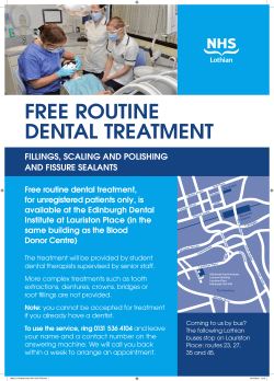

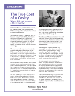
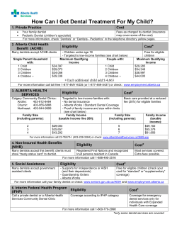

© Copyright 2026