
Surgical management of vitiligo and other leukodermas: evidence-based practice guidelines
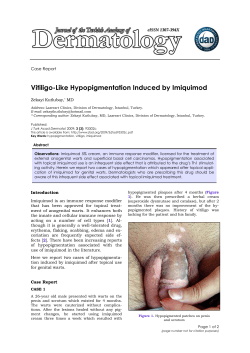
1405145218_4_009.qxd
08-08-2006
19:52
Page 69
CHAPTER 9
Surgical management of vitiligo and
other leukodermas: evidence-based
practice guidelines
Somesh Gupta, Tarun Narang, Mats J. Olsson and Jean-Paul Ortonne
Introduction
Both non-surgical and surgical therapies have been
employed in the management of vitiligo. However,
there are no uniformly agreed guidelines or recommendations available for surgical management of
vitiligo and other leukodermas. Current surgical
treatment practices are based on informal consensus meetings, expert opinions, results from uncontrolled clinical trials, and personal and/or institutional
experiences and infrastructures [1]. There is only
one randomized study available showing superiority of non-cultured melanocyte suspension over
placebo [2]. In the absence of randomized clinical
trials, the guidelines will be formulated on the basis
of meta-analysis of published uncontrolled clinical
trials.
Here a word of caution is necessary that this is
only an attempt to draw some conclusions from metaanalysis of a broad variety of reports published over
a long period, by different individuals. Often the
results are reported as subjective observations
without real measurement tools or statistical analyses. The procedures are performed using different
surgical equipment and bandage materials in distinct clinical settings. The patient populations are
from different parts of the world, so are genetically
heterogeneous. All these factors may bias the conclusions. A Consensus Report on the definition and
assessment of vitiligo has recently been worked out
by the Vitiligo European Task Force. This report will
be a valuable tool to make research outcomes on
this subject more uniform and comparable, but it
will still take many years until a sufficient number
of new publications has used the consensus assessment to make future studies more comparable and
meta-analyses more appropriate. In spite of inherent biases, meta-analyses of published literature
have been accepted as useful means to formulate
evidence-based practice guidelines [3].
Method
We searched two databases, Medline and Embase,
from 1966 to 2005 with Keywords “Vitiligo,”
“Leukoderma,” “Transplantation,” “Grafting,” and
“Surgery” in various combinations. A few leading
researchers were contacted to retrieve reports under
publication. Some Asian journals not indexed in the
literature were hand-searched for relevant reports.
Cross-references of some leading reviews on the
subject were also studied. All the studies pertaining
to transplantation procedures in vitiligo and other
leukodermas were reviewed and evaluated for the
data. Those studies providing inadequate data about
patients or outcome or analyzing data of “patches”
or “procedures” but not of “patients” were excluded.
Follow-up studies of previously published case series
were not included to avoid duplication of data. Data
for two different subtypes of vitiligo, namely bilateral
(generalized and acrofacial) and localized (segmental
and focal), was compared. Data for certain “difficult
to treat” sites was also analyzed. Adverse effects and
total treated area were also looked into.
69
1405145218_4_009.qxd
70
08-08-2006
19:52
Page 70
Chapter 9
The successful outcome was defined as greater
than 75% repigmentation, “excellent” repigmentation, repigmentation of “most” of the treated area,
or “complete” or “almost complete” repigmentation.
The percentage of patients with successful outcome
and 95% confidence intervals (CI) were calculated.
The mean number of adverse events per patient was
calculated to find out which procedures have a
better safety profile.
Results
A total of 96 studies were identified which were
suitable for inclusion of some data in the analyses
[4–99].
Procedures
The following transplantation procedures have been
found to be commonly used for hypopigmentation
disorders.
Tissue grafts:
1 Thin and ultra-thin split-thickness skin grafts
(STSG)
2 Suction blister epidermal grafts (SBEG)
3 Mini-punch grafts (MPG)
4 Hair follicular grafts (HFG)
Cellular grafts:
5 Non-cultured epidermal cell suspension (NCES)
6 Cultured “pure” melanocytes (CM)
7 Cultured epithelial grafts (CE)
In addition, two procedures, namely ultrasonic abrasion and seed grafts and flip-top technique of grafting
have also been tried in one study each [100,101].
In older literature, epidermal shave grafts have also
been used. These studies were not included in the
meta-analysis of the literature due to paucity of the
data. There were 14 studies in STSG, 27 studies in
SBEG, 20 studies in MPG, 12 studies in NCECS, 12
studies in cultured melanocytes, 12 studies in cultured epidermis/co-cultures, and 2 studies in follicular transplantation in patients with vitiligo.
seem to be the most effective procedures, with overall
success rates of 80.3% (CI 76.4–84.2%) and 77.9%
(CI 72.2–83.6%), respectively (Fig. 9.1). The least
successful method was hair grafting, with an overall
success rate of 58.3% (CI 38.6–78%); however, this
technique was especially useful in management of
leukotrichia. Among cellular grafts, all techniques
seem to be equally effective with success rates of
61.1% (CI 56.1–66.1%), 63.6% (CI 57.2–70%), and
63.6% (CI 55.8–70.6%) for non-cultured epidermal
cell suspension, cultured melanocytes, and cultured
epidermis, respectively. The success rates in bilateral
vitiligo including generalized and acrofacial vitiligo
were less than those in segmental or focal vitiligo
with all the procedures. The success rates varied from
25% (CI 0–67.4%) for hair grafting to 70.4% (CI
55.1–85.7%) for thin and ultra-thin split-thickness
grafts in bilateral vitiligo, while in segmental vitiligo,
these ranged from 70.6% (CI 48.9–92.3%) for hair
grafting to 100% (CI 100–100%) for thin and ultrathin split-thickness skin grafts. Overall, the transplantation procedures were successful in 73.1%
(CI 71.5–74.7%) of all cases of vitiligo, 84.4% (CI
81.8–87%) in patients with segmental or focal
vitiligo and 58.5% (CI 54.4–62.6%) in patients with
bilateral vitiligo.
Vitiligo on “difficult to treat” sites
As data pertaining to individual sites was not provided in most of the studies, the number of patients
for specific sites was too small to enable us to apply
methods of statistical analysis (Table 9.1).
Fingers and toes
Cultured melanocytes, thin or ultra-thin splitthickness grafts, mini-punch grafts, and suction blister
epidermal grafts are all highly successful ( 80%
success rate) on fingers and toes, which are otherwise very difficult to treat with medical therapies
and excimer laser. Cultured epidermis is not as successful on this site, with a success rate of 30.8%.
Palms and soles
Outcome
Vitiligo
Among all procedures, suction blister epidermal
grafts and thin and ultra-thin split-thickness grafts
There is only a single case report of successful
minigraft transplant on palm [68]. The donor site
used for this case was the skin of sole (instep). In the
literature, there are no reports of any other method
used for vitiligo of this site.
1405145218_4_009.qxd
08-08-2006
19:52
Page 71
2154/2946
632/749
117/184
178/280
217/355
41/68
12/17
88/116
95/108
67/85
1065/1400
28/67
50
71
14/24
325/556
60
98/175
82/138
70
57/78
19/27
80
Success rate (%)
404/499
90
159/204
34/34
186/208
100
150/181
Practice guidelines
40
1/4
30
20
10
0
STSG
SBEG
MPG
NCES
CM
CE
Hair
All methods
Transplantation method
Bilateral vitiligo
Segmental/focal vitiligo
All types
Fig. 9.1 Success rates of various transplantation methods in vitiligo, including its subtypes. The values given on
each bar represent number of successful patients/total number of patients. Error bars represent 95% CI.
Table 9.1 Summary of the outcome of literature analysis for surgical treatment of vitiligo at “difficult to treat” sites.
Site
STSG
(successful/
total)
SBEG
(successful/
total)
MPG
(successful/
total)
NCES
(successful/
total)
CM
(successful/
total)
CE
(successful/
total)
All methods
(successful/
total)
Fingers and toes
15/16
13/15
24/27
15/25
6/6
4/13
77/102
Lips
2/2
40/45
102/132
6/14
–
–
150/193
Eyelids
7/9
14/14
1/2
8/10
–
–
30/35
Nipples and areolas
–
2/2
–
2/4
–
–
4/6
Elbows and knees
1/1
–
4/6
39/78
12/27
1/1
57/113
Genitals
0/1
1/1
1/1
3/3
1/1
–
6/7
1405145218_4_009.qxd
72
08-08-2006
19:52
Page 72
Chapter 9
Lips
Table 9.2 Repigmentation of leukotrichia in vitiligo after
Thin or ultra-thin split-thickness grafts and suction
blister epidermal grafts are most successful in treatment of vitiligo of the lips, though very few cases
treated with the former procedure have been reported
in the literature. There is also a risk of mismatch in
texture with dermo-epidermal grafts; therefore,
presently the simplest and the most suitable procedure for this limited mucocutaneous transition area
is pure epidermal grafts obtained through suction
blisters. In terms of repigmentation, minigrafts are
also successful at this site, but high complication
rates in the form of cobblestoning which are associated with that method make it less desirable for this
cosmetically highly prominent site. Non-cultured
melanocyte suspension is less effective than tissue
grafts on lips (42.9% success rate). Concerning cultured cellular grafts no published data for procedures on lips are available but sometimes the lips
and eyelids are included in the data of face as a
whole, which makes it difficult to analyze when
scrutinizing the reports.
transplantation.
be successful on genitals, though data from very
few patients is available [70,81].
Eyelids
Leukotrichia in vitiligo
Suction blister epidermal grafts, thin or ultra-thin
split-thickness grafts, and NCEC are among the
most effective surgical treatments for eyelids [51].
No data for cultured cellular grafts are available for
this specific site.
The course of leukotrichia after transplantation has
been specifically studied in only a few studies (Table
9.2) [13,14,43]. STSG and SBEG appear to be more
successful in repigmentation of leukotrichia than
MPG. Sufficient data is not available for cellular
grafts. Transplantation of hair has been found to be
successful in about half of the patients in the treatment of leukotrichia.
Nipples and areolas
Suction blister epidermal grafts are reported to be
successful on the nipple and areola, though data
from very few patients is available. Non-cultured
melanocyte suspension has been found to be successful in about half of the patients.
Elbows and knees
These sites are difficult to treat because of their
mobility. Inadequate data are available for thin and
ultra-thin split-thickness skin grafts, SBEG, and
cultured epidermis. Minigrafts and non-cultured
melanocyte suspension appear to be successful in
about half of the patients.
Genitals
Suction blister epidermal grafts, non-cultured cellular
suspension and cultured melanocytes are reported to
Number of
patients with
successfully
repigmented
white hair
Transplantation
method
Total number
of patients
STSG
10
8
SBEG
28
27
96.4
MPG
63
16
25.4
NCES
1
1
100
CM
–
–
–
CE
1
1
100
13
6
Hair
Success
rate (%)
80
46.2
Size of the treated area
Cases with more extensive vitiligo vulgaris, involving greater than 30% body surface area, are generally
considered unsuitable for transplantation procedures
as chances of retention of the pigment are less [102].
This is not the case in segmental vitiligo, which
seems to respond equally well regardless of the size
of the involved area. Extensive areas may be best
treated with cellular grafts – theoretically, culture
methods would provide an unlimited number of
cells/tissue for transplantation, while NCEC would
provide up to 8–10 times donor-to-recipient expansion. Among tissue grafts, minigrafts are also based
on the principle of pigment spread and a 1.25-mm
1405145218_4_009.qxd
08-08-2006
19:52
Page 73
Practice guidelines
graft is expected to provide a pigmented halo of
5–10 mm, depending on the skin type of the patient
(more in darker skin type). This means a grafted area
of approximately 5 mm2 (or a graft of 1.25 mm) will
result in repigmentation of approximately 20 mm2
(pigment spread of 5-mm diameter) to 80 mm2
(pigment spread of 10-mm2 diameter) – 4–15 times
donor-to-recipient expansion. Therefore, moderately large areas may be treated with NCEC, thin
split-thickness grafts, and minigrafts. Small areas
do not require donor skin expansion through cultures or suspension, and therefore should be treated
with less complex tissue grafting procedures such as
suction blister epidermal grafts or thin split-thickness
grafts.
In literature, the mean area treated with each
procedure is as follows: STSG: 69.3 cm2, SBEG
4 cm2, MPG 21 cm2, NCES 31.6 cm2, CM 34.7 cm2,
and CE 161.5 cm2. In SBEG, data was available
from only one study. Therefore, it seems that larger
areas may be treated with cellular grafts and thin
and ultra-thin split-thickness grafts and moderate
areas may be treated cellular grafts and minigrafts.
Smaller areas may be easily treated with suction
blister epidermal grafts which gives good esthetic
results and is technically less challenging.
Adverse events
No serious adverse events have been reported with
any of the transplantation methods. Cellular grafts
appear to have the least frequency of adverse events.
Cultured melanocytes, cultured epidermis, and
NCEC have a mean of 0, 0.02, and 0.08, respectively, adverse events at recipient site, and 0.01, 0,
and 0.009, respectively, at the donor site (Tables 9.3
and 9.4). Tissue grafts are reported to be associated
with more adverse effects and the maximum number of adverse events on the recipient site are seen
when MPG and STSG methods are used.
Other leukodermas/hypopigmentation
disorders (Table 9.5)
Lip leukoderma due to recurrent herpes labialis
It was found to be manageable with thin or ultra-thin
split-thickness grafts. Minigrafting has been reported
73
Table 9.3 Mean number of adverse events per patient.
Recipient
Donor
STSG
0.5
0.5
SBEG
0.2
0.3
MPG
0.7
0.05
NCES
0.08
0.009
CM
0
0.01
CE
0.02
0
to be unsuccessful for this condition and reactivation
of herpes simplex virus infection has been thought to
be responsible for graft rejection [103]. Even when
minigrafts were successfully transplanted with the
help of concurrent acyclovir therapy, there was
no pigment spread from the grafts, which soon also
became depigmented. Micropigmentation (tattooing)
has been successful in such cases [104].
Piebaldism
In piebaldism, thin and ultra-thin split-thickness
grafting, NCEC, cultured melanocytes, and cultured epidermis have been tried and all these
methods yielded 100% success rates.
DLE leukoderma
In discoid lupus erythematosus (DLE) leukoderma,
thin split-thickness grafts, suction blister epidermal
grafts, and minigrafts gave 100% success rates [48];
however, the number of treated patients were few (1,
4, and 1, respectively). These patients were given
antimalarial therapy to prevent reactivation of disease or köbnerization.
Halo nevus
Recent studies have shown evidence that halo
nevus is a different entity from vitiligo [105]. Threequarters (75%) of patients with halo nevus will
show successful outcome with transplantation procedures. Non-cultured melanocyte suspension, cultured melanocytes, and cultured epidermis have been
found to be successful in the treatment of halo nevi.
1405145218_4_009.qxd
74
08-08-2006
19:52
Page 74
Chapter 9
Table 9.4 Nature of adverse events in transplantation procedures.
STSG
(n 76)
Recipient area
SBEG
(n 441)
NCES
(n 318)
Hyperpigmentation
2
53
312
3
Hypopigmented borders
4
1
5
20
Cobblestoning
CM
(n 259)
CE
(n 170)
Hair
(n 21)
1
397
Contact dermatitis to
topical medication
Inclusion cysts
6
3
7
244
9
3
20
Thick margins/stuck-on
appearance
9
Variegated appearance
Infection
2
Scarring
Donor area
MPG
(n 1295)
1
19
All adverse events
37
76
Hyperpigmentation
1
131
Scarring
30
983
2
24
3
60
Köbner phenomenon
4
Infection
3
3
1
All adverse events
31
135
64
3
Table 9.5 Summary of the outcome of literature analysis in other leukodermas.
Type of
leukoderma
STSG
(successful/
total)
SBEG
(successful/
total)
MPG
(successful/
total)
NCES
(successful/
total)
CE
(successful/
total)
All methods
(successful/
total)
–
–
10/18
2/2
7/7
39/39
Lip leukoderma
due to herpes
10/10
–
0/8
Piebaldism
14/14
2/2
4/4
DLE leukoderma
1/1
4/4
1/1
–
–
–
6/6
Contact leukoderma
–
1/1
1/1
1/1
1/1
–
4/4
Halo nevus
1/0
–
–
2/2
1/1
–
3/3
Nevus depigmentosus
–
1/1
–
1/2
–
–
2/3
Albinism
–
–
–
–
–
0/1
0/1
76/76
Post-burn leukoderma
Ideopathic guttate
hypomelanosis
60/60
–
11/11
4/4
–
CM
(successful/
total)
10/10
4/4
–
–
1/1
–
–
–
–
4/4
1405145218_4_009.qxd
08-08-2006
19:52
Page 75
Practice guidelines
75
Table 9.6 Guidelines for surgical management of vitiligo and other leukodermas.
First choice(s)
Alternative
Acral (fingers and toes)
CM, SBEG, STSG
MPG, NCES, CE
Palms
MPG
–
Lips
SBEG, STSG
MPG, NCES
Eyelids
SBEG, NCES, STSG
MPG
Nipple and areola
SBEG, NCES
Genitals
NCES, SBEG, CM
–
Small
SBEG, STSG
MPG, Hair
Moderate
NCES, STSG
MPG
Extensive
CM, CE, NCES
STSG
B. Herpes lip leukoderma
STSG
–
C. Piebaldism
Any – choice depends on the size of the
lesions. All are likely to be successful
D. DLE leukoderma
SBEG, STSG, MPG
E. Contact leukoderma
Any – all are likely to be successful
F. Halo nevus
NCES, CM, CE
G. Nevus depigmentosus
SBEG
NCES
H. Post-burn leukoderma
Any – STSG, SBEG, and MPG have been
tried with consistent success
–
I. Ideopathic guttate
hypomelanosis
Any – SBEG has been tried successfully
–
J. Albinism
Should never be treated with autologous
transplantation methods
–
A. Vitiligo
Site
Area
–
Nevus depigmentosus
Leukoderma after partial thickness burn injuries
There were only three cases with nevus depigmentosus reported to be treated with transplantation procedures – two with melanocyte suspension and one
with epidermal grafts [52,76,77]. It seems that there
is no pigment spread phenomenon in these patches
and the whole depigmented area needs to be replaced
with normally pigmented epidermal sheets [52].
Post-burn leukoderma is almost always successfully
managed with transplantation procedures. In the
literature, reports of use of tissue grafts are available and most reported cases have been treated
with split-thickness grafts [106]. This may be due to
familiarity of plastic surgeons with this procedure,
as most of the reported studies have been done in
1405145218_4_009.qxd
76
08-08-2006
19:52
Page 76
Chapter 9
that setup. Cultured epidermis has also been used
in post-burn leukoderma in a single case report.
Idiopathic guttate hypomelanosis
This has been successfully treated with suction blister epidermal grafts (4/4) [45].
Albinism
Albinism should never be managed with transplantation procedures as it is not likely to be successful.
Only one case of albinism has been reported in the
literature, in which cultured melanocyte transplantation was tried without any success [92].
Leukoderma due to contact with chemicals
It is manageable with transplantation methods –
cell suspension, suction blister epidermal grafts,
and minigrafts were all found to be 100% successful in these patients.
Guidelines
Based on reported success rates, adverse events, technical complexities, anatomical location, and size of
the areas, the guidelines have been developed (Table
9.6). Yet every patient should be evaluated individually to find out the best possible options. This depends
on expertise of the treating surgeon, economy of the
institute, infrastructure available, and patient’s preference. Patient should be counseled for all possible
therapeutic options and should be given liberty to
choose from the available modalities.
References
1 Njoo MD, Bossuyt PMM, Westerhof W. Management of vitiligo. Results of a questionnaire
among dermatologists in the Netherlands.
2 van Geel N, Ongenae K, De Mil M, et al. Doubleblind placebo-controlled study of autologous transplanted epidermal cell suspensions for repigmenting
vitiligo. Arch Dermatol 2004;140:1203–8.
3 Pai M, McCulloch M, Gorman JD, et al. Systematic
reviews and meta-analyses: an illustrated, step-bystep guide. Natl Med J India 2004;17:86–95.
4 Gupta S, Handa S, Kumar B. A novel scoring system
for evaluation of results of autologous transplantation methods in vitiligo. Indian J Dermatol Venereol
Leprol 2002;68:33–37.
5 Malakar S. Successful split thickness skin grafts in
stable vitiligo not responding to autologous miniature skin grafts. Indian J Dermatol 1997;42:215–8.
6 Olsson MJ, Juhlin L. Epidermal sheet grafts for
repigmentation of vitiligo and piebaldism, with a
review of surgical techniques. Acta Derm Venereol
1997;77:463–6.
7 Kahn AM, Ostad A, Moy RL. Grafting following
short-pulse carbon dioxide laser de-epithelialization.
Dermatol Surg 1996;22:965–7.
8 Bose SK. Modified Thiersch grafting in stable vitiligo.
J Dermatol 1996;23:362–4.
9 Kahn AM, Cohen MJ. Vitiligo: treatment by dermabrasion and epithelial sheet grafting. J Am Acad
Dermatol 1995;33:646–8.
10 Khandpur S, Sharma VK, Manchanda Y. Comparison
of minipunch grafting versus split-skin grafting in
chronic stable vitiligo. Dermatol Surg 2005;31:436–41.
11 Agrawal K, Agrawal A. Vitiligo: repigmentation with
dermabrasion and thin split-thickness skin graft.
Dermatol Surg 1995;21:295–300.
12 Bose SK. Thiersch grafting in recurrent herpesinduced lip depigmentation. J Dermatol 2005;32:
432–5.
13 Agrawal K, Agrawal A. Vitiligo: surgical repigmentation of leukotrichia. Dermatol Surg 1995;21:711–5.
14 Bose SK. Is there any treatment of leukotrichia in
stable vitiligo? J Dermatol 1997;24:615–7.
15 Al-Qattan MM. Surgical management of post-burn
skin dyspigmentation of the upper limb. Burns
2000;26:581–6.
16 Kahn AM, Cohen MJ. Treatment for depigmentation
following burn injuries. Burns 1996;22:552–4.
17 Kahn AM, Cohen MJ, Kaplan L, Highton A. Vitiligo:
treatment by dermabrasion and epithelial sheet
grafting – a preliminary report. J Am Acad Dermatol
1993;28:773–4.
18 Acikel C, Ulkur E, Celikoz B. Carbon dioxide laser
resurfacing and thin skin grafting in the treatment of
“stable and recalcitrant” vitiligo. Plast Reconstr Surg
2003;111:1291–8.
19 Behl PN. Treatment of vitiligo with autologous thin
Thiersch’s grafts. Int J Dermatol 1973;12:329–31.
20 Achauer BM, Le Y, Vander Kam VM. Treatment of
vitiligo with melanocytic grafting. Ann Plast Surg
1994;33:644–6.
21 Njoo MD, Nieuweboer-Krobotova L, Westerhof W.
Repigmentation of leucodermic defects in piebaldism by dermabrasion and thin split-thickness skin
grafting in combination with minigrafting. Br J
Dermatol 1998;139:829–33.
1405145218_4_009.qxd
08-08-2006
19:52
Page 77
Practice guidelines
22 Onur Erol O, Atabay K. The treatment of burn scar
hypopigmentation and surface irregularity by dermabrasion and thin skin grafting. Plast Reconstr Surg
1990;85:754–8.
23 Gupta S, Sandhu K, Kanwar A, Kumar B.
Melanocyte transfer via epidermal grafts for vitiligo
of labial mucosa. Dermatol Surg 2004;30:45–8.
24 Gupta S, Goel A, Kanwar AJ. Autologous melanocyte
transfer via epidermal grafts for lip vitiligo. Int J
Dermatol 2006;45:747–50.
25 Czajkowski R. Comparison of melanocytes transplantation methods for the treatment of vitiligo.
Dermatol Surg 2004;30:1400–5.
26 Gupta S, Kumar B. Epidermal grafting in vitiligo:
influence of age, site of lesion, and type of disease on
outcome. J Am Acad Dermatol 2003;49:99–104.
27 Pai GS, Vinod V, Joshi A. Efficacy of erbium YAG
laser-assisted autologous epidermal grafting in vitiligo.
J Eur Acad Dermatol Venereol 2002;16:604–6.
28 Gupta S, Kumar B. Epidermal grafting for vitiligo in
adolescents. Pediatr Dermatol 2002;19:159–62.
29 Oh CK, Cha JH, Lim JY, Jo JH, Kim SJ, Jang HS, Kwon
KS. Treatment of vitiligo with suction epidermal grafting by the use of an ultrapulse CO2 laser with a computerized pattern generator. Dermatol Surg 2001;27:
565–8.
30 Kim CY, Yoon TJ, Kim TH. Epidermal grafting after
chemical epilation in the treatment of vitiligo.
Dermatol Surg 2001;27:855–6.
31 Kim HU, Yun SK. Suction device for epidermal grafting in vitiligo: employing a syringe and a manometer
to provide an adequate negative pressure. Dermatol
Surg 2000;26:702–4.
32 Sachdev M, Shankar DS. Dermatologic surgery:
pulsed erbium:YAG laser-assisted autologous epidermal punch grafting in vitiligo. Int J Dermatol
2000;39:868–71.
33 Sachdev M, Krupashankar DS. Suction blister grafting for stable vitiligo using pulsed erbium:YAG laser
ablation for recipient site. Int J Dermatol 2000;39:
471–3.
34 Gupta S, Shroff S, Gupta S. Modified technique of
suction blistering for epidermal grafting in vitiligo.
Int J Dermatol 1999;38:306–9.
35 Gupta S, Jain VK, Saraswat PK. Suction blister epidermal grafting versus punch skin grafting in recalcitrant and stable vitiligo. Dermatol Surg 1999;25:
955–8.
36 Kim HY, Kang KY. Epidermal grafts for treatment of
stable and progressive vitiligo. J Am Acad Dermatol
1999;40:412–7.
77
37 Yang JS, Kye YC. Treatment of vitiligo with autologous
epidermal grafting by means of pulsed erbium:YAG
laser. J Am Acad Dermatol 1998;38:280–2.
38 Tang WY, Chan LY, Lo KK. Treatment of vitiligo with
autologous epidermal transplantation using the
roofs of suction blisters. Hong Kong Med J 1998;4:
219–24.
39 Suga Y, Butt KI, Takimoto R, et al. Successful
treatment of vitiligo with PUVA-pigmented autologous epidermal grafting. Int J Dermatol 1996;35:
518–22.
40 Skouge J, Morison WL. Vitiligo treatment with a
combination of PUVA therapy and epidermal autografts. Arch Dermatol 1995;131:1257–8.
41 Hann SK, Im S, Bong HW, Park YK. Treatment of stable vitiligo with autologous epidermal grafting and
PUVA. J Am Acad Dermatol 1995;32:943–8.
42 Mutalik S. Transplantation of melanocytes by epidermal grafting. An Indian experience. J Dermatol
Surg Oncol 1993;19:231–4.
43 Hann SK, Im S, Park YK, Hur W. Repigmentation of leukotrichia by epidermal grafting and systemic psoralen plus UV-A. Arch Dermatol 1992;128:
998–9.
44 Koga M. Epidermal grafting using the tops of suction
blisters in the treatment of vitiligo. Arch Dermatol
1988;124:1656–8.
45 Suvanprakorn P, Dee-Ananlap S, Pongsomboon C,
Klaus SN. Melanocyte autologous grafting for treatment of leukoderma. J Am Acad Dermatol 1985;13:
968–74.
46 Shenoi SD, Srinivas CR, Pai S. Treatment of stable
vitiligo with autologous epidermal grafting and
PUVA. J Am Acad Dermatol 1997;36:802–3.
47 Zachariae H, Zachariae C, Deleuran B, Kristensen P.
Autotransplantation in vitiligo: treatment with epidermal grafts and cultured melanocytes. Acta Derm
Venereol 1993;73:46–8.
48 Gupta S. Epidermal grafting for depigmentation due
to discoid lupus erythematosus. Dermatology 2001;
202:320–3.
49 Falabella R. Repigmentation of leukoderma by
autologous epidermal grafting. J Dermatol Surg Oncol
1984;10:136–44.
50 Falabella R. Epidermal grafting. An original technique and its application in achromic and granulating areas. Arch Dermatol 1971;104:592–600.
51 Nanda S, Relhan V, Grover C, Reddy BS. Suction
blister epidermal grafting for the management of
vitiligo of the eyelid: special considerations. Dermatol
Surg 2006;32:387–91.
1405145218_4_009.qxd
78
08-08-2006
19:52
Page 78
Chapter 9
52 Gupta S, Goel A. Letter to the editor: nevus depigmentosus needs transplant of epidermal sheets.
Dermatol Surg 2005;31:1746–7.
53 Malakar S, Lahiri K. Punch grafting for lip leucoderma. Dermatology 2004;208:125–8.
54 Sarkar R, Mehta SD, Kanwar AJ. Repigmentation
after autologous miniature punch grafting in segmental vitiligo in North Indian patients. J Dermatol
2001;28:540–6.
55 Falabella R, Barona M, Escobar C, Borrero I,
Arrunategui A. Surgical combination therapy for
vitiligo and piebaldism. Dermatol Surg 1995;21:852–7.
56 Singh AK, Bajaj AK. Autologous miniature skin
punch grafting in vitiligo. Indian J Dermatol Venereol
Leprol 1995;61:77–80.
57 Boersma BR, Westerhof W, Bos JD. Repigmentation
in vitiligo vulgaris by autologous minigrafting:
results in nineteen patients. J Am Acad Dermatol
1995;33:990–5.
58 Rathi TM, Singh AK. Punch grafting in the treatment of stable vitiligo. Indian J Dermatol Venereol
Leprol 1994;60:188–91.
59 Savant SS. Autologous miniature punch skin grafting in stable vitiligo. Indian J Dermatol Venereol Leprol
1992;58:310–4.
60 Falabella R. Treatment of localized vitiligo by autologous minigrafting. Arch Dermatol 1988;124:1649–55.
61 Falabella R. Repigmentation of segmental vitiligo by
autologous minigrafting. J Am Acad Dermatol 1983;
9:514–21.
62 Falabella R. Repigmentation of leukoderma by minigrafts of normally pigmented, autologous skin. J
Dermatol Surg Oncol 1978;4:916–9.
63 Garg T, Khaitan BK, Manchanda Y. Autologous
punch grafting for repigmentation in piebaldism. J
Dermatol 2003;30:849–50.
64 Falabella R. Repigmentation of stable leukoderma by
autologous minigrafting. J Dermatol Surg Oncol 1986;
12:172–9.
65 Hallaji Z, Daneshpazhooh M, Rezai-khiabanloo S.
Successful treatment of vitiligo with punch graft followed by outdoor topical psoralen plus ultraviolet A
radiation. Arch Iran Med 2003;6:86–90.
66 Shah BH, Joshipura SP, Thakkar JK. Surgical treatment in acrofacial vitiligo. Indian J Dermatol Venereol
Leprol 1994;60:26–27.
67 Malakar S, Dhar S. Acyclovir can abort rejection of
punch grafts in herpes-simplex-induced lip leucoderma. Dermatology 1999;199:75.
68 Kumar P. Autologous punch grafting for vitiligo of
the palm. Dermatol Surg 2005;31:368–70.
69 Malakar S, Dhar S. Acyclovir can abort rejection of
punch grafts in herpes-simplex-induced lip leucoderma. Dermatology 1999;199:75.
70 Mulekar SV, Al Issa A, Al Eisa A, Asaad M. Genital vitiligo treated by autologous, noncultured
melanocyte–keratinocyte cell transplantation. Dermatol
Surg 2005;31:1737–9.
71 Xu AE, Wei XD, Cheng DQ, et al. Transplantation of
autologous noncultured epidermal cell suspension
in treatment of patients with stable vitiligo. Chin Med
J (Engl) 2005;118:77–9.
72 Mulekar SV. Long-term follow-up study of segmental and focal vitiligo treated by autologous, noncultured melanocyte–keratinocyte cell transplantation.
Arch Dermatol 2004;140:1211–5.
73 Tegta GR, Parsad D, Majumdar S, Kumar B. Efficacy
of autologous transplantation of noncultured epidermal suspension in two different dilutions in the treatment of vitiligo. Int J Dermatol 2006;45:106–10.
74 Mulekar SV. Long-term follow-up study of 142
patients with vitiligo vulgaris treated by autologous,
non-cultured melanocyte–keratinocyte cell transplantation. Int J Dermatol 2005;44:841–5.
75 van Geel N, Ongenae K, De Mil M, Naeyaert JM.
Modified technique of autologous noncultured epidermal cell transplantation for repigmenting vitiligo:
a pilot study. Dermatol Surg 2001;27:873–6.
76 Olsson MJ, Juhlin L. Leucoderma treated by transplantation of a basal cell layer enriched suspension.
Br J Dermatol 1998;138:644–8.
77 Gauthier Y, Surleve-Bazeille JE. Autologous grafting
with noncultured melanocytes: a simplified method
for treatment of depigmented lesions. J Am Acad
Dermatol 1992;26:191–4.
78 Pandya V, Parmar KS, Shah BJ, Bilimoria FE. A
study of autologous melanocyte transfer in treatment
of stable vitiligo. Indian J Dermatol Venereol Leprol
2005;71:393–7.
79 Chen YF, Chang JS, Yang PY, et al. Transplant of
cultured autologous pure melanocytes after laserabrasion for the treatment of segmental vitiligo.
J Dermatol 2000;27:434–9.
80 Lontz W, Olsson MJ, Moellmann G, Lerner AB.
Pigment cell transplantation for treatment of vitiligo:
a progress report. J Am Acad Dermatol 1994;30:591–7.
81 Olsson MJ, Juhlin L. Transplantation of melanocytes
in vitiligo. Br J Dermatol 1995;132:587–91.
82 Kaufmann R, Greiner D, Kippenberger S, Bernd A.
Grafting of in vitro cultured melanocytes onto laserablated lesions in vitiligo. Acta Derm Venereol
1998;78:136–8.
1405145218_4_009.qxd
08-08-2006
19:52
Page 79
Practice guidelines
83 Olsson MJ, Moellmann G, Lerner AB, Juhlin L. Vitiligo:
repigmentation with cultured melanocytes after cryostorage. Acta Derm Venereol 1994;74:226–8.
84 Olsson MJ, Juhlin L. Repigmentation of vitiligo by
transplantation of cultured autologous melanocytes.
Acta Derm Venereol 1993;73:49–51.
85 Lerner AB, Halaban R, Klaus SN, Moellmann GE.
Transplantation of human melanocytes. J Invest
Dermatol 1987;89:219–24.
86 Chen YF, Yang PY, Hung CM, Hu DN. Transplantation
of autologous cultured melanocytes for treatment of
large segmental vitiligo. J Am Acad Dermatol 2001;
44:543–5.
87 Jha AK, Pandey SS, Gulati AK, et al. Inoculation of a
cultured autologous epidermal suspension containing
melanocytes in vitiligo. Arch Dermatol 1993;129:785–6.
88 Pianigiani E, Risulo M, Andreassi A, et al. Autologous
epidermal cultures and narrow-band ultraviolet B
in the surgical treatment of vitiligo. Dermatol Surg
2005;31:155–9.
89 Guerra L, Primavera G, Raskovic D, et al. Erbium:YAG
laser and cultured epidermis in the surgical therapy
of stable vitiligo. Arch Dermatol 2003;139:1303–10.
90 Guerra L, Capurro S, Melchi F, et al. Treatment of
“stable” vitiligo by Timedsurgery and transplantation of cultured epidermal autografts. Arch Dermatol
2000;136:1380–9.
91 Andreassi L, Pianigiani E, Andreassi A, Taddeucci P,
Biagioli M. A new model of epidermal culture for
the surgical treatment of vitiligo. Int J Dermatol
1998;37:595–8.
92 Kumagai N, Uchikoshi T. Treatment of extensive
hypomelanosis with autologous cultured epithelium. Ann Plast Surg 1997;39:68–73.
93 Falabella R, Escobar C, Borrero I. Transplantation of
in vitro-cultured epidermis bearing melanocytes for
repigmenting vitiligo. J Am Acad Dermatol 1989;21:
257–64.
94 Falabella R, Escobar C, Borrero I. Treatment of refractory and stable vitiligo by transplantation of in vitro
cultured epidermal autografts bearing melanocytes.
J Am Acad Dermatol 1992;26:230–6.
95 Toriyama K, Kamei Y, Kazeto T, et al. Combination
of short-pulsed CO2 laser resurfacing and cultured
96
97
98
99
100
101
102
103
104
105
106
79
epidermal sheet autografting in the treatment of
vitiligo: a preliminary report. Ann Plast Surg 2004;53:
178–80.
Guerra L, Primavera G, Raskovic D, et al. Permanent
repigmentation of piebaldism by erbium:YAG laser
and autologous cultured epidermis. Br J Dermatol
2004;150:715–21.
Stoner ML, Wood FM. The treatment of hypopigmented lesions with cultured epithelial autograft.
J Burn Care Rehabil 2000;21:50–4.
Malakar S, Dhar S. Repigmentation of vitiligo
patches by transplantation of hair follicles. Int J
Dermatol 1999;38:237–8.
Na GY, Seo SK, Choi SK. Single hair grafting for the
treatment of vitiligo. J Am Acad Dermatol 1998;38:
580–4.
Tsukamoto K, Osada A, Kitamura R, et al.
Approaches to repigmentation of vitiligo skin: new
treatment with ultrasonic abrasion, seed-grafting
and psoralen plus ultraviolet A therapy. Pigm Cell Res
2002;15:331–4.
McGovern TW, Bolognia J, Leffell DJ. Flip-top pigment transplantation: a novel transplantation procedure for the treatment of depigmentation. Arch
Dermatol 1999;135:1305–7.
Olsson MJ, Juhlin L. Long-term follow-up of leucoderma patients treated with transplants of autologous cultured melanocytes, ultrathin epidermal
sheets and basal cell layer suspension. Br J Dermatol
2002;147:893–904.
Lahiri K, Malakar S, Sarma N. Herpes-simplexinduced lip leucoderma revisited. Dermatology 2004;
208:182.
Malakar S, Lahiri K. Successful repigmentation of six
cases of herpes-labialis-induced lip leucoderma by
micropigmentation. Dermatology 2001;203:194.
de Vijlder HC, Westerhof W, Schreuder GM, et al.
Differences in pathogenesis between vitiligo vulgaris
and halo nevi associated with vitiligo is supported by
an HLA association study. Pigm Cell Res 2004;17:
270–4.
Grover R, Morgan BD. Management of hypopigmentation following burn injury. Burns 1996;
22:627–30.


© Copyright 2026