
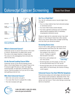
Polyp Guideline: Diagnosis, Treatment, and Surveillance
POSITION
PAPER
Polyp Guideline: Diagnosis, Treatment, and Surveillance
for Patients with Nonfamilial Colorectal Polyps*
John H. Bond, MD, for the Practice Parameters Committee of the
American College of Gastroenterology
• Objective: To outline the preferable approach to the
management of patients with nonfamilial colorectal
polyps.
• Data Sources: The human subject English language
literature for the past 15 years, searched using
MEDLINE and the terms "polyp-," "adenoma-," and
1
' polypectomy-colorectal.''
• Study Selection: The titles and abstracts of all pertinent articles were reviewed. All randomized controlled
trials and large case-control and cohort studies related
to colorectal polyps were reviewed in depth.
• Data Synthesis: Evidence was evaluated along a
hierarchy with randomized controlled trials receiving
the greatest weight. Conclusions and recommendations were reviewed by a large group of experts in
gastroenterology, radiology, and pathology and were
circulated for comment to primary care medical societies.
• Conclusions: Most patients with polyps should undergo colonoscopy to excise the polyp and search for
synchronous neoplasms. Small polyps (<0.5 cm) require individualization. A hyperplastic polyp found during proctosigmoidoscopy is not an indication for
colonoscopy. Large sessile polyps require careful follow-up to ensure complete resection. The need for
further treatment of a resected polyp with invasive
carcinoma depends on several well-defined clinical and
pathologic criteria. Follow-up surveillance after
polypectomy should be tailored to the individual risk
assessment for each patient. Initial follow-up should be
performed at 3 years for most postpolypectomy patients. After one negative result of a 3-year examination,
the interval can be increased to 5 years. Patients with
one small tubular adenoma do not have an increased
risk for cancer, and therefore follow-up surveillance
may not be indicated. Adoption of these recommendations should substantially reduce the cost of postpolypectomy surveillance and of screening for colorectal
cancer.
Ann Intern Med. 1993;119:836-843.
* This guideline has been officially endorsed by the American Society
for Gastrointestinal Endoscopy and the American Gastroenterological
Association. It is an official statement of the American College of
Gastroenterology. For a list of the members of the Practice Parameters
Committee of the American College of Gastroenterology, see end of
text.
836
Preamble
1 his guideline is intended to indicate preferable approaches to the management of patients with colorectal
polyps. It does not deal with either patients with known
colon cancer or familial polyposis. When only data that
will not withstand objective scrutiny are available, an
American College of Gastroenterology (ACG) recommendation is identified as a consensus of experts. The
guideline is applicable to all physicians who address
this subject without regard to specialty training or interests and is intended to indicate the preferable but not
necessarily the only acceptable approach to the patient
with colorectal polyps. The guideline is intended to be
flexible and must be distinguished from standards of
care that are inflexible and rarely violated. Given the
wide range of specifics in this common health care
problem, the physician must always choose the course
best suited to the individual patient and the variables in
existence at the moment of decision.
This guideline was developed under the auspices of
the American College of Gastroenterology and its Practice Parameters Committee and approved by the Board
of Trustees. It has been intensely reviewed and revised
by the committee, other experts in the field, physicians
who will use it, and specialists in the science of decision analysis (see Methods). The ACG recommendations are therefore considered valid at the time of their
publication based on available data.
Methods
The human subject English-language literature was searched
using MEDLINE and the following MeSH terms: polyp-, adenoma-, and polypectomy-colorectal. The titles and abstracts
of the articles were reviewed by the primary author. All randomized, controlled trials were read in depth, as were all large
case-control and cohort studies. In the resulting review, evidence was evaluated along a hierarchy with randomized, controlled trials receiving the greatest weight. Abstracts presented
at national meetings were used only in special circumstances in
which unique data from ongoing randomized trials were presented. When scientific data were lacking, recommendations
were based on expert consensus. During its preparation, the
guideline was submitted for review by the Practice Committees
and the Governing Boards of the American Gastroenterological
Association and the American Society for Gastrointestinal Endoscopy, and by selected authorities in colorectal neoplasia
including gastroenterologists, pathologists, and radiologists. All
recommendations resulting from this review were carefully
considered by the Committee and incorporated in the final
revision. In addition, the guideline was circulated for review
and comment to primary internal medicine and family practice
societies and to the membership of the American College of
Gastroenterology.
15 October 1993 • Annals of Internal Medicine • Volume 119 • Number 8
adenoma formation, growth, increasing dysplasia, and then
invasive carcinoma.
Clinical Considerations
A colorectal polyp is a circumscribed mass of tissue that
projects above the surface of the bowel mucosa. Grossly, a
polyp is classified as pedunculated or sessile depending on
whether it contains a discrete stalk. Polyps may ulcerate and
cause intestinal bleeding. Rarely, large polyps may cause
symptoms of partial bowel obstruction. Most polyps, however,
are asymptomatic lesions detected only by screening or diagnostic studies. This guideline addresses the management of
patients known to have one or more polyps; it does not address primary screening for colorectal neoplasia. Colorectal
polyps are extremely common in Western countries; they are
found in more than 30% of autopsies performed in people older
than 60 years (1, 2).
The main importance of polyps is their well-recognized relationship to colorectal cancer (3). After years of debate, it is
generally accepted that most colorectal cancers arise from benign, neoplastic polyps (adenomas). Although this adenomacancer sequence can probably never be proved directly, persuasive data exist indicating that colorectal neoplasia changes
through a continuous process from normal mucosa, to benign
adenoma, to carcinoma (4). Evidence of this sequence includes
the following:
1. There is a parallel prevalence of adenomas and carcinomas, with the average age of patients with adenomas being 5 to
7 years less than that of patients with carcinomas (5, 6).
2. Cancer is often contiguous with benign adenomatous tissue, whereas small carcinomas without adenomatous tissue are
rare (7, 8).
3. The adenomas of the familial polyposis syndrome, a wellrecognized premalignant state, are histologically similar to sporadic adenomas (9).
4. As adenomas grow, they exhibit increasing cellular atypia
and abnormal chromosomal patterns (5, 10).
5. The anatomic distribution is similar for adenomas and
carcinomas (11).
6. Adenomas are found in more than one third of surgical
specimens containing a colorectal cancer (12, 13).
Histologically, polyps are classified as neoplastic (adenomas)
or non-neoplastic (14, 15). Non-neoplastic polyps have no malignant potential and include hyperplastic polyps, hamartomas,
lymphoid aggregates, and inflammatory polyps. Neoplastic polyps or adenomas have malignant potential and are classified
according to the World Health Organization as tubular, tubulovillous, or villous adenomas, depending on the presence and
volume of villous tissue (16). Tubular adenomas are composed
of straight or branched tubules of dysplastic tissue; villous
adenomas contain fingerlike projections of dysplastic epithelium. Approximately 70% of polyps removed at colonoscopy
are adenomas (17). Seventy percent to 85% of these are classified as tubular (0% to 25%, villous tissue), 10% to 25% are
tubulovillous (25% to 75%, villous tissue), and fewer than 5%
are villous adenomas (75% to 100%, villous tissue).
Some degree of dysplasia exists in all adenomas. Most authorities recommend that dysplasia be classified as mild, moderate, or severe (18). Others prefer only two gradations, lowand high-grade dysplasis, because this classification reduces
the problem of interobserver variation (19). Severe, or highgrade, dysplasia includes the histologic changes previously
called "carcinoma in situ," "intramucosal carcinoma," or "focal carcinoma." Abandonment of these terms is recommended
because of concern for misinterpretation of the clinical significance that might lead to overtreatment, and thus they will not
be used in this guideline. Approximately 5% to 7% of patients
with adenomas have severe dysplasia and 3% to 5% have
invasive carcinoma at the time of diagnosis. Increasing dysplasia and, presumably, malignant potential correlate with increasing adenoma size, villous component, and patient age
(19). The likelihood of invasive carcinoma also increases with
increasing polyp size (15).
The development of colorectal adenomas and carcinomas
probably involves both environmental and genetic factors (10,
20-22). Environmental carcinogens appear to act on a genetically susceptible mucosa causing cellular proliferation followed
by oncogene activation and chromosomal deletions leading to
Diagnosis and Treatment
Colonic polyps are diagnosed by endoscopy or barium radiography. Because most polyps are asymptomatic, they are usually found incidentally. The singlecontrast barium enema examination is an inaccurate
method for detecting polyps in most patients. In one
large screening study, single-contrast barium enemas
found only 40% of neoplastic polyps detected on subsequent colonoscopy (23). Double-contrast techniques
greatly improve the accuracy of radiologic methods for
detecting polyps (24). A study comparing the accuracy
of both radiographic methods in 425 patients reported a
sensitivity for detecting polyps of 90% and 40% for
double- and single-contrast methods, respectively (25).
Several studies indicate that the double-contrast barium
enema can accurately detect most cancers and most
polyps that are larger than 1 cm in diameter (26, 27).
The main limitation of barium enema is that it does not
allow biopsy or polypectomy.
The most common use of flexible sigmoidoscopy is
for screening asymptomatic average-risk persons for colonic neoplasms. Flexible sigmoidoscopy done with the
standard 60-cm instrument detects two to three times as
many polyps and is more comfortable than is rigid sigmoidoscopy (28, 29). Sensitivity and specificity are very
high because few polyps within reach of the examination instrument are missed and the false-positive rate is
negligible. The combination of a double-contrast barium
enema and flexible sigmoidoscopy has been promoted
as an acceptable alternative to colonoscopy for patients
requiring a complete examination of the large bowel.
When a barium enema is used for surveillance, rigid or
flexible proctosigmoidoscopy should always be done to
ensure an adequate examination of the rectum. Flexible
sigmoidoscopy also provides a more accurate examination of the sigmoid colon, which is often a difficult area
for the radiologist to examine. Double-contrast barium
enema appears to be more accurate in the proximal
colon than in the distal colon (30). Although flexible
sigmoidoscopy allows biopsy of lesions, it should not be
used for electrosurgical polypectomy unless the entire
colon is prepared to eliminate the risk for electrocautery-induced explosion (31). Furthermore, detection of a
neoplastic polyp by screening flexible sigmoidoscopy is
usually an indication for colonoscopy, at which time the
polyp can be resected and a search made for synchronous neoplasia.
Colonoscopy is the best method for detecting polyps
accurately, especially those measuring less than 1 cm in
diameter, and it allows biopsy of lesions and resection
of most polyps (32, 33). A controlled, single-blinded
comparison study of double-contrast barium enema and
colonoscopy performed by expert examiners reported
an accuracy of 94% and 67% for diagnosing polyps for
colonoscopy and radiographic studies, respectively (34).
In a recent, similarly controlled investigation, tandem
colonoscopies performed by two experienced examiners
on 96 patients indicated a miss rate of 14.7% for polyps
measuring 8-mm diameter or less, but no polyps larger
15 October 1993 • Annals of Internal Medicine • Volume 119 • Number 8
837
than 8 mm were undetected by the first examination
(35). Most polyps found during colonoscopy can be
completely and safely removed by electrocautery (3638).
Despite its advantages for the diagnosis and treatment
of polyps, colonoscopy has some limitations. Areas adjacent to acute angulations or flexures and the ileocecal
valve may be difficult to observe. Further, in 5% to 10%
of patients, usually those with diverticulosis or previous
pelvic surgery, the endoscopist may not be able to pass
the instrument comfortably and safely to the cecum
(39).
Serious complications of perforation and bleeding occur in 0.1% to 0.2% of patients having diagnostic
colonoscopy (40). Major complications are appreciably
less common during barium enema (0.02%) and flexible
sigmoidoscopy (0.01% to 0.04%) (41, 42). The cost of
colonoscopy exceeds that of barium enema plus flexible
sigmoidoscopy by 40% to 60% in most centers. However, because 30% to 40% of patients undergoing barium enema examination for the purpose of detecting
neoplasia will be found to have lesions requiring the
subsequent performance of colonoscopy, the average
cost of the two alternative diagnostic strategies are approximately the same (43).
Uncontrolled, descriptive case studies have suggested
that removing colonic polyps reduces cancer mortality.
Long-term screening with rigid proctoscopy with removal of all rectal polyps reduced the incidence of
subsequent rectal cancer to 15% of that predicted and
eliminated rectal cancer mortality (44). In addition, a
recently reported retrospective, case-controlled study
indicated that screening rigid proctoscopy resulted in a
threefold reduction in fatal distal adenocarcinoma during the next 10 years (45). The National Polyp Study
recently reported the results of postpolypectomy
colonoscopic surveillance of 1422 persons who had undergone removal of one or more adenomas (46). During
more than 6000 person-years of follow-up surveillance,
only five cancerous lesions were detected. This represented a 58% to 87% reduced incidence compared to
that of three well-defined reference populations. Although available studies strongly suggest the effectiveness of polypectomy in reducing the incidence of colorectal cancer, no randomized trial has yet proved that
resecting adenomas reduces colorectal mortality. Some
caution regarding this issue is therefore justified pending
completion of ongoing trials such as the National Polyp
Study.
omy, which is indicated only when an experienced endoscopist is unable to resect a clinically significant
polyp safely or when a malignant polyp requires surgical resection (37, 38). Most pedunculated polyps and
medium- to large-sized sessile polyps are resected by
snare-polypectomy and the entire specimen is submitted
for pathologic evaluation. A total excisional biopsy is
desirable so the polyp can be properly classified and the
presence or absence of malignancy determined; and for
malignant polyps, the grade, vascular and lymphatic
involvement, and proximity to the margin of resection
of the cancer can be assessed. Small sessile polyps are
usually examined by biopsy and fulgurated. Large sessile polyps may require piecemeal snare resection, but,
again, every effort is made to retrieve all resected tissue
for pathologic analysis.
Small Polyps
The decision whether to perform colonoscopy for patients with polyps measuring less than 0.5 cm in diameter must be individualized depending on the patient's
age, comorbidity, and past history or family history of
colonic neoplasia. Most small polyps are adenomas with
some malignant potential, although the likelihood of
cancer already existing in a polyp this size is small
(<0.1%) (50, 51). Small polyps encountered during
colonoscopy are usually examined by biopsy and then
destroyed by fulguration. Representative biopsies
should be obtained when these small lesions are numerous.
When a small polyp is encountered during screening
flexible sigmoidoscopy, it should be examined by biopsy to determine if it is an adenoma. Small distal
adenomas are associated with an increased incidence of
proximal synchronous adenomas, although this risk appears to be less than that for larger distal adenomas
(52). Hyperplastic polyps have no proven malignant potential (15, 53). Several recent reports suggest that a
hyperplastic polyp found in the left colon may predict
the presence of an adenoma in the proximal colon (5457). Other large, prospective, controlled studies, some
performed in asymptomatic persons, have failed to confirm this association (49, 58, 59). The balance of evidence supports the recommendation that a hyperplastic
polyp found during flexible sigmoidoscopy is not, by
itself, an indication for subsequent colonoscopy (49).
Large Sessile Polyps
Initial Management of Polyps
Because of the adenoma-cancer relationship and the
mounting evidence that resecting adenomas prevents
cancer, most patients with polyps detected by barium
enema or flexible sigmoidoscopy should undergo
colonoscopy to excise the polyp and search for additional neoplasms. The incidence of synchronous adenomas in a patient with one known adenoma is 40% to
50% (47-49). Most polyps diagnosed during colonoscopy can be completely removed by electrocautery
techniques. The risk and cost of colonoscopic polypectomy are substantially less than resection at laparot838
Large sessile polyps (>2 cm) usually contain villous
tissue with a high malignant potential and tend to recur
locally after resection (53). For technical reasons, many
such lesions cannot be completely or safely excised
during colonoscopy and the patient should be referred
for primary surgical resection. A patient who has had
successful colonoscopic excision of a large sessile polyp
should undergo follow-up colonoscopy 3 to 6 months
later to determine if resection was complete (60). If
residual polyp is present, it should be removed and the
completeness of resection documented within another 3to 6-month interval. If complete resection is not possi-
15 October 1993 • Annals of Internal Medicine • Volume 119 • Number 8
ble after two to three examinations, the patient should
usually be referred for surgical therapy.
weighed against the surgical risk of colectomy for each
patient.
Recommendations for a Patient with a Malignant Polyp
The Malignant Polyp
A malignant polyp is a neoplasm that contains malignant cells that have penetrated through the muscularis
mucosae (61). Usually the term is used to describe an
endoscopically resected polyp that appears benign, but
on histologic analysis contains invasive carcinoma.
Questions that must be addressed when dealing with
these lesions are Does the patient require cancer surgery or is colonoscopic polypectomy adequate treatment? Is the risk for local recurrence of lymph node
metastasis greater than that of partial colectomy or is it
so small that no further treatment is indicated?
The risk for lymphatic spread from a malignant polyp
has been estimated by histologic study of resected specimens. Because lymphatics do not penetrate much beyond the muscularis mucosae, focal cancer that has not
invaded through this layer appears to have little or no
risk for lymph node spread (62). Prospective studies of
patients with resected polyps containing such superficial
carcinomas ("carcinoma in situ") confirm that colonoscopic polypectomy is definitive therapy for these lesions (63). In one large series, lymph node metastasis
occurred in about 10% of cases in which adenocarcinoma penetrated the muscular mucosae into the submucosa layers (7). However, all of these cancers involving
lymph nodes were poorly differentiated. Only 5% to
10% of all colorectal carcinomas are poorly differentiated (7).
In a large series of cases with malignant polyps, 60
patients were followed a minimum of 5 years after resection or until death (64). Two patients with incompletely excised polyps developed local recurrence, and
one patient with a poorly differentiated carcinoma developed lymph node metastasis. Therefore, if there is
no evidence of high-grade malignancy or incomplete
excision in a properly processed specimen, simple
polypectomy appears to be adequate treatment. A number of smaller series confirm these criteria (65-73).
Some of these add the stipulation that lymphatic or
vascular invasion in the polyp also requires surgery for
definitive treatment.
The risk for death from elective colonic resection
averages about 2% and varies from 0.2% in young,
healthy persons to more than 5% in elderly patients
(74-76). A recent analysis of published series of malignant polyps estimates a risk for residual cancer or nodal
metastases from endoscopically resected pedunculated
malignant polyps with favorable criteria of 0.3%, and
the risk with sessile malignant polyps with favorable
criteria of 1.5% (77). Another review of endoscopically
resected polyps with poor prognostic factors (poorly
differentiated cancer, margin involvement, or presence
of lymphatic or vascular invasion) reported residual
cancer in 8.5% of patients (78).
In summary, the literature dealing with malignant polyps indicates a low risk for residual cancer when the
criteria are favorable. Even with unfavorable criteria,
the likelihood of death from cancer is low and must be
Although these recommendations are not the result of
controlled prospective trials, they are the product of
considerable clinical experience and formal decision
analysis. The risk for local recurrence or of lymph node
metastasis from invasive carcinoma in a colonoscopically resected polyp is less than the risk for death from
colonic surgery, and therefore the ACG recommends no
further treatment if the following criteria are fulfilled:
1. The polyp is considered completely excised by the
endoscopist and is submitted in toto for pathologic examination.
2. In the pathology laboratory, the polyp is fixed and
sectioned so that it is possible to accurately determine
the depth of invasion, grade of differentiation, and the
completeness of excision of the carcinoma.
3. The cancer is not poorly differentiated.
4. There is no vascular or lymphatic involvement.
5. The margin of excision is not involved.
Patients with malignant polyps with favorable prognostic criteria should have follow-up colonoscopy in 3
months to check for residual abnormal tissue at the
polypectomy site, especially if the polyp was sessile
(79). After one negative follow-up examination, care
can revert to standard surveillance as performed for
patients with benign adenomas. Because the incidence
of recurrent cancer is small, no other follow-up laboratory or imaging studies are indicated for these patients.
When a patient's malignant polyp has poor prognostic
features, one should weigh the relative risks of surgical
resection against the risk for death from metastatic cancer. The patient at high risk for morbidity and mortality
from surgery should probably not have surgical resection. If a malignant polyp is located in that part of the
low rectum that would require an abdominal-perineal
resection, local excision rather than a standard cancer
resection is usually justified. Depending on the pathologic features of the resected specimen, further treatment (for example, radiotherapy) may be indicated.
Postpolypectomy Surveillance
The prevalence of synchronous polyps is 40% to 50%
(47-49). Some of these polyps, especially those measuring less than 1 cm in diameter, will be missed on the
initial colonoscopy (35). Metachronous adenomas are
reported in 20% to 50% of patients, depending on the
follow-up surveillance interval used (80-84). The National Polyp Study found that the rate of adenoma detection 1 and 3 years after initial adenoma resection was
28% and 42%, respectively (47). Recurrent adenomas
were mostly small, tubular adenomas with mild dysplasia and therefore were of marginal immediate clinical
significance.
A large series found no increased incidence of cancer
in 751 patients after resection of small colorectal polyps
(<1 cm) compared with that of the local community
from which these cases originated (85). However, the
relative risk for developing colon cancer was 2.7 times
15 October 1993 • Annals of Internal Medicine • Volume 119 • Number 8
839
that of the average-risk population if the index polyps
were larger than 1 cm, and was increased five times in
patients who initially had multiple polyps (86). Another
long-term, follow-up study of 1618 postpolypectomy patients also found no increased risk for cancer in patients
undergoing resection of single, small, tubular adenomas,
but an increased risk of 3.6 times in those with index
adenomas that were large (>1 cm) or contained villous
tissue and 6.6 times in patients with multiple adenomas
on their original examinations compared with the
known rates in the local community (87). Therefore,
clearly some, but not all, patients with adenomas have
a clinically significant risk for developing colorectal cancer and may benefit from postpolypectomy surveillance.
Most patients who have had resection of a colorectal
adenoma, therefore, have an increased risk for recurrent adenomas and subsequent cancer and may benefit
from long-term surveillance (47). The purpose of this
surveillance is to detect and resect synchronous adenomas missed during the initial colonoscopy and all subsequent metachronous adenomas, before they grow to a
size at which they might become malignant. The appropriate frequency of surveillance is yet to be precisely
determined by controlled trials. Therefore, follow-up
strategies are based on current knowledge of the polypcancer sequence, special risk factors, the sensitivity and
specificity of diagnostic tests, and the individual needs
and characteristics of each patient.
The appropriate frequency of surveillance was investigated by the National Polyp Study (88). Analysis of
the age distribution and colonoscopic findings of patients evaluated for this study suggests that the average
time it takes for a medium-sized adenoma to develop
from a grossly normal-appearing colon is about 5 years
and for a gross cancer to develop is about 10 years (6).
This supports the concept of a long natural history of
evolution of colon cancer through an intermediate adenoma stage and suggests that frequent surveillance is
not necessary if accurate methods are used to detect
developing neoplasia. In the National Polyp Study,
colonoscopy performed 3 years after initial colonoscopic removal of adenomatous polyps detected important colonic lesions as effectively as follow-up colonoscopy performed after both 1 and 3 years. At 3 years,
only 3.3% of patients had adenomas with advanced
pathologic features (defined as those >1 cm in diameter
and those with high-grade dysplasia or invasive cancer).
Therefore, this study recommends an interval of at least
3 years before follow-up colonoscopy after resection of
newly diagnosed adenomatous polyps. Because many
clinicians perform the first surveillance examination for
these patients at 1 year, national adoption of this new
recommendation should substantially reduce the cost of
postpolypectomy surveillance. Furthermore, reduction
in the frequency of postpolypectomy surveillance will
reduce the cost of screening for colorectal cancer because the cost of follow-up for patients found to have
asymptomatic adenomas contributes appreciably to the
overall cost of screening.
Recommendations for Postpolypectomy Surveillance
On the basis of all available data and the previous
discussion, the following program appears rational after
840
colonoscopic polypectomy (47, 89). Evidence that this
approach will reduce mortality from colorectal cancer
awaits the conclusion of long-term studies currently in
progress.
1. Complete colonoscopy should be done at the time
of polypectomy to detect and resect all synchronous
adenomas. Additional clearing examinations may be required after resection of a large sessile adenoma or of
multiple adenomas to ensure complete resection.
2. Repeated colonoscopy to check for missed synchronous and for metachronous adenomas should be
performed in 3 years for most patients with a single or
only a few adenomas, provided they have had a highquality initial clearing examination.
3. Selected patients with multiple adenomas or those
who have had a suboptimal clearing examination might
require colonoscopy at 1 and 4 years.
4. After one negative 3-year follow-up examination,
subsequent surveillance intervals may be increased to 5
years.
5. The presence of severe or high-grade dysplasia in a
resected polyp does not, per se, modify recommendations 1 to 4.
6. If complete colonoscopy is not feasible, flexible
sigmoidoscopy followed by a double-contrast barium
enema is an acceptable alternative.
7. Because patients undergoing resection of a single,
small tubular adenoma (<1 cm) may not have an increased subsequent risk for cancer, follow-up surveillance may not be indicated according to decision analysis of available data (90).
8. Follow-up surveillance should be individualized according to the age and comorbidity of the patient. Surveillance should be discontinued when it appears unlikely that continued follow-up is capable of prolonging
life expectancy.
Summary of American College of Gastroenterology
Recommendations
1. Initial Management
A. Most patients with polyps detected by barium
enema or flexible sigmoidoscopy should undergo colonoscopy to excise the polyp and
search for additional neoplasms.
B. The decision whether to perform colonoscopy
for patients with polyps less than 0.5 cm in
diameter must be individualized depending on
the patient's age, comorbidity, and past history
of colonic neoplasia.
C. Small polyps encountered during colonoscopy
are usually examined by biopsy and then destroyed by fulguration. Representative biopsies
are obtained when these small lesions are numerous.
D. When a small polyp is encountered during
screening flexible sigmoidoscopy, it should be
examined by biopsy to determine if it is an
adenoma and thus may be an indication for
colonoscopy. The balance of current evidence
supports the recommendation that a hyperplastic polyp found during flexible sigmoidoscopy
15 October 1993 • Annals of Internal Medicine • Volume 119 • Number 8
is not, by itself, an indication for subsequent
colonoscopy.
E. A patient who has had successful colonoscopic
excision of a large sessile polyp (>2 cm)
should undergo follow-up colonoscopy in 3 to
6 months to determine if resection was complete. If residual polyp is present, it should be
removed and the completeness of resection
documented within another 3- to 6-month interval. If complete resection is not possible
after 2 to 3 examinations, the patient should
usually be referred for surgical therapy.
2. The Malignant Polyp
A. No further treatment is indicated after colonoscopic resection of a malignant polyp if the
following criteria are fulfilled:
1. The polyp is considered completely excised
by the endoscopist and is submitted in toto
for pathologic examination.
2. In the pathology laboratory, the polyp is
fixed and sectioned so that it is possible to
accurately determine the depth of invasion,
grade of differentiation, and the completeness of excision of the carcinoma.
3. The cancer is not poorly differentiated.
4. There is no vascular or lymphatic involvement.
5. The margin of excision is not involved.
B. Patients with malignant polyps with favorable
prognostic criteria should have follow-up
colonoscopy in 3 months to check for residual
abnormal tissue at the polypectomy site, especially if the polyp was sessile. After one negative result of follow-up examination, the clinician can revert to standard surveillance as is
performed for patients with benign adenomas.
C. When a patient's malignant polyp has poor
prognostic features, the relative risks of surgical resection should be weighed against the
risk for death from metastatic cancer. The patient at high risk for morbidity and mortality
from surgery should probably not have surgical
resection. If a malignant polyp is located in
that part of the low rectum that would require
an abdominal-perineal resection, local excision
rather than a standard cancer resection is usually justified.
3. Postpolypectomy Surveillance
A. Complete colonoscopy should be performed at
the time of polypectomy to detect and resect
all synchronous adenomas. Additional clearing
examinations may be required after resection
of a large sessile adenoma or of multiple adenomas to ensure complete resection.
B. Repeated colonoscopy to check for missed
synchronous and for metachronous adenomas
is performed in 3 years for most patients with
a single, or only a few adenomas, provided
they have had a high-quality initial clearing
examination.
C. Selected patients with multiple adenomas or
those who have had a suboptimal clearing ex-
D.
E.
F.
G.
H.
amination might require colonoscopy at 1 and
4 years.
After one negative 3-year follow-up examination, subsequent surveillance intervals may be
increased to 5 years.
The presence of severe or high-grade dysplasia
in a resected polyp does not, per se, modify
recommendations A through D.
If complete colonoscopy is not feasible, flexible sigmoidoscopy followed by a double-contrast barium enema is an acceptable alternative.
Because patients undergoing resection of a single, small, tubular adenoma (<1 cm) may not
have an increased subsequent risk for cancer,
follow-up surveillance may not be indicated
according to decision analysis of available
data.
Follow-up surveillance should be individualized according to the age and comorbidity of
the patient. Surveillance should be discontinued when it appears unlikely that continued
follow-up is capable of prolonging life expectancy.
Appendix. Members of the Practice Parameters
Committee of the American College of Gastroenterology
James L. Achord, MD, Chair, University of Mississippi, Jackson, Mississippi; William D. Carey, MD, Cleveland Clinic, Cleveland, Ohio;
Richard J. Farmer, MD, Georgetown University, Washington, D.C.;
Frank L. Lanza, MD, Baylor College of Medicine, Houston, Texas;
David T. Lyon, MD, Long Island College Hospital, Brooklyn, New
York; Bergein F. Overholt, MD, Gastrointestinal Associates, Knoxville,
Tennessee; Marvin M. Schuster, MD, Johns Hopkins University School
of Medicine, Baltimore, Maryland; Michael M. VanNess, MD, Canton,
Ohio.
Requests for Reprints: American College of Gastroenterology, 4900 B
South 31st Street, Arlington, VA 22206-1656.
References
1. Correa P. Epidemiology of polyps and cancer in the human intestine. Gastroenterology. 1979;77:1245-51.
2. Williams AR, Balasooriya BA, Day DW. Polyps and cancer of the
large bowel: a necropsy study in Liverpool. Gut. 1982;23:835-42.
3. Morson BC. Evolution of cancer of the colon and rectum. Cancer.
1974;34:845-50.
4. Schottenfeld D, Winawer SJ. Large intestine. In: Schottenfeld D,
Fraumani J Jr; eds. Cancer Epidemiology and Prevention. Philadelphia: W.B. Saunders; 1982;703-27.
5. Muto T, Bussey HJ, Morson BC. The evolution of cancer of the
colon and rectum. Cancer. 1975;36:2251-70.
6. Winawer SJ, Zauber A, Diaz B. Temporal sequence of evolving
colorectal cancer from the normal colon [Abstract]. Gastrointest
Endosc. 1987;33:167.
7. Morson BC. Factors influencing the prognosis of early cancer of the
rectum. Proc R Soc Med. 1966;59:607-8.
8. Morson BC. In: Sherlock P, Zamcheck N; eds. Genesis of Colorectal Cancer. Philadelphia: W.B. Saunders; 1986:505-25.
9. Bussey HJ. Familial Polyposis Coli. Baltimore: Johns Hopkins University Press; 1975.
10. Vogelstein B, Fearon ER, Hamilton SR, Kern SE, Preisinger AC,
Leppert M, et al. Genetic alterations during colorectal-tumor development. N Engl J Med. 1988;319:525-32.
11. Granqvist S. Distribution of polyps in the large bowel in relation to
age. A colonoscopic study. Scand J Gastroenterol. 1981;16:1025-31.
12. Chu DZ, Giacco G, Martin RG, Guinee VF. The significance of
synchronous carcinoma and polyps in the colon and rectum. Cancer. 1986;57:445-50.
13. Eide TJ. Prevalence and morphological features of adenomas of the
large intestine with and without colorectal carcinoma. J Histopathol. 1986;10:111-8.
14. Fenoglio-Preiser CM, Hutter RV. Colorectal polyps: pathologic diagnosis and clinical significance. CA Cancer J Clin. 1985;35:322-44.
15 October 1993 • Annals of Internal Medicine • Volume 119 • Number 8
841
15. Fenoglio CM, Pascal RR. Colorectal adenomas and cancer: pathologic relationships. Cancer. 1982;50:2601-8.
16. Morson BC, Sobin LH. Histological typing of intestinal tumours. In:
International Histological Classification of Tumours, no. 15. Geneva: World Health Organization; 1976.
17. Konishi F, Morson BC. Pathology of colorectal adenomas: a colonoscopic survey. J Clin Pathol. 1982;35:830-41.
18. Day DW, Morson BC. Pathology of adenomas in the pathogenesis of
colorectal cancer. In: Morson BC; ed. Major Problems in Pathology. Philadelphia: W.B. Saunders; 1978:43-57.
19. O'Brien MJ, Winawer SJ, Zauber AG, Gottlieb LS, Sternberg SS,
Diaz B, et al. The National Polyp Study. Patient and polyp characteristics associated with high-grade dysplasia in colorectal adenomas. Gastroenterology. 1990;98:371-9.
20. Kern SE, Fearon ER, Tersmette KW, Enterline JP, Leppert M,
Nakamura Y, et al. Clinical and pathological association with allelic
loss in colorectal carcinoma. JAMA. 1989;261:3099-103.
21. Cannon-Albright LA, Skolnick MH, Bishop DT, Lee RG, Burt
RW. Common inheritance of susceptibility to colonic adenomatous
polyps and associated colorectal cancers. N Engl J Med. 1988;319:
533-7.
22. Kinzler KW, Nilbert MC, Vogelstein B, Bryan TM, Levy DB, Smith
KJ, et al. Identification of a gene located at chromosome 5q21 that
is mutated in colorectal cancers. Science. 1991;251:1366-70.
23. Gilbertsen VA, Williams SE, Schuman L, McHugh R. Colonoscopy
in the detection of carcinoma of the intestine. Surg Gyn Obstet.
1979;149:877-8.
24. Miller RE. Detection of colon carcinoma and the barium enema.
JAMA. 1974;230:1195-8.
25. de Roos A, Hermans J, Shaw PC, Kroon H. Colon polyps and
carcinomas: prospective comparison of the single- and double-contrast examination in the same patients. Radiology. 1985;154:11-3.
26. Feczko PJ, Halpert RD. Reassessing the role of radiology in Hemoccult screening. AJM Am J Roentgenol. 1986;146:697-701.
27. Jensen J, Kewenter J, Asztely M, Lycke G, Wojciechowski J. Double
contrast barium enema and flexible rectosigmoidoscopy: a reliable
diagnostic combination for detection of colorectal neoplasm. Br J
Surg. 1990;77:270-2.
28. Winawer SF, Leidner SD, Boyle C, Kurtz RC. Comparison of flexible sigmoidoscopy with other diagnostic techniques in the diagnosis
of rectocolon neoplasia. Dig Dis Sci. 1979;24:277-81.
29. Winnan G, Berci G, Panish J, Talbot TM, Overholt BF, McCallum
RW. Superiority of the flexible to the rigid sigmoidoscope in routine
proctosigmoidoscopy. N Engl J Med. 1980;302:1011-2.
30. Farrands PA, Vellacott KD, Amar SS, Balfour TW, Hardcastle JD.
Flexible fiberoptic sigmoidoscopy and double-contrast barium-enema examination in the identification of adenomas and carcinoma of
the colon. Dig Colon Rectum. 1983;26:725-7.
31. Bond JH, Levitt MD. Colonic gas explosion—is a fire extinguisher
necessary? [Editorial]. Gastroenterology. 1979;77:1349-50.
32. Rex DK, Weddle RA, Lehman GA, Pound DC, O'Connor KW,
Hawes RH, et al. Flexible sigmoidoscopy plus air contrast barium
enema versus colonoscopy for suspected lower gastrointestinal
bleeding. Gastroenterology. 1990;98:855-61.
33. Irving EJ, O'Connor J, Frost RA, Shorvon P, Somers S, Stevenson
GW, et al. Prospective comparison of double-contrast barium enema plus flexible sigmoidoscopy versus colonoscopy in rectal bleeding. Gut. 1988;29:1188-93.
34. Hogan WJ, Stewart ET, Geenen JE, Dodds WJ, Bjork JT, Leinicke
JA. A prospective comparison of the accuracy of colonoscopy vs
air-barium contrast exam for detection of colonic polypoid lesions
[Abstract]. Gastrointest Endosc. 1977;23:230.
35. Hixson LJ, Fennerty MB, Sampliner RE, Garewal HS. Prospective
blinded trial of the colonoscopic miss-rate of large colorectal
polyps. Gastrointest Endosc. 1991;37:125-7.
36. Gillespie PE, Chambers TJ, Chan KW, Doronzo F, Morson BC,
Williams CB. Colonic adenomas—a colonoscopy survey. Gut. 1979;
20:240-5.
37. Shinya H, Wolff WI. Morphology, anatomic distribution and cancer
potential of colonic polyps. An analysis of 7000 polyps endoscopically removed. Ann Surg. 1979;190:679-83.
38. Knutson CO, Max MH. Diagnostic and therapeutic colonoscopy. A
critical review of 663 examinations. Arch Surg. 1979;114:430-5.
39. Waye JD, Bashkoff E. Total colonoscopy: is it always possible?
Gastrointest Endosc. 1991;37:152-4.
40. Rankin GB. Indications, contraindications and complications of
colonoscopy. In: MV Sivak Jr; ed. Gastrointestinal Endoscopy.
Philadelphia: W.B. Saunders; 1987:873-8.
41. Dodd GD. The radiologic diagnosis of carcinoma of the colon in
gastrointestinal cancer. In: Stroehlein JR, Romsodahl MM; eds.
Gastrointestinal Cancer. New York: Raven Press; 1981:327-44.
42. Vellacott KD, Hardcastle JD. An evaluation of flexible fiberoptic
sigmoidoscopy. Br Med J. 1981;283:1583-6.
43. Barry MJ, Mulley AG, Richter JM. Effect of workup strategy on the
cost-effectiveness of fecal occult blood screening for colorectal cancer. Gastroenterology. 1987;93:301-10.
842
44. Gilbertsen VA, Nelms JM. The prevention of invasive cancer of the
rectum. Cancer. 1978;41:1137-9.
45. Selby JV, Friedman GD, Quesenberry CP Jr, Weiss NS. A casecontrol study of screening sigmoidoscopy and mortality from colorectal cancer. N Engl J Med. 1992;326:653-7.
46. Winawer SJ, Zauber AG, Gerdes H, Hornsby-Lewis L, O'Brien M,
Waye J, et al. Reduction in colorectal cancer incidence following
colonoscopic polypectomy: Report from the National Polyp Study
(NPS). Gastroenterology. 1991; 100:A410.
47. Winawer SJ, O'Brien MJ, Waye JD, Kronborg O, Bond J, Friihmorgen P, et al. Risk and surveillance of individuals with colorectal
polyps. Bull World Health Organ. 1990;68:789-95.
48. Morson BC, Konishi F. Contribution of the pathologist to the radiology and management of colorectal polyps. Gastrointest Radiol.
1982;7:275-81.
49. Rex DK, Smith JJ, Ulbright TM, Lehman GA. Distal colonic hyperplastic polyps do not predict proximal adenomas in asymptomatic
average-risk subjects. Gastroenterology. 1992;102:317-9.
50. Waye JD, Lewis BS, Frankel A, Geller SA. Small colon polyps. Am
J Gastroenterol. 1988;83:120-2.
51. Tedesco FJ, Hendrix JC, Pickens CA, Brady PG, Mills LR. Diminutive polyps: histopathology, spatial distribution, and clinical significance. Gastrointest Endosc. 1982;28:1-5.
52. Tripp MR, Morgan TR, Sampliner RE, Kogan FJ, Protell RL, Earnest DL. Synchronous neoplasms in patients with diminutive colorectal adenomas. Cancer. 1987;60:1599-603.
53. Muto T, Ishikawa K, Kino I, Nakamura K, Sugano HH. Comparative histologic study of adenomas of the large intestine in Japan and
England, with special reference to malignant potential. Dis Colon
Rectum. 1977;20:11-6.
54. Achkar E, Carey W. Small polyps found during fiberoptic sigmoidoscopy in asymptomatic patients. Ann Intern Med. 1988;109:880-3.
55. Stoltenberg PH, Kirtley DW, Culp KS, LeSage GD, White JG. Are
diminutive colorectal polyps (DCP) clinically significant? [Abstract].
Gastrointest Endosc. 1988;34:172.
56. Foutch PG, Mai HD, Pardy K, DiSario JA, Ruzkowski C. The index
hyperplastic polyp is a reliable marker for synchronous neoplasia in
the proximal colon [Abstract]. Gastrointest Endosc. 1990;36:212.
57. Ansher AF, Lewis JH, Fleischer DE, Cattau EL Jr, Collen MJ,
O'Kieffe DA, et al. Hyperplastic colonic polyps as a marker for
adenomatous colonic polyps. Am J Gastroenterol. 1989;84:113-7.
58. Provenzale D, Garrett JW, Condon SE, Sandler RS. Risk for colon
adenomas in patients with rectosigmoid hyperplastic polyps. Ann
Intern Med. 1990;113:760-3.
59. Zauber A, Winawer SJ, Diaz B. The National Polyp Study (NPS):
The association of colonic hyperplastic polyps and adenomas [Abstract]. Am J Gastroenterol. 1988;83:1060.
60. Cohen LB, Waye JD. Treatment of colonic polyps—practical considerations. Clin Gastroenterol. 1986;15:359-76.
61. Cooper HS. Surgical pathology of endoscopically removed malignant polyps of the colon and rectum. Am J Surg Pathol. 1983;7:
613-23.
62. Fenoglio CM, Kaye GI, Lane N. Distribution of human colonic
lymphatics in normal, hyperplastic, and adenomatous tissue. Its
relationship to metastasis from small carcinomas in pedunculated
adenomas. Gastroenterology. 1973;64:51-66.
63. Eckardt VF, Fuchs M, Kanzler G, Remmele W, Stienen U. Follow-up of patients with colonic polyps containing severe atypia and
invasive carcinoma. Cancer. 1988;61:2552-7.
64. Morson BC, Whiteway JE, Jones EA, Macrae FA, Williams CB.
Histopathology and prognosis of malignant colorectal polyps treated
by endoscopic polypectomy. Gut. 1984;25:437-44.
65. Shatney CH, Lober PH, Gilbertsen VA, Sosin H. The treatment of
pedunculated adenomatous colorectal polyps with focal cancer.
Surg Gyn Obstet. 1974;139:845-50.
66. Wolff WI, Shinya H. Definitive treatment of "malignant" polyps of
the colon. Ann Surg. 1975;182:516-25.
67. Coutsoftides T, Sivak MV Jr, Benjamin SP, Jagelman D. Colonoscopy and the management of polyps containing invasive carcinoma.
Ann Surg. 1978;188:638-41.
68. Rossini FP, Ferrari A, Spandre M, Coverlizza S. Colonoscopic
polypectomy in diagnosis and management of cancerous adenomas:
an individual and multicentric experience. Endoscopy. 1982; 14:
124-7.
69. Christie JP. Polypectomy or colectomy? Management of 106 consecutively encountered colorectal polyps. Am Surg. 1988;54:93-9.
70. Lipper S, Kahn LB, Ackerman LV. The significance of microscopic
invasive cancer in endoscopically removed polyps of the large
bowel. A clinicopathologic study of 51 cases. Cancer. 1983;52:
1691-9.
71. Fucini C, Wolff BG, Spencer RJ. An appraisal of endoscopic removal of malignant colonic polyps. Mayo Clin Proc. 1986;61:123-6.
72. Langer JC, Cohen Z, Taylor BR, Stafford S, Jeejeebhoy KN, Cullen
JB. Management of patients with polyps containing malignancy
removed by colonoscopic polypectomy. Dis Colon Rectum. 1984;
27:6-9.
15 October 1993 • Annals of Internal Medicine • Volume 119 • Number 8
73. Haggitt RC, Glotzbach RE, Softer EE, Wruble LD. Prognostic factors in colorectal carcinomas arising in adenomas: implications for
lesions removed by endoscopic polypectomy. Gastroenterology.
1985;89:328-36.
74. Wilcox GM, Beck JR. Early invasive cancer in adenomatous colonic
polyps ("malignant polyps"). Evaluation of the therapeutic options
by decision analysis. Gastroenterology. 1987;92:1159-68.
75. Richards WO, Webb WA, Morris SJ, Davis RC, McDaniel L, Jones
L, et al. Patient management after endoscopic removal of the cancerous colon adenoma. Ann Surg. 1987;205:665-72.
76. Greenburg AG, Saik RP, Coyle JJ, Peskin GW. Mortality and gastrointestinal surgery in the aged. Arch Surg. 1981;116:788-91.
77. Cranley JP, Petras RE, Carey WD, Paradis K, Sivak MV. When is
endoscopic polypectomy adequate therapy for colonic polyps containing invasive carcinoma? Gastroenterology. 1986;91:419-27.
78. Coverlizza S, Risio M, Ferrari A, Fenoglio-Presier CM, Rossini FP.
Colorectal adenomas containing invasive carcinoma. Pathologic assessment of lymph node metastatic potential. Cancer. 1989;64:193747.
79. Cohen LB, Waye JD. Colonoscopic polypectomy of polyps with
adenocarcinoma: when is it curative? In: Barkin JS, Rogers AI;
eds. Difficult Decisions in Digestive Diseases. Chicago: Year Book
Medical Publishers; 1989:528-35.
80. Henry LG, Condon RE, Schulte WJ, Aprahamian C, DeCosse JJ.
Risk of recurrence of colon polyps. Ann Surg. 1975;182:511-5.
81. Kirsner JB, Rider JA, Moeller HC, Palmer WL, Gold SS. Polyps of
82.
83.
84.
85.
86.
87.
88.
89.
90.
the colon and rectum: statistical analysis of a long-term follow-up
study. Gastroenterology. 1960;39:178-82.
Waye JD, Braunfeld SF. Surveillance intervals after colonoscopic
polypectomy. Endoscopy. 1982;14:79-81.
Fowler DL, Hedberg SE. Follow-up colonoscopy after polypectomy
[Abstract]. Gastrointest Endosc. 1980;26:67.
Matek W, Guggenmoos-Holzman I, Demling L. Follow-up of patients
with colorectal adenomas. Endoscopy. 1985;17:175-81.
Spencer RJ, Melton LJ 3d, Ready RL, Ilstrup DM. Treatment of
small colorectal polyps: a population-based study of risk of subsequent carcinoma. Mayo Clin Proc. 1984;59:305-10.
Lotfi AM, Spencer RJ, Ilstrup DM, Melton LJ 3d. Colorectal polyps
and the risk of subsequent carcinoma. Mayo Clin Proc. 1986;61:
337-43.
Atkin WS, Morson BC, Cuzick J. Long-term risk of colorectal
cancer after excision of rectosigmoid adenomas. N Engl J Med.
1992;326:658-62.
Winawer SJ, Zauber AG, O'Brien MJ, Ho MN, Gottlieb L, Sternberg SS, et al. Randomized comparison of surveillance intervals
after colonoscopic removal of newly diagnosed adenomatous polyps. N Engl J Med. 1993;328:901-6.
Fleischer DE, Goldberg SB, Browning TH, Cooper JN, Friedman E,
Goldner FH, et al. Detection and surveillance of colorectal cancer.
JAMA. 1989;261:580-5.
Ransohoff DF, Lang CA, Kuo HS. Colonoscopic surveillance after
polypectomy: Considerations of cost-effectiveness. Ann Intern
Med. 1991;114:177-82.
15 October 1993 • Annals of Internal Medicine • Volume 119 • Number 8
843


![endometriumcderived protein glycodelin when compared ... trol women without polyps [7]. ...](http://cdn1.abcdocz.com/store/data/000146135_1-b3b4ad3ae018f207712e6f4c4d8aa0b2-250x500.png)


© Copyright 2026