
Diagnosis and management of hyperprolactinemia Review Synthèse
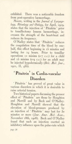
Review
Synthèse
Diagnosis and management of hyperprolactinemia
Omar Serri, Constance L. Chik, Ehud Ur, Shereen Ezzat
Abstract
PROLACTIN IS A PITUITARY HORMONE that plays a pivotal role in a variety of reproductive functions. Hyperprolactinemia is a common
condition that can result from a number of causes, including
medication use and hypothyroidism as well as pituitary disorders.
Depending on the cause and consequences of the hyperprolactinemia, selected patients require treatment. The underlying
cause, sex, age and reproductive status must be considered. We
describe the diagnostic approach and management of hyperprolactinemia in various clinical settings, with emphasis on newer diagnostic strategies and the role of various therapeutic options, including treatment with selective dopamine agonists.
CMAJ 2003;169(6):575-81
P
rolactin is a pituitary-derived hormone that plays a
pivotal role in a variety of reproductive functions. It
is an essential factor for normal production of breast
milk following childbirth. Furthermore, prolactin negatively modulates the secretion of pituitary hormones responsible for gonadal function, including luteinizing hormone and follicle-stimulating hormone. An excess of
prolactin, or hyperprolactinemia, is a commonly encountered clinical condition.1 Management of this condition depends heavily on the cause and on the effects it has on the
patient. In this review we summarize advances in our understanding of the clinical significance of hyperprolactinemia and its pathogenetic mechanisms, including the influence of concomitant medication use. Emphasis will be
placed on newer diagnostic strategies and the role of various therapeutic options, including treatment with selective
dopamine agonists, in various clinical settings.
Epidemiologic features
An excess of prolactin above a reference laboratory’s upper limits, or “biochemical hyperprolactinemia,” can be
identified in up to 10% of the population.1 Women with
oligomenorrhea, amenorrhea, galactorrhea or infertility,
and men with hypogonadism, impotence or infertility must
have serum prolactin levels measured.
The occurrence of clinically apparent hyperprolactinemia
depends on the study population. The prevalence has been
reported to range from 0.4% in an unselected healthy adult
population in Japan to 5% among clients at a family planning clinic.1 The rate is even higher among patients with
specific symptoms that may be attributable to hyperprolactinemia: it is estimated at 9% among women with amen-
orrhea, 25% among women with galactorrhea and as high as
70% among women with amenorrhea and galactorrhea.1
The prevalence is about 5% among men who present with
impotence or infertility.1
Regulation of prolactin secretion
Like most anterior pituitary hormones, prolactin is under
dual regulation by hypothalamic hormones delivered
through the hypothalamic–pituitary portal circulation
(Fig. 1). Under most conditions the predominant signal is inhibitory, preventing prolactin release, and is mediated by the
neurotransmitter dopamine. The stimulatory signal is mediated by the hypothalamic hormone thyrotropin-releasing
hormone. The balance between the 2 signals determines the
amount of prolactin released from the anterior pituitary
gland.2 Furthermore, the amount cleared by the kidneys influences the concentration of prolactin in the blood.2,3
Box 1: Clinical presentations of hyperprolactinemia
Premenopausal women
• Marked prolactin excess (> 100 µg/L [normally
< 25 µg/L]) is commonly associated with
hypogonadism,* galactorrhea and amenorrhea
• Moderate prolactin excess (51–75 µg/L) is associated
with oligomenorrhea
• Mild prolactin excess (31–50 µg/L) is associated with
short luteal phase, decreased libido and infertility
• Increased body weight may be associated with
5
prolactin-secreting pituitary tumour
• Osteopenia is present mainly in people with
associated hypogonadism
• Degree of bone loss is related to duration and severity
6
of hypogonadism*
Men
• Hyperprolactinemia presents with decreased libido,
impotence, decreased sperm production, infertility,
gynecomastia and, rarely, galactorrhea
• Impotence is unresponsive to testosterone treatment
and is associated with decreased muscle mass, body
7
hair and osteoporosis
*The degree of hypogonadism is generally proportionate to the degree of
prolactin elevation
CMAJ • SEPT. 16, 2003; 169 (6)
© 2003 Canadian Medical Association or its licensors
575
Serri et al
Medications
Neuroleptics: phenothiazines, haloperidol
Antihypertensives: calcium-channel blockers, methyldopa
Psychotropic agents: tricyclic antidepressants
Anti-ulcer agents: H2 antagonists
Opiates
Hypothalamic PRL stimulation
Primary hypothyroidism
Adrenal insufficiency
+
Inhibit dopamine release, thus leading
to reduced inhibition and increased
prolactin production
–
Dopamine
TRH
–
–
+
Neurogenic
Chest-wall injury
Breast stimulation
Breast-feeding
Via autonomic
nervous system
Optic
chiasm
Hypophyseal stalk
Estrogen
+
Physiologic causes
Pregnancy
+
Posterior
pituitary lobe
Anterior pituitary lobe
+
Prolactin
Other causes,
mechanism unclear
+
+
Increased PRL production
Ovarian: polycystic ovarian syndrome
Pituitary tumours:
Adenomas
Hypothalamic stalk interruption
Hypophysitis (inflammation)
Reduced PRL elimination
Renal failure
Hepatic insufficiency
+
Abnormal molecules
Macroprolactinemia
Normal
PRL binding
Adenoma
"Stalk
effect"
Hypophysitis
Myra Rudakewich
Normal
Macroprolactinemia
–
Inhibitory signal
+
Stimulatory signal
Fig. 1: Causes of hyperprolactinemia. Prolactin (PRL) is under dual control from the hypothalamus, where dopamine serves as an inhibitory signal, preventing PRL secretion, and thyrotropin-releasing hormone (TRH), under some conditions, stimulates increased PRL
production and release. Increased anterior pituitary hormone production can occur from a PRL-producing adenoma or from inflammation (hypophysitis). However, conditions that result in impaired dopamine delivery or enhanced TRH signalling, or both, will also result
in increased PRL release. In general, medications result in increased PRL production through their anti-dopaminergic properties. Chestwall injury and breast stimulation serve as peripheral triggers of autonomic control, which impinge on central neurogenic pathways
that attenuate dopamine release into the hypophyseal portal circulation. In some conditions, such as renal or hepatic insufficiency, PRL
is cleared less rapidly from the systemic circulation, which results in increased blood levels of PRL.
576
JAMC • 16 SEPT. 2003; 169 (6)
Management of hyperprolactinemia
stimulation of prolactin secretion.2 Furthermore,
prolactin elimination from the systemic circulation is reduced, which contributes to increased
prolactin concentrations.2 Primary hypothyroidism
can be associated with diffuse pituitary enlargement, which will reverse with appropriate thyroid
hormone replacement therapy.2
Box 2: Objectives of treatment of hyperprolactinemia
• Restoration and maintenance of normal gonadal function
• Restoration of normal fertility
• Prevention of osteoporosis
If a pituitary tumour is present:
• Correction of visual or neurological abnormalities
Pituitary tumours
• Reduction or removal of tumour mass
• Preservation of normal pituitary function
Pituitary tumours are common neoplasms that
exhibit a wide range of biological behaviour, as evidenced by hormonal and proliferative activities.2
Among pituitary adenomas, prolactin-producing pituitary tumours are the most common type. About
one-third of all pituitary tumours are not associated with hypersecretory syndromes but, rather, present with symptoms
of an intracranial mass, such as headaches, nausea, vomiting
or visual field disturbances. Because of suprasellar extension,
pituitary tumours may interrupt dopamine delivery from the
hypothalamus to the pituitary, resulting in loss of inhibition
of prolactin release, or the “stalk effect.” In contrast, tumours
that produce growth hormone (GH) may also secrete prolactin in nearly 25% of cases.2 This is a common source of
misdiagnosis, as the features of prolactin excess may capture
attention while the more subtle features of GH excess go unnoticed. In both cases the distinction is important. Surgery is
indicated for a nonfunctional pituitary adenoma that is large
enough to cause the stalk effect. For tumours that are secreting both GH and prolactin, therapy with GH-inhibitory
agents is the preferred treatment in most cases. Finally, an
autoimmune condition of the pituitary with lymphocytic infiltration can lead to hyperprolactinemia.4 This form of lymphocytic hypophysitis is typically noted in the postpartum
phase in women of childbearing age. Surgery is rarely indicated, and spontaneous resolution is common.4
• Prevention of progression of pituitary or hypothalamic disease
Causes of hyperprolactinemia
The differential diagnosis and causes of pathological hyperprolactinemia are summarized in Fig. 1. The presence
of a secondary cause and fluctuating degrees of hyperprolactinemia should raise the suspicion of a nontumorous
cause. Consideration of such secondary contributions can
obviate the need for unnecessary testing and inappropriate
treatment.
Macroprolactinemia
Asymptomatic patients with intact gonadal and reproductive function and moderately elevated prolactin levels
may have macroprolactinemia.3 This term should not be
confused with macroprolactinoma, which refers to a large
pituitary tumour greater than 10 mm in diameter. Macroprolactinemia refers to a polymeric form of prolactin in
which several prolactin molecules form a polymer that is
recognized by immunologically based serum assays. In
general, macroprolactin results from the binding of prolactin to IgG antibodies. The large prolactin polymer is unable to interact with
the prolactin receptor. Little, if any, bioBox 3: Medical therapeutic options for the managment of
logical effect of prolactin excess is noted.
hyperprolactinemia
If macroprolactinemia is suspected, the
laboratory should be notified, and the
• Dopamine agonists are currently the first therapeutic option (Table 1)
specimen can be subjected to polyethyl• Dopamine agonists have proven efficacy in reducing prolactin levels,
ene glycol precipitation before assessrestoring ovulation in premenopausal women and restoring gonadal
3
7,9
ment. If macroprolactinemia accounts
function in men
for most of the prolactin excess, no spe• Prolactin levels may remain above normal in about 20% of cases of
cific treatment is needed.
Hypothyroidism
The hyperprolactinemia of hypothyroidism is related to several mechanisms.
In response to the hypothyroid state, a
compensatory increase in the discharge
of central hypothalamic thyrotropinreleasing hormone results in increased
macroprolactinoma and about 10% of cases of microprolactinoma
9
despite dopamine agonist therapy
• Bromocriptine has been used the longest.
• Cabergoline has greater affinity and selectivity for pituitary dopamine
9–11
D2 receptors and longer duration of action. It is indicated in cases of
bromocriptine resistance or intolerance
10
• Quinagolide is an alternative dopamine agonist but with limited
access
CMAJ • SEPT. 16, 2003; 169 (6)
577
Serri et al
Clinical presentations
The clinical manifestations of prolactin excess (Box 1)
can be divided into 2 main categories: those that are mediated by prolactin excess directly and those representing the
consequences of the resulting hypogonadism.
Diagnosis
The evaluation is aimed at excluding physiologic, pharmacologic or other secondary causes of hyperprolactinemia
(Fig. 1). In the absence of such causes, imaging (preferably
MRI) of the pituitary fossa is recommended to establish
whether a prolactin-secreting pituitary tumour or other lesion is present. CT scanning may not be sensitive enough
to identify small lesions or large lesions that are isodense
with surrounding structures. Whereas serum prolactin lev-
els between 20 and 200 µg/L can be found in patients with
hyperprolactinemia due to any cause, prolactin levels above
200 µg/L usually indicate the presence of a lactotroph adenoma. In general, there is a relatively linear relation between the degree of prolactin elevation and the size of a
true prolactinoma. If a patient with only a mildly elevated
serum prolactin level has a pituitary macroadenoma, the diagnosis is more likely to be a non-prolactin-producing pituitary adenoma or other sellar mass causing the stalk effect. The approach to the diagnosis of hyperprolactinemia
is summarized in Fig. 2.
Natural history
Several series of patients with prolactin-secreting
microadenomas observed for long periods without treatment have shown that the risk of progression to macro-
Prolactin level
Macroprolactinemia
Repeat measurement
Pathological
hyperprolactinemia
Rule out
secondary causes
Correct underlying cause:
replace thyroid hormone,
remove/substitute potentially
offending medication
MRI of pituitary
Normal pituitary
Micro lesion (<10 mm)
Asymptomatic
Symptomatic
Follow-up prolactin
measurement
once yearly
Treatment
(see Fig. 3)
Fig. 2: Approach to diagnosis of hyperprolactinemia.
578
JAMC • 16 SEPT. 2003; 169 (6)
Macro lesion (≥10 mm)
Management of hyperprolactinemia
adenoma over 10 years is small (about 7%).8 In some cases,
prolactin levels returned to normal in patients who did not
receive treatment or who received treatment intermittently
with dopamine agonists. Women with prolactin-secreting
microadenomas who became pregnant during this interval
had a higher rate of remission than women who did not become pregnant (35% v. 14%).
Management
Medical therapy
Medical therapy has traditionally involved agonists of the
physiologic inhibitor of prolactin, dopamine (Box 3, Table
1). Although initially it was thought that patients would require dopamine agonist therapy all their lives, the current
use of these agents has evolved into a dynamic process depending on the patient’s needs and circumstances.
Surgical therapy
The objective of hyperprolactinemia treatment is to
correct the biochemical consequences of the hormonal
excess (Box 2). When present, the compressive features
of a large (macro) tumour must also be alleviated and the
tumour prevented from regrowing. The approach to the
management of hyperprolactinemia is summarized in
Fig. 3.
Surgical removal of tumours associated with prolactin
excess requires careful consideration of treatment objectives (Box 4). It is indicated in patients with nonfunctional
pituitary adenomas or other nonlactotroph adenomas associated with hyperprolactinemia and in patients in whom
medical therapy has been unsuccessful or poorly tolerated.
MRI of pituitary
Normal and
symptomatic
Micro and
symptomatic
Macro
Measure other pituitary
hormones to exclude associated
deficiency or excess
Dopamine agonist
therapy
Normal
prolactin level
Reduced
prolactin level
after 6 mo therapy
Asymptomatic
Measure
prolactin level
every 4–6 mo
Prolactin level
still elevated
after 6 mo therapy
Symptomatic
Consider pituitary
surgery
Isolated prolactin
excess
Stalk effect
(prolactin level not high
enough for size of tumour)
Pituitary surgery
recommended
Dopamine agonist
therapy
Normal
prolactin level
Reduced
prolactin level
after 6 mo therapy
Asymptomatic
No effect on
prolactin level
after 6 mo therapy
Symptomatic
despite prolactin
reduction
Measure prolactin
level every 4–6 mo;
MRI every 1–2 yr
Pituitary
surgery
Fig. 3: Approach to management of hyperprolactinemia.
CMAJ • SEPT. 16, 2003; 169 (6)
579
Serri et al
The best results with transsphenoidal resection of Monitoring and follow-up
the prolactinoma are limited to centres that have the
greatest experience. In one study, the apparent surgical
Biochemical and clinical improvements in response to
cure rate for prolactinomas, although good in the short dopamine agonist therapy are readily apparent in most paterm, decreased on re-evaluation during long-term fol- tients. In addition, tumour shrinkage can be expected in
low-up.12 Hyperprolactinemia recurred within 5 years about 80% of macroadenomas.17 However, a major drawafter surgery in about 50% of patients with micropro- back of medical therapy is the potential need for lifelong
lactinomas who were initially
treatment. Discontinuation of
thought to be cured.12 In other
bromocriptine therapy has
series, the rate of recurrence of
been shown to lead to recurBox 4: Indications for pituitary surgery
hyperprolactinemia following
rence of hyperprolactinemia in
in patients with hyperprolactinemia
initial cure by surgery ranged
most patients and to tumour
from 20% to 40%.13 However,
regrowth if treatment duration
• Surgery is indicated in cases of resistance or
recurrence of hyperprolactihas been less than 2 years. 18
intolerance to optimal medical therapy
nemia after surgery is not necPassos and associates18 reported
• Surgery should be considered in patients
essarily a permanent feature
maintenance of normal prowith intrasellar tumour for whom long-term
and does not inevitably indilactin levels and absence of
drug treatment is not acceptable
cate operative failure.13,14 Readenoma re-expansion after
• Surgical decompression may be required for
withdrawal of dopamine agoevaluation of long-term results
tumours pressing on the optic chiasm
nist therapy in 6.6% to 37.5%
indicates a success rate of about
• Surgery should be avoided in cases of
of patients. Recurrence usually
75% for surgical removal of
extrasellar (without optic chiasm
microprolactinoma. However,
occurs within months after
compression) expanding tumours because of
drug withdrawal. These authe results of surgery for
the low success rate
thors also reported reduced and
macroprolactinoma are poor,
normal prolactin levels after
with a long-term success rate
pregnancy in women who had
of only 26%.13
prolactinomas treated with dopamine agonists. Menopause
has also been suggested as a factor that increases the probaManagement of hyperprolactinemia in pregnancy
bility of maintaining normoprolactinemia after dopamine
The collaboration of various specialists, including an ob- agonist therapy is stopped.18 Unless there is evidence of
stetrician, is required for the careful planning of pregnancy growth of a prolactinoma or related symptoms, such as
in women with hyperprolactinemia (Box 5). Ideally, this headache, there is no indication to continue dopamine agoshould occur before conception, to permit a full assessment nist therapy after menopause.18 There are no significant difof the risks and benefits of dopamine agonist therapy dur- ferences in age, sex, initial dopamine agonist dose or length
ing pregnancy.
of treatment between those with continued normoproTable 1: Advantages, disadvantages and cost of various dopamine agonist agents
available in Canada
Agent
Main advantages
Disadvantages
Bromocriptine
Longest track record
2.5 mg/d
112.97
Cabergoline
High efficacy; low
frequency of
adverse events;
indicated in cases
of bromocriptine
resistance or
intolerance
Pituitary selectivity;
indicated in cases
of bromocriptine
resistance or
intolerance
Occasionally
beneficial in
resistant cases
High frequency
of gastrointestinal
upset and sedation
Experience during
pregnancy
relatively limited
0.5 mg/wk
139.50
Daily use; limited
access
0.075 mg/d
129.90
High frequency
of adverse events
0.25 mg/d
127.19
Quinagolide
Pergolide
580
JAMC • 16 SEPT. 2003; 169 (6)
Typical dose
Monthly cost, $
Management of hyperprolactinemia
Box 5: Management of hyperprolactinemia in
pregnancy
• There is no evidence of increased teratogenicity
associated with bromocriptine or cabergoline use
15,16
during pregnancy
• Similarly, there is no evidence of increased risk of
abortion or multiple pregnancies with dopamine
agonist use
• If the tumour size before pregnancy is < 10 mm,
dopamine agonist therapy is stopped during pregnancy
15
because the risk of tumour expansion is low
• If the tumour size before pregnancy is ≥ 10 mm before
pregnancy, bromocriptine use is advised during
15
pregnancy to avoid significant tumour expansion
• All patients should be evaluated every 2 months
during pregnancy
• Formal visual field testing is indicated in patients with
symptoms or a history of macroadenoma
• If visual field defects develop despite dopamine
agonist treatment, early delivery or pituitary surgery
15
should be considered
lactinemia and those with recurrence of hyperprolactinemia.18 We suggest that the dopamine agonist dose be decreased after 2 or 3 years of normal prolactin levels and
that therapy be stopped if the prolactin levels remain unchanged after 1 year at the reduced dose. The dose can be
reduced by half over the course of 3 months while prolactin levels are measured monthly. After complete discontinuation of treatment, regular monitoring of clinical
symptoms and prolactin levels is recommended. Given the
propensity for early recurrence, prolactin levels should be
measured monthly for the first 3 months and every 6
months thereafter.
This article has been peer reviewed.
From the Divisions of Endocrinology and Metabolism at the University of Montréal, Montréal, Que. (Serri), the University of Alberta, Edmonton, Alta. (Chik),
Dalhousie University, Halifax, NS (Ur), and the University of Toronto, Toronto,
Ont. (Ezzat)
Competing interests: The authors received an unrestricted educational grant from
Paladin Labs Inc.
Contributors: All of the authors contributed to the conception and design of the
paper, review of the data and drafting of the manuscript. Drs. Ezzat and Serri were
responsible for editing the manuscript.
References
1. Josimovich JB, Lavenhar MA, Devanesan MM, Sesta HJ, Wilchins SA, Smith
AC. Heterogeneous distribution of serum prolactin values in apparently
healthy young women, and the effects of oral contraceptive medication. Fertil
Steril 1987;47:785-91.
2. Asa SL, Ezzat S. The pathogenesis of pituitary tumours. Nat Rev Cancer 2002;
2:836-49.
3. Vallette-Kasic S, Morange-Ramos I, Selim A, Gunz G, Morange S, Enjalbert
A, et al. Macroprolactinemia revisited: a study on 106 patients. J Clin Endocrinol
Metab 2002;87:581-8.
4. Thodou E, Asa SL, Kontogeorgos G, Kovacs K, Horvath E, Ezzat S. Clinical
case seminar: lymphocytic hypophysitis: clinicopathological findings. J Clin
Endocrinol Metab 1995;80:2302-11.
5. Yermus R, Ezzat S. Does normalization of prolactin levels result in weight loss
in patients with prolactin secreting pituitary adenomas? [letter] Clin Endocrinol
(Oxf) 2002;56:562.
6. Biller BMK, Baum HB, Rosenthal DI, Saxe VC, Charpie PM, Klibanski A.
Progressive trabecular osteopenia in women with hyperprolactinemic amenorrhea. J Clin Endocrinol Metab 1992;75:692-7.
7. Di Somma C, Colao A, Di Sarno A, Klain M, Landi ML, Facciolli G, et al.
Bone marker and bone density responses to dopamine agonist therapy in hyperprolactinemic males. J Clin Endocrinol Metab 1998;83:807-13.
8. Schlechte J, Dolan K, Sherman B, Chapler F, Luciano A. The natural history
of untreated hyperprolactinemia: a prospective analysis. J Clin Endocrinol
Metab 1989;68:412-8.
9. Di Sarno A, Landi ML, Cappabianca P, Di Salle F, Rossi FW, Pivonello R, et
al. Resistance to cabergoline as compared with bromocriptine in hyperprolactinemia: prevalence, clinical definition and therapeutic strategy. J Clin Endocrinol Metab 2001;86:5256-61.
10. De Luis DA, Becerra A, Lahera M, Botella JI, Valero, Varela C. A randomized cross-over study comparing cabergoline and quinagolide in the treatment
of hyperprolactinemic patients. J Endocrinol Invest 2000;23:428-34.
11. Jeffcoate WJ, Pound N, Sturrock NDC, Lambourne J. Long-term follow-up
of patients with hyperprolactinaemia. Clin Endocrinol (Oxf) 1996;45:299-303.
12. Serri O, Rasio E, Beauregard H, Hardy J, Somma M. Recurrence of hyperprolactinemia after selective transsphenoidal adenomectomy in women with
prolactinoma. N Engl J Med 1983;309:280-3.
13. Serri O, Hardy J, Massoud F. Relapse of hyperprolactinemia revisited. N Engl
J Med 1993;329:1357.
14. Thompson JA, Gray CE, Teasdale GM. Relapse of hyperprolactinemia after
transsphenoidal surgery for microprolactinoma: lessons from long-term follow-up. Neurosurgery 2002;50:36-9.
15. Molitch ME. Management of prolactinomas during pregnancy. J Reprod Med
1999;44:1121-6.
16. Ricci E, Parazzini F, Motta T, Ferrari CI, Colao A, Clavenna A, et al. Pregnancy outcome after cabergoline treatment in early weeks of gestation. Reprod
Toxicol 2002;16:791-3.
17. Webster J, Piscitelli G, Polli A, Ferrari CI, Ismail I, Scanlon MF, for the
Cabergoline Comparative Study Group. A comparison of cabergoline and
bromocriptine in the treatment of hyperprolactinemic amenorrhea. N Engl J
Med 1994;331:904-9.
18. Passos VQ, Souza JJ, Musolino NR, Bronstein MD. Long-term follow-up of
prolactinomas: normoprolactinemia after bromocriptine withdrawal. J Clin
Endocrinol Metab 2002;87:3578-82.
Correspondence to: Dr. Shereen Ezzat, Mount Sinai Hospital,
Ste. 437, 600 University Ave., Toronto ON M5G 1X5;
fax 416 586-8834; [email protected]
CMAJ • SEPT. 16, 2003; 169 (6)
581


© Copyright 2026